Note: Descriptions are shown in the official language in which they were submitted.
CA 02270837 1999-OS-OS
Device for Optically Examining and/or Treating
the Human l
The invention concerns a device according to~ the preamble of patent claim 1.
Ophthalmologic examinations are preferably carried out using light. During
examinations
of the human eye, the forehead is typically pressed against a headband
arranged on a table and the
chin is supported on a chin support that is also arranged on the table. The
headband and chin
support serve as adjusting elements to immobilize the head of the patient in
the examination
position. A split lamp pivotally connected to the table is used as an
illumination source; the lamp
is connected in a suitable manner by means of a so-called biomicroscope. A
contact lens, which
the physician holds in the beam path of the biomicroscope and of the split
beam are also used in
many diagnoses. At the same time, the physician is :required to look through
the biomicroscope
and ensure that the patient remains in position.
1
CA 02270837 1999-OS-OS
From United States patent 4,477,159 is known a photocoagulator with which the
physician,
as explained above, holds a focusing or contact len:> with the hand and pushes
the same against
the surface of the eye. On its head is attached a telescope, with which he can
look into the eye
of the patient through the hand-held contact lens. The radiation of a laser is
faded into the
telescope by means of a beam conductor and is then guided out of the telescope
to the contact lens
as a free beam. Illumination radiation similar to laser radiation can also be
guided to the contact
lens .
The object of the invention is to make available to the physician a device
which allows safe
and problem-free examination and treatment of the human eye, preferably using
high-intensity
laser beams.
The object is attained in that, in contrast with known optical treatment
devices, which have
illumination and/or treatment optics that are no longer held by the hand of
the treating physician
on the eye to be treated, they are preferably applicable to the head of the
patient. The optics is
particularly displaceable manually by the physician (a remote-controlled
displacement is of course
also possible) and remains in the set position, whereby one of the surfaces of
the optic (contact
lens) remains applied on the eye surface. The optic is again displaced for a
new setting and then
remains self supported in the new set position. If the patient moves his/her
head, the adopted
position remains relatively unchanged relative to each eye. The head of the
patient need no longer
remain immobile in one position for a long period of time.
Treatments with laser beams (retina coagulation, etc.) can be carried out
easily when using
the device according to the invention since prolonged alignment of the eye
with the radiation
source is easily provided. The safety problems which occurred heretofore have
thereby been
2
CA 02270837 1999-OS-OS
eliminated. The physician also no longer requires a "third" or "fourth" hand
for holding the
observation or treatment optics.
The holding or guidance arrangement of the fevice according to the invention
can also be
attached to a free-standing patient head holder, by means of which the head
can be held in a
defined position by the forehead and chin.
The device should be configured as simply as possible. For this reason, the
radiation
sources for illumination and particularly for treatment of the eye should be
outside of the device
and the light or radiation (a form of radiation close to the visible spectral
range such as, for
example, infrared, is designated as "light" in the following) is guided by
means of light conductors
(light conductor beam).
Direct observation by means of a microscope is also eliminated in an
advantageous
manner. An image exposure of the eye including its background, the retina, the
chamber angle,
or other elements can take place with an image frame system having a CCD chip
that weighs
merely a few grams. In this way, a savings in weiglht is also obtained. The
physician can also
observe the patient or the apparatus better and can kf:ep the same under
control.
Another advantage of the device according to the invention is that it can be
used on a
patient who is sitting as well as lying down.
Working with light conductors for guiding illumination energy and treatment
energy as
well as an image transfer by means of wires results, aside from a considerable
savings in weight,
3
CA 02270837 1999-OS-OS
in freedom for the patient, since his/her head need no longer be immobilized,
for example, on a
treatment device arranged on a treatment table. ThE: patient can move his/her
head.
Measuring sensors (for example, for measuring eye movement, eye pressure,
etc.) can be
integrated into or within the device according to the invention in a simple
manner.
Light emitting elements such as LEDs can also be integrated into the contact
lens.
The device according to the invention can be used in an analogous fashion to
the
photocoagulator of United States patent 4,477,159 mentioned above. In contrast
to the known
photocoagulator, with the device according to the invention, the physician no
longer carries an
optic system on his/her head, which makes him irrunobile and limits his/her
field of vision.
Adjustment coordination of an observation and/or treatment optic due to the
patient's head
movements is no longer needed. Furthermore, the position of the units
necessary for treatment
is set even though the patient may move. The setting of the laser beam path in
a
photocoagulation, for example, is particularly advantageous. Inadvertent
slipping of the contact
lenses or faulty position of the lens, which could cause damage to the eye, is
no longer possible.
In the following, examples of the device according to the invention will be
described in
greater detail with reference to the drawings. Other advantages of the
invention result from the
following description. In the drawings:
Fig. 1 shows a perspective representation of the device according to the
invention;
Fig. 2 shows a variation of the device shown in Fig. 1;
Fig. 3 shows a schematic representation of the components of an optic head of
the device shown
in Fig. 2, for example;
4
CA 02270837 1999-OS-OS
Fig. 4 shows an enlarged view with respect to the representation of Fig. 2 of
a contact lens
holder, for example, in the view direction II:I indicated in Fig. 2;
Fig. 5 shows a variation of the optic head shown in. Fig. 3; and
Fig. 6 shows a cross section through an embodiment of a contact lens, for
example, which can
be used in the holder indicated in Fig. 4.
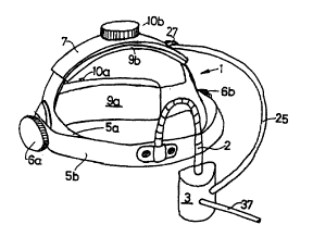
The device according to the invention shown in Figure 1 has a holding part 1
with a
housing 3 in which the illumination, observation and treatment devices shown
in Figure 4 are
located. The device can be placed on the head of the patient.
The holding part 1 has a headband Sa that runs around the head and a holding
band Sb,
between which the housing 3 is held by means of a so-called swan neck 2. The
swan neck 2 is
configured in such a way that it holds the housing :3 in pivoted-back position
without a spring
return. Furthermore, the holding part 1 has a top band 7. One end of the
headband Sa, the
holding band Sb and the top band 7 run together at one location to the left
and right (over the ears
in the situation where it is placed on the head) and are held together there
by means of a fixing
device 6a and 6b.
The headband Sa and the top band 7 are flexible, preferably configured with a
padding 9a
and 9b. The holding band Sb is rigid. The headband Sa and the top band 7 can
be adapted in
length to the head shape of the respective patient by nneans of a displacement
arrangement 10a or
10b.
Figure 2 shows an embodiment of the device shown in Figure 1. The housing,
referred
to herein as 3, is removably attached to the holding band Sb with a clamping
arrangement 11. It
S
CA 02270837 1999-OS-OS
can be displaced from one eye to the other in the direction of the double
arrow 12 in Figure 2
along the holding band Sb. The removability of tJhe housing 3 allows for its
use on the head
holder. The housing 3 has a positioning arrangement 13 for a three-dimensional
setting of an
optic head 15 by means of handles 22a and 22b. 7.'he optic head 15 contains
the illumination,
observation and optical treatment devices described below.
At approximately the location of the clamping (and displacement) arrangement
11, the
housing 3 has a positioning arrangement 13 with a self inhibiting pivoting
arrangement 19, which
can be stopped with a stop head 20 and can be unlocked by pushing on a head
lying on the other
side (not shown in Figure 2). Pivoting arrangement 19 makes possible a
pivoting around an axis
that is approximately parallel to the connection axis of both displacement
arrangements 6a and 6b.
In Figure 2, under the pivoting arrangement 19, the positioning arrangement 13
has a self
inhibiting displacement direction 21, by means of which the optic head 15 can
be displaced
vertically to the pivot axis of the pivoting arrangement 19. The displacement
arrangement 21 can
be fixed by means of a left and right stop lever 22a or 22b. This makes it
possible for the treating
physician to work with either the left or right hand. Likewise, the stop
handle 20 can also be laid
out for work with both hands. The optic head 15 itself is also three-
dimensionally displaceable
and is self inhibiting pivotally held on the ball pivot. The displacement of
the optic head 15
serves for fine tuning, particularly during eye examination and/or treatment.
The pivoting and both displacement arrangements are configured to clamp with a
spring
tension in such a manner that a self inhibition is obtiiined. The spring force
for clamping is set
along a spectrum ranging from completely loose to firm by means of the stop
handle as well as
the stop lever.
6
CA 02270837 1999-OS-OS
In Figure 2, on top of the housing 3, the elecaric supply lines, the signal
lines, and a light
conductor or a light conductor beam 37 described below are gathered into a
cable 25 and are
guided away by means of a plug 26. The light conductor 37 for the laser
radiation is guided away
separately, but could also be gathered into the cable 25 by means of another
embodiment of the
optic head 15. The cable 25 is held by means of a clamp 27 for purposes of
stress reduction at
the top band 7.
The optic head 15, shown schematically in Figure 2 pushed against an eye 29,
has a
contact lens 30 on the side nearer to the eye 29. The contact lens 30 held in
a tubus 23 has, as
shown in Figure 4, for example, an upper and a lower lid deflector 28a or 28b
for the upper or
lower eyelid. The tubus 23 can have respective adapter or coupling elements
for the different
contact lenses mentioned below. As an alternative, it can preferably be held
in a ring holder.
Different contact lenses are used for certain areas of the eye (retina fundus,
retina periphery,
chamber angle, etc. ). The surface of the contact lens 30 to be placed on the
eye 29 has, as shown
in Figure 6 with respect to the embodiment described herein, two different
curve radii rF and r~.
The central part 33a of the surface has a smaller curve radius rG than the
edge region 33b with rF.
The smaller radius r~ is, depending upon the application area, between 6 mm
and 8 mm, and the
radius rF is between 10 mm and 14 mm. The ring holder for the contact lens 30
is placed in the
tubus 32. Of course, one with only a single radius c;an be used instead of the
contact lenses 30
with the both radii rF and rG described herein. The contact lens 30 described
herein, however,
results in better optical imaging and observation properties.
For illuminating the eye 29, as shown in Figure 3, the illumination radiation
is fed by
means of the light conductor 37 in the cable 25 and b;y means of a plug 36.
The placement of the
illumination source outside of the optic head 15 reduces its weight,
facilitates an exchange of the
7
CA 02270837 1999-OS-OS
illumination source, produces free space in the optiic head 15 for other units
and sensors to be
aligned on the eye 29, and also reduces heat generation in the direct vicinity
of the eye 29 and the
hand or hands of the physician. A beam forming arrangement is also available
for the illumination
source, which is represented symbolically as a lens 39. A filter arrangement
can also be provided
for the illumination radiation, which however may also be assigned to an
external light source.
The illumination beam 40 passes, after beam forming and an eventual filtering
(color, intensity,
polarization), a first deflection mirror 41, is then directed onto a focusing
lens 44 and from there
by means of a contact lens 30 into or onto eye 29. The first deflection mirror
41 is now tempered
or arranged so that the radiation of the illumination light is transmitted
almost unimpeded
(preferably 99 % ); laser radiation as described below, however, is deflected.
Beam splitting can
take place by tempering, but also by a suitable selection of the beam paths.
The second deflection
mirror 43 allows part (as little as possible) of the illumination radiation to
be transmitted and
deflects the rest of the radiation toward the lens 44. The laser radiation is
here also (almost)
completely deflected. The radiation that comes fronn the illuminated areas of
the eye is imaged
on an image emitting element (CCD chip) 45 as observation arrangement with the
lens 44 through
the second deflection mirror 43 with a radiation loss of less than 50 % . The
electric signals of the
image emitting element 45 are displayed as images on a monitor and at the same
time are stored
in a computer, where they are available for further processing . The computer
storage can of
course also be eliminated. The monitor can now be configured as an independent
(stand-alone)
device or as a so-called headband monitor. This small headband monitor would
preferably be
worn on the head by the treating physician.
If necessary, the electronic image emitting element 45 can be removed and the
illuminated
inside of the eye or its surface can be observed by means of a microscope (so-
called
biomicroscope).
8
CA 02270837 1999-OS-OS
Aside from the illumination and observation arrangements, a beam-physical
processing
arrangement is installed in the optic head 15. The radiation for the
processing arrangement is
guided by means of a separate light conductor 47 to~ a sensor 49, with which
the laser radiation
transported by the light conductor 47 is simultaneously direction-manipulated
for processing. The
sensor 49 can, as indicated in Figure 3, be configured as a three-dimensional
pivotable, preferably
self inhibiting, ball bearing. Furthermore, a beam forming arrangement 51 for
producing
different beam cross sections is available for the laser light that comes out
of the light conductor
47. The laser radiation is guided toward the eye by means of the mirrors 41
and 43 and the
focusing lens 44 as well as the contact lens 30.
A helmet that is pulled over the head of the patient can also be used instead
of the above-
described device. However, the above device is preferred because it impairs
movement less. An
advantage of the helmet would be, however, that a hearing capsule could be
installed in it with
which the patient could listen to music, information, or instructions.
A beam path as shown in Figure 5 can be selected for a non-central processing,
for
example, in the chamber angle of the eye. A beam forming arrangement 52 (as a
rule a settable
opener) for the processing laser beam 53 of a laser is indicated symbolically
by a lens pair 52.
The imaging optic for the image conversion element, designated herein as 55,
is indicated by a
lens pair 56. A mirror 57 positioned in the laser beam path is "transparent
coated" to the laser
radiation (on both sides). The observation beam path 59 deflected by the
mirrors 53 and 59 is
guided by means of a mirror 61 arranged sideways 1:o the eye axis 62 through a
contact lens 65
lying on the eye surface 63 sideways through the chamber angle of the eye. In
contrast to the
above embodiments, no especially configured beam forming optic must be used
for illumination;
the end of a light conductor 66 connected to an illumination source is
inserted in such a manner
9
CA 02270837 1999-OS-OS
in a breakthrough or in a threaded hole 67 in the contact lens 65 that it
comes to lie near the eye
surface 63 when the contact lens 65 is in place. (The illumination could be
configured as a ring
illumination). The contact lens 65 is configured preferably similar to the
contact lens 19 [sic].
Correspondingly adapted contact lens optics are used for the regions of the
eye which can
be observed under preset angles.
The device according to the invention can also be configured as glasses with a
face shell
similar to goggles instead of the embodiment shown :in Figure 2. In this face
shell are integrated
the observation, illumination and, if necessary, processing arrangement
similar to the above-
described embodiment.
Instead of holding the optic head 15 with the: pivot and both displacement
arrangements
19 and 21 at the holding band Sb, a mounting can also take place with a so-
called swan neck or
manipulator. The optic head 15 can also be arranged with a similar swan neck
or manipulator on
a fixed head holder on which are immobilized the forehead and chin of the
patient.
Aside from the above-mentioned contact lens there are a variety of other
contact lens
embodiments . The above-described optic head 15~ or its tubus 23 can be
provided with a
corresponding adapter in such a manner that these different contact lenses can
be applied
(flanged). With these other contact lenses, different enlargements and image
fields of different
dimensions can be obtained. Contact lenses may also be used which are adapted
to the juvenile
eye as well as the eyes for persons of various ethnicities.
CA 02270837 1999-OS-OS
Depending upon the treatment and examinatiion requirements, the contact lens
30 or 65
may also be eliminated, whereby an observation then takes place by means of
the image
conversion element 45 or SS belonging to the now-modified optics system.
In Figure 3, the illumination and treatment beams (laser) are gathered by
means of the
mirror 41 into one beam path, which is then superposed with the observation
beam by means of
the mirror 43. In Figure 5 are gathered the treatment and observation beams by
means of the
mirror 57. The illumination takes place by means of a separate beam path. The
illumination,
observation, and treatment beam paths may, depending upon need, be gathered or
split by beam-
gathering or beam-splitting elements, among others by using spectral filters.
A gathering and
splitting can also be undertaken by using beam polarization. As splitting and
gathering elements,
aside from the described mirrors, beam splitting prisms or binary optics can
be used.
If a semiconductor laser is used, then the same can already be integrated into
the optic head
15, whereby the light conducting supply 47 would be eliminated. However, the
increased weight
as well as the additional space requirement must be taken into consideration.
Additional electric
energy supply cables must also be available for the laser.
The device according to the invention can be easily operated in such a way
that it can be
used not only for the treatment of humans but also for animals, whose
movements are often
unpredictable.
11