Note: Descriptions are shown in the official language in which they were submitted.
CA 02271018 1999-OS-OS
WO 98I20957 PCTlIJS97121787
INTEGRATED CARDIOTOMY .AND VENOUS BLOOD RESERVOIR
BACKGROUND ~OF THE INVENTION
The invention relates to a new and improved integrated cardiotomy and
venous blood reservoir device, and morn specifically to such a device which is
compact, provides for ease of assembly, and improved filtration. Further
embodiments relate to methods of manufacturing the reservoir device and
systems
employing the reservoir device and a blood oxygenator/heat exchanger.
Blood reservoirs are well recognized in the prior art. Blood reservoirs are
commonly used, for example, during open heart surgery by a perfusioni st and
are
coupled to a cardiopulmonary bypass circuit which takes over the function of
the
heart and Lungs. The blood reservoir stores and filters blood in the bypass
circuit.
Cardiotomy blood comes from the surgery situs or chest cavity, and often
includes debris such as bone chips, saline solution, and liquids applied to
the heart.
The cardiotomy blood must be filtered t~efore being returned to the patient.
Venous
1 S blood comes directly from the vena cave: or right atrium and does not
include the
debris found in cardiotomy blood. Venous blood does not require filtration.
Defoamers are used with both the venous and cardiotomy blood to remove foam
from the blood. Defoamers and filters ~~re commonly used with blood reservoirs
to
filter particulate material and foam from the blood. In recent years, the
cardiotomy
and venous reservoir components have been combined in a single unit which
requires separate chambers for the cardiotomy and venous blood. Examples of
such
integrated devices are described in U.S. Patent Nos. 4,642,089 and 5,1 S8,533.
Integrated cardiotomy/venous blood reservoirs are often used by a
perfusionist in an extra-corporeal blood circuit during such surgical
procedures as
open heart surgery to store, trap air, and filter blood. During arterial
incapacitation,
the blood is transferred and stored in a reservoir. The cardiotomy portion of
the
reservoir holds and filters blood salvaged from the patient's chest cavity or
surgery
CA 02271018 1999-OS-OS
WO 98/20957 PCT/US97/21787
2
situs. The venous portion of the reservoir holds and filters blood directly
from the
right atrium or vena cave. The cardiotomy portion must provide more filtration
than the venous portion because blood transferred from the chest cavity often
contains debris such as bone pieces, skin, etc. Blood from the vena cave
typically
requires less filtration than cardiotomy blood salvaged from the chest cavity
because venous blood is not contaminated with debris from the surgery situs.
SUMMARY OF THE DISCLOSURE
It is an object of preferred embodiments of the present invention to integrate
the cardiotomy and venous reservoir and filtration components in a single unit
and
at the same time provide more filtration for the cardiotomy component. It is a
further obj ect to provide an improved structure and method of manufacturing
the
cardiotomy and venous blood chambers and providing separation therebetween. It
is
still a fiwther obj ect to assemble a filter/defoamer core assembly and affix
the
filter/defoamer assembly within the reservoir without using any adhesives or
bonding chemicals.
These and other objects and advantages are achieved in an integrated
cardiotomy and venous blood reservoir wherein a generally cylindrical, hollow
support structure separated by a separator disposed within the support
structure is
used to form both a cardiotomy chamber and venous blood chamber. The support
structure has a first end, a second end, and a wall therebetween with an
aperture in
the wall. The axial length of the support structure is divided into a first
portion
extending from the first end along the axial length, and second portion
extending
the remaining axial length to the second end. Preferably, the support
structure is a
generally cylindrical, hollow cage having ribs extending the axial length of
the
generally cylindrical structure.
The support structure is disposed within a generally cylindrical, hollow
blood defoamer which extends along the first and second portions of the
support
structure. Preferably a fabric sock extends around the outer defoamer to
maintain
the outer defoamer compressed against the support structure. A separator with
an
opening is disposed within the support structure and is located between the
first and
second portions of the support structure. In a preferred embodiment a
generally
CA 02271018 1999-OS-OS
WO 98l20957 PCT/US97/21787 - -
3
cylindrical, hollow depth filter is disposed within the support structure,
between the
first end and the separator. However, in alternative embodiments, the support
structure may be disposed within the depth filter or both the outer defoamer
and
depth filter may be disposed within the support structure. Further, a first
inner
defoamer may be disposed within the depth filter in the first portion of the
support
structure and a second inner defoamer :may be disposed within the second
portion
of the support structure. In preferred embodiments, the inner defoamer
disposed
within the second portion of the suppou structure is part of the outer
defoamer
wrapped around the second end of the support structure.
A tube extends axially through l:he first portion of the support structure,
through the opening in the separator and into the second portion of the
support
structure. One end of the tube is in fluid communication with the outer
defoamer in
the second portion of the support structure, or in fluid communication with a
second inner defoamer disposed within the second portion of the support
structure
the if there is such a second inner defoarner.
In preferred embodiments, the support structure, defoamer, filter, separator
and tube, are disposed within a generally cylindrical, hollow outer shell
having a
first end, a second end, and a wall therebetween, wherein the outer defoamer
is
spaced apart from the annular wall of the outer defoamer. A first blood inlet
and a
second blood inlet are located on the first end of the outer shell, and a
blood outlet
is located on the outer shell. Preferably, the first end is comprised of a
cover which
is bonded to the wall of the shell. The rube is in fluid communication with
the first
blood inlet and the second blood inlet is in fluid communication with the
depth
filter.
In this way, a cardiotomy chamber is formed by the separator, depth filter,
and inner defoamer in the first portion of the support structure. Thus, the
second
blood inlet, which is in fluid communication with the depth filter, functions
as a
cardiotomy blood inlet. A venous bloodl chamber is formed by the separator and
defoamer in the second portion of the support structure. The first blood inlet
in
communication with the tube functions as a venous blood inlet.
CA 02271018 1999-OS-OS
WO 98I20957 PCT/US97/21787 _ .
4
In preferred embodiments, a deflector having an opening is located between
the first end of the support structure and the first end of the outer shell.
The tube
extends from the first blood inlet through the opening in the deflector. The
deflector guides blood from the second blood inlet into the cardiotomy
chamber.
Embodiments of the present invention are also directed to methods of
manufacturing the above apparatus. The filter/defoamer core assembly is formed
by
first disposing the support structure within the outer defoamer. Preferably, a
fabric
sock is then placed around the outer defoamer to maintain the outer defoamer
compressed against the support structure. The depth filter is then placed in
the first
portion of the support structure. The separator is then placed in the support
structure, between the first and second portions of the support structure. The
first
and second inner defoamers may then be placed within the first and second
portions of the support structure, respectively.
The tube for communication with the first blood inlet is then inserted
through the first portion of the support structure, through the opening in the
separator, and into the second portion of the support structure. The tube is
then
affixed to the first end of the outer shell to be in fluid communication with
the first
blood inlet. The first end of the outer shell is then affixed to the outer
shell. In this
way, the filter/defoamer core assembly may be manufactured without the use of
adhesives or other bonding chemicals.
CA 02271018 1999-OS-OS
W0 98/20957 PCT/US97/21787
BRIEF DESCRIPTION OF DRAWINGS
Figure 1 is a perspective view showing an embodiment of a system
composed of an integrated venous and cardiotomy reservoir and an integrated
oxygenator/heat exchanger.
5 Figure 2 is a cross sectional diagram of the integrated venous and
cardiotomy reservoir of Figure 1.
Figure 3 is an exploded layout of a preferred embodiment of the integrated
venous cardiotomy reservoir of Figure 1.
Figures 4a, 4b, 4c, 4d are different cross-sectional views of a preferred
embodiment of the filter/defoamer core assembly.
Figures Sa and Sb are top and side views, respectively, of a preferred
embodiment of the cover.
Figure 6 is a side view of a preferred embodiment of the outer shell of the
blood reservoir.
CA 02271018 1999-OS-OS
WO 98I20957 PCTIiTS97/21787 -
6
DETAILED DESCRIPTION OF PREFERRED EMBODIMENTS
Figures 1-6 show preferred embodiments of an integrated
venous/cardiotomy blood reservoir 4 and a system composed of the integrated
venous/cardiotomy blood reservoir 4 and an integrated oxygenator/heat
exchanger
2. In the detailed description below, references made to the "top," "bottom,"
"upper," or "lower" portions of the blood reservoir 4, or elements thereof,
are made
with reference to the orientation of the structures shown in the drawings and
are
not intended to limit the scope of the invention where such limitations are
not
otherwise required.
As shown in the system embodiment of Figure 1, the venous/cardiotomy
reservoir 4 is preferably used in combination with a membrane oxygenator and
heat
exchanger 2 which receives blood transferred from the reservoir 4 and
oxygenates
and alters the temperature of the blood. Preferably, the membrane
oxygenator/heat
exchanger 2 is of the type described in the U.S. Patent Application titled
Integrated
Oxygenator and Heat Exchanger, filed under U.S. Express Mail label no.
EM419214600US, on November 7, 1996, (incorporated herein by reference) and
assigned to the assignee of the present invention. However, other suitable
oxygenators or integrated oxygenator/heat exchangers may be used with the
reservoir 4 in other system embodiments. In preferred embodiments, blood
reservoir 4 is a single use reservoir for intraoperative perfusion of adult
patients.
From the reservoir 4, filtered blood is then transferred to the
oxygenator/heat
exchanger 2 via tubing (not shown). A pump, such as a peristaltic pump (not
shown), may be applied to the tubing connecting the blood reservoir 4 and
oxygenator/heat exchanger 2. The pump is preferably controlled to provide the
blood at a desired pressure to the oxygenator/heat exchanger 2. The reservoir
4 is
preferably positioned higher than the' heat exchanger/oxygenator 2 to maintain
positive pressure on the oxygenator when blood flow is stopped.
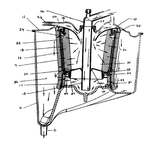
A preferred embodiment of the blood reservoir is described with reference
to Figures 2-6. Referring to Figures 2 and 3, j the venous/cardiotomy blood
reservoir 4, when assembled, can be characterized as comprising six concentric
CA 02271018 1999-OS-OS
WO 98l20957 PCTlLJS97/21787
7
shells, each shell having a substantially cylindrical shape. The innermost
shell is a
generally cylindrical, hollow inner defoalner 24. The second shell is a
generally
cylindrical, hollow depth filter 22. The third shell is a generally
cylindrical, hollow
support structure 18, wherein the axial length of the support structure 18 has
an
upper first portion 26 and a lower second portion 28. The inner defoamer 24
and
depth filter 22 each extend the length oil the first portion 26 of the support
structure
18. The fourth shell is a generally cylindrical, hollow outer defoamer 16. The
fifth
shell is a fabric sock 8. The sixth shell its a generally cylindrical, hollow
outer
shell 6.
Figures 2 and 6 show a preferred embodiment of the outer shell 6. The
lower end of the outer shell 6 inverts upward in the center thereby forming an
annular funnel channel converging downward toward one side of the shell. At
the
lower most point of the annular funnel of the shell 6 is a blood outlet 5.
With
reference to Figure 2, a blood flow channel 36 is formed between the outer
shell 6
and the outer defoamer 16, which may be covered by the fabric sock 8. In a
preferred embodiment, the outer shell 6 is made of a clear, injection molded
polycarbonate and is not taller than about sixteen inches. The blood flow
channel
36 is visible to the perfusionist or user of the reservoir, through the outer
shell 6. It
should be appreciated that alternative sh~~pes or sizes of the outer shell 6
may be
used, and that the outer shell may be comprised of any suitable material.
Further,
the blood outlet 5 may be placed at other locations on the outer shell 6 than
that
shown in the preferred embodiment shown in the drawings.
The depth filter 22 is comprised .of a material having a density greater than
the density of the inner and outer defoanners 24, 16. The depth filter 22 and
defoamers 24, 16 are preferably comprised of polyester or polyurethane
materials
well known in the art. In a preferred embodiment, the depth filter is disposed
within the first portion of the support structure. However, it should be
appreciated
that, in alternative embodiments, the depth filter 22 may be located on the
outside
surface of the support structure or both the outer defoamer 16 and depth
filter 22
may be disposed within the support structure 18. In preferred embodiments, the
hollow inner defoamer 24 is disposed within the depth filter 22.
Alternatively, the
CA 02271018 1999-OS-OS
WO 98I20957 PCT/ITS97/21787
8
inner defoamer 24 may be comprised of a sponge defoaming material that fills
the
space formed within the annular depth filter 22.
The support structwe 18 provides support for the filters and defoamers. The
support structwe 18 may be formed of a polyolefin or other thermoplastic
material.
With reference to Figwes 3, 4a, and 4b, the support structwe 18 has an upper
first
end and a lower second end. In preferred embodiments, the support structwe is
a
generally cylindrical, hollow cage having a plwality of axial ribs extending
the
axial length of the cage. In should be appreciated that alternative shapes or
designs
of the support 18 may be provided. For instance, the support structwe 18 may
be
comprised of a generally cylindrical, hollow wall with a plwality of apertures
or
may have a non-cylindrical shape. With reference to Figwes 2 and 3, the first
portion 26 of the support structwe 18 defines a cardiotomy blood chamber 30
and
the second portion 28 defines a venous blood chamber 32.
With reference to Figwes 2 and 4a-d, the inner surface of the support
structwe 18 has an annular dividing ledge 34 located between the first and
second
portions 26, 28. In preferred embodiments shown in Figwes 4b, 4c, and 4d, a
separator 20, has an upper flange 38, a lower flange 37, and a center opening
40.
The separator 20 and depth filter 22 are disposed within the support structure
18.
The upper flange 38 of the separator 20 rests on the dividing ledge 34 and
compresses the depth filter 22 against the support structwe 18. In this way,
separator 20, support structwe 18, and filter 22, compressed between the
separator
20 and support structwe 18, form a separate dividing wall separating the first
and
second portions 26, 28 of the support structwe 18 to define separate
cardiotomy 30
and venous 32 blood chambers.
Figwe 4d shows a plwality of bendable tabs 35 attached to the annular
dividing ledge 34 that extend toward the center of the support structure 18.
When
the separator 20 is inserted in the support structwe and the upper flange 3 8
rests on
the dividing ledge 34, the tabs 35 extend between the upper flange 38 and
lower
flange 37 of the separator 20 to firmly maintain the separator 20 and the
inner
section 17 of the outer defoamer 16 in place.
CA 02271018 1999-OS-OS
WO 98/20957 PCT/US97/21787 . . -
9
It should be appreciated that alternative structures may be used to separate
the cardiotomy 30 and venous 32 chambers or bonding agents may be used to
maintain the separator in place.
In preferred embodiments, the outer defoamer 16 extends the axial length of
the support structure 18. With reference: to Figures 2 and 4a-c, a lower end
1? of
the outer defoamer 16 wraps around thc; bottom end of the support structure 18
and
extends upward the length of the second portion 28 to the separator 20. It
should
be appreciated that in alternative embodiments the defoamer on the inside
surface
of the venous chamber 32 may be comprised of a separate defoamer instead of
being a portion 17 of the outer defoame;r 16 that wraps around the bottom of
the
support structure 18. T'he inner defoame;r in the second portion may be a
generally
cylindrical, hollow defoamer or a defoaming sponge that fills the second
portion of
the venous chamber 32. The fabric sock: 8 (Figures 2 and 3) extends around the
outer defoamer 16 and maintains the defoamer 16 positioned against the support
structure 18.
In this way, the cardiotomy chamber 30 is bounded by the inner defoamer
24, the depth filter 22, the outer defoarner 16, and the fabric sock 8. The
lower
venous chamber 32 is bounded only by the outer defoamer 16 doubled over. The
cardiotomy chamber 30 preferably includes the additional layer of the depth
filter
22 because more debris is typically present in blood from the chest cavity or
surgery situs than in venous blood that ;goes to the venous chamber 32.
A preferred embodiment of a cover 12 is shown in Figures 2, 3, and 5. The
cover 12 is attached to the top end of the outer shell 6 by bonding, welding
or
other suitable attachment means. Figure Sa shows the cover having six
cardiotomy
inlets 42 that open laterally into the deflector 10. Figure Sa shows an
embodiment
of the cover 12 having additional inlets, leurs, and vents used to sample
blood,
introduce medication, or vent trapped ai.r.
A funnel-shaped deflector 10 is located between the first end of the support
structure and the cover. The deflector 10 is in fluid communication with the
second
blood inlets 42 and the cardiotomy chamber 30.
CA 02271018 1999-OS-OS
WO 98I20957 PCT/US97/21787 -
With reference to Figures 2 and 3, a venous center tube 13 extends
downward from the center opening in the cover 12, through the center opening
in
the deflector 10, through cardiotomy chamber 34, through the center opening 40
in
the separator 20, and into the venous chamber 32. In preferred embodiments,
the
5 venous center tube 13 opens near the bottom of the venous chamber 32. A
venous
inlet 14 is attached to the cover 12 and is in fluid communication with the
upper
end of the venous center tube 13. The venous inlet 14 is adapted to be linked
with
a Iine (not shown) to the vena cava or right atrium of a patient's heart.
In operation, with reference to Figure 2, venous blood travels from a line
10 connected to the right atrium, through the venous inlet 14, through the
venous
center tube 13 and into the venous chamber 32. Venous blood then flows
laterally
through the wrapped around portion i 7 of the outer defoamer 16 and through
the
main portion of the outer defoamer 16 into the blood flow channel 36. The
filtered
venous blood then exists the reservoir through the blood outlet 5.
With reference to Figure 2, cardiotomy blood from the surgery situs travels
from a line (not shown) to the cardiotomy inlets 42. Blood flows through the
inlets to the deflector 10, which directs the blood into the cardiotomy
chamber 30
along the outside of the venous center tube 13. Cardiotomy blood then flows
laterally through the inner defoamer 24, through the depth filter 22, through
the
support structure 18, and then through the outer defoamer 16, through the
fabric
sock 8, and into the blood flow channel 36. The filtered cardiotomy blood then
exits the reservoir through the blood outlet S.
As described in detail above, embodiments of the present invention provide
a compact housing integrating both the cardiotomy and venous blood reservoir
components within a single support structure. The integrated housing provides
an
additional layer of filtration for the cardiotomy blood. In this way, a higher
level
of filtration of the cardiotomy blood is performed, while at the same time
minimizing trauma to the venous blood which typically needs less filtration
than
cardiotomy blood.
Embodiments of the present invention are designed to provide
improvements and benefits with respect to the ease of manufacture. In
preferred
, ~ ,
CA 02271018 1999-OS-OS
WO 98I20957 PCT/US97/21787 ~.
embodiments, a filter/defoamer care as:>embly 44 is manually assembled as
shown
in Figures 3 and 4a- c, where the support structure 18 is placed within the
annular
outer defoamer 16. The sock 8 is then placed over the outer defoamer 16 to
maintain the defoamer 16 in contact with the support structure 18. As shown in
Figure 4b, the separator 20 and depth falter 22 are then placed into the first
portion
26 ofthe support structure 18. The separator 20 is placed in the depth filter
22 and
positioned to rest on the dividing ledge 34. The separator 20 compresses an
end of
the depth filter 22 against the support structure. The inner defoamer 24 is
then
placed within the hollow depth filter 22. It should be appreciated that steps
of the
manufacture of the filter/defoamer core assembly 44 may be performed with
automation. In addition, in preferred embodiments, the filter/defoamer core
assembly 44 is assembled without the use of adhesives or bonds. In this way,
the
fitter core may be manually assembled quickly, in a cost effective manner.
In preferred embodiments showr.~ in Figures 2 and 3, the inner defoamer 24,
depth filter 22, and the first portion of t:he support structure 26 are
tapered toward
the bottom. Accordingly, the lower part of these generally, hollow shell
components has a circumference that is less than the upper part of the element
in
which they are placed. The tapered shape makes it easier to insert the
generally
cylindrical shell components 24, 22, and 26 one inside the other.
As part of the manufacturing process, the venous center tube 13 is bonded
to the venous blood inlet 14. The blood inlet 14 is snapped into the cover 12.
An
o-ring 27 may be used to provide rotational resistance between the blood inlet
14
and the opening in the cover 12, which seals the cardiotomy section 30. In
preferred embodiments, the cover 12 has an annular wall 46 and the deflector
10
also has an annular wall 48. The funnel-shaped deflector 10 is inserted into
the
space formed by the annular wall 46 of the cover 12 and maintained in place by
means of a compression fit with the anrmiar wall 46. The filter/defoamer 44
core
assembly is then placed in a channel 50 formed between the annular wall 46
ofthe
cover 12 and the annular wall 48 ofthe deflector 10. When the filter/defoamer
core
assembly 44 is positioned with respect to the cover 12, the venous center tube
13
CA 02271018 1999-05-05
WO 98120957 PCT/US97/21787
12
extends through the center opening of the deflector 10 and the center opening
of
the separator 20. The cover 12 is then bonded to the open end of the outer
shell 6.
In preferred embodiments, the components of the filter/defoamer core
assembly 44 are not bonded with adhesives or bonds to the shell 6, but are
instead
maintained in place with a tight fit with the outer shell 6 and annular walls
46 and
48 extending from the cover 12 and deflector 10. In preferred embodiments, the
venous portion of the reservoir is capable of handling blood flow rates of one
to
seven liters per minute. The cardiotomy portion is capable of handling blood
flow
rates of one to five liters per minute and the combined venous/cardiotomy
components can handle blood flow rates of one to seven liters per minute.