Note: Descriptions are shown in the official language in which they were submitted.
CA 02271462 1999-OS-11
WO 98I21578 PCT/C1S97120486
DERMAL PATCH FOR DETECTING LONG-TERM ALCOHOL CONSUMPTION AHD METHOD OF USE
FIELD OF THE INVENTION
The present invention relates to detecting an analyte in perspiration, and
particularly relates to a
dermal patch far collecting and retaining volatile analytes such as ethanol in
perspiration for detection of
consumption over a period of hours to several days.
BACKGROUND OF THE INVENTION
Ethanol in alcoholic beverages is consumed regularly by about half of all
adults and is the most
frequently abused drug worldwide. Alcohol abuse results in substantial
morbidity and mortality with the
associated costs of medical care, accidents and lost productivity. Laws
generally restrict driving under the
influence of alcohol and or employees in safety-sensitive jobs (e.g., heavy
machinery operators or mass transit
drivers) are prohibited from performing their jobs with limited blood alcohol
levels or within a few hours of
consuming alcohol.
Ethanol distributes evenly throughout body fluids with the ethanol
concentration proportional to the
body fluid's water content. A single ethanol dose (1 glkg body weight) raises
the blood-alcohol level from
the normal endogenous ethanol concentration of about 0.000t5 gldl to about 0.1
gJdl in about an hour on
an empty stomach (Basalt, R. & Cravey, R., Deposition of Drugs and Chemicals
in Man, 4th ed., p. 293,
Chemical ToxicoL Inst., 1994). During the post-absorption phase) the ratio of
ethanol in urine compared to
whole blood averages 1.3, whereas the breath to whole blood ratio averages
about 2180 and is related to
the partition coefficient between blood alcohol in the lungs and alcohol vapor
in air (Payne, J.P, et al., Nature
217:963, 1968; Heise, A.H., J. for. Sci. 12:454, 1967; Jones, A.W., J. Stud
Alc. 39:1931, 1978).
Alcohol in body fluids is commonly measured in a laboratory or in the field
using enzyme assays,
immunoassays) gas chromatography [GC), chemical oxidation and photometry,
electrochemical oxidation with
fuel cells) infrared spectrometry or solid-state semiconductor sensing. Breath
and saliva ethanol
measurements are commonly used for non-invasive instantaneous analysis and
monitoring of alcohol use
(Jones, A.W., J. Ann. Tox. 19:169, 1995I. These methods provide an instant
measurement of body fluid
ethanol at the sampling time and are useful for determining the subject's
condition at the specimen collection
time, but provide no information on the subject's long-term or cumulative
alcohol consumption. This is
because the ethanol half-life in the body is relatively short, averaging 8 hr
in breath, blood and urine (Basalt,
R. & Cravey, R., id 1. Thus, instantaneous measurements provide no information
on alcohol use unless the
sample is collected shortly after consumption.
' Individuals who abuse alcohol often underreport the amount they consume.
Moreover, blood
carbohydrate-deficient transferins (CDT) that correlate with chronic alcohol
abuse are only detected after
relatively extreme levels of ethanol consumption (Bean, P. et al., Clin. Chem.
41(6):858, 1994). Thus,
methods for monitoring long-term abstinence or limited alcohol consumption are
needed for monitoring
compliance with forensic andJor treatment programs.
CA 02271462 1999-OS-11
WO 98I21578 PCT/US97/20486
2
Biological analytes can exit the body in either insensible or sensible
perspiration. Insensible
perspiration results from passive diffusion of water and other volatiles
through the skin. Insensible
perspiration varies with location on the body and skin temperature but is
relatively similar on the upper arms,
back, and lower chest, In contrast, sensible perspiration is actively secreted
from eccrine and apocrine sweat
glands (located, respectively, throughout the skin and in the axilla, pubic
and mammary areas).
Many drugs, including ethanol) are excreted in sweat (Nyman, E. & Palmlov, A.,
Scand Arch.
Physiol. 74:155, 1936). During absorption, alcohol concentration of insensible
sweat lags behind that of
blood and breath but after complete absorption, the ethanol concentrations in
insensible perspiration, breath
and blood are similar. At the beginning of the post-absorption phase, when
blood and breath levels begin
to drop, the alcohol concentration of insensible sweat is slightly higher.
During post-absorption, the alcohol
elimination rate constant from skin is similar to that of blood and breath
(Brown, D., Meth, find Exptl. Clin.
Pharmacol. 7(51:Z69, 1985; Brown, D., Meth. Find Exptl. Clin. Pharmacoh 7(i
0):539, 1985).
Because perspiration can be collected noninvasively, it is preferable to blood
collection, an invasive
procedure, or urine collection which involves privacy concerns and samples
that can be readily adulterated.
Collecting perspiration samples for analyte analysis is known. For example,
clothing worn by an individual
can be extracted and analyzed for drugs (Smith et al., J. Forensic Sci. 36:582-
585, 1981; J. forensic Sci.
31:1269-1273, 19B6). Perspiration induced by exercise, thermal stress or
pilocarpine iontophoresis (a
procedure involving small amounts of electrical current) can be collected and
analyzed. A sensor placed on
the skin and attached to a battery-operated device can electrochemically
measure and record the skin ethanol
vapor concentration every two to five minutes (Swift, R.M., et al.,
Alcoholism: Chi. Exp. Res 16(4):7Z1,
1992).
Occlusive dermal patches for collecting and retaining perspiration and
analytes therein have been
used to monitor exposure to chemicals including alcohol as described in U.S.
Patent Nos. 4,329,999,
4,732,153 and 5,396,901. These occlusive transdermal dosimeters utilize a
waterproof dermal adhesive
patch for collecting, storing and processing perspiration. Occlusive dermal
patches to collect perspiration
generally have significant disadvantages. Hydration alters the skin's steady-
state pH, affects transepidermal
water loss and carbon dioxide emission rates, promotes growth of microbial
species that colonize the skin
and results in skin irritation. After three to five days, the pH of skin, the
transepidermal water loss and
carbon dioxide emission rates, and the number of microbes under an occius-rve
patch all increased significantly
(Aly et al., J. Incest. Oermatol. 71(6):378-381, 1978; Aly, et al., Am. J.
Infec. Control l6(3):95-100, 1988).
In some cases, antifungal and antimicrobiai agents have been included in
occlusive patches to inhibit microbial
growth and glycolysis by microbes growing in or under the patch (U.S. Patent
Nos. 4,329,999 and
4,732,153). Perspiration also tends to leak from some occlusive patches
affecting analyte analysis results.
Other dermal patches that collect components of perspiration have been
described in U.S. Patent Nos.
5,203,327 and 4,957,1O8.
Activated carbon (charcoal) has been used to selecfrvely adsorb voiatiVe
solutes in gas or liquid that
CA 02271462 1999-OS-11
WO 98/21578 PCT/US97/20486
3
contacts the carbon particles. Charcoal is activated by exposing it to high
temperatures in a controlled
atmosphere to produce microscopic pores in the carbon crystalline lattice.
These pores are responsible far
adsorption of compounds. Some dermal patches have included activated charcoal
as a binding material. For
example, U.S. Patent No. 4,732,153 discloses a transdermal dosimeter to
monitor exposure to chemical
agents by providing an unbroken fluid link between tissue fluids in the skin
and the fluid collecting component
which may include activated charcoal as a binding material. U.S. Patent No.
5,396,901 describes a
watertight transdermal dosimeter connected by a fluid bridge to the skin for
storing collected fluid and
chemical substances in a tamper-resistant container. U.S. Patent No. 4,756,314
describes an osmotically-
driven absorbent sweat collection pad, which may contain activated charcoal,
in a patch that stores fluid
phase water and substances in perspiration for determining the presence of low
molecular weight substances
in sweat. U.S. Patent No. 4,909,256 discloses a transdermal patch that
includes a charcoal-containing
binding reservoir in an airtight adhesive cover for monitoring exposure to
chemical substances including
ethanol. U.S. Patent No. 4,960,467 describes an occlusive patch for collection
of liquid transdermal
substances in a wettable substance binding reservoir of activated charcoal
powder immobilized in a gel matrix.
Carbon-containing wound dressings are also known. These include KALTOCARB'"
(Britcair), made
of alginate and charcoal; OPRASORB~' (Lohmann GmbH & Co. KG, Nuweid, Germany),
an activated charcoal
cloth; and LYOFOAM"" (Seton Healthcareh a charcoal-containing polyurethane
foam (Dover et al., Brit. J.
Plastic Surgery 48:230235, 1995; Wollina et al., Skin Phaimocol. 9:35-42,
1996).
The non-occlusive dermal patch of the present invention overcomes many of the
disadvantages
associated with other transdermal patches and methods of monitoring alcohol
consumption. This dermal
patch collects and retains volatile alcohol in vapor phase perspiration during
the entire period the patch is
worn, thus providing a system of monitoring ethanol consumption for over a
week without collecting and
storing liquid perspiration or causing skin irritation.
SUMMARY OF THE INIfENTION
According to the invention, there is provided a dermal patch to be worn on the
skin of a subject
mammal for determining the presence of an analyte in the subject's
perspiration. The dermal patch includes
a first adsorption pad for collecting vapor phase perspiration from a
subject's skin and retaining a vapor
phase analyte present in vapor phase perspiration. The first adsorption pad
includes a first side, a second
side and an outer perimeter, and the first side is adapted to be in fluid
communication with the subject's
skin. The dermal patch also includes a first gas permeable film having a first
side and a second side and
an outer perimeter, with the first side of the gas permeable film located
adjacent to the second side of the
adsorption pad, and the second side of the first gas permeable film is adapted
to be in fluid communication
with the subject's environment. The first gas permeable film has a first
moisture vapor transmission rate
(MYTR) that allows vapor phase perspiration to escape from the dermal patch
through the first gas permeable
film. The derma! patch also includes a second gas permeable film having a
first side and a second side and
art outer perimeter, with the first side of the second gas permeable film
adapted to be in fluid communication
~ I
CA 02271462 1999-OS-11
WO 98I21578 PCT/US97/20486
4
with the subject's skin, and the second gas permeable film has a second MV1R
that is less than or about
equal to the first MVTR. In one embodiment, the dermal patch also includes a
second adsorption pad for
collecting the vapor phase analyte from the subject's environment and
retaining the analyte, and a separator
layer having a first side and a second side. The second adsorption pad has a
first side and a second side
and an outer perimeter, with the second side adapted to be in fluid
communication with the subject's
environment, and the separator layer is located between the first adsorption
pad and the second adsorption
pad. In another embodiment, the first adsorption pad is located proximate to
the second gas permeable film
and the second adsorption pad is located proximate to the first gas permeable
film, with the first side of the
separator layer located proximate to the second side of the first adsorption
pad and the second side of the
separator layer located proximate to the first side of the second adsorption
pad. In a different embodiment,
the first adsorption pad is located proximate to the second adsorption pad in
a side-by-side arrangement such
that the perimeter of the first adsorption pad is proximate to the perimeter
of the second adsorption pad,
with the separator layer located adjacent to the perimeter and the first side
of the second adsorption pad,
thereby separating the proximate perimeters of the first and second adsorption
pads and separating the
I S second adsorption pad from the second gas permeable film. In another
embodiment, the first adsorption pad,
the second adsorption pad or both are capable of retaining ethanol as the
vapor phase analyte. In a
preferred embodiment, the first adsorption pad, the second adsorption pad or
both are made of an activated
carbon-containing material. In another embodiment, the dermal patch also
includes an adhesive layer located
on the first side of the first gas permeable film, the first side of the
second gas permeable film or both, and
the adhesive layer is capable of reversibly attaching the dermal patch to the
subject's skin. In another
embodiment, the first gas permeable film, the second gas permeable film or
both are of polyurethane.
Embodiments may also includes a release liner located between the first gas
permeable film and the first
adsorption pad such that the release liner is adjacent to the second side of
the first adsorption pad. Indicia
may be included for identifying the dermal patch. The dermal patch may also
include an outer protective liner
located adjacent to the second side of the first gas permeable film, adjacent
to the first side of the second
gas permeable film, or both.
According to another aspect of the invention, there is provided a method of
determining the presence
of an analyte contained in the perspiration of a subject mammal, comprising
the steps of providing a dermal
patch as described above, attaching the dermal patch to a subject's skin,
passing vapor phase perspiration
containing the vapor phase analyte expressed from the subject's skin through
the first adsorption pad for a
period of time sufficient to adsorb the analyte, then removing the dermal
patch from the subject's skin, and
determining the amount of analyte adsorbed in the first adsorption pad. Where
the dermal patch used
includes a second adsorption pad, the method also includes collecting the
vapor phase analyte in the subject's
environment in the second adsorption pad during the period when vapor phase
perspiration is passing through
the first adsorption pad, and determining the amount of the analyte adsorbed
in the second adsorption pad.
One embodiment of the method also includes the step of comparing the amount of
analyte in the second
CA 02271462 1999-OS-11
WO 98I21578 PCT/US97l20486
adsorption pad with the amount of analyte in the first adsorption pad to
determine an amount of the analyte
in the subject's perspiration during the period when vapor phase perspiration
passed through the first
adsorption pad. In the method, the removing step preferably occurs about one
hour to about ten days after
the attaching step. In a preferred embodiment of the method, the analyte is
ethanol or a metabolite of
5 ethanol. In another embodiment, the determining step includes extracting the
analyte from the first
adsorption pad, from the second adsorption pad or both, to produce an extract.
The method may be
- practiced by measuring the analyte in the extract by gas chromatography.
BRIEF DESCRIPTION OF THE DRAWINGS
FIG. ~ is a plan view of a dermal patch with a single adsorption pad for
ethanol collection from
perspiration.
F1G. 2 is a cross-sectional view of the dermal patch taken along the line 2-2
of FIG. 1.
FIG. 3 is an exploded perspective view of the dermal patch including outer
protective liners.
FIG. 4 is a perspective view of a dermal patch having two side-by-side
adsorption pads, one for
monitoring environmental exposure and one for collecting ethanol in
perspiration.
1 S F1G. 5 is a cross-sectional view of a dermal patch taken along the tine 5-
5 of FIG. 4.
FIG. 6 is an exploded perspective view of a dermal patch having an upper
adsorption pad for
monitoring environmental exposure and a lower adsorption pad for collecting
ethanol in perspiration.
FIG. 7 is a bar graph showing the ethanol content (mean and standard
deviation) of patches
removed from eight subjects at day 0 (pre-dose) and days 1 ( 3, 5 and 7 post-
dose.
FIG. 8 graphically shows the relationship of breath ethanol measurements to
the ethanol dose
measured 30 min post-dose for each of the four subjects (subject A: ~; subject
B: ~; subject E: ~; and
subject G: X ). FIG. 9 graphically shows the relationship of ethanol content
from dermal patches (mean
and standard deviation of six patches for each of seven ethanol doses)
relative to the ethanol dose for each
of the four subjects (subject A: ~; subject B: ~; subject E: ~; and subject G:
O ).
FIG. l0 graphically shows a typical kinetic relationship of ethanol measured
in blood and saliva ( 0,
solid line), breath (~, solid line), urine (x, shaded line), and collected
from perspiration using the dermal
patch (~, shaded line) for one subject.
DETAILED DESCRIPTION OF THE PREFERRED EMBODIMENT
The dermal patch of the present invention includes a carbon-containing
adsorbent material to collect
analytes including ethanol excreted in perspiration, where the water and
substances contained in perspiration
enter the adsorbent material in vapor rather than liquid form. For
perspiration collection, the non-occlusive
dermal patch is attached to the skin of the user with an adhesive material.
The adsorbent material is
preferably activated carbon that is kept dry by a non-occlusive film
separating the adsorbent material from
liquid perspiration present on the skin and another non-occlusive film to
prevent exposure of the adsorbent
material to liquid in the environment while allowing vapor phase perspiration
to exit the patch. Thus, the
pas permeable dermal patch allows volatilized perspiration substances to enter
the patch where the adsorbent
CA 02271462 1999-OS-11
WO 98l21578 PCT/US97/20486
6
material collects and holds analytes including ethanol while allowing vapor
phase water to escape from the
patch. Gas permeability allows the patch to be worn for relatively tong
periods of time (many days) to
collect volatilized substances in the adsorbent material without becoming
saturated with liquid perspiration
and uncomfortable to the wearer. Because volatilized ethanol present in
perspiration can be collected and
held in the dermal patch over the course of many days, the patch is useful for
detecting ethanol consumption
by the patch wearer during the entire wearing period and provides a means of
measuring total ethanol
consumption during the wearing period. The collected ethanol may be detected
and measured by chemical,
electrochemical, immunochemical or chromatographic methods well known in the
art.
The term "non-occlusive" is used herein to describe a material that will
permit the passage of vapor
phase water and other volatilized small molecules in perspiration such as
volatile ethanol but will exclude
liquid phase water and other larger molecules. In contrast, "occlusive" refers
to materials that are
substantially vapor impermeable and liquid impermeable. The term "gas
permeable" is used to describe
material that permits the passage of gases, including the vapor phase of
fluids expressed from the skin.
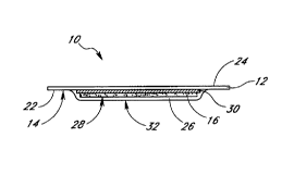
Referring to FIGS. 1 and 2, one embodiment of the dermal patch 10 includes a
first non-occlusive
film 12 with an attached adhesive layer 14 and an adsorption pad 16 made of an
adsorbent material that
is a porous layer of activated carbon 18 (i.e., activated charcoal)
immobilized in an inert matrix 20, such as
nylon, polyester, polyurethane) polytetrafluoroethylene (PTFE), polystyrene
and other known polymers.
Preferably the first non-occlusive film 12 is a very thin polyurethane (e.g.,
about 0.001 inch) film with an
acryiate adhesive applied to a first lower surface 22 of the film 12. The non-
occlusive film 12, when applied
to the skin using the adhesive 14 covers the skin but allows water content of
perspiration to evaporate,
exiting from a second upper surface 24 of the film. A preferred non-occlusive
film is a hypoallergenic) water-
resistant polyurethane film with an adhesive layer (e.g., as used in TEGADERM-
1625~ dressing; 3M Health
Care, St. Paul, MNI. Preferably the non-occlusive film 1 Z is attached to the
skin by the adhesive layer 14
for periods up to about eight to ten days without causing adverse
dermatological reactions. The polyurethane
film 12 is preferably 6 cm x 7 cm and 0.025 mm thick.
The dermal patch 10 may be removably attached to the subject's skin using the
adhesive layer 14
such that the adsorption pad 16 is located adjacent to the wearer's skin and
is held in place by the first
non-occlusive film 12. The dermal patch 10 also includes a second non-
occlusive film 26 covering a lower
surface 28 of the adsorption pad 16, such that the second non-occlusive fihn
26 separates the adsorption
pad from the wearer's skin when the patch 10 is placed on the subject's skin
and prevents liquid perspiration
from contacting the activated carbon of the adsorption pad because liquid
water in perspiration could
potentially desorb ethanol retained in the pad. The second non-occlusive film
26 also prevents discoloration
of the wearer's skin resulting from direct contact with the activated carbon 1
B of the adsorption pad 16.
Thus, the adsorption pad 16 is sandwiched between an upper first non-occlusive
film 12 and a lower second
non-occlusive film 26. When the patch is worn on a subject's skin, the first
film is exposed to the
environment and the second film is adjacent to the wearer's skin. Preferably,
both films are of the same
CA 02271462 1999-OS-11
WO 98I21578 PCTlUS97120486
7
non-occlusive polyurethane material.
Preferably, the adsorption pad 16 is a rectangle (3.18 cm x 4.76 cm X 1.0 mm)
with rounded corners
made of activated carbon 18 immobilized in an expanded polytetrafluoroethylene
(PTFE) matrix. The preferred
adsorption pad 16 has an area of about 14 cmz and contains about 540 mg of
activated carbon, with no
other chemicals or substances needed to retain ethanol. A preferred material
for the adsorption pad is
DARCO G-60° medium (3M Health Care) St. Paul, MN).
The dermal patch 10 may include indicia 29 to identify the patch. For example,
as shown in FtG.
1, a muhi-digit serial number or bar code may be printed underneath the first
film 12 so that the
identification indicia can be read through the film when the dermal patch is
applied to a wearers skin.
Alternatively, the indicia may be placed under the second film, to be visible
before the patch is applied or
after it is removed from the subject's skin. Such indicia are useful for chain-
of-custody identification of the
dermal patch.
Referring to FIG. 2, the dermal patch 10 may also include a release liner 30
to allow removal of
the adsorption pad 16 from the adhesive layer 14 following dermal patch use.
The release liner 30 shields
the adsorption pad 16 from the adhesive layer 14 and prevents the adsorption
pad from sticking to the
adhesive. Preferably, the release finer 30 is a very thin (0.003 mm) medical
grade cellulosic tissue (e.g.,
1-ply 17~ drape, James River Corporation, Gouvemeur, NY). The release liner 30
is preferably slightly larger
than the adsorption pad 16 (about 3 cm x 5 cml.
The lower adhesive side 32 of the second non-occlusive film 26 is proximate to
the wearer's skin
when the dermal patch 10 is adhered to a subject, thus adding to the total
adhesive surface area of the
patch. The second non-occlusive film 26 is adhered to the first non-occlusive
film 3 2 having an adhesive
layer 14 by adhesion between the two films in a portion of the border area 34
around the perimeter of the
pad 16 or by any of a variety of other methods (e.g., spot welding).
Preferably, the entire border area 34
around the perimeter of the adsorption pad is about 30 mm wide.
As shown in FIG. 2, the adsorption pad 16 is sandwiched between the two non-
occlusive films 12,
26. The second non-occlusive film 26 protects the adsorption pad 16 from
contact with liquid perspiration
that may appear under the dermal patch 10 during use. The absolute humidity
between the two non-
occlusive films 12, 26 is controlled in a relatively narrow range by the
moisture vapor transmission rates
(MYTR) of the two films, defined as the amount of water vapor passing through
a specified area of film in
a specified time (e.g., g~m~124 hr) under specified conditions. This property
improves the ability of the dermal
patch 10 to retain captured ethanol in the adsorption pad 16. The MUTR of the
first non-occlusive layer
12, the outer layer when the patch is worn, should be greater than or about
equal to the MVTR of the inner
second non-occlusive film 26 to keep the activated carbon of the adsorption
pad 16 sufticiently dry to
efficiently retain collected ethanol. If the MVTR of the outer first non-
occlusive film is significantly less than
the MYTR of the inner second nornocclusive film, the carbon of the adsorption
pad may become saturated
with perspiration water vapor which can condense to form liquid water that can
desorb ethanol from the
CA 02271462 1999-OS-11
WO 98!21578 PCT/US97/20486
8
adsorption pad thus creating an inaccurate measurement of the collected
perspiration ethanol. Thus, by
choosing two non-occlusive films that have appropriate permeability
characteristics, the dermal patch under
normal usage conditions (i.e., substantially at body temperature) adsorbs
vapor phase perspiration components
that pass through the patch, releases nonadsorbed components from the patch in
vapor phase at a rate
sufficient to prevent condensation of perspiration within the patch, and
prevents liquid components of
perspiration or from the environment from contacting the adsorptive material
within the patch. For example,
a patch made of a material having an MYTR of about 450 to 850 g(mZ124 hr,
preferably 640 to 8t0 glmzl24
hr, is envisioned, where the MIITR of the first outer non-occlusive layer 12
layer is greater than or about
equal to the MIITR of the inner second non-occlusive film.
FIG. 3 illustrates an embodiment of the dermal patch in an exploded view
showing the first non-
occlusive film 12, the adsorption pad 16, the second non-occlusive film 26 and
two outer protective liners
33. Outer protective liners 33 are protective barriers for use during storage
and handling of the dermal
patch. Preferably there are two outer protective liners 33, one liner covering
each non-occlusive film 12, 26
of the dermal patch. The protective liners may be made of paper, woven or
nonwoven fabric, plastic film
or other similar materials. The protective liners are removed before dermal
patch application. The outer
protective liners may be attached to each other by an adhesive layer (e.g.,
around the perimeter of each liner)
acrd may include tabs 35 for easily gripping the individual liners to aid in
their removal.
FIGS. 4 and 5 illustrate another embodiment of a dermal patch 10 with two
adsorption pads
arranged side-by-side in the patch. A first adsorption pad, the environmental
indicator pad 36, monitors
alcohol in the wearer's environment and serves as an internal measurement of
potential contamination of the
second pad, the collection pad 38, that collects the wearer's perspiration for
measurement of the wearer's
alcohol consumption. The two adsorption pads 36, 36 are bath directly under
the first non-occlusive film
12 with the adhesive layer 14 and arranged side-by-side so that a portion of
the perimeter 40 of the
environmental indicator pad 36 is proximate to a portion of the perimeter 42
of the collection pad 38. To
avoid perspiration from entering the environmental indicator pad 36, a first
separator layer 44 is located
adjacent to the bottom surface 46 and second separator layer 48 is located
substantially around the
perimeter 40 of the environmental indicator pad 36 to effectively separate the
pad 36 from the wearer's skin
during use and prevent fluid communication of perspiration into the
environmental indicator pad 36. The
second separator layer 48 also prevents cross-contamination between the
environmental indicator pad 36 and
the collection pad 38 because it is located between the adjacent perimeter
portions 40, 42 of the two pads.
The first and second separator layers may be integral, formed from a single
continuous layer or may be
separate but cooperating components with one component adjacent to the bottom
surface 46 of the
environmental indicator pad 36 and a second component essentially
perpendicular to the first component and
surrounding the perimeter 40 of the environmental indicator pad 36. Preferably
the separator layers 44, 48
are made of an occlusive material that covers only the environmental indicator
portion of the patch so that
the patch 10 remains substantially nornocclus'rve and allows vapor phase
perspiration to escape from the
CA 02271462 1999-OS-11
WO 98I21578 PCTII1S97J20486
9
patch. The separator layers 44, 48 may be made of any of a variety of well
known pliable occlusive
materials such as films of metal foil or polymers such as polyvinylidene
chloride, polyester) polyethylene
terephthalate, polyvinyl fluoride or polyvinyl chloride, polyolefin,
polyethylene, polypropylene, PTFE or
nitrocellulose. Suitable occlusive materials have an MVTR that is about 0.01 %
to about 1 °h that of the
nonocculsive film and are readily available (e.g., ALUREX CX°, St.
Regis Paper Co. or SARAN°) Dow
Chemical).
The patch 10 having both environmental indicator 36 and collection 38 pads is
applied and worn
as described for the single-pad patch. After the subject has worn the patch
for the required testing period,
the environmental indicator and collection pads are independently removed from
the patch as described for
the single-pad patch and analyzed independently for their ethanol content.
Because both pads have been
exposed to potential environmental contaminants, but only the collection pad
has been exposed to the
subject's perspiration, the ethanol concentration detected in the
environmental indicator pad measures
environmental contamination of the dermal patch and is a baseline measurement
for ethanol detection by the
collection pad. That is, the amount of ethanol detected in the environmental
indicator pad is subtracted from
that detected in the collection pad to determine the subject's ethanol
consumption during the wearing period.
If the collection pad contains significantly more ethanol than the
environmental indicator pad, then the
difference in the amounts of ethanol detected represents a measure of the
subject's ethanol consumption
during the wearing period. But if the amount of ethanol detected in the
environmental indicator pad equals
or exceeds the ethanol detected on the collection pad, then the subject's
perspiration did not contain any
detectable ethanol during the wearing period, meaning the subject did not
consume ethanol then.
FIG. fi shows another embodiment of the dermal patch 10 having two adsorption
pads, a first upper
environmental indicator pad 36 and a second lower collection pad 38 with
separator 44 between the two
pads. "Upper" and "lower" refer to the relative positions of the two pads when
the patch is attached to
a subject's skin. The separator 44 between the upper and lower pads 36, 38 is
preferably of the same non-
occlusive polyurethane film as used in the first non-occlusive film 12. The
environmental indicator pad 36
collects ethanol and other volatiles primarily from the external environment
and serves as a standard for
environmental contamination during the time the dermal patch 10 is worn. The
collection pad 38 collects
ethanol in perspiration and serves as a measure of alcohol consumption during
the wearing period. Because
of the efficiency of adsorption in the collection pad 38, ethanol in
perspiration is retained in the collection
pad 38 and does not substantially escape into the upper pad 36 despite the non-
occlusive nature of the
separator 44. Both the first and second adsorption pads 36, 38 are preferably
made of highly porous layer
of activated carbon 18 immobilized in an inert matrix 20, such as PTFE, nylon,
polyester, polyurethane,
polystyrene or other known polymers. The dermal patch includes a first non-
occlusive film 12, preferably
of very thin polyurethane with an attached acrylate adhesive layer 14 applied
to a first lower surface 22
of the fikn 12. This embodiment also has a second lower non-occlusive film 26
with an adhesive lower
surface 32 adjacent to the collection pad 38 such that the adhesive lower
surface 32 contacts the wearer's
CA 02271462 1999-OS-11
WO 98I21578 PCT/US97/2a486
skin when the patch is attached to a subject. The second lower non-occlusive
film 26 is preferably of the
same type of polyurethane as the first nomocclusive film 12. The second non-
occlusive film 26 protects the
collection pad 3B from liquid perspiration on the wearer's skin and its
adhesive lower surface 32 provides
additional adhesive surface for attachment of the dermal patch to the wearer's
skin. The er~bo~rr~rtt
5 includes a release liner 30 located between the environmental indicator pad
36 and the adjacent non-occlusive
film 12 to prevent the pad from sticking to the adhesive Payer 14 and aid in
removal of the environmental
indicator pad 36. Preferably, the release liner is made of thin (0.002 to
0.010 mm) medical grade cellulosic
tissue and is slightly larger than the dimensions of the adjacent
environmental indicator pad.
When the dual pad dermal patch shown in FIG. 6 is applied to a wearer's skin,
vapor phase
10 perspiration enters the patch through the second non-occlusive film 26 and
encounters the collection pad 38
where volatile ethanol and other analytes are adsorbed by the activated
charcoal. The water vapor and other
volatile non-adsorbed substances then pass through the non-occlusive separator
layer 44, the environmental
indicator pad 36, the release liner 30, and the first non-occlusive film 12 to
exit the patch to the
environment. The environmental indicator pad 36 primarily encounters
substances present in the wearer's
environment outside of the patch which enter the patch through the first non-
occlusive film and the release
liner 30. Any ethanol from perspiration that is not adsorbed by the collection
pad 38 may be adsorbed by
the environmental indicator pad 36 as the volatile substances pass from the
subject's skin through the patch.
This would result in a somewhat higher baseline for ethanol detected in the
environmental indicator pad.
For the patches with an environmental indicator pad 36 and a collection pad
38, such as illustrated
in FIGS. 4-6) the ethanol detected in the environmental indicator pad is
subtracted from that of the collection
pad to determine if the subject has consumed ethanol during the testing
period. That is, the amount of
collection pad ethanol minus the amount of environmental pad ethanol
correlates with the level of ethanol
consumption. For example, if the subject consumes one dose of ethanol
(equivalent to 1 glkg) the level of
ethanol detected in the collection pad is typically 50 nl. If the same subject
was not exposed to significant
levels of environmental alcohol, then the level detected in the environmental
indicator pad is typically less
than 10 nl and the difference of these two levels (50 nl - 10 nl - 40 nl)
suggests that the subject consumed
sufficient ethanol during the testing period to accumulate detectable ethanol
(40 nl) in the dermal patch. If
the subject is exposed to environmental ethanol and the level of ethanol
detected in the environmental
indicator patch substantially equals or exceeds that of the collection pad, it
suggests that the subject has
not consumed ethanol during the test period, although the subject may have
been exposed to some
environmental volatile ethanol such as present in cosmetics or household
cleaners. For example, if the
collection pad contains 15 nl of ethanol and the environmental indicator pad
contains 14 nl of ethanol, then
the difference is negligible (15 nl - 14 nl = 1 nl), suggesting the subject
did not consume ethanol during the
testing period. Similarly, a negative difference between the ethanol detected
in the collection pad and the
environmental indicator pad suggests that the subject did not consume ethanol
during the testing period but
was exposed to some environmental volatile ethanol. For example, if the
collection pad contains 10 nl of
CA 02271462 1999-OS-11
WO 98l21578 PCT/US97/20486
11
ethanol and the environmental indicator pad contains 20 nl of ethanol, then
the negative difference (10 nl -
20 nl - - 7 0 nl) suggests that the subject did not consume ethanol during the
testing period. If the amount
of ethanol in the environmental indicator pad (e.g., 1.000 nl) greatly exceeds
that of the collection pad (50
nll but the amount of ethanol in the collection would otherwise suggest
consumption, then the difference (50
nl - 1,000 nl - - 950 nl) formally suggests that the subject did not consume
ethanol during the testing
period, but also suggests considerable exposure of the patch to environmental
alcohol, possibly due to patch
tampering.
The measurements made using by the dermal patch as illustrated in FIGS. 4 and
5, for the side-by-
side collection and environmental indicator pads may differ somewhat from
those made using the dermal
patch as illustrated in FIG. 6, where the environmental indicator pad is
located above the collection pad when
the patch is worn by a subject. That is, the side-by-side arrangement allows
use of an occlusive separator
layer separating the environmental indicator pad from the skin and from the
collection pad while maintaining
the substantially nonocciusive nature of the dermal patch. In contrast, the
embodiment shown in FIG. 6 is
completely nonocclusive and relies on the collection pad to collect all of the
ethanol from the perspiration
1 S before water vapor from perspiration enters the environmental indicator
pad, and the environmental pad to
collect all of the environmental ethanol before it can cross the nonocclusive
separator layer and enter the
collection pad. Thus, because a nonocclusive separator layer separates the two
pads in the embodiment as
shown in FIG. 6, there is the possibility that some ethanol from one pad may
leak into the other pad.
The dermal patch may also be provided with a pouch (not shown) that is
preferably a foil-lined
vapor-barrier envelope to surround the dermal patch and protective liners (if
includedl. The pouch is useful
for storing one or more dermal patches and preventing patch exposure to
environmental volatiles before
application. A dermal patch may be sterilized in the pouch such as by gamma
irradiation and the pouch may
include the sterilization date, although the dermal patch itself has no known
expiration period. The pouch
may be resealable and can be used to store and protect a dermal patch after it
has been removed from a
wearer and during shipping to an analytical laboratory at ambient temperature
(e.g. by surface mail?.
Because the preferred polyurethane film of the dermal patch is gas permeable,
volatile water and
ethanol molecules from the skin can cross the film. When perspiration
containing volatile analytes including
ethanol crosses the second non-occlusive film into the adsorption pad, the
activated carbon adsorbs the vapor
phase analytes while the water vapor passes through the adsorptive pad and
escapes through the first outer
non-occlusive film to the environment. Body heat volatilizes the water in
perspiration driving it through the
dermal patch. Larger non-volatile molecules (e.g., liquid water) cannot pass
through the second inner non-
occlusive film and remain trapped against the skin. Liquid water cannot cross
the gas permeable film from
either the skin or the environment to reach the adsorption pad and vapor phase
water in perspiration escapes
to the environment, eliminating many disadvantages of an occlusive patch.
Moreover, because the dermal
patch of the present invention is non-occlusive, equilibrium between the
dermal patch and the skin is never
reached and the analytes including ethanol accumulate in the adsorption pad
during the entire wearing period.
CA 02271462 1999-OS-11
WO 98I21578 PCT/US97120486
12
At the end of the monitoring period, the dermal patch is removed and the
adsorption pad is separated from
the dermal patch. The adsorption pad contents are eluted into an aqueous
buffer and analyzed.
The dermal patch collects insensible and sensible perspiration from the skin
covered by the
adsorption pad. Total insensible perspiration from a 1.75 mz body surface has
been measured at 381 ) 526
and 695 mltday at 22°C, 27°C and 30°C, respectively,
whereas sensible perspiration varies with an
individual's response to thermal, physical or emotional stress iEamke, L.O.,
Scand J. CI~~, lab. Invest.
37:325, 1977). Insensible perspiration for an individual experiencing minimal
thermal, physical and emotional
stress is the minimum amount of perspiration that the dermal patch processes
during a monitoring period.
Thus, using the measurements indicated above, a dermal patch with an
adsorption pad of about 14 cmz
processes a minimum of 300 ~I of perspiration per day at 22°C average
temperature. The subject's alcohol
consumption during the monitoring period determines the ethanol content in
insensible perspiration.
Use of the Patch for Detective Ethanol Consumption
The dermal patch is applied to and removed from the subject's skin, preferably
by a trained
technician who chooses a suitable location on the subjects body: avoiding
areas of excessive body hair,
lesions, abrasions, wounds, scars or dermatological irritations. The dermal
patch is preferably attached on
either upper arm, the back or tower chest because these skin areas have about
the same level of
permeability. It will be appreciated that other portions of the body are also
appropriate to attaching the
derma! patch, such as, for example, the leg, ankle, top or sole of the foot,
top or palm of the hand, forearm,
neck, upper chest and buttocks, depending on the condition of the skin and
other considerations such as
privacy concerns. The skin of the selected site is cleaned with an agent to
remove surface contaminants
(e.g., a standard alcohol wipe containing 70~ isopropanol) and the dermal
patch is placed on the dry cleaned
skin, contacting the adsorption pad and adhesive layer to the subject's skin
and then gently pressing the
dermal patch to the skin and pressing the adhesive layer gently around the
border area of the patch. The
adhesive layer becomes translucent with no air bubbles remaining between the
adhesive layer and the skin
when adhesion is complete. Moreover, complete adhesion produces a subtle
puckering of the polyurethane
fgm and the underlying skin, producing a slightly rippled or wavy appearance
on the surface of the applied
patch. The person applying the dermal patch may also record information such
as the application date, the
application location) the dermal patch condition and identification number or
similar information.
The subject wears the dermal patch for a monitoring period of about 1 hr to
several days.
Generally, the subject wears the dermal patch for at least 24 hr and up to ten
days. The subject is
instructed not to rub the dermal patch (e.g., with a towel after washing, but
otherwise, no other precautions
must be followed while wearing a dermal patch. At the end of the monitoring
period, the technician removes
the dermal patch.
Before removing the dermal patch, the technician determines visually whether
the dermal patch has
been compromised during wear by detecting a change in patch appearance
generally associated with patch
removal or tampering. Because the preferred adhesive layer can be applied
securely to a subject's skin only
CA 02271462 1999-OS-11
WO 98I21578 PCTIL1S97/20486
13
once, removal and reapplication to skin is readily detected by the patch's
degree of adhesion and
transparency. Exfoliated stratum corneum cells stick to the preferred adhesive
during patch removal making
secure adhesive reattachment impossible. Moreover, the retained skin cells
produce a cloudy nontransiucent
appearance to the polyurethane layer with adhesive lower surface at the
patch's border. Similarly, rips or
punctures to the patch, portions of cloudy appearance in the patch's perimeter
border area, discoloration of
the patch or surrounding skin, or inflammation around or under the patch are
readily detectable evidence of
tampering. If removal or tampering is detected, the technician records that
information for use in interpreting
the analysis results.
During removal, the technician avoids contamination of the adsorption pad by
using gloved hands or tweezers to remove the pad and place 'rt into a clean
container that is then sealed.
The technician removes the patch by gently prying under an edge of the patch
to loosen a portion of the
adhesive layer from the subject's skin and lifting the patch from the
subject's skin to a clean surface where
the pad is removed. Alternatively, the dermal patch may be partially removed
by loosening a portion of the
adhesive and peeling a portion of the patch away from the skin to expose the
adsorption pad which remains
partially attached to the subject's skin. The adsorption pad is then removed
from between the subject's skin
and the outer portion of the patch and placed into a clean container as above.
After removal of the dermal patch, the trained person further inspects the
removed patch and skin
for evidence of prior removal or tampering of the dermal patch. Such evidence
includes detection of
punctures, rips or tears in the patch or adsorption pad (e.g., detected by
holding the patch to a light sourceh
discoloration of the patch or underlying or surrounding skin, and changes in
the patch identification number
or other indicia. Any changes are recorded and the patch is discarded.
The removed adsorption pad is used for detection of an analyte such as ethanol
retained in the
activated carbon. Analysis may be done immediately after removing the pad from
the dermal patch or the
pad can be stored andlor transported to another facility for analysis after
the pad has been sealed into a
suitable container. Storage and transport of the pad before ethanol analysis
may be at ambient temperature
and does not require special conditions because ethanol in the adsorption pad
is stable at temperatures up
to 60°C for 3 days and at ambient temperature for at least one month.
Once ethanol is adsorbed onto the activated carbon of the adsorption pad, it
remains there until it
is desorbed by displacing it with another compound such as liquid water, an
aqueous solution or an organic
solvent that has a higher chemical potential for binding with the carbon. The
extracted ethanol can be
analyzed by well known methods such as) for example, enzymatic assays,
immunoassays, gas
chromatography, chemical oxidation and photometry, electrochemical oxidation
with fuel cells, infrared
spectrometry or solid~state semiconductor sensing.
Although collection and detection of ethanol expressed the skin is the most
direct means of
detecting ethanol consumption by an individual during the period the dermal
patch is wom, the dermal patch
of the present invention can also be used to collect and detect a metabolite
of ethanol that is expressed
CA 02271462 1999-OS-11
WO 98I21578 PCTIUS97/20486
14
through the skin in perspiration, such as acetaldehyde. Acetaldehyde is also
adsorbed by activated charcoal
in the dermal patch and can be similarly extracted and detected using
conventional methods. The level of
acetaldehyde produced from consumed alcohol and expressed through the skin is
less than that of ethanol
expressed through the skin, but is still within detectable levels. Moreover,
detection of an ethanol metabolite
such as acetaldehyde provides a measure of alcohol consumption even when the
individual has been exposed
to an amount of environmental ethanol sufficient to negate or interfere with
accurate detection of ethanol
expressed through the skin in perspiration. That is, a positive detection of
acetaldehyde and ethanol in the
adsorption material of the dermal patch would suggest alcohol consumption,
whereas detection of ethanol
without acetaldehyde would suggest environmental exposure to ethanol.
The invention can be better understood by way of the following examples which
are representative
of preferred embodiments. All volunteers who participated in the clinical
investigations provided informed
consent. Efficacy of the dermal patch was demonstrated in clinical trials
using dermal patches such as
illustrated in FIGS. 1-3. These studies included controlled administration of
ethanol to volunteers wearing
dermal patches and negative control studies of dermal patches worn by non-
drinking subjects. Based on
1 S these studies, the relationship between a specific ethanol dose and the
amount of ethanol collected by a
worn dermal patch and detected using conventional methods was determined.
These studies show that the
dermal patch may be used to monitor human subjects for exposure to ethanol,
with sensitivity sufficient to
collect, retain and detect ethanol from as little as a single ethanol dose.
EXAMPLE 1: Dermal Patch Ahnfication and Removal
A technician wearing sterile gloves cleaned application sites on the subjects'
upper arms, back and
lower chest with 70~ isopropanol alcohol swabs and applied dermal patches as
described above. Subjects
stretched their skin slightly just before and during dermal patch application
by flexing the biceps Ifor arm
application), bending forward (for back application), and bending backward
(for lower chest application) to
prevent dermal patch from putting tension on the relaxed skin. The technician
examined the dermal patch
after it was applied. The peripheral adhesive border area of the patch became
uniformly translucent when
the patch was properly applied (the adsorption pad remained opaque) and the
patch had a slightly rippled or
wavy appearance on its surface. The technician replaced the dermal patch if it
did not appear to be properly
and completely adhered to the subject's skin, e.g., it had raised edges or
channels in the adhesive perimeter
area where contact was interrupted or incomplete. Close~up photographs of
dermal patches were taken just
after application and before removal of each dermal patch to assess and record
dermal patch integrity;
specimen codes, dates and other pertinent information were recorded with the
photograph.
After application, the subjects were instructed to wear the patch for a
designated time period
without disturbing it. Otherwise, subjects performed their normal activities
including swimming, manual labor
and daily exercise. After the designated time period, subjects returned to the
test facility for patch removal.
The technician examined the dermal patch before removal to verify that the
dermal patch was securely
affixed throughout the wearing period and remained free of holes or tears.
That is, the adhesive perimeter
CA 02271462 1999-OS-11
WO 98l21578 PCTIUS97/20486
portion of the patch retained its translucent appearance with no obvious
channels in the adherent film
spanning the area between the adsorption pad and the outer perkneter edge of
the dermal patch. The
technician noted the degree of force needed to remove the dermal patch because
a secured dermal patch
generally required some effort to remove it from the skin, whereas a partially
or completely removed patch
5 was removed with little effort.
The technician wore sterile gloves to remove the dermal patch. As the derma!
patch was removed,
the adsorption pad was separated from the first and second polyurethane film
layers and placed in a clean
container which was stoppered and crimp capped. The remaining parts of the
dermal patch were removed
from the subject and discarded. After patch removal, the technician further
observed and recorded the
10 appearance of the skin that had been under the dermal patch. In a properly
adhered patch, the adherent film
compressed the skin slightly, forming ridges about 2-3 mm apart that were
retained temporarily after patch
removal. In contrast, if the adherent film had been removed or disturbed
earlier, the skin ridges were absent.
Dermal patches that exhibited any characteristics of removal or nonadherence
during the use period
15 or otherwise abnormal appearance or tactile characteristics when removed
were noted and retained. The
technician appropriately noted anomalous conditions detected during patch
removal before sending the pads
far analysis, excluding those that had failed to adhere properly. Fewer than
2% of 1,342 dermal patches
used in the investigations were excluded because of adherence failure. Only
six of 90 patches worn for four
to 14 days by subjects who perspired profusely during exercise during the
wearing period showed incomplete
adherence when the patches were removed. Adsorption pads were stored at -
15°C to about 20°C during
transport to a laboratory for extraction and analysis.
El(AMPLE 2: Adsorption Pad Extraction and Analysis
To avoid potential problems of analyte nonuniformity on the pad, the entire
adsorption pad was
extracted. Before pad extraction, vials containing the pads were allowed to
warm to room temperature.
The adsorption pad was removed and placed in a 20 mi glass vial (e.g., a
scintillation viall. 2 ml of deionized
water was added, the vial was capped and the pad was vortex mixed with the
water for 20 seconds. After
mixing, the pad and water incubated at room temperature for 4 ht with
occasional mixing to form an extract.
A 4 hr extraction was considered optimal for speed and aqueous recovery based
on results from extractions
for 5 min i67% of maximal recoveryh 4 hr (88%1, and 48 hr (100%); heat or
solvent extraction may increase
recovery but require additional equipment and reagents. After the 4 hr
extraction, 50 ,ul of extract was
transferred into a labeled conical polypropylene vial, 15,u1 of a 0.005%
propionitrile internal standard (Aldrich
Chemical Co.. Milwaukee, WA was added, and the vial was crimp capped and
vortex mixed.
A 1 ,ul sample of the mixture was analyzed by gas chromatography (GCI using
flame-ionization
detection (FID) to detect the presence and amount of ethanol with a Hew~tt-
Packard (HPI 5790A gas
chromatometer with FID, an HP 3390A Integrator, and an HP 7673A Auto Sampler.
The GC column (6 ft
x 1l8 in) was packed with 80t100 Carbopack C with 0.2% Carbowax 1500 (Supelco,
Bellefonte, PA); the
CA 02271462 1999-OS-11
WO 98I21578 PCT/US97I20486
16
carrier gas was He 130 mllmin). H I45 mllmin) and air (240 mllmiy; the oven
temperature was 90-100°C
isothermal; the injector temperature was 359°C; and the detector was
300°C. Under these conditions, the
retention times were: methanol, 0.48 min; ethanol. 0.73 min; acetone, Q.97
min; isopropanol 1.19 min, n-
propionitrile (internal standard). 1.30 min; and n-propanol) 1.54 min. A
ethanol calibrator (Clinical Standard
S Solution, College of American Pathologists, Northfield) ILh equivalent to 50
nl ethanolldermal patch, was used
as a standard and control samples from pads containing no ethanol or 30, 50,
100 or 200 nllpad of ethanol
were included. Ethanol was quantitated by comparing the ratio of the ethanol
peak height to the propionitrile
peak height, utilizing the calibration curve for known ethanol concentrations.
EXAMPLE 3: Controlled Sinnle Dose Studv
lfolunteers received dosages of ethanol in the study and were instructed to
avoid contact with
ethanol-containing drinks, foods, cosmetics and other substances containing
ethanol during the test period.
As independent measures of ethanol consumption, breath ethanol was measured
using an AIcoSensor III
(Intoximeters, Inc., St. Louis, M01 and saliva ethanol was measured with the
QED A150'" test (STC
Diagnostics, Bethlehem PA) whenever a dermal patch was removed.
On the day before the ethanol dose, fifteen subjects each received fourteen
dermal patches applied
as follows: three on left back, two on right back, two on left chest, three on
right chest and two on each
upper arm. A single dose of ethanol (contained in 40% Vodka diluted with fruit
juice or equivalent) was 1.0
glkg body weight for male subjects, and 0.85 glkg body weight for female
subjects. Subjects fasted
overnight (at least 12 hr) before receiving the ethanol dose and then were
given a low fat breakfast (cereal,
low fat milk and a banana). The ethanol dose, administered 30 min after the
breakfast, was consumed over
a 30 min period.
One dermal patch was removed from the left back before the ethanol dose was
consumed (pre-dose).
Then the dose was administered and the time clock was started after the
subject had consumed the dose.
At 1, 2.5, and 5.5 hr after the ethanol dose (post-dose), dermal patches were
removed from the right chest,
left chest, and left back respectively. Dermal patches were removed over the
ensuing week as follows: on
day 1 (24 hr post-dose), one each from the back, arm and chest; on day 3 (72
hr post-dose), one each from
the back, arm and chest; on day 5 (120 hr post-dose) one from the arm; and on
day 7 (168 hr post-dose)
one each from the back, arm and chest. A total of 3D0 patches were used: 210
pre-dose and 90 post-dose.
On day 1 post-dose, two fresh dermal patches were applied to the chest and
back. These two
dermal patches were removed on day 3 and two fresh dermal patches were applied
in their place. On day
5 post-dose, the two dermal patches added on day 3 were removed, and replaced
with two fresh dermal
patches. On day 7, the added dermal patches from day 5 were removed. These six
additional dermal
patches were used to monitor the subjects' contact with ethanol following the
control-administered dose.
Breath alcohol levels were highest at 1 hr post-dose (mean: 0.092%! and
dropped by 5.5 hr post-
dose (mean: 0.027%) and remained undetectable thereafter. Similarly, saliva
alcohol levels were highest at
portion of the patch reta
CA 02271462 1999-OS-11
WO 98/21578 PCT/L1S97120486
17
1 hr post-dose (mean: 0.104%) and dropped by 5.5 hr post-dose (mean: 0.035%)
and remained undetectable
thereafter.
Mean levels of ethanol collected and detected on the dermal patches were: 3 nl
for time 0, about
14.3 nl at 1 hr post-dose; about 27.3 nl at 2 hr post-dose; 56 nl at 5.5 hr
post-dose; 52 nl at 24 hr post-
s dose; 49 nl at 72 hr post-dose; 59 nl at 120 hr post-dose; and 63 nl at 168
hr post-dose. Median levels
were 39 nl at Z4 hr post-dose; 45 nl at 72 hr post-dose; 68 nl at 120 hr post-
dose; and 73 nl at 168 hr
post-dose. Three sets of six additional post-dose dermal patches used to
monitor the subjects' contact with
ethanol following the control-administered dose all had mean and median levels
of less than 15 nllpatch
suggesting that the subjects avoided ethanol exposure. These results show that
ethanol adsorption by the
adsorption pad was delayed relative to breath and saliva ethanol and that the
pads retained ethanol as long
as seven days after the single dose, whereas the information on ethanol
consumption was lost for breath
and saliva measurements by one day post-dose.
EXAMPLE 4: Cumulative low Dose Study
On the day before the ethanol dose was given, eight subjects each received
thirteen dermal patches
applied as follows: two on the left back) two on the right back, two on the
left chest, three on the right
chest and two dermal patches on each upper arm (totaling 104 patches). One
dermal patch was removed
from the right chest before the ethanol dose was administered (pre-dose).
The ethanol dose was equivalent to 0.5 glkg body weight, contained in a volume
of commercially
available beer (i.e., 24 oz155 kg body weight), with no dosage difference
dependent on gender. Subjects
consumed the beer over a 30 minute period on an empty stomach at the same time
of day for seven
consecutive days beginning on day 0. The beer was obtained from a single keg,
so a consistent alcohol
concentration (5.0% by vol) was assured for al! subjects over the test period.
Breath and salvia ethanol were
measured at 30 min post-dose. On days 1, 3, 5 and 7, one patch each from the
back, chest, and arm were
removed.
The breath ethanol levels of the eight subjects varied between less than
0.0375% to greater than
d.0525%) with significant variation between individuals and between different
test samples for a single
individual. As shown in F16. 7, the mean ethanol content of adsorption pads
from patches for the eight
individuals increased relatively steadily over time. For example, the mean pad
ethanol content at day 3 (three
doses totaling 1.5 glkg) was 45 nl. By day 5 (5 doses totaling 2.5 gfkg), 75%
of the pads (18 patches)
showed a cumulative ethanol content over 50 nl. These results show that the
absorbent pads can
accumulate ethanol over time, providing a measurement of the total ethanol
consumed over a period of up
to seven days.
EXAMPLE 5: Dose Response Studv
On the day before ethanol administration, four subjects received six dermal
patches (two each on
the back, chest and arml; each subject wore a control set of dermal patches
for 24 hr pre-dose. The ethanol
dosages were given in increasing amounts equivalent to 0.17, 0.33, 0.5, 0.67,
0.83 and 1.0 glkg body
CA 02271462 1999-OS-11
WO 98l21578 PCT/US97/20486
18
weight) administered as in Example 3, with doses given 24 hr apart for the
first three, and 48 hr apart for
the last three. Breath and saliva ethanol measurements were taken at 30 min
post-dose. At 24 hr after
each dose, but before the next dose. the six dermal patches were removed, and
replaced by six fresh dermal
patches. Thus, for each dose, six dermal patches were applied and a total of
168 pads were analyzed.
FIG. 8 shows the relationship of breath ethanol measurements to the ethanol
dose for each of the
four subjects. The data points show significant variation between the breath
ethanol detected and the dose
administered, with correlation coefficients (R2) for the data points and the
lines varying from 0.69 to 0.97
(0.94, 0.92, 0.97 and 0.69 for subjects A, B. E and G, respectively). Similar
results were obtained for the
saliva ethanol tests (data not shown. F1G. 9 shows a similar plot for the
ethanol content (mean of six
patches, two each from the arm, back and chest for each subject) of the
absorbent pads for the same four
subjects compared to the ethanol dose, with correlation coefficients (RZ)
varying from 0.87 to 0.97 (0.97,
0.B7, 0.95 and 0.93 for the data points and lines for subjects ~k, B, E and G,
respectively). These results
show the difficulty of quantitating ethanol consumption based on breath
ethanol, saliva ethanol or ethanol
adsorbed by a dermal patch.
EXAMPLE 6: Nenative Control Studv
Twenty-one subjects participated. Eighteen wore eight dermal patches for seven
days, applied as
follows: four on the arms, two on the back, and two on the chest. The other
three subjects wore four
patches on the upper arms for 3 days. No ethanol was administered and subjects
were asked to abstain
ftom alcohol consumption for 24 hr before dermal patch application and while
the dermal patches were worn,
and to avoid using products containing ethanol or SD alcohol during that time.
Breath and saliva ethanol
measurements were taken on day 0 as the dermal patches were applied and when
the dermal patches were
removed (day 3 or day 7). Absorbent pads were assayed for ethanol content
using GC with FID as described
above.
Of 146 patches analyzed (10 were compromised during wearing and not analyzed),
the mean alcohol
content was 26 t 12 nl, and only four patches had over 50 nl. These resuhs
probably reflect accumulation
over a week of endogenous ethanol normally produced in the body and
inadvertent exposure to ethanol in
cosmetics or household chemicals. When the median ethanol content measurements
were compared for the
different body locations of the patches, they were indistinguishable (at a 95%
confidence level; p < 0.05).
While there was some variation between patches obtained from an individual
subject, most subjects' patches
showed similar ethanol levels independent of the patch location.
EXAMPLE 7: Dermal Patch Placement Studv
Three subjects each received eighteen dermal patches applied on day 0 as
follows: three each on
the right and left arms, right and left back, and right and left chest (54
total). Subjects were given a single
1.0 glkg ethanol dose as in Example 3 and patches were removed on day 3 post-
dose and absorbent pads
analyzed to ethanol content by GC as described above. Breath and saliva
ethanol measurements were taken
pre-dose, 30 minutes post-dose, and at day 3 during dermal patch removal.
CA 02271462 1999-OS-11
WO 98/21578 PCTIUS97I20486
19
The ethanol content (mean and median) of the pads from the six different
locations did not differ
tat a 95% confidence level; p < 0.05), although for a single location the
largest range was seen for the
right arm and the smallest range variation was seen for the left back.
Moreover, there was a high
correlation ( >- 0.84, generally >_ 0.90) between the ethanol dose detected
and all of the patch locations.
Thus, all of the application locations are equivalent for collecting and
retaining ethanol in the patch.
EXAMPLE 8: Kinetic Studv: Seauential Accumulation Over a 48 hr Period
On the day before ethanol dosing, four subjects each received three dermal
patches applied as
follows: one each to the chest, back and arm. During this study, subjects
received additional sets of patches
just before the dose Rime O) and at 1, 2, 4, B, 12, 24, 48 and 72 hr post-dose
(a total of 27 patches per
subject and 108 for the study). Dosing was as described in Example 3. Before
the ethanol dose was
administered, the three dermal patches were removed, breath and saliva ethanol
levels were measured, and
three fresh dermal patches were applied in the same locations. At 1 hr post-
dose, breath and saliva
measurements were made, dermal patches were removed and fresh dermal patches
were applied. This
procedure was repeated at 2, 4, 8, 12, 24, and 48 hr post-dose. The ethanol
collected on the dermal
patches in this phase represents the ethanol accumulation over the interval of
dermal patch wear.
The mean breath and saliva ethanol levels were 0.094% and 0.103°Yo;
respectively, a 1 hr post-dose.
At 2 hr post-dose, the mean breath ethanol decreased to 0.0909'o and the mean
saliva ethanol level increased
to 0.119%, but by 4 hr post-dose both breath and saliva ethanol levels were
about half of their highest
values and were undetectable by 8 hr post-dose. Ethanol detected in the
adsorption pads measured (mean
values): 8 nl at 1 hr post-dose, 10 nl at 2 hr post-dose, 23 nl at 4 hr post-
dose) 22 nl at 8 hr post-dose,
and undetectable thereafter. After B hr post-dose, no ethanol above baseline
was detected in the absorbent
pads. Thus, the dermal patch can be used detect ethanol in approximately the
same time range as detected
by breath and saliva testing (1 to 4 hr post-dose), but can also be used
detect ethanol between 4 hr and
B hr post-dose, when no ethanol was detected by breath and saliva testing.
Although there is some delay
in detection of ethanol in perspiration compared to breath and saliva testing,
ethanol can be collected from
perspiration up to 48 hr post-dose, when breath and saliva would no longer be
detectable) and the patch of
the present invention can retain the perspiration ethanol for up to 8 days
post-dose providing a much longer
period for detection of ethanol consumption that possible with breath and
saliva testing.
FIG. 10 shows a typical kinetic relationship of ethanol in blood, urine,
saliva and collected from
perspiration using the dermal patch. The Y-axis presents the ethanol content
of the dermal patch (nl); blood,
saliva and urine (mgldll and breath (%) for a subject who received 1 gfkg of
ethanol at time 0. The blood
ethanol was measured up to 5.5 hr post-dose and interpolated from there based
on published ethanol
metabolism rates (Jones A.W. et al., Clin. Chem. 38(5I:743, 1992; Sidell, F.R.
& Press, J.E.,
Psychopharmacol. 19:246, 1971 ); urine content was based on the blood ethanol
levels and the published
relationship between blood and urine ethanol levels (Biasotti, A.A. &
Valentine, T.E., J. for. Sci. 30(11:194,
1985). FIG. 10 shows that blood, saliva and breath ethanol levels rise more
quickly relative io that collected
CA 02271462 1999-OS-11
WO 98l21578 2 a PCT/US97l2048b
in the dermal patch and then drop after about 2 hr post-dose, returning to
baseline by 24 hr post-dose. In
contrast, ethanol collected in the dermal patch is cumulative) peaking at
about 9 to 12 hr post-dose, and
retained until the patch is removed.
Based on the clinical studies, the best ethanol concentration for the GC
cutoff calibrator, the
sensitivity and specificity of GC for possible cutoffs (10, 20, 30, 35, 40,
50, 60 and 70 nllpad) were
calculated by analysis of the number of true positives, false positives, false
negatives and true negatives for
791 patches from 35 subjects. The optimum cutoff concentration, 30 nllpad,
resulted in the fewest false
positives and false negatives, thus giving the highest sensitivity and
specificity for the assay.
The limit of quantitation (L00) is the lowest concentration of ethanol that
can be reliably
quant'rtated by GC for known ethanol concentrations in dermal patch extracts.
The LOO was 16 nllpatch
based on seven of ten assays in which more than 50% of the samples were within
t 50% of the known
concentration of ethanol concentration for the sample. Clinical and diagnostic
sensitivity and specificity were
determined using standard procedures ("Proposed Guideline: Assessment of
clinical sensitivity and specificity
of laboratory tests" NCCLS Document GP-10) Ilol. 7, No. 6, 1987; Galen, R.S. &
Gambino, S.R., "Beyond
Normality: The predictive value and efficiency of medical diagnoses," Whey
Biomedical Publications, 1975
(John Wiley & Son. New York, NY); and Spiehler, U.R. et al., Clin. Chem.
33:1535, 1988). Using a GC cutoff
of 30 nllpatch, the diagnostic sensitivity was 90.38% (determined from 364
dermal patch results after 24
hr minimal wear from subjects administered known ethanol doses) and the
diagnostic specificity of 85.95%
(determined from 427 dermal patch results from subjects who received no
ethanol dose and said they had
not consumed alcohol during the wear period). The clinical sensitivity and
specificity were determined from
dermal patch results in controlled studies using known doses (0.85 glkg for
females, 1.0 glkg for males),
producing a positive predictive value for ethanol consumption of 86.5% and a
negative predictive value for
ethanol abstinence of 90.9%.
EXAMPLE 9: Dermal Patch Analysis After Immersion in Liquids
immersion studies were used to determine whether ethanol would leave the
dermal patch during
wear it the dermal patch were soaked in water at relatively high temperatures
(40 t 1 °C in liquid). Blank
derma! patches were spiked with about 100 nl of ethanol and then attached to
petri dishes to simulate a
dermal patch attached to skin. A control set of three patches was stored dry
at 40 t 1 °C and tire test
sets of three patches each were immersed in a water bath at 40 t 1 °C
containing ordinary tap water with
or without 5 PPM chlorine, 0.9% NaCI (common salt water) or 0.18% bubble bath
soap. For comparison,
swimming pool water containing 3 PPM chlorine has a strong chlorine smell and
irritates eyes; and 0.18°Yo
bubble bath soap is equivalent to adding an 8 ounce bottle of bubble bath soap
to a forty gallon bathtub of
water. At the end of 3 fir and 24 hr, the control patches and test patches
were dismantled and the
adsorption pad recovered and assayed for ethanol using the standard procedures
essentially as described
above. The results of these tests) shown in Table 1, show that ethanol in the
patches was recoverable even
after exposure to high temperatures.
CA 02271462 1999-OS-11
WO 98I21578 21 PCT/US97I20486
Table 1: Mean and standard deviation of recovered ethanol (nllpatch)
Conditions 3 hr Exposure 24 hr Exposure
Control (dry) 109 15 104 t 10
Tap water 101 t 14 105 t 22
5 PPM Chlorine solution107 t 12 108 t 6
NaCI solution 116 t 3 103 t 24
Soap solution 123 t 13 147 t 7
Immersion in water, chlorine solution and salt solution did not significantly
affect the detectable ethanol
in the pads showing that the outer polyurethane layer of the dermal patch
effectively isolates the adsorption
pad from the environment. immersion for hours in bubble bath soap solution
increased the ethanol
concentration probably because the bubble bath soap used contained ethanol
(2146 mg96) which was
adsorbed across the outer polyurethane film. Avoidance of ethanohcontaining
household products during
dermal patch use would prevent such uptake of ethanol from the environment.
Alternatively, a dermal patch
with two adsorption pads as illustrated in FIGS. 4-6 and discussed above may
be used to monitor
environmental ethanol exposure and serve as an internal control (baseline) for
the perspiration collection pad.
EXAMPLE 10: Microorganism Studies
These studies were conducted to (1 ) confirm that the adsorption pad encased
in the first and
second polyurethane layers of a sterilized dermal patch remains sterile during
seven days of dermal patch
wear; (2) identify and quantitate the specific microorganisms recovered from
underneath the dermal patch
worn for seven days; and (3) evaluate the effects of the identified
microorganisms on ethanol contained on
the adsorption pad. The ability of an identified microorganism to produce or
metabolize ethanol in a dermal
patch environment was based on known biosynthetic and catabolic
characteristics of the identified
microorganisms.
Standard arm skin swabs from eight subjects were taken in the approximate
location and area size
where the adsorption pad would contact the skin. Using a template to limit the
area, a swab specimen of
the skin was collected. The same arm location was then prepared for dermal
patch wear by following the
standard isopropanol skin prep protocol. After the isopropano) evaporated, a
second skin swab specimen was
taken from the treated area, again using the template. Two standard alcohol
dermal patches were then
applied, one directly over the prepared swab zone, and the second next to it.
The patches were worn for
seven days during which the subjects were instructed to avoid contact with
ethanol. At the end of the
seven days, the first dermal patch was removed) and the skin under the
adsorption pad was swabbed as
described above. The second dermal patch was removed) and the adsorption pads
were isolated using aseptic
procedures. Swab samples from the day of dermal patch application, from the
day of patch removal) and
the worn adsorption pads were extracted with growth media and the extract was
cultured on nutritional agar
plates (3 days, 37°C) to promote growth of aerobic microorganisms using
standard microbiology procedures.
CA 02271462 1999-OS-11
WO 98/21578 2 2 PCTIUS97/20486
The types and numbers of colony forming units (CFU) of microorganisms
(staphylococci, micrococci,
diphtheroids and bacilli) were determined using standard microbiological
techniques.
The pads generally remained sterile during the test period. No fungi or other
yeast (i.e., < 5
CFUlsample) were detected in any sample. Only one of the eight subjects had
consistently high CFU on the
skin and under the dermal patch (25-40 x 1 O' CFUlsample, identified as
Staphylococcus epidermis). Other
samples showed a mixture of significantly lower numbers (about < 10Z
CFUlsample) of gram positive cocci
and rods, consistent with the types of normal microbial flora associated with
skin. The isopropanol
disinfection reduced the number of CFU recovered about 10-fold lower. Based on
the types and numbers
of microorganisms recovered from patches applied to the isopropanol
disinfected areas, it is extremely unlikely
that organisms recovered from the skin under the patch or the pad itself would
affect the alcohol
concentration detected from an adsorption pad recovered from a dermal patch.
These results show that the nonocclusive patch does not increase bacterial
growth or affect the
microflora type during the wearing period. That is, for skin that was not
isopropanol disinfected before patch
application, the number and type of microorganisms was approximately the same
as samples taken from
uncovered skin. For isopropanol-disinfected skin before patch application, the
number of microorganisms was
decreased relative to samples taken from uncovered skin and the flora type
detected did not change
significantly.
In contrast, occlusive patches are known to increase bacterial growth about
100-fold to 10,000-fold
compared to exposed skin when worn for three to five days (Aly et al., J.
Invest. Dermatol. 71(6):378-381,
1978; Aly et al., Am) J. Infect. Control 16I31: 95-100, 1988).
EXAMPLE 11: Comparative Quantitative Assessment of Skin Under Nonocclusive and
Occlusive
Patches
Changes in skin under a nonocclusive patch as described herein are compared to
changes to skin
under an occlusive patch having the same dimensions as the nonocclusive patch
but made of three layers of
vinylidene polymer plastic film. Specifically, the changes assessed include:
pH at the skin surface,
transepidermal water loss ("TEWL"), and COZ emission rates. Alf measurements
are begun immediately after
the dermal patch is removed from the skin. The skin pH is measured using a
calibrated pH meter with a flat
surface electrode using standard procedures. After the dermal patch is removed
at the time described below,
the electrode is dipped in a pH 7 phosphate buffer, applied to the skin
formerly covered by the patch and
held in place until the pH reading stabilizes. Two to four measurements per
dermal patch are taken at the
time intervals described below and the arithmetic mean of the measurements is
recorded. TEWL is measured
with an electrolytic water analyzer as previously described (AIY et. al.) J.
Invest. Dermatal. 71161:378-3S1,
1978). Briefly, a 0.64 cmz cup is placed on the skin and high purity NZ gas is
passed at 100 cclmin from
a molecular sieve to remove residual water into the measuring cup and the
effluent is directed into a water
analyzer. Measurements are taken 20 minutes after cup placement on the skin to
remove water from
perspiration trapped under the dermal patch. The COZ emission rate is measured
using an infrared analyzer
CA 02271462 1999-OS-11
WO 98I21578 2 3 PCT/US97I20486
as previously described IAiy et al., id 1. Briefly, a 9.6 cmz cup is applied
to the skin and high purity NZ gas
is passed at 50 cctmin through the cup to collect COz emissions which are
measured by the analyzer at 20
min after the cup is placed on the skin.
Six dermal patches are applied to both arms of five volunteers (using the
standard isopropanol skin
S prep protocol as described above): three occlusive patches per arm and three
nonocclusive patches per arm.
The patches are worn for up to seven days during the testing period, with one
arm used to take
measurements periodically during the testing period and one arm used to take a
single measurement at the
seventh day (i.e., the set of patches are worn for the entire seven days
without interim removal of patchesl.
For the periodically tested skin areas the following protocol is used. After
about 24 hr, one of each type
of patch is removed for measuring each of the three parameters (pH, TEWL and
COz emission, one
measurement per patch-covered area) from one arm and measurements are taken
and recorded for a "day
1" measurement. After the measurements are taken, the same types of patches
are reapplied to the same
areas from which the "day 1" measurements are taken and the second set of
patches remains in place for
an additional two days. Then, the patches are removed and "day 3" measurements
are taken for each of
the patches as described above. After the measurements are completed, the
patches are again replaced as
described above and after an additional two days, the patches are removed and
"day 5" measurements are
made as described above. After the measurements are completed, the patches are
again replaced and worn
for an additional two days when "day 7" measurements are taken as described
above. Day 7 measurements
are also made for the areas of skin under that patches that were worn without
removal far the complete
seven day period. As controls, skin that was not under any patch was also
measured for each of the three
parameters at each measuring day (days 1, 3, 5 and 7).
The pH of uncovered skin is moderately acidic 13.8 to 4.5) for each of the
measurement times. The
pH of skin under the nonocclusive patch does not differ significantly from
that of the uncovered skin for any
time during the seven day period and skin under the removed-and-replaced patch
does not differ significantly
from the skin under the continuous-wear patch. The pH of the skin under the
occlusive patch increases to
a relatively neutral pH over the testing period. The pH is about 4.5 at day t,
about 6.5 at day 3, about
6.8 at day 5) and about 7.0 at day 7. The skin under the occlusive patch worn
for the entire seven days
without removal does not differ significantly from that under the occlusive
patch that is removed and replaced
during the seven days.
The TEWL measurements are about 0.5 to 0.6 mglcmZlhr for uncovered skin at all
measurement
times. The TEWL measurements for the skin under the nonocclusive patches does
not differ significantly
from those for the uncovered skin during the testing period and does not
differ significantly whether the
patch is removed and replaced or remains in place for the full seven days. The
TEWL measurements for the
skin under the occlusive patches increased significantly from those for the
uncovered skin during the testing
period. By day 1, TEWL is about 1.2 mglcmZJhr, at day 3 is about 1.6
mgJcmZlhr, at day 5 is about 1.8
mglcm2lhr and at day 7 (for both day 7 patches) is about Z.D mglcm2lhr.
~ , I
CA 02271462 1999-OS-11
WO 98I21578 2 4 PCT/ITS9?I20486
The COZ emission rate is about 25 nllcmZlmin for uncovered skin throughout the
testing period. For
the skin under the nonocclusive patches, the COZ emission rate does not differ
significantly from that of the
uncovered skin during the testing period and does not differ significantly
whether the patch is removed and
replaced or remains in place during the entire testing period. In contrast,
the COZ emission rate for skin
under the occlusive patch increases during the testing period. On day 1, the
rate is about 80 nllcmZlmin;
on day 3, is about 105 nllcm~lmin; on day 5, is about 120 nllcm~lmin; and on
day 7 (for both day 7
measurements) is about 125 nl)cm~(min.
These results, taken together, show that there are significant and
quantitative differences between
the nonocclusive patch of the present invention and an occlusive patch of the
same dimensions. These
differences significantly contribute to the comfort level of the wearer,
allowing the wearer to comfortably
wear the nonocclusive patch during the longer periods that are desirable for
collecting and monitoring alcohol
usage.
The dermal patch and method of the present invention are useful for collecting
and detecting volatile
analytes including ethanol in perspiration, and can serve as an alternative or
adjunct to urine specimen
collection and analysis. The dermal patch is useful for monitoring a subject's
ethanol use during an extended
time period during which the patch is worn. Thus, it is useful for determining
abstinence from ethanol use
over time in people such as alcoholics, parolees, persons receiving particular
medical treatments or who
perform safety~relatad jobs such as machinery or vehicular operation. The
dermal patch and method of
analysis for alcohol consumption are also useful for discovery of new
therapeutic drugs or treatments to
preclude or limit alcohol consumption by individuals who should avoid alcohol
consumption. The invention
is useful for monitoring people being treated with drugs or therapy designed
to make alcohol unpalatable to
the treated subject to determine if treatment results in alcohol avoidance.
Unless defined otherwise, a11 scientific and technical terms used herein have
the same meaning as
commonly understood by those skilled in the relevant art. Unless mentioned
otherwise) the techniques
employed or contemplated herein are standard methodologies well known to one
of ordinary skill in the art.