Note: Descriptions are shown in the official language in which they were submitted.
CA 02271595 1999-OS-21
- 1 -
MODULAR HOME DIALYSIS SYSTEM
This application is a divisional of copending patent
application serial number 2,168,629 filed on February l, 1996.
FIELD OF THE INVENTION
The invention relates to dialysis machines, their
constituent components and subsystems, and their methods of
operation. The dialysis machine of the present invention is
particularly suitable for use outside of a conventional
dialysis clinic, e.g., in a home, self-care clinic, or nursing
home environment.
BACKGROUND OF THE INVENTION
Dialysis, including hemodialysis and peritoneal dialysis,
is a treatment mode for patients that suffer from inadequate
kidney function. In hemodialysis, blood is pumped from the
patient's body through an extracorporeal artificial kidney
(dialyzer) circuit, where blood-borne toxins and excess water
are filtered out of the blood through a semipermeable membrane
into an electrolyte (dialysate) medium. A commonly used form
of dialyzer comprises a large number of semipermeable hollow
fiber membranes, which greatly increase the surface area
available for dialysis to facilitate diffusion and convection
across the membranes.
Existing dialysis systems typically consist of two
parts; one comprising an extracorporeal blood flow circuit and
the other comprising a dialysate circuit or flow path.
Typically, the entire blood flow circuit is disposable and
comprises: 1) an arterial and venous fistula needle, 2) an
arterial (inflow) and venous (outflow) tubing set, 3) a
CA 02271595 1999-OS-21
- la -
dialyzer, 4) physiologic priming solution (saline) with
infusion set, and 5) an anticoagulant, such as heparin or
sodium citrate with infusion set. The arterial needle
accesses blood from the
CA 02271595 1999-OS-21
-2-
patient's blood access site and is connected to the arterial blood tubing set,
which conveys
blood to the dialyzer.
The arterial line typically comprises: a pumping segment with interfaces to a
rotary
or peristaltic blood pump on the hemodialysis machine, pressure monitoring
chambers
including tubing which interfaces to pressure transducers on the machine to
monitor the
pressure pre-pump and/or post pump, inlet ports for saline and anticoagulant,
and one or
more injection sites for drawing blood or injecting drugs.
The dialyzer itself typically comprises a case which encloses a bundle of
hollow fibers
having a semi-permeable membrane. The blood is circulated on the inside of the
hollow
fibers while dialysis solution is circulated on the outside, so that the two
never come into
direct contact. Toxins diffuse out of the blood and into the dialysis solution
owing to the
concentration gradient. Excess water in the patient's blood enters the
dialysate as a result of
a pressure gradient. The membrane is made from cellulosic derivatives or
synthetic
polymers.
The venous line and needle carry the newly dialyzed blood away from the
dialyzer
and back into the patient's circulatory system. The venous set is comprised of
a pressure
monitoring chamber with tubing leading to another pressure transducer in the
machine,
injection sites, and a segment of tubing which interfaces to an air detection
assembly in the
machine in order to prevent air emboli from passing to the patient.
Dialysis solution is typically prepared continuously on-line in present-day
machines
by combining water which has first been purified by a separate water treatment
system and
liquid concentrates of electrolytes. Over the past decade the dialysate
concentrates have
evolved from a single formulation which contained acetate as the physiologic
buffering agent
CA 02271595 1999-OS-21
-3-
for the correction of circulatory acidosis, to two containers where
bicarbonate replaces
acetate as the buffering agent. Two proportioning pumps are required, the
first to mix the
bicarbonate concentrate with water and the second to proportion this mixture
with the
concentrated electrolytes to achieve the final, physiologically compatible
solution.
Most contemporary hemodialysis machines continuously monitor the pressure at
the
blood outlet side of the dialyzer by way of the pressure transducers connected
to the blood
sets and also in the dialysate circuit. Microprocessors calculate an estimated
transmembrane
pressure (TMP) which correlates to the amount of water transmission through
the membrane.
These machines may also have means of measuring the amount of dialysis
solution entering
and leaving the dialyzer, which allows the calculation of net water removal by
ultrafiltration
from the patient. By electronically comparing the amount of water entering or
leaving the
blood with the transmembrane pressure, the system is able to control actively
the water
removed from the patient to a desired target previously programmed into the
system. When
low-water-transmission cellulosic membranes are employed, negative pressure
must be
generated on the dialysate side of the membrane by the machine in order to
accomplish
sufficient water removal. Because suction may be applied to the dialysate as
it transits the
dialyzer, it must first be placed under a greater vacuum in a degassing
chamber so that air
bubbles are not generated within the dialyzer that would cause errors in the
calculation of
ultrafiltration by the sensors and also reduce the efficiency of the dialyzer.
On the other
hand, when high-water-transmission, synthetic membranes are used, it is
frequently
necessary to apply positive pressure on the dialysate side to control the
otherwise excessive
rate of ultrafiltration.
The majority of dialyzers are reused in the United States. The trend worldwide
is
CA 02271595 1999-OS-21
-4-
towards reusing dialyzers. There are numerous procedures for reusing dialyzers
both
manually and automatically. In centers, special machines for simultaneous
multiple dialyzer
reprocessing are used.
These procedures must be conducted in a biohazard environment since there is
always
the potential for exposure to human blood, and hepatitis and AIDS are
relatively prevalent
in the dialysis population. Also, the OSHA and EPA stipulate various working
environment
regulations owing to the hazardous sterilizants and cleaning agents used.
Reprocessing of dialyzers and lines may be performed on the dialysis machine.
The
Boag patent, U.S. No. 4,695,385, discloses a cleaning apparatus for dialyzer
and lines. The
device is permanently or semipermanently connected into the dialysis machine
system.
Finally, the dialysis machine fluid circuits must be periodically cleaned and
disinfected. There are two reasons for this. The first relates to the fact
that the dialysate has
historically not been sterile. From the very beginning of dialysis as a
therapy, the dialyzer
membrane has been relied upon to be a sterile barrier between dialysate and
blood. This is
certainly true for whole bacteria, but concern has been growing over the past
several years
that with the use of synthetic membranes and their more porous structure,
endotoxins, or
components thereof, may by permeating these membranes and activating
inflammatory
processes within the patients. When dialysate containing bicarbonate is used,
calcium
carbonate inevitably precipitates and accumulates on the plumbing and must be
dissolved
with an acidic solution.
Historically, many artificial kidneys have utilized a proportioning system for
producing dialysis solution and delivering it into a hemodialyzer. In the
early years of
hemodialysis only a so-called tank or batch system was used. The machine was
provided
CA 02271595 1999-OS-21
-5-
with a large tank where purified water was premixed with dry chemicals to make
dialysis
solution, which was warmed and recirculated through the dialyzer dialysate
path.
Bicarbonate was used as a buffer; CO~ was bubbled through the solution, or
lactic acid was
added to the solution to prevent calcium/magnesium carbonate precipitation.
With inefficient
dialyzers, a dialysis time of 12 hours or more was used. Warm dialysate was an
excellent
culture medium for bacterial growth. Long dialysis treatment time magnified
the problem.
Ta overcome this problem a proportioning system was designed whereby the
solution was
being prepared ex tempore from purified water and concentrate. The concentrate
contained
acetate as the physiologic buffering agent because bicarbonate tended to
precipitate with
calcium and magnesium if present in the same concentrate.
As of the mid-1990's there are approximately 180,000 patients on dialysis in
the
United States, almost 500,000 worldwide. Most of them dialyze in hemodialysis
centers and
approximately 17 % are on home peritoneal dialysis with less than 3 % on home
hemodialysis.
Typically, in-center hemodialysis is performed three times per week for
between two and
four hours. The more physiologically desirable four times per week dialysis
sessions are used
only with patients with severe intolerance to three times weekly dialysis,
generally due to
cardiovascular instability. Home hemodialysis is also typically performed
three times
weekly.
Three dialysis sessions per week is considered a standard schedule in the
majority of
dialysis centers, yet there is considerable scientific evidence that more
frequent dialysis for
shorter periods of time is more beneficial. Whereas the normal human kidneys
function
continuously to produce gradual changes in total body fluid volume and
metabolic waste
levels, three times weekly dialysis schedules produce abnormal physiological
fluctuations
CA 02271595 1999-OS-21
_6-
which yield considerable stress on the patient's systems.
The amount of time consumed travelling to and from the center, and the
dialysis
procedure itself, is mostly tolerable for the patients who perform three
sessions per week.
Consequently, only those patients who experience unbearable intolerance of
body fluid
volume fluctuations, and the associated symptoms, agree to more frequent (four
times
weekly) dialysis sessions. For home dialysis patients, more frequent dialysis
than three times
per week would mean more stress on the relatives who help with set-up and who
monitor
the patient and on the patient who does most of the work for set-up, tear-
down, and
cleaning. Accordingly, the use of home hemodialysis on a frequent basis (four
or more
times per week) has, at least heretofore, not been widely practiced.
Many patients have enormous difficulties achieving a "dry" body weight if they
accumulate three, four, or more kilograms of fluid between dialysis
treatments. Some
patients, especially those with heart disease, poorly tolerate even a two
kilogram fluid weight
gain; they are short of breath before dialysis, have muscle cramps and
hypotension during
dialysis, and feel "washed out" and are extremely weak, needing several hours
to
"equilibrate" and become functional. Serum concentration of highly toxic
potassium
frequently reaches dangerous levels (more than seven mEqIL), particularly
preceding the first
dialysis after a longer interval (weekend). To mention only a few others,
calcium and pH are
too low before dialysis or too high after dialysis in many patients.
Empirically, in many
hemodialysis units, these patients are placed on a four times weekly dialysis
schedule.
Historically, artificial kidney systems were developed according to the
assumption
that the machine should be very sophisticated and automated during dialysis
and less so for
preparation and cleansing. This assumption was valid for long and infrequent
dialysis
CA 02271595 1999-OS-21
_7_
sessions where compared to the total dialysis time the time for setup and
cleansing of the
machines was relatively short.
More efficient dialyzers were eventually designed, and time of a single
dialysis
session gradually decreased to 8, 6, 5, 4, 3, and even 2 hours. With very
efficient dialyzers,
acetate was delivered to the patient in excess of the body ability to
metabolize it, which
caused cardiovascular instability. An answer to this problem was to return to
bicarbonate
as a buffer but within an overall design of proportioning system. Because of
chemical
incompatibility of bicarbonate with calcium and magnesium, two proportioning
pumps are
required, the first to mix the bicarbonate concentrate with water and the
second to proportion
this mixture with the concentrated electrolytes to achieve the final,
chemically compatible
solution. However, a short daily dialysis ~sessiom of 1-3 hours offers a
possibility of
abandoning the proportioning system. If short daily hemodialysis is done in a
dialysis
clinic, the travel time, inconvenience and expense incurred by the patient
increases
dramatically. If such a practice is adopted by a large number of the center's
patients, the
staff at the treatment center is also burdened. Additionally, the dialysis
facility's capacity
for performing this number of incremental treatments would have to be
increased, requiring
capital expansion. Consequently, the patient's home is a desirable location
for this treatment
modality.
U.S. Patent No. 5,336,165 to Twardowski describes techniques for overcoming
many
of the problems associated with conventional dialysis devices. This patent
describes a
hemodialysis system which has a built-in water treatment system; automatic
formulation of
batch dialysis solution; automated reuse; automated set-up; automated cleaning
and
disinfection of blood and dialysate circuits; and reduction in storage space
by utilizing dry
CA 02271595 1999-OS-21
- 8 _
and concentrated chemical reagents. This system is suitable for home dialysis.
The failure of home hemodialysis to achieve the widespread popularity is due
partly
to the failure in the art to produce a user-friendly, efficient, and
affordable home
hemodialysis system that relieves the patient and the patient's family from
time-consuming
and tedious pre-treatment and post-treatment set-up and teardown of the home
hemodialysis
equipment. The present inventive machine remedies this situation, offering
patients a
hemodialysis system particularly suitable for short daily hemodialysis in the
home
environment.
The present invention relates to a modular hemodialysis machine especially
suitable
for use in the home environment that provides for a cost-effective,
transportable, simple and
highly reliable home hemodialysis system that automates substantially the
entire process and
requires a minimum of patient input and labor. By substantially reducing the
labor intensity
and disposables cost associated with prior art home hemodialysis treatment
equipment, the
present invention is intended to open up the availability of short daily
hemodialysis in the
home environment to a larger pool of hemodialysis patients. These patients, by
practicing
the present invention, can avail themselves of this treatment modality, which
has proven to
yield outstanding clinical benefits, without having the inconvenience of
travel to remote
treatment centers.
CA 02271595 1999-OS-21
-9-
SCTMMARY OF THE I1WENTION
The reader is directed to the pending claims, wherein numerous embodiments of
the
present invention are defined with particularity. In an overall aspect, the
present invention
is a hemodialysis machine with integral water preparation, dialysate
preparation and
extracorporeal circuit modules under central computer control from a patient
interface and
control module. A water pretreatment module for the machine is installed in
the home and
connected to household hot and cold water pipes. The water pretreatment unit
contains a
temperature-controlled mixing valve, water filtration, carbon and possibly
other purifying
or conditioning agents depending on the composition and nature of the local
water supply.
Also contained is an integral pressure relief valve with water sample removal
port for
sampling the treated water for any residual chlorine or chloramines. The water
pre-treatment
unit supplies the machine with treated water at a temperature of at or below
approximately
30 degrees C and at a substantially constant pressure.
The dialysate preparation module contains a substantially noncompliant
dialysate
preparation tank with a novel and advantageous chemical addition and
dispersion subsystem
mounted to the tank which automatically adds the dialysate chemicals to the
dialysate
preparation tank. The chemical addition and dispersion subsystem includes
dialysate
chemicals in bottles with machine-readable identifiers affixed to their
exterior surface. If the
patient installs the wrong chemical bottles to the addition and dispersion
subsystem, an alarm
is activated and the user is prompted to replace the bottle with the correct
bottles.
The operation of the water treatment, dialysate preparation and extracorporeal
circuit
modules feature novel and advantageous process control methods to insure
reliability and
efficiency during the dialysis process. Automatic testing of the integrity of
the ultrafiltration
CA 02271595 1999-OS-21
- 10 -
system, the dialysate filter (or "ultrafilter/pyrogen
filter"), the dialyzer, the extracorporeal circuit, the
dialysate circuit and the clearance of the dialyzer prior to
dialysis is performed. Further, calibration of key pressure
sensors and the blood pump is performed automatically prior to
dialysis to insure their accuracy. In order to accomplish
these and other process control features, novel and
advantageous subsystems, flow paths, and system subcomponents
have been built into the design of the machine.
The machine further features a hot purified water
disinfection process, without the use of disinfection
chemicals, for the entire machine, including the water
treatment module. The computer-controlled water disinfection
process takes advantage of strategically placed thermistors in
the flow paths of the machine to monitor water temperature and
provide feedback control for the process. The system features
a back-up chemical disinfection capability.
The result of these and other features of the invention
is a highly efficient, robust, and user-friendly machine that
requires a minimum of user involvement. As such, the machine
is particularly suitable for use in a short, daily
hemodialysis therapy regime. The entire machine also contains
design features resulting in a transportable machine, making
the machine particularly suitable for use outside of a
traditional dialysis clinic, and in particular in a home,
nursing home or self-care clinic environment. It will be
further noted that many of the inventive process control
techniques, subsystems and components of the machine are
CA 02271595 1999-OS-21
- 11 -
applicable (either directly or by slight modification) to
other types of therapies besides hemodialysis, such as
hemodiafiltration, hemofiltration and peritoneal dialysis.
According to the invention there is provided a method of
operating a dialysis machine having an extracorporeal circuit,
a dialysate circuit, and a dialyzer having a membrane
separating said extracorporeal circuit from said dialysate
circuit, comprising the steps of: filing said extracorporeal
circuit with a priming fluid and removing air bubbles which
may be present in said extracorporeal circuit; and withdrawing
at least a portion of said priming fluid from said
extracorporeal circuit by transporting said at least a portion
of said priming fluid through said dialyzer membrane into said
dialysate circuit.
These and many other aspects, features and advantages of
the invention are explained in the following detailed
description of the invention and the best mode known to the
inventors of practicing the invention.
CA 02271595 1999-OS-21
-12-
BRIEF DESCRIPTION OF THE DRAWINGS
In the following detailed description of the presently preferred embodiments
of the
invention, reference will be made to the accompanying drawings, wherein like
numerals in
the drawings refer to like elements in the various views, and wherein:
FIG. 1 is a schematic block diagram of the overall system, showing the
relationship
between a water pretreatment module, a modular home dialysis machine and the
patient;
FIG. 2 is a detailed schematic diagram of the water pretreatment module of
FIG. 1;
FIG. 3A is a perspective view of the water filtration unit 40 of FIG. 2,
showing the
path of water through the water filtration unit;
FIG. 3B is a perspective view of a preferred secondary water filtration unit
84 of the
water treatment module 24;
FIG. 3C is sectional view of the secondary water filtration unit 84 of FIG.
3C,
showing the flow of water through the unit;
FIG. 3D is a perspective view of an alternative filtration unit 84, showing
the flow
of water through the unit;
FIG. 4A is a cross-sectional view of the pressure relief valve 78 with
integral sample
removal port of FIG. 2;
FIG. 4B is a detailed view of the upper portion of the pressure relief valve
78 of
FIG. 4A during removal of a sample from the valve;
FIG. 4C is a perspective view of an alternative construction for the central
member
146 of FIG. 4A;
FIG. 4D is a perspective view of the insert of FIG. 4A;
FIG. 5 is a detailed schematic diagram of the water treatment module 24 of
FIG. 1;
CA 02271595 1999-OS-21
r
c
FIG. 6 is a detailed schematic diagram of the
hydraulic or dialysate preparation module 26 of FIG. 1;
FIGS. 7A-7F are several views of the chemical loading
platform 250 of FIG. 6;
FIGS. 8A-8C are several views of the chemical
applicator system 260 of FIG. 6;
FIGS 9A-9C are several views of the mounting member
354 of the chemical applicator 260 of FIGS. 8A-8C;
FIGS. l0A-lOF are several views of the chemical bottle
270 of FIG. 6;
FIG. 11A is a plan view of the noninvasive
conductivity cell 426 of FIG 6;
FIG. 11B is a plan view of an alternative noninvasive
conductivity cell 426;
FIG. 11C is a plan view of an alternative noninvasive
conductivity cell 218;
FIG. 12 is a sectional view of the chemical applicator
and bottle of FIG. 6 during rinsing of the bottle;
FIG. 13 is a schematic diagram of the extracorporeal
circuit module 28 of FIG. 1;
FIGS. 14A-14B are several views of the noninvasive
pressure sensor 500 of the extracorporeal circuit module 28 of
FIG. 13;
FIG. 14C is a sectional view of the diaphragm of FIG.
14A;
FIGS 15A-15D are several views of a cassette-style
debubbler for use in the extracorporeal circuit module 28 of FIG.
13;
FIG. 16 is a block diagram of the user interface and
- 13 -
76909-43
CA 02271595 1999-OS-21
control module 25 of FIG. 1, showing its relationship to the
various sensors and components of the machine;
FIG. 17 is a flow diagram of the sequence of steps of
the operation of the machine;
FIG. 18 is a flow diagram of the sequence of events
during the disinfect step of FIG. 17;
- 13a -
76909-43
CA 02271595 1999-OS-21
- 14-
FIG. 19 is a flow diagram of the sequence of events during the prepare
dialysate step
of FIG. 17;
FIG. 20 is a flow diagram of the sequence of events during the initiate
dialysis step
of FIG. 17;
FIG. 21 is a flow diagram of the sequence of events during the dialyze step of
FIG.
17, showing in particular the periodic backflush of the dialyzer 404 during
dialysis;
FIG. 22 is a flow diagram of the sequence of events during the rinseback step
of FIG.
17;
FIG. 23 is a flow diagram of the sequence of events during the clean and rinse
step
of FIG. 17;
FIG. 24A -24B are two views of a technique for securing silicone tubing such
as that
used in the present invention to a hardware component, such as, for example, a
pump or
valve;
FIG. 25A is a schematic diagram of the blood leak detector 428 of F1G. 13;
FIG. 25B is a diagram of the flow of signals in the blood leak detector of
FIG. 25A;
FIG. 26 is a diagram of conductivity as a function of time measured by the
conductivity sensor 426 during the clearance test 743 of FIG. 19;
FIG. 27A is an elevational view of the extracorporeal circuit module 28 of
FIG. 13,
with the arterial 432 and venous 492 blood tubes shown in dashed lines
connected to the
disinfection manifold 494, as they would be when the dialysis session has been
completed;
FIG. 27B is an elevational view of an alternative embodiment of the
extracorporeal
circuit module 28 of FIG. 27A, with two of the disinfection manifold ports
497, 499 moved
to lower location in the housing and the arterial and venous lines connected
to the ports, as
CA 02271595 1999-OS-21
-15-
they would be during disinfection of the machine;
FIG. 27C is an elevational view of an alternative embodiment of the
extracorporeal
circuit module 28 of FIG. 27A, with two of the disinfection manifold ports
497, 499 moved
to lower location in the housing and the arterial and venous lines connected
to the patient,
as they would be during dialysis;
FIG. 28 is a detailed perspective view of the disinfection manifold 494 of
FIG. 13;
FIGS. 29A-29C are several views of an male luer 550 of a tubing connection
terminal
for use with the disinfection manifold of FIG. 36, FIG. 29D is a sectional
view of the male
luer 550 of FIG. 29A engaging a female luer 559;
FIG. 30A is a perspective view of the male luer 550 of FIG. 37 shown prior to
insertion of an outer piece 570 thereover, the male luer and outer pieces
forming a unitary
tubing connector;
FIGS. 30B-30C are several view of the outer piece 570 of FIG. 30A;
FIG. 30D is an elevational view and partially broken away of the tubing
connector
of FIG. 30A in an assembled contion;
FIGS. 30E-G are illustrations of alternative constructions of the connector of
FIG.
30A;
FIG. 30H is a perspective view of an alternative male luer of FIGS. 30G;
FIG. 30I is a plan view of the male luer of FIG. 30H;
FIGS. 30J and 30K are sectional views of the luer of FIG. 30H;
FIG. 31A-31C are several views of preferred design for the ports of the
disinfection
manifold 494 of FIGS. 13 and 28;
FIG. 31D is a sectional view of the port of FIG. 31C with the tubing connector
of
CA 02271595 1999-OS-21
FIG. 30D installed therein;
FIG. 31E is a perspective view of the construction
of FIG. 39D partially broken away;
FIGS. 32A-32E are several views of the knob 641 of
the port 499 of FIG. 31E;
FIG. 33 is an illustration of a hemofiltration with
pre-dilution embodiment of the invention;
FIG. 34 is an illustration of a hemofiltration with
post-dilution embodiment of the invention; and
FIG. 35 is an illustration of a hemodiafiltration
with post-dilution embodiment of the invention.
- 16 -
76909-43
CA 02271595 1999-OS-21
- 17-
DETAILED DESCRIPTION AND BEST MODE OF
PRACTICING THE INVENTION
Referring to FIG. 1, a preferred embodiment of the overall inventive machine
and
system is shown in block diagram form. The modular dialysis machine 22
receives water
from a water pretreatment module 20. The pretreatment module 20 and modular
dialysis
machine 22 are shown installed, for purposes of example and not limitation, in
a patient's
home environment. The primary functions of the water pretreatment module 20
are to
provide preliminary treatment of water from a household water supply, to
provide treated
water at a predetermined warmed temperature and pressure to the dialysis
machine 22, and
to carry system drain and waste water from the dialysis machine 22 to a
household drain.
The dialysis machine 22 is a preferably a moveable unit, mounted on wheels,
that houses
three functionally discrete modules: a water treatment module 24, a dialysate
preparation or
hydraulic module 26 and an extracorporeal circuit module 28. The patient in
need of dialysis
(not shown) is connected to the extracorporeal circuit module 28 in
conventional fashion with
two lines designated "arterial" and "venous".
The dialysis machine 22 further includes a patient interface and control
module 25
including a display and a touch screen (or other patient input means, such as
a keyboard or
voice-activated system) connected to one or more central processing units. The
interface and
control module 25 exercises supervisory control over the operation of the
system, displays
the current status of the machine, prompts the user to input commands and
information,
receives data from the various sensors and other passive components of the
system, records
the data in memory, controls the operation of the active components of the
machine (such
as valves, pumps, heaters, etc.), alerts the patient to abnormal or failure
conditions in the
CA 02271595 1999-OS-21
;,
- 18-
machine with alarms or other indicators, calculates parameters relating the
hemodialysis, and
performs additional tasks as discussed in detail below. Additionally, the
interface and
control module 25 may be provided with additional hardware components to
permit the
machine 22 to send patient dialysis information during or after the treatment
to a central
monitoring station electronically, such as by modem.
I. Water Pretreatment Module 20
Referring now to FIGS. 1 and 2, the water pretreatment module 20 is shown
installed
in a cabinet 32 under a sink 34 (FIG. 1). The water pretreatment module 20
could also be
a mobile unit, in which flexible lines connect the module 20 to the household
hot and cold
water. Referring in particular to FIG. 2, hot and cold water is tapped off a
household water
system and fed to a temperature-controlled mixing valve 36, where the water is
mixed to
maintain a constant temperature of 28 to 30 degrees C in the output line 37. A
suitable
temperature-controlled mixing valve is available from Grohe, part no. 34 448.
The warm
water is passed through a water pressure regulator 38 past a manually operated
valve 39 to
a replaceable integral water filtration and treatment unit 40. A preferred
pressure regulator
38 can be obtained from Norgren.
A preferred water treatment unit 40 is the ROPAK unit from Millipore, part no.
MSPB00168. Referring to FIG. 3A, the water treatment unit 40 has a unitary
housing 47
containing four chambers 49A-49D. The water enters the chamber 49A via water
inlet 41A.
Chamber 49A is loaded with a particle filtration agent 42 that filters the
water for particulate
matter. After passing through the particulate agent 42, the water is passed
through a second
chamber 49B and a third chamber 49C loaded with a carbon filtration agent 44
which
CA 02271595 1999-OS-21
- 19-
removes organic material and dissolved gasses from the water. The water then
passes into
a fourth chamber 49D containing a polyphosphate water conditioning agent 43
and passes
through the polyphosphate water conditioning agent and out the outlet 45A. The
columns
or chambers in the unit 40 can be precon~gured as necessary to meet the
requirements of
local conditions.
Water is sent out of the water filtration unit 40 in line 46 and sent to a
pressure
relief valve 78 with an integral port for manual removal of samples of water
to test for the
presence of chlorine or chloramines in the water in the line 46. An outlet 50
directs the flow
of water from the water pretreatment module 20 to a water inlet 52 in the
dialysis machine
22 via a flexible hose 54. The water pretreatment module 20 has a drain inlet
56 that
receives effluent from the dialysis machine 22 via flexible hose 58, and sends
such effluent
through a drain line 62, past check valve CV3 to a household drain 60. It may
be advisable
to switch input and output hoses 54, 58 periodically to avoid buildup of any
organic matter
in the input hose S4, which might occur since the water going to the machine
normally
contains no chlorine and the effluent will be hot in certain points in time.
The provision of a temperature-controlled mixing valve 36 to mix household hot
and
cold water offers numerous advantages. The water temperature that is input
into the dialysis
machine 22 at inlet port 52 is controlled and maintained at a constant
temperature (ideally
28 to 30 degrees C). This decreases the power consumption of the machine 22,
since the
machine 22 heating load is minimized, as the machine 22 does not have to heat
up cold
water. Further, the temperature-controlled mixing valve 36 supplies water into
the water
treatment module 24 close to the temperature at which the reverse osmosis
filter 100 (FIG.
5) membrane is most efficient. This maximizes the throughput of water into the
machine 22,
CA 02271595 1999-OS-21
i
-20-
thereby reducing water consumption. It should be noted that the temperature-
controlled
mixing valve 36 could be installed in the inlet circuit of the water treatment
module 24 in
the event that a water pre-treatment module 20 is not used, for whatever
reason, with the
benefits still obtained.
The pressure regulator 38 further supplies water to the dialysis machine 22 at
a
substantially constant pressure. A pressure relief valve 78 with integral
water sample
removal port provides a means for permitting the removal of water from the
line 46
downstream of the water treatment unit 40 and to thereby allow for testing of
a water sample
for the presence of chlorine or chloramines in the water. The sample port
allows a fluid
sample to be taken from the fluid flow path (i.e., water in line 46) without
contaminating the
sample. The sample is taken with a syringe or other suitable implement.
The pressure relief valve 78 with integral sample removal port 138 is shown in
a
cross-section in FIG. 4A. The valve 78 consists of a standard adjustable
pressure relief
valve housing having an adjustment member 130 which screws clockwise or
counterclockwise relative to housing 133, thereby adjusting the force that the
pressure relief
spring 144 applies to the plastic plunger 142 and elastomeric diaphragm 140.
The
elastorneric diaphragm 140 provides a lower boundary to an upper chamber 131.
The relief
valve housing member 132 has a fluid inlet tube 134 and a fluid outlet tube
136. An integral
sample removal port 138 is provided at the base of the housing 132.
A cylindrical member 146 is placed within the principal fluid passage chamber
137
with the top rim 139 normally flush against the bottom of the diaphragm 140,
thereby
preventing entry of fluid over the rim and into the cylindrical member 146 and
out the
sample removal port 138 under normal pressure conditions in the unit 78.
Preferably, the
CA 02271595 1999-OS-21
-21 -
cylindrical member is integrally formed with the housing 132 of the pressure
relief valve.
In the alternative construction of FIG. 4C, the cylindrical member 146 is
shown as a separate
piece and is threaded onto the base of the housing 132 just above the sample
removal port
138.
S A cylindrical plastic insert 148 with a lower tip 152 and an upper surface
154 is
placed within the cylindrical member 146. The insert 148 is shown isolated in
perspective
view in FIG. 4D. The purpose of the insert 148 is to transmit forces from the
tip of a
syringe 135 inserted into the sample removal port against the base of the
diaphragm 140 to
lift the diaphragm above the rim 139 of the cylindrical member 146, thereby
allowing fluid
to escape over the rim 139 down into the sample removal port 138.
FIG. 4B is a detailed view of the upper portion of the chamber 137 when the
insert
l48 is pushed by the tip of the syringe 135 into an upper position. Referring
to FIGS. 4A
and 4B, when the user wishes to remove a sample, the user inserts the tip 150
of a syringe
135 into the sample removal port 138. The tip 150 of the syringe 135 pushes
against the
bottom tip 152 of the cylindrical insert 148, causing the upper portion 154 to
push the
diaphragm 140 upwards (FIG. 4B). Fluid in the chamber 137 now flows over the
rim 139
into the interior region of the cylindrical member 146 (see arrows) and down
into the region
156 surrounding the insert 148 and into the sample removal port 138, from
where it is pulled
into the syringe 135.
Chlorine and chloramines have a high level of toxicity to hemodialysis
patients, hence
their removal from the water used in the dialysate is imperative. The carbon
filter agent 44
of water filtration unit 40 removes such substances from the water line, but
in the event that
the carbon filter agent 44 has exhausted its capacity to remove chloramines or
chlorine, the
CA 02271595 1999-OS-21
-22-
user will need to replace the water filtration unit 40. After each use of the
machine, the user
inserts a syringe into the sample removal port, withdraws a sample of the
water, and applies
the sample to a chloramines or chlorine reagent test strip to see if a color
change in the test
strip occurs, indicating that chlorine substances are in the sample. A
preferred source for
the test strips is Serim Research Corporation, P.O. Box 4002, Elkhart, Indiana
46514-0002.
The presence of chlorine or chloramines in a household water supply is
ordinarily
attributable to municipal water treatment efforts. If the carbon filter agent
44 of the water
pre-treatment unit 40 is working properly, the chloramine level in line 46 is
normally zero.
However, if the carbon filter 44 is exhausted, the secondary carbon filter 88
in water
treatment module 24 (FIG. 5) removes the chloramines from the water, insuring
safety of
the system. Ideally, the user checks for chloramines daily after each dialysis
treatment,
thereby insuring that in the case that the primary chloramine filter agent
(e.g., filter 44) is
exhausted, the backup secondary carbon filter 88 does not also become
exhausted.
Thus, the present invention provides a method for treating water used for the
preparation of a dialysate solution in a dialysis machine, comprising the
steps of passing
water through a first filter (e.g., carbon filter 44) having chlorine removal
properties and
passing filtered water into a line, removing water from the line and
periodically sampling
the removed water for the presence of chlorine or chloramines, the presence of
chlorine or
chloramines indicating that the filtration capacity for chlorine of the first
filter is substantially
exhausted, filtering the water downstream from the sample location in a second
filter (e.g.,
carbon filter 88) also having chlorine removal properties, and replacing the
first filter if
chlorine or chloramines detected during the sampling step.
In the event that~a single water filtration unit 84 (FIGS. 3B and 3C) is used
(no
CA 02271595 1999-OS-21
- 23 -
pretreatment, as in the case where the patient is traveling with the machine
22 but not the
pretreatment module 20), the filter unit 84 has sufficient capacity to be used
as the sole
source of water pretreatment for up to two weeks depending on the chlorine
content in the
incoming water. If the chloramine content of the tap water and the filtration
capacity of the
carbon filtration agent are known, an estimate of the life expectancy of the
filtration unit 84
can be arrived at and the replacement of the unit 84 scheduled accordingly.
Further, a
sample of reverse-osmosis water may be taken at a sample port in the dialysate
preparation
module 26 of the machine 22, e.g., at the pressure relief and sample unit 210
(FIG. 6) for
determination of chlorine of the water while in the travel mode.
II. Water Treatment Module 24
Referring now to FIG. 5, the water treatment module 24 of the dialysis machine
22
will be discussed in detail. The water treatment module ~4 includes a water
line 70
connected to the water inlet 52 that receives water from the water
pretreatment module 20.
The flow of water into the water treatment module 24 is controlled by a valve
72 (such as
Sirai part no. D111 V 14 Z723A), three way valve 83 (such as Sirai part no.
311 V 14
Z723A)~ and check valve CV6.
The valve 83 has a normally closed (NC) port in communication with a feed line
F
that supplies water to the ultrafiltration tank 244 for the purpose of rinsing
polyphosphate
water conditioning agents, if present, from the feed side of the reverse
osmosis membrane
100 prior to heat disinfection. The flushing of all ionic species from the
feed loop avoids
the buildup of insoluble compounds and prevents fouling of the RO membrance.
A thermistor 74 (lOKohm from Thermometrics) and pressure transducer 76
CA 02271595 1999-OS-21
- 24 -
(Microswitch part no. 26PC X-98752-PC) monitor the temperature and pressure of
the
incoming water in the line 72. A check valve CV 1 is placed on return line 73.
A three-way valve 80 (such as Sirai part no. 311 V 14 Z723A) is provided
connecting
drain line 71 and inlet line 70 via return line 73. With port 81 in a closed
condition, water
is shunted into line 82 where it is passed to a pressure transducer 76 to the
secondary water
filtration and treatment unit 84.
A preferred embodiment of the secondary water filtration and treatment unit 84
is
shown in FIGS. 3B and 3C. The water treatment unit 84 is shown in a
perspective view in
FIG. 3B, and has a cap C with an inlet and an outlet, shown by the arrows. The
cap screws
onto a generally cylindrically shaped housing H, which houses carbon and
particulate filter
elements. The unit 84 is shown in a vertical cross-sectional view in FIG. 3C
along the lines
3C-3C of FIG. 3B. The cap C screws onto the housing H with an O-ring seal OR
preventing any leakage therebetween. Water from the inlet line 82 is led to
circumferential
inlet chamber P that surrounds a central filtration unit F consisting of a
particulate filter 42
disposed in a first vertically disposed chamber 86 and a carbon filter 44
placed in a second
concentric vertically disposed chamber 88. The inner surface of the chamber 88
is lined
with lining L having holes LH permitting filtered water to enter the central
discharge
chamber D. The central discharge chamber D is in fluid communication with the
outlet and
passes water into the outlet line 90. The central filtration unit F is
retained securely in the
housing H by washer seals W at the top and bottom. The housing H is
preferrably made out
of a high temperature resistant material, such as RYTON TM high temperature
plastic. When
it is time to replace the filter elements of the unit 84, the user unscrews
the cap C from the
housing, and lifts the filter unit F out of the housing H and replaces it with
a new one.
CA 02271595 1999-OS-21
- 25 -
Connectors A and B allow for quick changeout of unit 84.
In an alternative embodiment shown in FIG. 3D, the water filtration and
treatment
unit 84 is of the same basic construction and design as the water filtration
and treatment unit
40 of the pretreatment module 20. In particular, the housing for the'
secondary water
filtration and treatment unit 84 is given dimensions such that it can be
interchangeably
installed in either the water pretreatment module 20 or the water treatment
module 24.
Referring to FIG. 3D and FIG. 5, the water is first passed through a first
chamber 86 (86A
in FIG. 3D) containing a particle filter agent 42 and then a second chamber 88
(88A in FIG.
3D) containing a carbon filter agent 44 that removes organic matter and
dissolved gasses and
any residual chlorine or chloramines in the water. The water then flows
through a screen in
the chamber 88 and through a polyphosphate water conditioning agent 43.
' In the embodiment of FIG. 3D, the chambers 86B and 88B of water filtration
and
treatment unit 84 are filled with the same filtration agents as chambers 86A
and 88A,
respectively. In the event that any of filtration agents in chambers 86A and
88A are
exhausted, the user simply reconnects the inlet and outlet lines from 41A and
45A to the
inlet 41B and outlet 45B. This arrangement makes it relatively easy for the
user to remedy
the situation of an exhausted filter without having to replace an entire
filter assembly, and
gives the user time to make arrangements for the delivery of a replacement
water treatment
unit 40.
After treatment by the filter 84, the treated water is then fed on output line
90 to
water pressure sensor 92 (same as 76) and to an invasive conductivity cell 94
(such as the
Pulsa Feeder part no. E-2A). The conductivity cell 94 measures the ion content
of water in
the line 90. periodically the pressure sensors 76 and 92 are compared to
determine when
CA 02271595 1999-OS-21
-26-
filter unit 84 is ready for replacement due to a blocked particulate filter
42.
If the embodiment of FIG. 3D is implemented, a three way bypass valve and
bypass
line beteen line 82 and line 90 is provided on line 82 at the inlet to the
filter 84 such that,
during the disinfection cycle of the machine, hot water bypasses the water
.filtration and
treatment unit 84 to prevent hot water from adversely impacting the integrity
of the
polyphosphate water conditioning agent in the treatment unit 84. Polyphosphate
water
conditioning agents are known to degrade when subjected to water at high
temperatures for
an extended period of time. The normally closed port NC and normally open port
NO of .
the valve allows incoming water from the water pretreatment module 20 to pass
through the
water filtration and treatment unit 84, but when the condition of these ports
is reversed,
water is shunted through a bypass line around the filter 84 to the output line
90.
Still referring to FIG. 5, a pump 96 (such as Procon part no. C016505AFV and
Bodine motor) is located in the line 90 to pump the water past a pressure
sensor 98 to a
reverse osmosis filter 100 (Dow FilmTek XUS 50454.00 filter). A flow
restrictor 95 is
placed across the pump 96 to avoid deadhead failure conditions. A valve 112,
flow
constrictor FC2 and check valve CV4 are place in return line 110. An
adjustable pressure
regulator 114 is placed in parallel with the high pressure valve 112 (Sirai,
same as above).
The pressure regulator 114 provides back pressure for t he reverse osmosis
filter 100 to
force water to cross the membrane. High pressure valve 112 bypasses flow to
regulator 114
minimizing back pressure in certain operating modes and failure conditions.
Flow
constrictor FC2 provides about lOpsi back pressure to RO filter 100 during the
hot water
disinfection, described in detail below. Lines 110 and 116 are drain lines
which drain water
rejected by the reverse osmosis filter 100 through valve 80 to drain line 71.
CA 02271595 1999-OS-21
-27-
Water that passes through the reverse osmosis RO filter 100 is passed through
a line
102, past a thermistor 104, past a conductivity cell 106 (same as 94), to a
three way valve
108 having a normally open port NO connected via check valve CV 14 to drain
lines 109 and
116. When the normally closed port NC of valve 108 is open, reverse osmosis
water is fed
via line 111 to the dialysate preparation module 26 (FIG. 1, 6). This occurs
when a
comparison of conductivity cells 94 and 106 verifies proper function of
reverse osmosis filter
100. If the comparison yields improper function of reverse osmosis filter 100,
the water is
diverted to drain through the normally open port of valve 108, and lines 109,
116 and 71.
Line 107 and check valve CVS provide a pathway for the flow of drain fluids
and
heated 'water from the dialysate preparation module 26 to the water treatment
module 24.
Depending on the condition of three-way valve 80, fluids from line 107 are
directed through
line ?1, or line 73. It will be further appreciated that the valve network in
water treatment
module 24 permits the selective flow of water through every fluid pathway in
the module 24,
including a bypass of the water filtration and treatment unit 84. Check valve
CVS further
prevents water from being passing through the line 107 when rejected water
from reverse
osmosis filter 110 is returned to drain line 71.
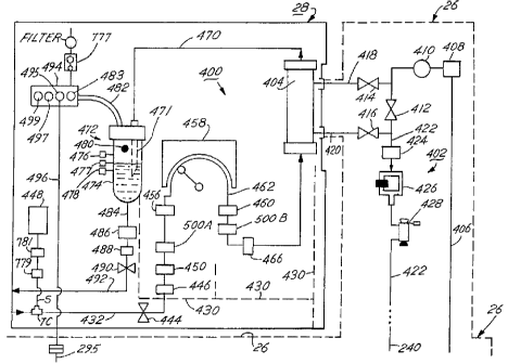
III. Dialysate Preparation (or Hydraulic) Module 26
Referring now to FIG. 6, the dialysate preparation module 26 will be discussed
in
detail. An overall function of the dialysate preparation module 26 is to
automatically mix
and prepare the dialysate solutions and deliver the solutions to the dialyzer
404. The
dialysate preparation module 26 has an inlet line 200 connected to line 111
(FIG. 5)
receiving filtered water from the water treatment module 24 via valve 108
(FIG. 5). The
CA 02271595 1999-OS-21
-28-
line 20Q carries the water past check valve CV 10 to a chemical mixing tank
202, preferably
cbnstructed _ from polypropylene. A chemical addition and dispersion subsystem
204 is
attached to the side of the tank 202 in fluid communication therewith. The
loading platform
250 of chemical addition subsystem 204 is illustrated in FIGS. 7A-7F. The
chemical
applicator 260 of the chemical addition subsystem 204 is illustrated in FIGS.
8A-8C and 9A-
9C. The chemical vessels (ideally bottles) 270 are illustrated in FIGS. l0A-
IOF.
The addition and dispersion subsystem 204 preferably includes two .chemical
applicators 260, each for opening a vessel 270 containing an individual batch
quantity of
dialysis chemicals placed directly above it. One vessel 270 typically contains
chemicals in
liquid form and the other in powdered form. The batch of chemicals are
provided in
individual batch vessels, preferably polyethylene and/or polypropylene bottles
270. When
the tank 202 is filled with purified water to the proper level, the chemical
applicators 260
pierce the bottles 270 from below with a spike, and the chemicals in the
bottles fall out of
the bottle by gravity and rinsing into the interior of the loading platform
250. As explained
in detail below, a sprayer 285 rinses the chemicals from the loading platform
250 into the
tank 202 where the chemicals are dissolved and mixed with water to form the
dialysate
solution. Additionally, bottle rinsing nozzles are preferably provided within
the chemical
applicators 260. The nozzles that are disposed below bottles containing dry
dialysate
chemicals eject water into the bottles in a series of short bursts to
gradually flush the
chemicals out of the bottles. After the chemicals are dispensed on the loading
platform 250,
the nozzles flush any remaining chemicals in the bottles 270 from the bottles
onto the loading
platform 250. A third chemical applicator 260 and third vessel 270 ark also
preferably
provided above the platform 250. The chemicals in the third vessel will
typically either be
CA 02271595 1999-OS-21
-29-
a salt which~can be added to the dialysate solution on demand to adjust the
chemistry of the
dialysate solution, or else a chemical cleaning or disinfecting agent that is
added to the tank
during the disinfection cycle. Other possible chemicals for the third bottle
270 are
medications, and vitamins and other nutritional supplements. As described
below, we prefer
S to use a hot pure water disinfection process, without chemicals, to clean
the fluid circuits of
the machine 22. However, if for some reason the hot water disinfection is not
sufficient,
an alternative mode may be entered.whereby the disinfecting chemicals in the
third vessel
are added to the tank and circulated throughout the machine to achieve
cleaning andlor
disinfection. Of course, additional chemical applicators and vessels could be
added to the top
of the loading.platform 250, if desired. '
The tank inlet tube 203 is placed at the bottom of the tank 202 and . oriented
tangentially to the walls of the tank 202 in a horizontal plane such that the
incoming water
is swirled about the side of the tank in the direction of the orientation of
the inlet 203 to
create a vortex, thereby stirring the water in the tank 202. A spray washer
20S similar to
1S a dishwasher sprayer is provided in the upper region of the tank 202, and
is operative during
cleaning of the tank 202 and mixing of the dialysate chemicals the tank 202.
The force of
the water through spray washer 20S causes the spray washer 20S to rotate and
spray water
into the tank 202 in the same direction as the flow of water in the vortex
created by water
inlet 203. The cooperation of the spray washer 20S and water inlet 203 create
good mixing
action in the tank 202, promoting effective dispersion and dissolution of the
chemicals
introduced into the tank 2D2 from the loading platform 250, and preventing the
settlement
of chemicals on the bottom of the tank.
The tank 202 itself is preferably made from a lightweight, biocompatible,
chemically
CA 02271595 1999-OS-21
-30-
compatible, and sterilizeable and substantially non-compliant (i.e., rigid and
not suxeptible
to expansion or contraction due to pressure, temperature or other condition)
material, that
is given the shape shown in FIG. 6. Other shapes are of course possible. A
tank made from
polypropylene with the shell reinforced with fiberglass windings on the
outside of the shell
meets these requirements. The polypropylene is chosen because of its chemical
inertness,
light weight and ability to be exposed to hot water for long periods without
any effect. An
alternative material for the shell is polyvinylidene fluoride (PVDF). The
reinforcing
fiberglass threads significantly improve the non-compliance (or stiffness) of
the tank 202.
As discussed in detail below, non-compliance of the tank is important for
improving the real-
time measurement of fluid removed from the patient during dialysis. The
fiberglass threads
are wound around the exterior of the walls of the tank 202 in overlapping
diagonal layers,
with an additional layer wrapping about the mid-section of the tank 202 in a
horizontal
manner. A suitable tank can be obtained from Structural North America in Ohio.
Other
possible reinforcing fibers may be suitable, such as composite fibers, carbon
fibers and
kevlar, which may be integrated into the shell body itself or wound on the
outside of the
shell. Heating elements may be built into or wrapped around the upper portion
of the tank
shell to assist in the dialysate separation technique discussed below.
A pressure transducer LT (Microswitch part no. 26PC X-98493-PC) is provided at
the bottom of the tank 202 in line 206 for the purpose of determining the
level of water in
the tank 202. Line 206 is isolated (static, with no fluid flowing through the
line) when the
NO port of valve V 17 for line 206 is closed and the NC port in line 209 is
open, permitting
the level transducer to read the level in the tank 202. This would be the case
when the tank
202 is being filled. During the filling and mixing of the tank, water is
circulated from the
CA 02271595 1999-OS-21
r
4
-31-
line 209 to V 17 to V9 through pump 212, valves 220 and 232, line 231 to valve
V 15, and
sprayer 205 in the tank 202, which assists in the mixing of the tank 202.
The tank 202 has a mesh filter FTB (130 micron) molded into a flat plate with
a
polypropylene frame at the bottom of the tank 202. A pump filter FP2
(preferably 50 to 200
microns) is placed on the degassing line 209. Any air or gas which may have
been
introduced into the dialysate is removed by pumping the dialysate through the
filter FP2.
The filter FP2 creates a negative pressure which causes entrapped air to come
out of the
water.
The tank outlet line 206 carries dialysate solution to a pump 212. When the
chemicals are released from the chemical addition subsystem 204 to the tank
and are being
mixed in the tank 202, the circulation of fluid is though line 206 (with
degassing line 209
static).
A three-way valve V 17 is placed at the intersection of lines 206 and 209 and
determines which line 206, 209 is static. The pump 212 (such as Micropump EG
series, 0-3
Llmin.) pumps the solution past a pressure transducer 214 (Microswitch PN 26PC
X-98752-
PC),
Referring again to FIG. 6, a three-way valve 220 controls the flow of fluid
through
output line 226 and return line 236. Water or solution in line 226 is fed to a
heater assembly
228. Heater assembly 228 is a temperature controlled, 1300 watt, flow-through
heater, such
as the Heatron no. 23925 heater. The heater assembly 228 is used for heating
dialysate up
to body temperature as it is passed to the extracorporeal circuit module 28
(FIG. 1). The
heater is also used for heating water up to a disinfection temperature of at
least 80 degrees
C., and preferably at least 85 degrees C., and maintaining the water at that
temperature for
CA 02271595 1999-OS-21
r.
-32-
more than an hour during the water disinfection of the fluid paths of the
machine 22, as
dixussed in detail below.
After passing through the heater 228, the water passes through a flow meter
241
(such as the Digiflow'~"'' unit) which measures the flow rate of the solutions
in line, and a
safety~thermistor THS. A second thermistor 230 is used to control the
temperature of the
fluids in the line 226. A three-way valve 232 controls the flow of fluid
through the tank
return line 231 and the output line 233. A dialysate filter such as
ultrafilter/pyrogen removal
filter 234 is provided for removal of any pyrogenic matePals and particulate
matter from the
dialysate. A preferred filter 234 is the Minntech RenaguardTM pyrogen filter.
No dialysate
solution goes to the dialysate circuit 402 during dialysis treatment without
first passing
through the filter 234. The condition of three-way valve 236 controls whether
fluid exits
from the ultrafilter/pyrogen filter 234 through line 238 or out dialysate
circuit input line 406.
A check valve CV 12 is placed between line 238 and 206. Line 238, check valve
CV
13 and valve V22 allow air to come out of the ultrafilter 234 (i.e., the
outside of the fibers
in the filter 234) during the priming of the ultrafilter 234 and pumping of
dialysate through
the ultrafilter 234 to the dialysate circuit 402.
We have devised a pre-treatment fiber bundle integrity test for the
pyrogen/ultrafilter
234. The integrity of the ultrafilter 234 is important to insure that there
are no leaks. The
pyrogen/ultrafilter is pressurized on the "blood" side (that is, the interior
of the fiber bundles
in direct fluid communication with the dialyzer 404) of the ultrafilter 234
prior to dialysis,
and the rate of pressure decay is measured. A' rapid pressure decay, or
inability to
pressurize the pyrogen/ultrafilter, will cause an alarm to sound, warning the
patient of the
CA 02271595 1999-OS-21
a
- 33 -
need to replace the pyrogen/ultrafilter 234. To accomplish this, we first
evacuate fluids from
the blood side of the pyrogen/ultrafilter 234 by operating the OF pump 242 in
the reverse
direction to pump air back through the valve 236, through bypass valve 412 in
the dialysate
circuit 402, through line 406 into the lumen or blood side of the
pyrogen/ultrafilter 234.
Once water has been evacuated from the blood side of the pyrogen/ultrafilter
234, the blood
side starts to pressurize (assuming there are no leaks in the
pyrogenlultrafilter 234). The OF
pump 242 pumps until the pyrogen/ultrafilter 234 is pressurized to 500 mm Hg.
If there are
any leaks, air will leak into the dialysate side of the filter 234. The air
pressure is measured
with the pressure sensor 410 in the dialysate circuit 402. If pressure sensor
410 never
pressurizes, then a severe leak is present. A slow decay in pressure indicates
there is no
leak. The rate of decay indicative of a leak requiring replacement of the
pyrogen/ultrafilter
is a function of the physical properties of the filter's membrane, and will
accordingly vary
depending upon which filter. is used. For most filters 234, we expect the
threshold decay
rate indicative of a failure to be greater than 10-25 mm Hg/30 seconds,
depending on the
type of filter.
The pressurization of the pyrogen/ultrafilter 234 can also be correlated to
the
maximum pore size of the filter. As the pyrogen/ultrafilter 234 is pressurized
to higher and
higher pressures, a maximum pressure will be reached above which the pressure
drops
suddenly indicating that the surface energy of water in the pores of the
filter is less than the
force due to the pressure. By knowing the pore size from the maximum pressure,
the
filtration capacity for certain pyrogens and other materials may be
determined.
Referring to FIG. 16, it will be appreciated that the analog board 614 and
central
processing unit 610 of the central control module receive the pressure data
from the pressure
CA 02271595 1999-OS-21
!. .
f~
-34-
sensor 410. Pressure readings indicative of a leak, such as where the rate of
decay is greater
than a predetermined threshold limit, will cause the CPU 610 (or safety CPU
616) to issue
an alarm, such as by issuing a message on the patient interface, or activating
the audio or
visual indicators 604 or a buzzer.
During the filling of the tank 202, after the chemicals are added, the machine
22
determines when to stop adding water to the tank by monitoring the fluid
sensor 288 in the
line coming out of the top of the tank 202. When fluid sensor 288 sees fluid,
the flow of
water is stopped by closing off valve 108 (FIG. 5).
The return flow of old solution (i.e., solution that has passed through the
dialyzcr)
from the dialyzer 404 is through return line 240, valve V 18 and dialysate
inlet 243. Valves
V 19, V 15 and V6 are closed, directing dialysate through to the dialysate
inlet 243. An
integral pressure relief valve with sample removal port 210 is placed in the
line at the top
of the tank 202 leading to valve V6.
We have invented a technique of maintaining the separation of new and old
dialysate
in the tank 202 by taking advantage of the differences in density in dialysate
when the
dialysate is at different temperatures. Our technique is an improvement over
the technique
described in the Terstegen Patent, U.S. No. 4,610,782. The preparation and
mixing of
dialysate in the tank 202 takes place with the dialysate at a temperature of
28 to 30 degrees
C. This temperature is controlled, in the preferred embodiment, by the
temperature-
controlled mixing valve 38 in the water pretreatment module 20. During
dialysis, dialysate
is heated in the heater 228 ~to body temperature, generally 37 degrees C, and
sent to the
dialyzer 404 in the extracorporeal circuit module 28 (FIG. 13). New (i.e.,
fresh) dialysate
is withdrawn from the bottom of the tank 202 and old dialysate is returned at
the top of the
CA 02271595 1999-OS-21
-35-
tank 202 in inlet 243 at a temperature of about 37 degrees C, or perhaps a
degree or two
cooler due to radiative and conductive heat loss in the tubing and hardware in
the dialysate
circuit 402.
The old dialysate is returned to the top of the tank 202 in a manner so as to
substantially prevent turbulence of the "old" or returned dialysate, that is,
in a manner to
gently introduce the returned dialysate into the top of the tank to prevent
mixing of the
returned and fresh dialysate. A preferred method of accomplishing this is to
provide a
circular inlet tube at the center of the top of the tank 202 with a series of
small holes in the
tube pointed radially inward. As the returned dialysate enters the inlet tube,
it gently passes
through the holes into roughly the center of the top of the tank. An
alternative embodiment
accomplishes the introduction of returned dialysate with mirrimal turbulence
by orienting an
inlet 243 slightly upward and towards the side walls,of the tank 202. The
returned dialysate
forms a zone above the fresh dialysate with a thermocline boundary layer
separating the
retuned and fresh dialysate due to the temperature differential (and resulting
density
differential) between the dialysate in the two zones. As the dialysis process
continues, the
boundary zone migrates down the tank 202 as the volume of fluid in the upper
zone of
returned dialysate increases and the volume of fresh dialysate diminishes.
This rrr~hod
works best when the temperature differential between the upper zone and lower
zone is at
least 5-7 degrees C, or greater, but will work acceptably down to 3 degrees C.
Ordinarily,
this differential will be present when the dialysate is heated as described.
An improvement to this technique is to heat one to two liters of new dialysate
above
the temperature of the dialysate in the tank 202 (by preferably at least 5
degrees C) prior to
the initiation of dialysis, and introducing the heated dialysate into the top
of the tank in a
CA 02271595 1999-OS-21
-36-
substantially non-turbulent manner. This sets up the temperature differential
zones such that
when the old (used) dialysate in introduced into the tank, it enters the upper
zone, further
minimizing the likelihood of substantial mixing of the old and new dialysate.
The heating can
be performed by heater 228, and the return of the heated dialysate is through
valve 232,
return line 231 and valve V 18. Valves V6, V IS and the NO ports of valves 236
and 232
should be closed to direct the heated dialysate into the tank 202.
The separation of returned and fresh dialysate in the tank 202 offers a number
of
advantages. First, it allows a closed loop ultrafiltration control methodology
to be used.
Second, the fluids being dialyzed from the patients are collected in the tank
202 separate
from other solutions, permitting the old dialysate to be sampled, measured,
and visually
observed in a tank with a window or a sample-removal port. Thirdly, the closed
loop
ultrafiltration permits the machine to operate, during dialysis, without the
machine being
connected to a water source and a drain. This gives more mobility to both the
machine 22
and the patient, a feature particularly advantageous in the hospital, home and
nursing home
environments. Fourthly, separation of old and new dialysate improves the
efficiency of
clearance of uremic toxins for a batch system.
A OF (ultrafiltration) pump 242, connected to the return line 240 through
valve 236,
pumps dialysate solution to and from the OF tank 244, the direction of flow
being a function
of whether the OF pump 242 is operated in a forward or reverse direction. NC
port of valve
V9 is closed and NO port of valve V 13 is open providing the pathway for the
solution to
enter the bottom of the tank 244. The OF pump 242 is also used to pump priming
solution
from the extracorporeal circuit 400 back to the OF tank 244.
The OF tank level sensor PUH precisely measures the fluid volume in the OF
tank
CA 02271595 2001-05-09
76909-43G
37
244. The OF tank 244 is used to store fluid removed from the
dialysate circuit commensurate with the fluid removed from the
patient. The fluid removed from the patient is the difference
in the volume of fluid in the OF tank before and after the
dialysis procedure. The rate of fluid removal into the OF tank
244 (and hence total volume when multiplied by time) is
controlled by the pump rate of OF pump 242. A sterile barrier
air filter AF (such as Pall EMFLON II) open to the atmosphere
is installed at the top of the tank 244. Background
information on ultrafiltration control in hemodialysis is
described in U.S. Pat. Nos. 3,974,284 and 3,939,069 assigned to
Rhone-Poulenc (both now expired).
The pressure transducer PUH is mounted at the bottom
of the OF tank 244. The transducer PUH measures the pressure
and hence level of fluid in the tank 244. The level sensor PUS
acts as a safety backup and watchdog for the OF pump 242 to
verify the amount of ultrafiltration during dialysis.
Specifically, the sensor PUH measures the hydrodynamic pressure
of dialysate in the ultrafiltration tank 244 and responsively
generates a measurement signal (sent to the control module 25)
indicative of the volume of fluid within the OF tank 244.
Alternatively, the rate of transport of fluid by OF pump into
the OF tank 244 is continuously monitored. A further
alternative is knowing the output volume per revolution of the
OF pump, and the time elapsed during dialysis. This
information allows the central control module 25 (FIG. 16) to
determine the expected volume of dialysate in the OF tank 244.
By comparing the measurement signal from the sensor PUH with
the expected volume of dialysate in the OF tank, the pump rate
of pump 242 is verified.
76909-43G
CA 02271595 2001-05-09
37a
In one possible implementation of this technique, a
decision as to the adjustment of the dialysate transport rate
into the OF tank (i.e., the pump 242's pump rate) can be made.
CA 02271595 1999-OS-21
-38-
For example, if sensor PUH indicates that there is 350 ml of fluid in the tank
244 but a
calculation of the expected volume of fluid in the OF tank based on pump rate
and elapsed
time is 385 ml, the pump 242 is pumping about 10 % too slow and the pump speed
should
be increased to meet the ultrafiltration target in the expected dialysis time.
After the tank 202 and fluid circuits of the dialysate preparation module 26
have been
filled with dialysate, positive pressure is created with pump 212 in line 209.
Dialysate is
conducted from tank 202 through valve V9, through OF pump 242, to valve 236,
down
through CV 12, line 206, valve 220, valve V8 and into pyrogen/ultrafilter 234.
The dialysate
is sent . up through flow meter 241 to the dialysate circuit 402, where the
solution goes
through bypass valve 412, through return line 422, line 240, valve V 18 and
back to the tank
202. During dialysis, dialysate is pumped from the dialysate circuit into the
ultrafiltration
tank 244 via line 240, valve 236 and valve V 13 in accordance with the target
ultrafiltration
volume for the patient.
Air and drain paths 282 and 280 are provided in the module 26 for collecting
any
fluid or condensate from .the tank 202. An optical fluid sensor 288 is used to
detect when
the tank 202 is full during the tank fill mode, to detect failure of valve V6
during dialysis,
and detecting water or dialysate in the hose portion (solid line) from valve
V6 to air filter
AF.
In our design, the housing of the dialysate preparation module 26 includes a
floor or
base for the entire machine 22, including the other modules 24 and 28. Any
fluid such as
blood, water or dialysate that leaks from the modules 24, 26 or 28 collects in
a catchment
basin 284 at the bottom of the entire machine. Leaks will drip on any
arbitrary path, shown
schematically as broken lines 280 and 282. The floor of the housing for the
machine 22 is
CA 02271595 1999-OS-21
-39-
horizontally non-planar to facilitate the catchment of fluid, in a fashion
similar to an oil pan
for an engine. The floor of the machine may be bowl shaped or given any other
suitable
contour to provide a lower catchment basin 284. A fluid sensor 286 is placed
in the vicinity
of the catchment basin 284 to detect the presence of fluid in the catchment
basin 284. If
fluid is detected, the user is alerted by an audio or visual indicator, and
the machine is
checked for leaks.
Referring now to the left-hand side of FIG. 6, a line 283 is provided for
conducting
water to the chemical application system 260 for rinsing the dialysate
chemical bottles 270
after they have been opened, and for disinfection of the bottle's seal. Line
281 is a return
line from the chemical application system 260 to valve V 13. Line 291 also
provides water
from three-way valve 287 to a fountain or sprayer 285 in the chemical loading
platform 250.
Drain lines 236A and 236B provide a pathway for dialysate or disinfection
water to exit from
the extracorporeal circuit module 28 via the disinfection manifold 494 (see
FIG. 36 also)
through valve V 14, and thermistor 293. A pressure relieflsample port 215 is
placed in a
drain line 236C connected to the valve 220. The pressure relieflsample port
215 is a
combination pressure relieve valve and integral sample removal port of the
same design as
pressure relief/sample port 78 (see FIG. 4); and is used to take fluid samples
from the
system. Lines 289 and 289A provide a fluid pathway into the extracorporeal
circuit
module 28 via valve CV 11. Line 295 connects the disinfection port 495 of the
disinfection
manifold 494 (FIG. 36) via line 496. Thermistor 293 monitors the temperature
of the fluid
returning from the extracorporeal circuit 400 during the hot water
disinfection cycle.
A. The Chemical Loading Platform 250
CA 02271595 1999-OS-21
-40-
The chemical loading platform 250 of FIG. 6 is illustrated in detail in FIGS.
7A-7F. FIG. 7A is a perspective view of the platform 250 shown positioned
against the side
of the tank 202. FIG. 7B is a top plan view of the platform 250. FIG. 7C is a
sectional view
of the platform 250 along the lines 7C of FIG. 7B. FIG.7D is a sectional view
of the
platform 250 along lines 7D of FIG. 7B. . FIG. 7E is an elevational view of
the platform
250. FIG. 7F is a sectional view of the platform 250 along the line 7F of FIG.
7E. In the
figures, the platform 250 is an integrally molded housing mounted to the side
of the tank 202
and having a top 304 with four apertures. Apertures 306, 308 and 310 provide
passageways
for chemicals from the chemical application assemblies 260 which are installed
above the top
304 of the platform 350. Aperture 312 is for a line 291 (FIG. 6) to provide
water to a
sprayer 285 suspended within the platform 250 for rinsing chemicals from the
shelf 320 into
the tank 202. (See FIG. 6). Shelf 320 is inclined at an angle of between 10
and 30 degrees
(preferably 17 degrees) relative to the horizontal to promote dispersion of
chemicals
delivered onto shelf 320 into the tank 202. Note that the tank 202 has
fiberglass windings
314 wrapped around a polypropylene shell 316.
The platform further has a rim 302, 326 and sidewalls 318 and 319. The
chemicals
are placed in fluid communication with the interior of the tank by virtue of
the open side 324
of the platform 250, which is aligned with an opening (not shown) in the tank
202.
Referring to FIG 6: and 7D, the sprayer 285 sprays fluid (e.g. dialysate or
purified
water) in the direction of the lower shelf 320 to assist in washing dialysate
chemicals off the
shelf 320 and. into the tank 202, thereby promoting effective dissolution of
the chemicals
within the tank 202 and the avoidance of buildup of chemicals on the shelf
320.
Referring in particular to FIGS. 7C and 7D, an aluminum plate 322 is mounted
to
CA 02271595 1999-OS-21
-41 -
the top of the platform 250 to provide a mounting base for the chemical
application
assemblies 260 (FIGS. 6, 8A-8C).
B. The Chemical Applicator 260
Referring now to FIGS. 8A-8C, the chemical applicator 260 will be explained in
detail. The chemical applicators 260 (three in all in the preferred
embodiment) are installed
on the aluminum plate 32z directly above the apertures 306, 308, 310 (FIG.
7A). FIG. 8A
is an elevational view of the applicator 260, and FIGS. 8B and 8C are
sectional views of the
applicator 260 with the spike 330 in upper and lower positions, respectively.
When the spike
330 is in the upper position (FIGS. 8A and 8B), the tip 331 of the spike
pierces the bottle
270 which is installed in an upside-down orientation in the upper region 332
of the applicator
260, causing the chemicals in the bottle 270 to pour out through the
'applicator 260 and
apertures 306 (or 308 or 310) and onto the shelf 320 of the loading platform
250 (FIG. 7).
The applicator 260 has a cylindrical housing 334 mounted to. a base member 336
affixed to the aluminum plate 322. The housing 334 has an open interior region
338. A
threaded drive collar 340 is mounted to the housing 334. The spike 330 is
reciprocable
within the interior region 338 between upper and lower positions. The spike
330 pierces the
seal on the bottle 270 or other vessel containing the dialysate chemicals (or
other contents
of the bottle 260) when the spike 330 is moved to the upper position (FIG.
8B). The spike
330 has an integral cylindrical body 342 concentric with the housing 334 with
an open
interior for permitting passage of dialysate chemicals therethrough after the
spike 330 has
pierced the seal of the bottle 270. A pair of thread blocks 344 are mounted to
the side walls
of the spike 330 which engage the threads 346 on the drive collar 340. A drive
belt 348
CA 02271595 1999-OS-21
- 42 -
(one or two) or other suitable means (such as a cog) engages the threaded
drive collar 340
(FIG. 8A). As the belt 348 rotates the collar 340, the thread blocks 344 are
rotated, causing
the spike. 330 to move between the upper and lower positions depending on the
direction of
movement of the drive belt 348.
Referring in particular to FIGS. 8B and 8C and FIG. 12, a nozzle 350 is
disposed
within the cylindrical housing 334 in communication via line 281 with the
water inlet line
200. The cylindrical body 342 of the spike has a vertical slit to accommodate
the nozzle 350.
The tip 352 of the nozzle 350 is oriented upward in the direction of the
bottle 270 when the
bottle 270 is mounted to the housing 332. The flow of water through the nozzle
250 on
demand ejects water towards the interior of the bottle 270 after the bottle
has been opened
by the spike 330, thereby rinsing the interior of the bottle 270 and promoting
the release by
gravity of'the entire contents of bottle 270 through the aperture 306 (or 308,
310) and into
the tank 202. To control the dispensing of dry dialysate chemicals from the
bottle, and
prevent clogging of chemicals at the base of the bottle, we prefer to pulse
water through the
nozzle 350 over a period of time. For example, we pulse water through the
nozzle for one
second (with a pressure greater than 10 psi), then pause briefly while some of
the chemicals
fall through the interior of the spike 330, then pulse again, pause, and then
continue the
process until all the chemicals have fallen out of the bottle. This pulsing
may occur for
perhaps 50 times over a ten minute period. This pulsing action prevents all of
the chemicals
from being dumped at once onto the shelf of the loading platform. When the
bottle is
substantially empty, the nozzle rinses out the bottle with a continuous stream
of water of 5
to 10 seconds duration.
The nozzle 350 also ejects heated water (or water treated with disinfecting
chemicals)
CA 02271595 1999-OS-21
-43-
onto the outside surface of the seal 372 of the bottle 270 during the
disinfection cycle of the
machine, thereby disinfecting the interface between the chemicals in the
bottle 270 with the
dialysate preparation tank 202.
An O-ring 329 is provided around the base 335 of the spike 330. When the spike
is
in the lower position, outlet tube 337 leading to line 281 (FIG. 6) is open
and the tank 202
is closed off through ports 306, 308 and 310.
C. The Chemical Vessel (Bottle) 270 and Automatic Identification System
Referring now in particular to FIGS. 8C and 9A-9C. together with FIG. 10A, a
bottle
mounting member 354 is placed above the housing 334 of the applicator 260 to
insure that
the bottle 270 is mounted in alignment with the spike 330 to the applicator
260. The
mounting member 354 is shown in a top plan view in FIG. 9A (i.e., as it would
be seen
when looking down from above in the direction of the top of the spike), in a
bottom plan
view in FIG. 9B, and in side elevational view in FIG. 9C. The mounting member
354 has
a central opening 360 through which the head of the bottle 270 is inserted. A
button through
hole notch 356 accommodates a touch button 362 (FIG. 10A) containing coded
information
as to .the contents of the bottle 270 that is affixed to the neck of the
bottle 270. The touch
button 272 is about a half inch in diameter. The member 354 has a pawl 357
(that is
retractable by operation of an electric solenoid 358) for automatically
removing the touch
button 362 when the bottle 270 is removed from the mounting member 354.
During installation of the bottle 270, the head of the bottle 270 (turned
"upside
down") is placed within the opening 360 and rotated in the direction of the
arrow of FIGS.
9A and 9C. The touch button 362 Aides past the pawl 357 into contact with a
touch button
CA 02271595 1999-OS-21
-44-
reader. When the bottle 270 is removed from the applicator, the bottle must be
rotated in
the opposite direction. Pawl 357 is activated by solenoid 358 to an extended
position. When
the bottle is rotated such that the touch button is rotated past the pawl 357,
the pawl 357
pushes the touch button 362 off of the bottle 270, causing the touch button to
fall. A
suitable catchment structure is provided about the applicators 260 and
aluminum shelf 322
(FIG.B) to catch and collect the fallen touch buttons. The user of the machine
22 collects
the buttons and sends them back to a collection center for reprogramming and
reuse.
Alternatively, the buttons 362 could be collected by a service technician
during a service
visit.
The structure of the bottle 270 is shown in detail in FIGS. l0A-IOF. FIG. 10A
is an
elevational view of the bottle 270 with the touch button 362 removably affixed
to the neck
region 364 of the bottle 270. FIG. 10B is a sectional view of the neck region
364 of the
bottle 270 showing the polyethylene or polypropylene shell 380 and a
polypropylene cap 370
that is attached in any convenient fashion to the shell, such as by threaded
engagement. An
alternative embodiment is one in which the cap 370 is snapped onto the shell
380 via
circumferential complementary snap elements. A polypropylene seal 372 integral
with the
cap 370 closes off the bottle 270. Polypropylene is chosen for the material
for the cap 370
since the cap 370 is subject to hot water disinfection during the disinfection
cycle of the
machine 22. Specifically, when the bottle 270 is installed within the mounting
member 354
above the applicator 260, hot water is applied via nozzle 350 (FIG. 8C) to the
exterior
surface of the polypropylene seal 372. While polyethylene is a preferred
material for the
bottle shell 380, it tends to soften when subjected to hot water for an
extended period of
time.
CA 02271595 1999-OS-21
- 45 -
FIG. lOC is a detailed elevational view of the upper portion of the bottle
showing
the pinch semicircular rim 366 that retains the touch button 362. FIG. lOD is
similar to
FIG. lOC with the cap 370 rotated 90 degrees. A retaining bead 368 helps keep
the touch
button 362 in place. FIG. 10E shows the opposite side of the cap 370 from
FIG.. IOC. The
threads 367 engage the upper portion of the housing 334 of the applicator 260
(FIG. 8).
FIG. lOF is a plan view of the seal area 372 of the bottle 270. The seal 372
comprises a frangible section 374 and a hinge section 376. The mounting member
354 (FIG.
9), and in particular the notch 356, acts as a means for insuring that the
bottle 270 can be
inserted only one way onto the applicator 260 and aligning the upper tip of
the spike with
the frangible section 374 of the seal opposite the hinge section 376, so that
the tip 331 of the
spike 330 (FIG. 8B) contacts the region 384 of the seal 372. The uppermost rim
of the spike
330 tears through the frangible section 374, with only the hinge portion 376
uncut, when the
spike 330 is moved to its upper position (FIG. 8B). By virtue of the stiff
properties of the
polypropylene material, and by virtue of the support from the spike 330 from
below when
the spike 330 is in the upward position, the broken seal 372 maintains an
generally upward
orientation, allowing the chemicals in the bottle 270 to exit from the bottle
270 and
permitting the nozzle 350 to spray into the interior of the bottle 270 to
rinse out any
remaining chemicals in the bottle 270. The rinsing action of a bottle 270
containing dry
dialysate chemicals is shown in FIG 12.
When the bottle 270 is mounted to the applicator 260, the touch button 362 is
placed
in touching contact with a touch button reader mounted in any suitable fashion
above the
applicator 260. The reader retrieves information coded in the button 362 (such
as the
contents of the bottle, a date code, a lot code, and other information) and
passes the
CA 02271595 1999-OS-21
-46-
information to the central. processing unit of the control module 25 (FIG. 1).
The control
module 25 has a memory storing information such as the correct dialysate
chemicals for the
patient, the patient's dialysis prescription, and software for processing the
information from
the touch button. If the bottle is not the proper chemical for the patient,
the control module
25 alerts the user, such as by activating a suitable alarm. The user, thus
alerted, removes
the incorrect bottle 270 prior to commencement of the next dialysis procedure
and replaces
the bottle 270 with the proper bottle, and the process goes forward. If a
rernoveable touch
button is used for the indicator, it should not be detached when the bottle is
removed from
the applicator. Touch buttons, readers and supporting materials suitable for
use with the
present invention can be obtained from the Dallas Semiconductor Corp., 4401 S.
Beltwood
Parkway, Dallas Texas. The above-described identification technique promotes
safety and
the prevention of unintentional introduction of the vi~rong dialysate
chemicals into the tank.
It will be appreciated that other types of indicators besides touch buttons
may be
applied to the bottles such that the indicator is read by a machine when the
bottle is about
to be used. For example, bar codes, two and three dimensional bar or dot
matrices, radio
frequency transmitters or magnetic strips may be affixed in any suitable
fashion to the sides
of the bottles and read by the appropriate machine in a well known manner.
Ideally, the
reading occurs during or immediately after installation of the bottle and
prior to the opening
of the bottles and introduction of the chemicals to the tank 202, so that in
the case that the
wrong bottle was installed, the patient is alerted and corrective action can
be taken.
D. The Dialysate Circuit 402
The dialysate preparation module 26 further includes a dialysate circuit 402
that
CA 02271595 1999-OS-21
-47-
circulates dialysate from the tank 202 to the dialyzer 404 and back. The
dialyzer 404 (such
as the Fresenius F-80 filter) filters blood to remove toxins and excessive
water buildup in
the patient's blood. The patient's blood is introduced into the machine via
the extracorporeal
circuit 400 (FIG. 13).
S The inlet line 406 carries dialysate solution to a thermistor 408, which
monitors the
temperature of the fluids in the line 406. A pressure transducer 410
(Microswitch 26PC X-
98752PC) monitors the pressure in the line 406. Bypass valve 412, and input
and output
valves 414 and 416 control the flow of dialysate into and out of the dialyzer
404 via input
line 418 and output line 420.
During dialysis, the thermistor 408 data is fed to the safety CPU 616 (FIG.
16) to
insure that the temperature of the dialysate is less than a maximum critical
temperature, in
the present example 39 degrees C. If the temperature is greater than the
critical temperature,
the safety CPU 616 closes off valves 414 and 416 and opens up bypass valve
412.
Conductivity sensor 426 data (FIG. 6) is also fed the safety CPU 616 and if
abnormal
conductivity readings are sensed, the valves 414 and 416 are closed and bypass
valve 412
is opened.
The return flow of old dialysate is via line 422. A noninvasive conductivity
monitor
426 and a blood leak detector 428 are provided in the line 422. Blood leak
detector 428
detects a leakage of blood from the dialyzer 404 into the dialysate. The
presence of blood
in the line 422 also causes valves 414 and 416 to close and valve 412 to open,
to prevent any
additional loss of the patient's blood.
The noninvasive conductivity cell 426 is illustrated in detail in FIG. 11A.
The inlet
line 422 is divided into first and second fluid channels 423 and 427 integral
with the inlet
CA 02271595 1999-OS-21
- 48 -
line 422. The channels 423 and 427 are constructed such that the flow of fluid
is evenly
split between the passages to minimize response time. In the embodiment of
FIG. 11A, the
channels 423 and 427 branch in directions 90 degrees from each other. The
purpose of this
consctruction is to insure equal flow paths in the two channels. The channels
are each
oriented at an angle of approximately 135 degrees relative to the inlet line
422. The
channels 423, 427 form a rectangular loop with the inlet line 423 and the
outlet line 422' at
opposite corners. A conductivity measurement sensor (e.g., Great Lakes no. 697
E sensor)
224 with leads 225 is circumferentially disposed about one of the fluid
channels. The leads
225 from the sensor 224 are fed to the central processing unit 610 or 616 of
the user
interface and control module 25 (FIG. 16). An alternative construction is
shown in FIG.
11B, where channels 423A and 425A are shorter than channels 427 and 423. With
either
construction, the conductivity cell 218 is preferrably installed in a vertical
or. vertically
inclined orientation such that fluid flows upwards through the channels 423
and 427 which
prevents the entrapment of air bubbles in the fluid line 427.
The construction of FIG. 11 B provides a minimum path-length to cross-
sectional area
ratio for the fluid channels 423 and 427. This construction generally
maximizes the
sensitivity of the sensor 426 and reduces response time.
An alternative embodiment of the conductivity sensor is shown in FIG. 11C. The
inlet
422 is connected to a molded polysulfone Y fitting containing two passageways
for the two
channels 423 and 427. The channels meet at the opposite Y fitting and go out
the outlet
422'. The conductivity sensor 224 is placed around one of the tubes,
preferrably made from
a hard plastic such as KYNAR, between the Y fittings. The Y fittings split the
incomming
fluid into two substantially equal channels 423 and 427. The entire assembly
426 is
CA 02271595 1999-OS-21
preferably mounted in a vertical orientation so as to prevent
the entrapment of air bubbles in the line channel 427.
E. The Blood Leak Detector 428
Background information on blood leak detectors known
in the prior art can be found in U.S. pat. Nos. 4,925,299;
4,166,961; 4,087,195; 4,087 ,85 and 4,017,190. Our presently
preferred design, based on absorbency of light by the blood,
is shown in schematic form in FIG. 25A. A light emitting
diode (LED) 530 is pulsed between an OFF condition and an ON
condition, during which it emits light at which the absorbency
of the light by blood is at a maximum, such as 880 nm. The
light from the diode 530 passes through a mirror-coated beam
splitter 532. The resulting light of intensity P is split
into two portions. One portion is directed toward a reference
photodiode 534, and the other portion is directed through a
chamber or.cuvette 536 containing dialysate solution and onto
a second blood detector photodiode 538 identical to the
reference photodiode detector 534. The reference photodiode
534 also receives light from external interfering light
sources pEXT2 directed onto the diode 534. The reference
diode 534 is used for generating a light intensity correction
factor, as discussed below. Photodiode 534 is connected to an
operational amplifier 540 having a resistor 542. The output
voltage of the op. amp. 540 is represented by VpD2.
External interfering light sources PEXT1 also
impinge on the photodiode detector 538. The photodiode
detector 538 is connected to operational amplifier 546 having
resistor 533 connected across the output and negative
- 49 -
76909-43
CA 02271595 1999-OS-21
terminals as shown. The resulting output voltage signal is
represented by VpDl. Suitable beam splitter and optical
diffusing glass components
- 49a -
76909-43
CA 02271595 1999-OS-21
-50-
for the detector of FIG. 25A can be obtained from Edmund Scientific Co., of
Barrington,
New Jersey.
The housing for the blood leak detector (not shown) is constructed so as to
deflect
any air within the cuvette 536 away from the light path 535. Curved entry
paths or baffle
plates may be used for this purpose. The goal is to prevent air bubbles for
adhering to the
transmitting and receiving windows 537 and 529, respectively on the sides of
the cuvette
536. Maintenance of turbulence of the fluid within the cuvette 536 should
accomplish this,
but the above-cited blood leak detector patents disclose additional techniques
for the
avoidance of air-bubbles along the light path of a sensor.
The signal flow of the blood leak detector 428 of FIG. 25A is shown in FIG.
25B.
The following is a table of the legends used in FIG. 25B.
P = LED 530 Light Intensity
ICS = Light Attenuation of Cuvette 536 and Dialysate
Ks = Attenuation Coefficient Due to Blood in Dialysate
Pte.,.,, P~ = External Interfering Light Sources
KPDI KPD2 = Photodiode 538, 534 Sensitivity Coefficients, respectively
Vow,, VoF~ = Electronics Offset Coefficients for 546, 540, respectively; and
~PDI = (P ' Kc ' Ks'I' PFxri) ~cnn- Vom
Prior to dialysis, a control solution consisting of dialysate solution free
from blood
in introduced into the cuvette 536, the light source 530 is pulsed on and off
and
measurements of the light intensity in the reference and blood leakage
photodetectors 534,
538 respectively are made. The measurements during the light off condition are
stored and
subtracted from the next light-on measurement. The process is repeated during
the
CA 02271595 1999-OS-21
-51 -
conduction of dialysate solution from the dialyzer 404 during dialysis. A
calculation of an
attenuation coefficient indicative of the presence of blood in the dialysate
is made repeatedly
during dialysis, and an alarm is sounded if blood is detected. The provision
of the reference
detector 534 permits the removal of any offset or drift conditions in the
electronics or
variation in the light intensity from the light source 530.
' The photodiodes 534, 538 must be shielded from extraneous light sources such
as
incandescent bulbs or fluorescent lights. Any residual light appearing on the
detectors P~.,.l
and P~ is low-pass filtered. The filtering passes only the DC component of the
extraneous
signal. The DC component of the extraneous signal is removed in the on-off
pulsing that
removes electronic offset and drift.
Any additional lighUpaths from source to detector must also be minimized.
Light
paths other than that through the dialysate will cause the measurements to
deviate from the
expected levels for a given concentration. This cannot be corrected since the
offset
measurement is taken with the light source off. The extraneous light path can
be measured
by a given cuvette by replacing the dialysate with a fluid of virtually no
light transmittance.
Then the light source is varied from 0 to maximum output and the detector
output monitored.
The blood concentration measurement is as follows. Prior to treatment, the
light
intensity at the photodiode detectors 538 and 534 is measured before the
dialysis treatment.
(1) No Blood KS = 1.0, Light Off P=O
VPDIOFFI PEXTI KPDI + VOFFI
CA 02271595 1999-OS-21
- 52 -
(2) No Blood KS = 1.0 , Light on P
VPDIpNI - ~~ + P~CTI) KPDI + VOFPI
If P~-,.1, KPDI, Vin Constant Between 1 + 2
V - V = PICc KPD1
PDIpN~ PDlppgf
During treatment, the light intensity at the detector 538 is measured. The
ratio of the
intensity before treatment over the intensity during treatment is computed. In
addition, the
reference photodiode detector 534 is measured to correct the intensity
readings during
treatment from offset, drift, and extraneous light sources. Measurements and
calculations
are as follows:
(3) Blood KS, Light Off P = 0
VPDIpFF PP~CZ' KPDI + VOFFI
(4) Blood KS, Light On P
vPDlON - (PKcKs + PFXf1) KPDI + VOFFI
If Pte.,, KPD1, VOFFI Constant Between 3 + 4
VPDION - VPOIOFF - PKcKsKPDI
The attenuation coefficient due to the presence of blood in the dialysate, KS,
is as follows
(assuming ICS, KPDI are constant during the treatment):
VPDIpN - VPDIpFF PKcKsKPDI
- - Ks
~PDIpN~ - VPDIpFF PKCKPD1
CA 02271595 1999-OS-21
- S3 -
Suppose that the light intensity from the LED S30 increased S09o after the
initial
measurements prior to dialysis were made, for whatever reason. The attenuation
coefficient
K9 is the same. The manufacturers' variations for light intensity P,
photodiode sensitivity
KpDi and KPDI will not effect the calculation of the attenuation coefficient,
since they cancel
S out.
Thus:
No Blood, Before Treatment
VPDIpH~ - VPDIp~I = PKcKPDI
Blood, During Treatment
1O ~PDIpN - VPOIpFF P(1.S) KC KS KPDI
The reference detector 534 is used for correction:
No Blood, Before Treatment
vPDIpN~ - VPDIpFp/~ - P KPDI~
Blood, During Treatment
IS VPD2pN - VPD2pFF P(1.S) KPDZ
o Attenuation Calculation (If ICS, KPDI, KPDI, Constant During Treatment)
VPDIDN - vPDIpPP VPDIpN~ - VPD2pFP,
vPDIpN~ - VPDlpplr vPDIpN - VPD2ppp
P(1.S) IC~KSKPD, X PKPDI _ K
S
PKcKPDI P(I.S)KPDI
CA 02271595 1999-OS-21
-54-
~t,witt additionally be noted that the above calculations of KS assume the
beam sputter
532 is a 509& splitter directing light in two paths of equal intensity P. A
different ratio of
the light intensity could be used with a conversion factor used in the KS
calculation.
If the sensitivity of the diodes 534 and 538 varies differently from each
other during
the treatment, then the attenuation coefficient KS will vary. This situation
will be avoided
by choosing the same type of photodiode detectors 534, 538. Additionally, the
photodiode
sensitivity coefficient variance is typically small.
The blood leakage detector can be tested by varying the intensity of the light
source
530 and making sure that the reference and blood sensor photodiode detectors,
534 and 538,
respectively, track within limits. The supporting electronics for the blood
leakage detector
428 should be low-noise with high stability. Calibration tests must be
performed to
determine the expected light levels detected , for various dialysate flow
rates and blood
concentrations. The path length of the light.within the cuvette determines the
sensitivity of
the optical density measurements.
As an alternative approach, instead of pulsing the light from LED 530 on and
off,
the light intensity can be sequenced OFF, LOW and HIGH. If a blood
concentration in the
cuvette 536 causes a low reading near the noise floor of the electronics, the
next higher
detector reading corresponding to the high light source intensity can be used.
IV. The Extracorporeal Circuit Module 28
Referring now to FIGS. 13 and 27A, the extracorporeal circuit module 28 will
be
described in detail. The patient's blood is introduced into the extracorporeal
blood circuit
400 in arterial line 432. If a saline bag 448 is used as discussed below, the
saline is
CA 02271595 1999-OS-21
- 55 -
introduced into the arterial line 432 at three-way connector TC with rotating
male luer lock
and two female luer lock such as Haemotronics Part No. B-82 or, alternatively,
~a four-way
injection site with rotating male luer lock/double female luer lock, such as
Haemotronics Part
No. CR-47. The saline bag 448 is optionally provided, and is connected to the
arterial tine
432 by a saline infusion line S having an optional fluid/air sensor 781 and a
clamp ?79. The
saline bag 448 has several potential uses: for priming air out of the
extracorporeal circuit
400, for replacing lost fluid during therapy and rehydrating the patient, and
for rinsing back
blood to the patient. Our reverse osmosis water and ultrapure dialysate,
introduced to the
extracorporeal circuit 400 by causing a pressure differential to exist at the
membrane of the
dialyzer, serves these functions as well, thus the saline .bag 448 is for an
alternate method
of priming and rinseback. The fluid sensor 781 detects when the saline bag is
empty, and
permits automatic identification of this condition to the patient, obviating
the need for
periodic checks of the saline bag 448. When air is sensed by the sensor 781,
the clamp 779
is closed. An alternative to the use of a fluid sensor 781 is an in-line
infusion filter in the
infusion set which obviates the need of a fluid sensor.
A clamp 444, an ultrasonic air bubble detector 446, a pressure monitor SOOA,
and
an optional injection site (needle or needleless type) 456 are placed in the
line 432. Blood
pump 458 pumps blood into line 462, via special pump section tubing (from
Pharmed TM
material or silicone) past optional injection site 460 and pressure monitor
SOOC and (optional)
expansion chamber 466 to the dialyzer 404. The blood is returned to the
patient via line 470
to an air-separating and pressure monitoring chamber 472 having an inlet tube
471 at the top
or bottom, with the top preferred.
Referring in particular to FIGS. 13 and 27A, the air-separating and pressure
CA 02271595 1999-OS-21
-S6-
monitoring chamber 472 has a chamber 474, an upper and a lower blood level
sensors 476
and 478 respectively, and an optional injection site 480 (one or more). A
third blood level
sensor 477 placed at the optimum blood level is preferrably provided with the
chamber 472
for monitoring or controlling the blood level. The chamber 474 is in air
communication via
S line 482 to a connection port 483 in a disinfection manifold unit 494, which
is further
connected to a line 491 having a pressure sensor 775, an air pump 777 a filter
and then open
to atmosphere. Because the fluid in the chamber 474 is normally under positive
pressure
during dialysis, the level may be raised (when identified as being too low by
the level sensor
478) by operating the pump 777 to remove air from the chamber 472 until the
level is raised
to the level of sensor 476. The level may be lowered by stopping the occlusive
blood pump
458, and operating the air pump 777 to add air to the chamber 472. The bottom
of the
chamber 474 is connected to a line 484 having an ultrasonic air bubble
detector 486, a blood
sensor 488 and a clamp 490 and is connected to the venous line 492 which leads
to the
patient.
1S A pressure transducer isolator (a disc shaped unit) 493 (FIG. 36) is
installed at the
interface between , the line 482 and the port 483. The isolator 493 has a
microporous
membrane which allows no fluid to escape out of the line 482 but which allows
air to escape
and enter the line 491. An alternative construction to the air pump 777 is to
place an air
pressure adjustment valve and a clamp in the line 491 from the port 483 which
is open to
atmosphere.
Referring to FIG. 13 and FIG. 28, the disinfection manifold 494 includes
disinfection
ports 495, 497 and 499. Port 49S is connected at the back side of the manifold
494 to
disinfection line 496, which carries disinfection fluids (e.g., hot water) to
the extracorporeal
CA 02271595 1999-OS-21
-57-
circuit 404. Ports 497 and 499 receive the connectors at the end of the venous
and arterial
lines 432 and 492, respectively after the,dialysis session is completed. Ports
497 and 499
are connected to each other via valve V20 (FIG. 6). Port 497 and port 495 are
connected
via a T fitting on the back side of the disinfection manifold 494. These
connections provide
a path for the flow of disinfection fluid (i.e., hot water or water treated
with disinfection
chemicals) through the entire extracorporeal circuit 400, including the blood
side of the
membrane in the dialyzer 404. The port 483 is not in fluid communication with
the other
ports 495, 497, 499. When the dialysis session is completed, the patient
reconnects the
bubble trap line 482 from the port 483 to the disinfection port 495. While the
disinfection
manifold 494 could be formed as a unitary housing, it may also simply be
composed as an
array of connectors having the fluid communication pathways described herein
(or
equivalents). Referring now also to FIG. 6 and 36, it will be seen that lines
289A and 236A
connect at the back side of the disinfection manifold 494 to ports 499, 497.
The back side of the disinfection .manifold 494 is connected via return lines
236A and
289A to valves V 14, V20, check valve CV 11 and thermistor 293 (FIG. 6). Lines
236A and
289A connect through the disinfection manifold to the arterial 432 and venous
492 lines,
respectively, of the extracorporeal circuit, when the lines 432 and 492 are
connected to the
ports 499, 497 of the disinfection manifold 494, as shown in FIG. 27A.
An alternative embodiment of the extacorporeal circuit is shown in FIGS. 27B
and
27C. FIG. 27B shows the venous and arterial lines connected to their
respective disinfecdon
manifold ports, and FIG. 27C shows the lines as they would be when they are
connected to
the patient. The only difference between the embodiment of FIG. 27A and the
embodiment
of FIG. 27B-C is that the venous and arterial disinfection manifold ports 497
and 499,
CA 02271595 1999-OS-21
-5g-
respectively, are located lower on the machine, facilitating easier patient
connection. The
tubing connecting the ports 497 and 499 to the dialysate preparation module 26
is behind the
extracorporeal circujt bulkhead B.
It is common in extracorporeal blood circuits to have a blood filter which
filters the
blood as well as eliminates air or gas bubbles. The typical application for
these filters is in
major surgical procedures. The blood flow rates for these surgical procedures
range from
3, to 6 liters per minute. The typical blood flow rate for hemodialysis is
only 200 to 600
milliliters per minute.
It is well known that air contact with blood often causes clotting. This is
one of the
draw backs of traditional bubble traps. Bubble traps also require level
sensors and a valuing
scheme and control system to allow the collected air escape. By using a
hydrophobic
microporous membrane to allow the air to escape passively, less clotting
occurs, less sensing
and valuing hardware is needed, and fewer set manipulations by the patent or
machine
operator are needed. 1n addition, the unit is simpler to clean and sanitize.
Thus, an
alternative to the air separating and pressure monitoring chamber 472 is a
cassette-type
debubbler 1000, shown in FIGS. 15A-15D. FIG. 15A is an exploded view showing
the
front or blood side of the debubbler 1000. FIG. 15B is an exploded view
showing the rear
or air side of the debubbler 1000. FIG. 15C is a cross-sectional view of the
debubbler 1000
through the blood outlet 1020 in an assembled condition, with the unit in a
vertical
orientation as it would preferably be installed in the extracorporeal circuit
400 (FIG. 13).
FIG. 15D is a perspective view of the debubbler 1000 partially broken away in
section in
the same plane as FIG. 15C. Also for this application the debubbler 1000 is
designed to be '
cleanable, sanitizable and reusable.
CA 02271595 1999-OS-21
-59-
Referring to FIG. 15A, the debubbler 1000 has a front cover 1002, a fluid
circuit
board 1004, two microporous membranes having a blood contact portion 1006 and
a
secondary air vent 1006A, a back cover 1008 with pressure transducer opening
1016A and
an over molded support portion 1010 having a series of parallel support ridges
1024
separated by adjacent parallel apertures 1115. Thc: fluid circuit board 1004
has a blood
chamber 1014 and an optional pressure transducer opening 1016 disposed
therein. A
retaining ring 504 and pressure transducer comprising diaphragm 506 with metal
disk 508
are mounted within the opening 1016 for measuring the blood pressure in the
blood chamber
1014. The magnet, rod and strain guage elements of the pressure transducer are
described
in construction with FIG. 14A-C below.
A condensate outlet 1018, a blood inlet 1022 and a blood outlet 1020 are
provided
at the bottom of the fluid circuit board 1004. An air port 1012 is also
provided at the
bottom for conducting air passed through the filter 1006A and 1006 out of
circuit hoard
1004. Referring to FIG. 15B, which is a exploded view of the debubbler 1000
seen from
the rear or air side, the fluid circuit board 1004 also has a condensate
chamber 1032 and
hole 1038 covered by membrane 1006A, a membrane peripheral seal area 1028
where the
microporous membrane 1006 is sealed to the fluid circuit board 1004, a raised
rib 1026 and
a channel 1030 for collecting condensate. Condensate is passed out of the unit
1000 from
condensate outlet 1018.
It is well known that hydrophobic microporous membrane filters will allow air
to
escape a chamber while preventing aqueous liquids to escape through the
membrane. This
also works with blood. PTFE, also known as TEFLON T"', microporous membrane
filters
have been used successfully for many years. However, if blood flow is
deadheaded against
CA 02271595 1999-OS-21
a PTFE microporous .membrane.filter, in a short period of time the membrane
will become
coated with a biofilm that impedes air escapement. It is well known that PTFE
attracts lipids
and proteins. It is also well known that if the blood flow is allowed to flow
past the
microporous membrane in a tangential manner, the flow minimizes the build up
of a biofilm
and better maintaining air escape efficiency.
Prior art air venting blood filters for surgical use typically employ an
essentially
horizontal inlet port that passes blood tangentially across a horizontal
hydrophobic
membrane. The blood flow across and around and then down through a blood clot
filter and
out. There are several variations to this theme. There are reports that claim
the clot filters
cause more clots than they eliminate from the flow stream. All these surgical
units are
designed for single use and are relatively expensive. For our hemodialysis
application, no
clot filters will be used.
Recently, two microporous membrane manufacturers, Pall and Millipore, have
introduced PVDF (polyvinylidene fluoride) also known as KYNAR TM membranes
that have
superior hydrophobic properties over PTFE and which reportedly do not have the
protein and
lipid attraction that PTFE has. PVDF is the preferred material for the
membranes 1006,
1006A of the cassette-debubbler 1000 of FIG. 15.. PVDF has the following
properties:
sealability to the PVDF microporous membrane, blood compatibility, natural
hydrophobia,
moldability,, heat sealability, translucency and can be compliant under
pressure. Polysulfone
could also be used as an alternate material with difficulty and with a trade
off in properties.
To achieve tangential flow, our microporous membrane filter 1006 is installed
in an
essentially vertical orientation. The blood inlet port 1022 is also vertical,
as is the outlet
1020. The inlet port 1022 can be from either the top, bottom or side. Our
preferred
CA 02271595 1999-OS-21
-61 -
embodiment is having the inlet 1022 enter from the bottom.
The covers 1002, 1008 are heat sealed to the ribs 1027, 1026 using a heat seal
process. The fluid circuit board 1004 is fabricated preferably by injection
molding with ribs
1027, 1026 forming channels on both sides of a central plane 1029 of material.
Three holes
S are provided in the fluid circuit board. ' The hole 1016 is for the silicone
diaphragm pressure
transducer. The silicone diaphragm or membrane 506 is to be captured and
secured by
welding a retaining ring 504 over the edge of the silicone to a mounting
structure 1036 on
the fluid circuit board 1004.
The hole 1034 is covered with the microporous membrane 1006 and sealed around
the edges to the fluid circuit board 1004. This can be accomplished several
ways. The
membrane 1006 can be placed in the part and heat sealed in place at membrane
peripheral
seal area 1028 or held mechanically in place. The preferred embodiment insert
molds the
membrane in place. To insure even better seal integrity, the insert molded
membrane 1006
can be over molded with support member 1010. Coincident with the over molding
would
be the addition of suitable support ribbing 1024.
The membrane 1006 is placed over the hole 1034 from the back side of the fluid
circuit board 1004 (FIG. 15B), the back side being the non-blood contact side.
Placing the
membrane 1006 from the back is necessary because the objective is to be able
to eliminate
all the air from the chamber 1014 on the blood side (FIG. 15A). If the
periphery seal was
accomplished from the blood side, then the seal area at the top of the chamber
would be
higher than the active membrane area, making total air elimination impossible.
Total air
removal is important for sanitization purposes. .
Usually, insert molded filters are designed such that the direction of flow
applies
CA 02271595 1999-OS-21
-62-
force perpendicular to the peripheral seal area and against the seal area.
Because of the total
air removal issue, the fluid flow in this instance applies a peel force to the
peripheral seal
when the membrane is placed from the back. Integrity of the filter membrane
1006 and seal
is dependent on the membrane strength and the peel seal strength. A second
insert molding
of an overseal around the periphery of the membrane 1006 is used to better
secure the
membrane 1006 and eliminate the possibility of the pressure exceeding the peel
force
strength. The membrane 1006 ends up sandwiched between two layers of plastic.
With the
over molding 1010 of the seal, support members 1024 are also added to further
support the
membrane 1006 against the flow pressure preventing distortion and possible
rupture of the
membrane 100b. Alternately, this can also be accomplished mechanically. The
membrane
filter 1006 material may or may not have a polymer screen mesh incorporated
into its
structure to improve membrane strength.
Blood enters the blood side of the chamber 1014 through the inlet port 1022.
The
flow is directed- to the center of the chamber 1014 to gently disrupt the flow
pattern and
allow the previously entrained bubbles to contact the microporous membrane
1006 and
escape. To enhance contact time with the microporous membrane 1006, the
distance
between the front cover 1002 of the chamber 1014 and the microporous membrane
1006 is
preferrably 1/8 inch or less. For typical bubble traps the volume and
geometry'of the
chamber requii-ed is considerably larger. This is necessary in order to slow
the blood flow
down and give the entrained bubbles time to escape the viscous blood.
The back side of the fluid circuit board 1004 (FIG. 158) manages the air that
passes
through the microporous membrane 1006 and condensate formed during
sanitization. Air
and condensate is allowed to flow into condensate region 1030 and down and out
the exit
CA 02271595 1999-OS-21
-63-
port 1018 to drain via silicone tubing, a connector, a pinch valve and
suitable internal
machine plumbing (not shown).
Air is let in or out through the air port 1012 at the bottom of the cassette
via silicone
tubing, a connector, a pinch valve and suitable internal machine plumbing.
Some condensate
is also allowed to exit the cassette through port 1012. In the preferred
embodiment of the
cassette, the air will be directed through the hole 1038 back to the front of
the fluid circuit
board, down a channel 1040 (FIG. 15A) and out the bottom of the cassette at
air port 1012.
The hole 1038 is covered by a microporous membrane 1006A. This membrane could
be
an extension to the original membrane or a separate piece as shown. The second
membrane
1006A serves as a safety mechanism. Should the primary membrane 1006 fail, the
patient
could suffer a potentially catastrophic loss of blood. With the secondary
membrane 1006A,
if the primary membrane 1006 ruptured, the blood would be stopped by the
second
membrane. A Mood detection sensor (not shown) is provided to sense the
presence of blood
on the back side of the cassette and the activate an alarm and stop the
machine. The blood
detection sensor is similar to the blood leak detector 428 described above.
Because the
volume of space between the two membranes is sterile (ETO or radiation
sterilization post
assembly), the patient will not be at risk of infection should the primary
membrane rupture.
During sanitation, if condensate occurs on the air side of the membranes 1006,
it can
be removed by allowing the condensate and air to exit the cassette from the
bottom via port
1018 utilizing gravity. If condensate occurs on the down stream side of filter
1006A it can
be removed by allowing the condensate and air to exit via port 1012, Otherwise
a
condensate build-up could act as a shutoff valve and block all air passage in
or out.
The preferred design of the connection terminals for the lines 432 and 492 is
shown
CA 02271595 1999-OS-21
-
in FIG. 29A-C and 30A-D., A preferred design of the ports of the disinfection
manifold 494
are shown in FIG. 31A-C. Referring to FIG. 29A-C, an integral inner piece or
male luer
with luer lock 550 is shown ~in an end view in FIG. 29A, a cross-sectional
view in FIG. 29B,
and in a elevational view with a tube 552 in phantom in FIG. 29C. The male
luer 550
receives the end of a silicone tube 552 by insertion of the tube over the
cylindrical tubing
port 554. A secondary silicone oversleeve could also be placed over the tube
552. The male
luer 550 has a locking hub 556 with threads 560 disposed on its inner surface.
The
connector further includes a second elongate spout or tube portion 558
integral with the wall
562 and tubing port 554. A pair of apertures 551 are provided in the side
walls of the
locking hub 556 to allow air to vent out of the interior of the locking hub
556. At least one
aperture is needed on connectors with integral nonrotating locking hubs. The
aperture can
be anywhere on the locking hub shoulder.
Referring to FIG. 29D, in operation, male luer 550 locks onto female luer 559
by
virtue of threaded engagement of flange 561 of female luer 559 with threads
560 of male
luer 550 and rotational movement of locking hub 556 relative to female luer
559. In FIG.
29D, an alternative construction is shown in which the locking hub 556 is a
separate spinning
hub piece that snaps over a circumferential ridge 555. Air vents out of the
hub 556 by
virtue of the clearance 553 between the locking hub and the integral tube 557.
The connection terminal further includes a separate outer piece 570, shown in
FIGS.
30A-D. FIG. 30A is a perspective view of the outer piece 570 prior to pushing
the outer
piece over the male luer 550 to secure the two pieces together. FIG. 30B is an
end view of
the outer piece 570. FIG. 30C is a sectional view of the outer piece. The
generally elongate
cylindrical outer piece 570 has a housing 572 with a recessed notch portion
574 on its outer
CA 02271595 1999-OS-21
-65- '
surface, a series of axially disposed raised ridges 576 circumferentially
disposed on the
housing, with or without spaces 577 between the ridges 576. A slanted shoulder
region 578
is disposed adjacent to the end region 579 of the piece 570. The interior
region of the piece
570 is dimensioned to provide compression on the tube 552 preferrably 360
degrees around
the tube and male luer 550 when the outer piece 570 is pressed in a friction
fit over the tube
and luer 550. The recesses 577 can be omitted with housing 572 smooth at the
thickness of
ridges 576.
Referring to FIG. 30A, the outer and inner pieces 570 and 550 are secured
together
by inserting the outer piece 570 over the end of the tube 552 and firmly
pressing the outer
piece 570 onto the inner piece 550 (see arrow, FIG. 30A) such that the
interior region snugly
compresses the silicone tube 552, resulting in the construction shown in FIG.
30D.
Alternatively, and referring to FIG. 30E, oversleeve 583 Eomprising a short
tubing segment
can be installed over the end of the tube 552, and the interior surface 584
can have 3 or 4
longitudinal ribs 581 projecting inwaidly from the interior surface 584 that
securely grip the
tube segment 583 and tube 552 when the second piece 570 is snugly inserted
over the male
luer 550.
Alternatively, the outer and inner pieces 570 and 550 could be formed as a
single
integral unit, with tubing port 554 extending rearwardly past the end of the
cylindrical
housing 572 to allow insertion of the end of the tube 552 onto the tubing port
554. See
FIGS.30F-3G.
A slight variation of the patient connector embodiment of FIG. 30G is shown in
a
persepctive view in FIG. 30H, a plan view in FIG. 30I, and sectional views in
FIGS 30J and
30K. The connector 570 is a one piece construction with an elognate entrance
tube S54 and
CA 02271595 1999-OS-21
-66-
integral locking hub 556. The basic elements of the connector 570 are as
described above.
The connector does not have a vent hole. Cleaning of the connector in situ is
accomplished
by using the flow of fluid through the connector, and pulsing valves or clamps
in circuit with
the connector to force air out of the connector. The pressure surges are
created with the OF
pump and pulsing of valves in the extracorporeal circuit 400.
The connection terminal of FIG. 30D is applied to the ends of the arterial and
venous
lines 432 and 492. The terminals are inserted into the preferred manifold port
design, shown
in FIGS. 31A-C. In FIG. 31A, the connection port 499 there illustrated is the
same as the
other ports 497 and 493. The port 499 is shown in a elevational view in FIG.
31A, and end
view in FIG. 31B, and in a sectional view in FIG. 31C. The connection terminal
of FIG.
31D is installed in the connection port 499 as shown in FIGS. 31D and 31E.
Referring to FIG. 31 A, the port 499 consists of a housing 632 defining axis
661 with
a flange 634 for mounting the port 499 to the disinfection manifold 494
housing (or perhaps
to the side of the machine if the disinfection manifold is arranged as an
array of ports).
Screw threads 636 are provided for accommodating a threaded nut for securing
the housing
632. Six apertures 638 are circumferentially spaced about the housing 632 with
steel
bearings 637 placed therein. Upper and lower projection elements 640 lock the
knob 641
in place when the knob 641 is pushed against the force of the biasing spring
643 in the
direction of the flange 634 and rotated. The notch 642 retains a retaining
ring 695 for knob
641 in place. An elastomeric O-ring 650 is placed in the interior 654 of the
port 494. The
tube end 644 of the port 644 includes an optical detector comprising a light
generation unit
646 and a sensor 648 with a lead going to CPU 610. Sensor 648 detects the
presence of a
connection terminal within the port 499. The tube end 644 accommodates a
silicone tube
CA 02271595 1999-OS-21
-67-
(such as line 289A) in the manner described below in conjunction with FIG. 24.
Referring to FIGS. 32A-32E, the knob 641 is shown isolated from the rest of
the port
499. Knob 641 is shown in a side elevational view in FIG. 40A, with surface
699 oriented
towards flange 634 and surface 645 oriented cowards the outside as shown in
FIG. 31. FIG.
32B is an end view of the knob. FIG. 32C is a sectional view of the knob along
the line
32C-32C of FIG. 32B. FIG. 32D is an opposite end view of the knob, with
recessed
portions 653 fitting over projections 640 of FIG. 31A. FIG. 32E is a sectional
view of the
knob 641 along the line 32E-32E of FIG. 32C. Races 657 accommodate the
projections 640.
The outer turn of the biasing spring 643 seats against t>~e inner wall 655 of
the knob. The
spring biases the knob 641 to an outer position. The knob 641 locks on to
projections 640
when the knob 641 is pushed to an inner position such that the projections 640
pass into
recessed portion 653, and the knob is turned such that projections are rotated
into race
regions 657. ,
Referring to FIGS. 31D and 31E, the connector assembly of FIG. 30D is.shown
installed in the port 499. To establish the connection, the user inserts the
connector 550,
570 into the port 654. To lock the connector 550, 570 in place, the user
pushes the knob
641 against spring 643 such that portion 647 is positioned over the bearings
637, pushing the
bearings 637 radially inward into notch region 574 of the outer piece 570. The
shoulder 578
seats against the O-ring 650, with male luer 550 projecting into the region
652 of the port
499 where it can be sensed by the sensor 648. The knob 641 is rotated
clockwise over the
projection 640 (FIG. 31B) into a locked position. The bearings 637 are
securely positioned
within the notch 574 of the outer piece, preventing removal of the connection
assembly
550/570.
CA 02271595 1999-OS-21
-68-
When the connector assembly 550/570 of FIG. 30D is installed as shown in FIGS.
31D and 31E, it will be appreciated that complete disinfecdon of the interior
and exterior
surfaces locking hub 556 is accomplished when disinfection fluids are
circulated within the
port 499. In particular, if the patient contaminates (as by touching) locking
hub 556 or spout
558 of the male luer 550 when disconnecting from the arterial or venous lines
from the
fistula needle, these surfaces of the male luer 550 are subject to hot water
disinfection when
the connector 550, 570 is installed on the port 499 during the disinfection
cycle. Moreover,
by reason of the clamping engagement of the outer piece 570 onto the tube 552
and seating
of the shoulder region 578 of the outer piece 570 against the O-ring 650, and
the locking
engagement of the outer piece 570 to the port 499, fluids will not escape past
the O-ring 650
into the chamber 654.
The pressure sensors 500A-C of FIG. 13 are of the same design, which is
illustrated
in detail in FIGS. 14A-14C. FIG. 14A is a cross-section view of the sensor
500, FIG. 14B
is a top plan view of the sensor 500 in an assembled condition, and FIG. 14C
is a sectional
view of the diaphragm element 506. The sensor 500 includes a housing 502 and a
retaining
ring 504 which retains the diaphragm 506. The diaphragm 506 is placed opposite
a wall
520. The diaphragm 506 is a preferably a circular resilient silicone membrane
(or the
equivalent) having an upper surface 526 and a lower surface 524 in. contact
with fluid within ..
~:.
the chamber 522 of the sensor 500. A circumferential retaining rim 528
integral with the
upper surface 526 of the diaphragm 506 retains a metal disk member 508 on the
upper
surface 526 of the diaphragm. The magnetic metal member 508 is placed into
contact with
a magnet 510 mounted to ttie distal portion 514 of a rod 512. The metal member
508 may
be coated to prevent corrosion or leaching of chemicals. It could also be made
from plastic
CA 02271595 1999-OS-21
-69-
impregnated with metal. The metal must be magnetic. The rod 512 includes a
lever
member 516 connected to a strain gauge 518 that measures the back and forth
movement of
the rod due to the movement of the diaphragm 506 caused by pressure variations
in the
chamber 522. If a ferromagnet is chosen for the magnet 510, the magnet S 10 is
in
S continuous contact with the metal member 508. When the magnet 510 is an
electromagnet,
the magnet 510 would come into contact with the metal member 508 when the
magnet is
energized by an electric current.
The required magnetic force per unit area for the present application is about
11.6
pounds per square inch. For a disc 508 with a diameter of 0.441 inches, the
preferred
design, the required magnetic force is 1.77 lbs. The ideal force is a little
greater, about 2
pounds.
The pressure sensors SODA, SOOB monitor the pressure in the arterial line 432.
If for
some reason the arterial fistula needle gets accidentally positioned against
the wall of the
patient's blood vessel, the pressure will generally drop. The CPU 616 (FIG.
16) monitors
the readings of sensors SODA, 5008 and, if the pressure drops, it prompts the
patient to
move about to free up the needle or adjusts the blood pump 458 to bring the
pressure to
acceptable limits.
The efficiency of a dialyzer in removing toxins is maximized if the dialysis
time is
made as short as possible. The faster clearance of urea requires a faster flow
rate of the
patient's blood. We achieve a faster flow rate by taking advantage of a lower
limit of
pressure to be monitored by pressure sensor SOOB that is safe for conducting
dialysis. This
pressure limit would be set by the patient's physician. As long as the
pressure is above this
limit, the pump rate of the blood pump 458 is gradually increased. If the
pressure drops
CA 02271595 1999-OS-21
-70-
below the limit, the blood pump is slowed or stopped if the pressure fails to
rebound. When
the pressure rebounds, the pump is speeded up. This feedback control of a
blood pump 458
by pressure monitors in the arterial line will permit the system to generally
shorten the
dialysis time, to inform the patient of the expected time for dialysis, and to
update the time
based on any significant slowing or speeding of the blood pump 458. During
this process,
the pressure sensor SOOA provides data in case of a malfunction in sensor
SOOB. Ordinarily,
the pressure sensors SOOA and 500B have the same readings. The pressure
sensors SOOA-B
are calibrated against the reference sensor 410 in the dialysate circuit 402
as described below
in conjuction with the pressure test of the extracorporeal circuit.
The blood sensors 446 and 486 are of the same basic design as the blood leak
detector, but without the beam splitter and reference photodetector. The
sensors 446 and
486 serve two purposes: (1) to detect blood when blood is first introduced
into the
extracorporeal circuit 400, thereby permitting calculation of the time elapsed
during dialysis,
and (2) permitting automatic rinse back control by automatically ending the
rinsing back of
the blood when the light transmission levels detected by the sensors 486 and
446 rises to a
threshold value. As dialysate (or saline) is pumped through the dialyzer 404
during
rinseback, the blood concentration in the lines 432 and 492 diminishes. When
the blood
concentration has been diluted to a threshold level, as determined by the
blood sensors 446,
486, rinseback is deemed to have been completed. Clamps 444, 490 close, the
blood pump
is stopped, the input and output valves 414 and 416 for the dialyzer 404
close, and bypass
valve 412 opens. During rinseback, the time and flow rate of the
ultrafiltration pump 242
and blood pump 458 must be coordinated to insure equal pressure in the lines
432, 492.
Generally, the ultrafiltration pump 242 pumps at twice the pump rate of the
blood pump 458.
CA 02271595 1999-OS-21
-71-
This creates the pressure differential in the dialyzer 404 and a split flow of
blood/dialysate
in the arterial and venous lines of the extracorporeal circuit 400. Further,.
by knowing the
flow rate and the volume of blood in the extracorporeal circuit 400, it is
possible to
determine the time for rinseback and blood can be automatically rinsed back
without
monitoring the concentration of blood in the arterial and venous lines: As
another
alternative, the blood may be rinsed back with saline from a saline bag with
blood
concentration measured in the venous line. This technique is discussed in
detail below.
Leakage from the various lines and hardware components of extracorporeal
circuit
module 28 out of the tubing or hardware components is indicated by a leak path
430 (dotted
lines). In use, the module 28 is placed above the other modules of the machine
22. A
suitable drain and drain tube are provided from the extracorporeal circuit
module 28 to the
bottom of the housing of the entire machine 22, where such leakages may sensed
by the fluid
sensor in the catchment basin of FIG. 6. Alternatively, a blood sensor and
fluid leak
detector may be installed in the base of the extracorporeal circuit module 28
for leakage
detection in situ.
The tubing (lines) used in the various modules 20, 24, 26, 28 is preferably a
silicone
tubing, as silicone tubing is biocompatible, translucent, susceptible to
disinfection by hot
water, oxidation chemicals and other disinfecting chemicals, and has a long
operational life.
Note, however, for the section of tubing used in the blood pump 458 we prefer
to use a
tubing that has superior anti-spalling characteristics, such as the PharMedTM
polyolefin-based
thermoplastic elastomer tubing .from Norton Chemical, or the equivalent.
Silicone tubes are inert to most bonding solvents, so a way of fastening the
tubes to
the hardware was invented. A preferred technique for connecting the silicone
tubes to the
CA 02271595 1999-OS-21
-72-
various hardware or rigid components of the machine (such as the pumps,
valves,
thermistors, tanks, filters, etc.) is shown in FIGS. 24A and 24B. A generic
silicone tube
900 is shown connected to an arbitrary piece of hardware 902 by insertion of
the free end
of the tube 906 over an entry port 904 for the hardware 902. To keep the free
end 906
S securely installed on the port 904, we use a short section of tubing 908
typically having the
same diameter as the tube 900 and insert the segment 908 over the other end
901 of the tube
900, spread the segment 908 apart with any suitable implement such as a tubing
expander,
and thread the segment over the tube to the end 906 until the segment 908
covers the port
904 and end 906, as shown. An alternative method of making the clamping
connection is
to first thread the segment 908 over the free end 906 of the tube 900, expand
the segment
908 and end 906 of the tube with a tubing expander, and place the free end 906
and segment
908 over the port 904.
Different wall thicknesses and diameters of the segment 908 and tube 900 may
still
be used. The segment 908 can be the same tubing as the silicone tube 900. This
1S construction gives good clamping results. We have found it particularly
advantageous to
have the segment 908 installed relative to the port 904 such that the outside
end 903 of the
segment 908 extends past the end 90S of the port, as shown in FIG. 24A. This
construction
creates a slight circumferential bulge 907 on the inside of the tube 900,
preventing fluids
from leaking around the edge 90S of the port.
V. The User Interface and Control Module 25
Referring now to FIG. 16, the user interface and control module will now be
described. The module 2S includes a display 600 which displays messages and
information
CA 02271595 1999-OS-21
-73-
concerning the status of the system to the patient. A touch screen 602 (or
alternatively a
keyboard or voice-activated system) interfaces with the patient and is
provided for inputting
commands or.information from the patient into a human interface (HI) board
608.
Indicators 604, including lights and audio indicators, and a speaker 606,
alert the
patient to abnormal conditions in the machine 22, and provide information as
to the status
of the modes of operation of the machine.
1fie module 25 includes a host central processing unit 610 connected via high
speed
digital data busses 611 and 613 to a driver board 612 and an analog board 614.
The central
processing unit 610 has an associated memory (not shown) that stores the
operating software
for the machine 22 and for other operational requirements, such as storing
data from the
sensors, and storing data input from the patients. Analog board 614 contains
analog to
digital converters for converting incoming analog signals from the passive
sensors in the
machine 22 into digital signals. The driver board 612 receives commands from
the CPU 610
and sends the commands to the valves, pumps, heaters, motors, and other active
components
of the machines (represented by 620) to cause the components to change their
status, e.g.,
commence or cease operation or change rate, as in the case of a pump, or open
and close,
as in the case of a valve. The signals from the passive components 622 of the
system, for
example, the conductivity sensors, touch button readers, pressure transducers,
thermistors,
provide their inputs to the analog boards 614 and 618. The CPU 610 and driver
board 612
together act as a controller for the active components.
Analog board 618 provides digital information on bus 617 to a safety CPU 616.
Safety CPU acts as watchdog of critical system sensors, and provides enable
signals to the
driver 612 that allow certain driver commands to issue to the active
components 620 (such
CA 02271595 1999-OS-21
-74-
as enable signals to the motor to move the spike in the chemical applicator
260 to open the
bottle when the correct indicator has been read on the side of the bottle).
Communications
between the CPU 616 and host CPU 610 are passed on data bus 609. The safety
CPU 616
activates a buzzer if certain alarm conditions are present in the machine. A
backup 24 volt
battery (not shown) is provided in case of a power~failure.
VI. System Operation
The operation of the constituent components of the machine 22 is controlled by
a
software program resident in the memory of the host CPU 610. FIGS. 17-23
illustrate in
flow diagrams the individual routines and subroutines of the software (or,
equivalently,
operational sequences and modes of the machine 22). These routines and
subroutines, the
inputs and outputs to the CPU, and the operation of the other modules 24, 26
and 28 of the
machine 22 are described in detail in this section.
Before describing the sequences and modes in detail, the system-in-progress
and self-
check routines that are performed when entire machine 22 is turned on or when
power is
restored after a temporary power interruption will be described first.
Upon power on, the machine performs self checks necessary to ensure correct
operation. If there is an error in any portion of the machine, the user is
notified, as by
displaying messages on the display 600, illumination of indicator or warning
lights 604, or
other suitable means consistent with AAMIIIEC standards. An indicator light
604 for power
is preferably provided, allowing distinction between the absence of power and
a system
failure. It is preferred that the machine 22 be set up with auxiliary
equipment, such as a
faxlmodem, for reporting the results of the dialysis treatments to a central
monitoring
CA 02271595 1999-OS-21
' w -75-
station, a blood pressure cuff, a scale for weighing the patient, and heparin
infusion
apparatus. The self check routines should determine the status of these
features as well.
After the self checks have been performed, the unit 22 performs a cycle-in-
progress
check to determine whether it was in mid-process (~gi, clean, disinfect,
dialyze) when
power was withdrawn and the backup battery had been exhausted. If the system
was in mid-
process and the power-off time was minimal, the system will continue the
process.
If the disinfecting process was being performed, the CPU 610 can be
preprogrammed
to either continue or display message for operator to press "Resume". Default
is that it
continues, showing status. When continuing, the temperature of the system must
be
checked. Preferably there is a method of determining, based upon time elapsed
without
power and the current temperature of the device, whether the heat cycle is to
be merely
continued, lengthened, or completely rerun with a possible flush. The result
must he that
the disinfection cycle achieves the required limits of bacterial presence.
If the tank 202 was being filled, the CPU determines, based upon time elapsed
without power, whether the existing water should be drained or whether the
fill should be
continued from the existing level. If bacteriologically safe to continue from
existing level,
the system continues filling, showing status. If not safe, the system drains
and begins filling
again. Depending upon time elapsed, it may be necessary to rerun the disinfect
cycle.
If the dialysate was being mixed, the system determines, based upon the time
elapsed
without power, whether the existing batch is "safe" from bacterial growth and
precipitation.
If not, the operator is to be notified that the batch must be discarded.
Preferably, the system
is user programmable as to whether this is an audible as well as visual alarm.
Default is
audible as well as visual. If "safe", the mixing process continues, showing
status.
CA 02271595 1999-OS-21
-76-
If the extracorporeal circuit was being primed, the system determines, based
upon the
time elapsed without power, whether the existing prime is "safe" from
bacterial growth and
precipitation. If not, the operator is to be notified that the prime must be
disregarded, and
that an entirely new batch of dialysate must be prepared. If "safe", the
priming process
continues.
If the clearance test process was being performed, the system notifies user
that valid
clearance test data could not be obtained (only the sophisticated user may be
interested, but
the treatment report given to the center should indicate the lack of clearance
test data). If
a short enough time period has elapsed the system will continue dialyzing
against water in
the blood side until a proper. electrolytic concentration and temperature are
assured on the
blood side. If too much time had elapsed, the system notifies the user that
the prime must
be discarded, and that an entirely new batch of dialysate must be prepared.
If the "initiate dialysis" process was being performed, the system determines,
based
upon the time elapsed without power, whether the existing prime is "safe" from
bacterial
growth and precipitation. If not, the operator is notified that the prime must
be discarded,
and that an entirely new batch of dialysate must be prepared. If "safe", the
system continues
recirculating the dialysate and maintaining its temperature.
If the dialyzing process was being performed, the system checks to see if
bloodlines
are connected to the machine. If bloodlines are connected to the machine, it
determines time
elapsed since removal of power. If safe time for bacterial growth, it asks if
it should begin
a cleaning cycle or if the user wants to reconnect. The system should only
allow patient re-
connection (and/or allows dialysate to be taken out of bypass) when the
dialysate is at the
correct temperature and conductivity. If the temperature gradient no longer
allows for
CA 02271595 1999-OS-21
_Tj_
separation (if that method is used), it must account for this in reporting of
therapy adequacy.
If too much time elapsed for the dialysate to be "safe", the system asks to
begin a cleaning
cycle. It may be programmed to begin automatically, as long as bloodlines 432,
492 are
connected to the machine at 495, 497 (FIG. 13). If the bloodlines are not
connected to the
machine, (~, probably connected to the patient), the system asks the patient
if they wish
to resume dialysis, rinse back blood, or merely disconnect. If resuming or
rinsing back
blood, it notifies the user that they are to verify that safety clamps are put
back in operating
position (i.e. not opened manually). The system also verifies the temperature
and
conductivity of the dialysate. If the patient is continuing treatment, the
treatment continues
from where it was interrupted.
If the rinsing back blood process was being performed, the resume procedure is
the
same as the dialyzing process.
If the waiting for patient disconnection process was being performed, the
system
checks to see if the bloodlines are connected to the machine. If not, it asks
the patient to
disconnect. If so, the system asks to start the cleaning cycle; but it could
be programmed
to start cleaning automatically if the bloodlines are connected to the
machine.
If the taking of blood pressure was being performed, the system begins blood
pressure measurement again. The system looks to see the time elapsed since
power was
removed. The system may need to delay the number of minutes before retaking
the blood
pressure, due to rebound in the patient's body.
Referring now to FIGS. 16 and 17, after the system and in-progress checks have
been
performed, the system enters an idle state 702. An overview of the sequences
of operation
of the machine represented by FIG. 17 is described here. In the idle state
702, the machine
CA 02271595 1999-OS-21
_78_
22 waits for a user input to commence dialysis. The machine 22 monitors the
time elapsed
since the last dialysis treatment. If the time since the last disinfection is
greater than an
experimentally validated dwell period (perhaps 48 hours), the machine enters a
disinfect
sequence 704. In the disinfect sequence 704, the entire machine is disinfected
with hot water
at a high level disinfection temperature (e.g., greater than 80 degrees C) for
sufficient period
of time to disinfect the machine, for example at least an hour at 80 degrees
C. If the
thermistors in the modules 24, 26 and 28 report temperatures of greater than
80 degrees C
to the CPUs 610 and 616 for one hour, the machine initiates the prepare
dialysate sequence
706. After the dialysate has been prepared, the machine commences the initiate
dialysis
sequence 708. When the priming of the extracorporeal circuit has been
completed, the
machine enters a dialyze sequence 710, where blood and dialysate are
circulated to through
the extracorporeal circuit and dialysate circuits 400, 402, respectively. When
the
ultrafiltration volume, and/or KT/V parameter andlor dialysis time objectives
have been met
for the dialysis session, the machine commences the rinseback sequence 712, in
which
remaining blood in the extracorporeal circuit 400 is returned back to the
patient. When this
sequence has been completed, a rinse sequence 714 is performed. After the
rinse has
completed satisfactorily and waste fluids have been flushed from the machine
out the drain
line, the machine returns to the idle mode 702 and waits for a command or the
scheduled
treatment time to occur and repeats the process.
As an alternative embodiment, the system could perform the disinfect sequence
704
after the rinse sequence 714, thereby immediately disinfecting the machine
after the dialysis
session has ended. After the disinfect sequence is performed, the machine
would enter the
idle state and wait for the next dialysis session to commence (or disinfect
again if the dwell
CA 02271595 1999-OS-21
-79-
period between sessions was greater than a predetermined period.
It should be further noted that after a dialysis session has been completed,
the arterial
and venous lines, 432 and 492 (FIG. 13) are connected to their respective
ports 497, 499 of
the disinfection manifold 494. This connection provides a pathway for reverse
osmosis water
from the dialysate preparation module 26 to be introduced into the
extracorporeal circuit 400,
since the ports 497, 495 are connected to lines 289a and 236a (FIG. 6),
linking the two
modules together. This connection is important for performance of a number of
specific
functions relating to the extracorporeal circuit as described later.
It should also be noted that the disinfection temperature of the hot water (80
degrees
C) and the time for the hot water circulation throughout the machine 22 (1
hour) is not the
only possible choice. The achievement of high level disinfection of fluid
circuitry with water
is ,a function of the water temperature and the length of time of hot water
circulation.
Generally, hotter water will require less time for circulation and cooler
water more time.
In practice, a high level disinfection temperature will generally be
determined or selected in
advance and controlled by the operation of the water heater 228 in the machine
and
strategically placed thermistors, and the circulation time controlled by a
clock in the CPU
of the control module 25 and the operation of the pumps and valves of the
machine.
A. Disinfect Sequence 704
FIG. 18 is detailed flow diagram for the disinfect sequence 704. During this
sequence, the system decontaminates the dialysate preparation, water
treatment, and
extracorporeal circuit modules, 26, 24, 28 respectively, within a
bacteriologically acceptable
window prior to the next treatment. Reference should be made to FIGS. 5, 6, 13
and 18 in
CA 02271595 1999-OS-21
$~ -
the following discussion.
At step 716, the system checks to see that the chemical loading mechanism 260
is
closed (i.e., the spike is in the lower position) and that the drain outlet of
the machine 22
is connected to a drain source. The valve 72 in the water treatment module 24
is switched
to allow water to enter the water filtration unit 84 (FIG. 5). Pressure sensor
98 is monitored
to see if water pressure is present at the inlet of the reverse osmosis filter
100. If the water
pressure is below a specified level, an indicator or alarm is activated. The
pressure drops
across the primary and secondary pre-filters are calculated. The reverse
osmosis filter output
valves 112, 108 and 80 are directed to drain water to the drain line 71. The
feed side of
the reverse osmosis filter 100 is flushed with water.
At step 718, the reverse osmosis filter 100 is put in a mode to create
filtered water.
The RO filter 100 valves are directed to bypass to drain. In a preferred
embodiment, heated
water is passed through the filter unit 84. In an alternative embodiment using
a water filter
84 arrangement shown in FIG. 3D, the filter unit 84 is then bypassed with a
bypass valve.
Valve 81 is toggled a number of times to prime the recirculation loop (lines
110 and 116).
The RO filter 100 valves are directed to bypass to drain. The system waits
until the
rejection conductivity exceeds a threshold level.
At step 720, the RO filter 100 is placed in a generate product mode. The RO
filter
100 inlet and outlet conductivity and inlet pressure are monitored and an
alarm is sounded
if necessary. The tank 202 is then filled with water, and lines 206 and 209
(FIG. 6) are
primed via valve 232. The OF pump 242 primes the dialyzer 404 via the valve
236 and
lines 240, 422. The valves in module 26 are then directed to prime the
pyrogen/ultrafilter
234 (not through the dialyzer 404) back to the tank 202. The OF pump 242 is
then stopped,
CA 02271595 1999-OS-21
-81-
and the dialyzer 404 is primed in the forward direction.
At step 724, the blood pump 458 is operated and the valves in the module 26
are
directed to transfer water from the tank 202 through the pyrogen/ultrafilter
234 to the
dialyzer 404. The similarity of the thermistor readings of thermistors 408,
424, 216, 230
are compared and an alarm is activated if necessary.
At step 724, the RO filter 100 is directed to produce water. The valves in
modules
24 and 26 are directed to send filtered water to the tank 202. The valves of
module 26 are
directed so that water from the tank 202 goes through the pyrogen/ultrafilter
234 and back
to the tank 202. The valves are directed so that water bypasses the dialyzer
404. The
similarity of the thermistor 408, 424, 216, 230 readings are compared and an
alarm is
sounded if necessary.
At step 726 a series of hot fill sequences. are performed. The CPU 610 begins
accumulating time data for the thermistors. Valve V9 and check valve CV 12 are
primed
using OF pump 242. The RO filter 100 is directed to produce water and fill the
tank 202.
The heater 228 is directed to heat water to 85 degrees C. The valves are
directed so that
water bypasses the filter 234 and backfilters through the dialyzer 404 through
valve 416. The
blood pump 458 is turned on in the reverse direction to recirculate water
through the
extracorporeal circuit 400. Heated water is sent through the chemical
applicators 260 and
through valve CV9. When the fluid sensor senses an adequate level in the tank
202, the RO
filter 100 is directed to an idle mode. Water is directed through the filter
234 and dialyzer
404. The OF tank level sensor PUH is monitored and the OF tank is filled, with
sensor
288 triggering, indicating the tank 244 is full. The RO filter is stopped. At
the end of this
mode, the tank 202 contains RO filtered water at a temperatures of at least 80
degrees C.
CA 02271595 1999-OS-21
- 82 -
During the hot fill modes, heated water is not circulated through the water
preparation
module 26. .
After the time in which the tank 202 is filling, a flush of the feed side of
the reverse
osmosis membrane is then conducted. Because the primary pre-filter 40 contains
polyphosphate in the preferred embodiment, the feed side of the RO membrane
will be rinsed
with RO water from the OF tank 244 to drain. This will prevent precipitation
of the
polyphosphate onto the membrane when disinfection temperatures decrease the
solubility of
the phosphate. The water to flush the RO membrane will come from the OF tank
via valve
83 and flush all components downstream to valve 80. Next, the water in the OF
tank will
be heated prior to the circulation of heated water in the disinfection
sequence.
At steps 728, the water heated to the high level disinfection temperature is
circulated
throughout the water treatment module, dialysate preparation module, and
extracorporeal
circuit module for at least an hour. The paths 3, 4, 5, 1, 2 indicate that due
to the particular
valuing and fluid line network in the machine, the water cannot be passed
through every
fluid circuit at once, and that certain flow paths must be disinfected first
before others can
be disinfected. The description of the parts is provided below in the
discussion of the rinse
mode and FIG. 23.
Step 736 indicates that in the event that any of the thermistors report a
temperature
of less than 80 degrees C, the water is heated further and the cycle of steps
728 is repeated.
As an alternative, the water could be heated additionally above 80 degrees C
and the flow
path affected, e.g. flow path "3", repeated a second time. As another
alternative, an alarm
could be activated or a chemical disinfection mode could be entered if the
high level
disinfection is not attained.
CA 02271595 1999-OS-21
-83-
After the disinfection cycles have been performed, the machine enters a drain
mode
738, where fluid is directed from the OF tank 244 through the dialysate module
26 to the
extracorporeal circuit and the drain line 107 in water treatment module 24.
When the sensor
PUH reads 0, mode 740 is entered, in which tank 202 is drained.
S The machine then enters a fill mode 742, in which the RO filter 100 sends
water to
the tank 202. In the rinse mode steps 750, the water is circulated through the
same fluid
pathways as for the hot recirculation steps 728, except that the water is not
heated. The
entire machine is rinsed, including the chemical disinfection ports in the
loading platform
250, the tank 202, dialysate circuit, OF tank, etc: After the machine plumbing
has been
rinsed, the fluid lines are drained to the machine drain. The five steps in
750 indicate that
some fluid circuits may have to be rinsed before others due to the particular
valve and tubing
network in the machine. .
B. Prepare Dialysate Sequence 706
After the disinfection mode, the system enters a dialysate preparation
sequence 706,
described in detail in FIG. 19. At step 717, the process described with step
742 above is
performed. At step 719, the RO filter 100 is placed in a produce water mode.
The RO
alarm monitoring RO conductivity in cell 106 is activated. R0 filtered water
is then directed
to the tank 202. Pump 212 is run at top speed in the forward direction. The
tank 202 is
placed in a recirculation and duration mode, in which water circulates out the
tank 202
through degassing line 209, through valves V9 and 220 and back to the tank via
valve 232
and line 231 and valve V 15. The temperature at thermistor 230-should read a
temperature
of 30 degrees C. The OF tank 244 is filled with 500 ml of water using the OF
pump 242.
CA 02271595 1999-OS-21
-84-
The tank 202 is filled with reverse osmosis water up to the level at which
chemicals are
added to the tank 202, and then the RO filter 100 is turned off.
At step 721, a test of the integrity of dialysate circuit, and ultrafiltration
control
system, is performed. When the test is initiated, the level of water in the
tank 202 is up the
S to level of the chemical loading platform, the RO filter is in an idle mode
and the arterial
444 and venous 490 clamps of the extracorporeal circuit 400 are open. The
valves of module
26 are switched to direct water away from the dialyzer 404 and to isolate the
tank 202. The
fluid lines of the dialysate circuit 402 are completely full. This fluid
circuit is a closed
system, with valves 414 and 416 closed with bypass valve 412 open. The
ultrafiltration tank
244 contains some reverse osmosis water. The OF pump 242 is operated in the
reverse
direction to pump water into line 240 in the dialysate pathway. This increases
the volume
of water in the closed system, causing an increase in pressure. Pressure
sensor 410 in the
dialysate circuit 402 monitors the increase in pressure. Any failure or
leakage in the system
will be detected by the rate of decay in the pressure monitored by sensor 410,
activating an
alarm. The pressure in the extracorporeal circuit 400 is also monitored and
slowly reduced
with the blood pump 458.
At step 723, the pressure sensors 500A-B in the extracorporeal circuit are
calibrated
against pressure sensor 410 in the dialysate circuit. Pressure variations in
the dialysate
circuit 402 are achieved by moving volumes of fluid between the tank 202 and
the
ultrafiltration tank 244, with the introduction of fluid into the tank 202
causing an increase . '-
~" . , ':;
in pressure. Similarly, pressure variations in the extracorporeal circuit 400
are achieve by .
introducing additional volumes of fluid into the extracorporeal circuit via
the dialyzer. This .
calibration test is advantageous in that it permits the use of disposable, off
the shelf pressure
CA 02271595 1999-OS-21
- 85 -
transducers to be used in the extracorporeal circuit 400. It also permits high
accuracy of the
monitoring of the blood pressure in the extracorporeal circuit 400 during
dialysis. To
accomplish this, the valves are switched to direct dialysate-side fluid
through the dialyzer 404
and pressure sensor 410. The valves are switched to isolate the tank 202 from
the dialysate
pathway. OF pump 242 is run in reverse to direct fluid from the OF tank to the
dialyzer
404 and the extracorporeal circuit and to pressurize, the dialysate circuit
402 to 300 mm Hg.
If the pressure sensors SOOA-B in extracorporeal circuit 400 fail to
pressurize or the rate of
decay exceeds a predetermined limit, an alarm is activated, indicating a
leakage in the
extracorporeal circuit 400. Assuming no leakage, the pressure reading of
sensor 410 is used
to calibrate the pressure sensors SODA-SOOB in the extracorporeal circuit 400.
The OF pump 242 is then run in the forward direction, removing fluid from the
dialysate circuit 402, and the pressures is stabilized at about 10 mm Hg. A
second
calibration of the pressure sensors SODA-B is then done, and gain and offset
values for the
sensors are determined. If a failure of the pressure sensor occurs, an alarm
indicating a
- failure of the pressure sensors is activated. The OF pump is run in the
forward direction
until negative pressure is developed, and the additional calibration of the
pressure sensors
is performed. Additional negative pressure is generated and another
calibration is
performed. Then, OF pump 242 is run in reverse, the pressures are stabilized
at 0 mm Hg.
and the tank 202 is vented to atmosphere.
At step 725, the air filter AF integrity is verified using a pressure decay
test. The
air filter AF is a sterile barrier membrane that filters all air entering or
leaving the fluid
pathways of the machine 20. The integrity is verified by filling the OF tank
244, closing
valve V6, and pressurizing the tank 244 to approximately 12 psi via the OF
pump 242.
CA 02271595 1999-OS-21
-86-
Pressure will be monitored using the safety ultrafiltration tank pressure
transducer PUS. If
the pressure transducer fails to record an adequate pressure, or the decay
rate is too great,
the air filter is deemed to have failed the integrity test and the user is
alerted of the need to
replace the filter.
At step 727, a test of the integrity of the fibers in the dialyzer 404 is
performed to
insure that the dialyzer 404 does not have any leaks. We perform this test
with air pressure,
similar to the fashion in which the ultrafilter/pyrogen filter 234 is tested.
To perform this
test, the clamp 490 is closed and valve V 14 is closed. Air is pumped by the
blood pump'458
from the ultrafiltration tank 244 into arterial line 432 (via disinfection
manifold 494) up
through the dialyzer 404 to displace any fluid through valve 414 until the
fluid is
substantially removed from the lumen side of the dialyzer 404. The pressure
sensor SOOB
in the extracorporeal circuit 400 monitors the pressurization of the dialyzer
404 and the
pressure decay in the line 462. If the sensor SOOB fails to record an adequate
pressure, or
the decay rate is too great, the extracorporeal circuit is deemed to have
failed the test and
the user is alerted to the need to replace the extracorporeal circuit.
At step 729, the integrity of the pyrogen/ultrafilter 234 is tested. This test
was
described in detail above in the discussion of the dialysate preparation
module 26.
At step 731, the extracorporeal circuit 400 is' filled with water. The RO
conductivity
and pressure are monitored. The OF tank 244 is filled with approximately 1
liter of water
using OF pump 242. The RO filter 100 is placed in an idle mode. R0 water is
directed
through the dialyzer 404 from the OF tank 244 back to the tank 202 using the
blood pump
458. Then, water is back-filtered through valve 416 while venous clamp 490 is
pulsed to
fill the air separating chamber 474 (FIG. 13). Valve V 13 is pulsed to clear
air from the
CA 02271595 1999-OS-21
_ 87 _
arterial extracorporeal circuit line 432. When the OF tank 244 is empty, the
clamps 444,
490 are closed and the blood pump 458 is closed.
At step 733, RO water is pumped from the tank 202 to the dialyzer 404. The
dialyzer 404 is bypassed for a short period of time and then water is
backfiltered across the
membrane of the dialyzer 404 into the cxtracorporeal circuit 400 and back to
the tank 202
to prime the extracorporeal circuit 400. During these steps, the blood pump
458 is run in
reverse during bypass and then forward during backfiltration.
At step 735, the extracorporeal circuit 400 is flushed with fresh reverse
osmosis water
to eliminate air and bubbles from the circuit. The automatic priming process
may be
implemented as a sequence of steps pre-, during, and post-dialysate
preparation, depending
on the most effective and efficient way to achieve priming and dialyzer
clearance test
requirements. A new extracorporeal circuit will be required immediately when
the
extracorporeal circuit 400 is determined to have a leak. Unacceptable
performance of the
dialyzer during the clearance test will require replacement of the dialyzer
prior to the next
treatment. If the dialyzer clogs during treatment, it is replaced in mid-
treatment.
During the prime mode 735, water is pumped through the dialyzer 404 (with
valves
412, 414 closed and valves 416, 232 and V 15 open). The extracorporeal circuit
lines are
put in a recirculation mode with V20 closed. To shear any remaining bubbles
from the
fibers of the dialyzer 404, pressure surges (or spikes) are induced i~r the
arterial line 432.
This is accomplished by opening and closing in rapid succession the clamps 490
and 444 and
varying the flow direction of the blood pump 458. Pressure is increased in the
lines when
the clamps are closed and the blood pump 458 continues to pump, and when the
clamps are
opened the release of pressure within dialyzer 404 shears the bubbles from the
fibers. Valve
CA 02271595 1999-OS-21
_ g8 -
416 is also pulsed to cause backftltration to shear bubbles from the fibers.
Priming is also assisted by periodic backfiltration of water across the
dialyzer 404.
The backfiltration of fluid across the dialyzer is also accompanied by the
introduction of
pressure pulses in the dialysate circuit 402. The pressure pulses in the
introduction of fluid
across the membrane causes air bubbles to be sheared off the blood side of the
dialyzer
membrane. The air bubbles are then conducted from the blood side of the
dialyzer out of
the extracorporeal circuit 400 via the bubble trap air line 482. The dialysate
is pumped at
a high flow rate through the dialysate circuit 402, and a valve in the
dialysate circuit is
opened and closed to thereby introduce pressure pulses in the fluid. The
backfiltration may
occur in synchrony with the pressure pulses introduced in the dialysate
circuit 402.
The system then enters a blood pump calibration mode. Because the blood pump
tubing segment will tend to take a heat set during the disinfection cycle, the
blood pump 458
is calibrated before every treatment to ensure proper fluid flow rate for a
given blood pump
rotation rate and fluid pressure. The flow rate will be determined by a change
in volume
of the fluid in the OF tank 244 over the calibration time. The blood pump
rotation rate will
be determined by the blood pump speed and direction sensors located above the
pressure
transducers in the blood tubing set. A check valve may is needed to close out
the line
downstream of valve V 14 and the disinfection manifold port on the machine
side of the port.
Fluid flows from the OF tank 244 through valve 236, check valve CV 12, valve
220, line 236
to V21 up through V 14 to the blood pump 458. The fluid will filter through
the dialyzer and
return via line 240 back to the dialysate tank 202. The blood pump rate is
such that the pre-
pump pressure is maintained at the specific calibration pressure when the OF
pump is at the
specified calibration flow rate.
CA 02271595 1999-OS-21
r
i
-89-
At step 737, the RO conductivity and pressure is monitored and the dialysate
tank 202
is filled with RO water unless the level is above the level of the chemical
loading platform
250. The OF tank 244 is drained. Water is pumped to the filter 234 and away
from the
dialyzer 404, with the water heated by heater 228 to 37 degrees C. The tank
return valve
V 18 is closed and water is directed from the tank 202 though valve 236 into
the OF tank 244
using the OF pump 242. The OF tank 244 is filled. The tank return valve V 18
is opened.
At step 739, the RO conductivity and pressure are monitored and RO water is
sent
to tank 202 until the proper level for addition of dialysate chemicals is
reached. The bottle
270 containing powdered chemicals is pierced by the chemical applicator 260,
and chemicals
are purged from the bottles 270 by periodic short bursts of water from the
nozzles 350 in
the applicators 260 (FIG. 12). The sprayer 285 in the loading platform 250
rinses the
chemicals off of the shelf of the platform 250 into the tank 202. As water is
circulated
through the tank and outlet line 206, the conductivity sensor 426 monitors the
conductivity
of the solution. Additional water is added to the tank 202 if necessary. The
additional
dialysate chemicals in the second and third chemical bottles are then released
onto the
platform 250 by operation of the chemical applicators 260. The liquid
chemicals are added
just before the fluid level reaches the level of the nozzle 352 in the
applicator 260. The tank
202 is then filled completely with water.
At step 741, the system enters a mix mode in which the dialysate chemicals are
mixed
in the tank 202. The chemicals are mixed in the tank using the process
previously
described. During the mixing mode, conductivity sensor 218 monitors the
conductivity of
the dialysate in the line 206 and reports the measurements to the CPU 610. A
safe
concentration of chemicals is verified by conductivity measurements in
conductivity sensor
CA 02271595 1999-OS-21
-90-
426 and/or by sampling the dialysate in sampler 210. Preferably the dialysate
is circulated
from the tank outlet, through the conductivity sensor 426 and back into the
top of the tank
202 via sprayer 205 during the mixing process. When the conductivity of the
dialysate
remains constant for a sufficient period of time, the solution is deemed
mixed.
At step 743, a conductivity test is performed with the purpose to verify
conductivity
in the in the sensor 426. Dialysate is pumped from the tank 202 through the
ultrafilter 234
and dialysate circuit 402, through bypass valve 412 and back to the tank 202.
An alarm is
activated if the conductivity reading is not within a predetermined range.
Also, the readings
of the thermistors 424 and 408 are compared, and an alarm is activated if the
readings of
dialysate temperature are not substantially the same, indicating a failure of
one of the
thermistors.
At step 745, a dialyzer clearance test mode is entered. Prior to conducting
dialysis,
the integrity of the dialyzer 404 and the diffusion rate through the membrane
of the dialyzer
should be checked. On average, extracorporeal circuits are reused from 12-15
times before
they must be discarded. In order to determine whether the extracorporeal
circuit should be
replaced, its clearance must be tested. We perform the clearance test after
dialysate
chemicals have been mixed in the tank 202, and with the ultrafiltration tank
244 filled with
approximately 4 liters of reverse osmosis water heated to a temperature of 37
~ 1 degrees
C. The dialysate temperature is about 30 degrees C. The extracorporeal circuit
400 is filled
with reverse osmosis water.
The machine 22 tests the clearance of the dialyzer 404 by taking advantage of
several
properties of the Na+ ion: the Na+ ion is about the same size as the urea
molecule, that
Na+ is the dominant ration in a dialysate solution, and that Na+ is very
conductive and able
CA 02271595 1999-OS-21
-91 -
to be monitored with precision with a conductivity monitor, such as the
noninvasive
conductivity cell 426 in the dialysate preparation module 26. The Na+ ion is
used as a
substitute for urea. The conductivity sensor 426 measures the conductivity of
the dialysate
coming out of the dialyzer 404.
The blood pump 458 continuously pumps pure reverse osmosis water through the
blood side of the dialyzer 404 (i.e., single pass). The water flows from OF
tank 244
through valve V 13, through line 289 and 289A to the port 499 in the
disinfection manifold
494 (FIG. 13) of the extracorporeal circuit module 28, then into the arterial
line 432 and
through the circuit 400 and dialyzer 404, out the venous line 492 to port 497
of the
disinfection manifold 494, and back to the drain. Simultaneously, pump 212
pumps fresh
dialysate through heater 228 where it is heated to 37 ~ 1 degrees C and pumped
through the
dialysate circuit 402 and back to the tank 202. At the end of the dialyzer
clearance mode
745, about 500 ml of RO water remains in OF tank 244 at a temperature of 37 ~
1 degrees
C.
The measurements of conductivity are sent to the CPU 610 of the interface and
control module 25. The difference in conductivity between an initial condition
with the
bypass valves to the dialyzer closed, and an equilibrium or stable condition
with the bypass
valves open, correlates to a measure of the urea clearance of the dialyzer
404. As shown
in FIG. 26, the conductivity measured by conductivity cell 426 drops when the
process is
initiated, but soon levels off. When the conductivity measured by cell 426 has
leveled off,
the clearance of the dialyzer 404 in units of ml of sodium cleared per minute
can be
calculated by the central processing unit 610. A minimum and maximum
conductivity level
759 may be determined for the sensor 426, and if the sensor does not record a
minimum
CA 02271595 1999-OS-21
-92-
conductivity below this level at steady state, a clearance test failure may be
deemed to have
occurred.
An alternative method of determining whether the dialyzer needs Lo be replaced
is to
compare the clearance coefficient K for the dialyzer with the value of K when
the dialyzer
was new. Let Cm = an initial conductivity measurement prior to the test, with
the bypass
valves closed. Let Co", = Conductivity on outlet side of dialyzer with the
valves open,
measured by sensor 426. Let K = [(C;, - C~/C~ X flow rate in ml/min.]. Let
K~;, = the
initial measurement of the clearance coefficient when the dialyzer was new.
Before every
dialysis session, K; is determined as set forth above. When K; < 0.9 K~;" the
dialyzer is
deemed to be in condition for replacement prior to the next treatment.
As a redundant safety measure, the machine 22 performs the clearance test
twice
before conducting dialysis. If the dialyzer 404 fails both times, a
replacement message is
displayed at the user interface advising the user of the need to replace the
extracorporeal
circuit and dialyzer 404 prior to the next dialysis. The CPU 610 records a
failure of the
dialyzer including the clearance value and the date at which the failure
occurred.
At step 747, a mixing mode is entered for the purpose of bringing the
extracorporeal
circuit 400 and OF tank 244 fluids up to the correct conductivity. Dialysate
is circulated
through the L1F tank 244 with the temperature controlled to 29 ~ 2 degrees C.
After a
certain amount of time, the conductivity of the OF tank dialysate becomes
stable, and an
alarm is activated if the conductivity is outside of an expected range. The
valves are
switched to direct dialysate out of the tank 202 though the
pyrogenlultrafilter 234 through
the dialyzer 404 and into the extracorporeal circuit by backfiltration. The
extracorporeal
circuit dialysate flow.is directed through the disinfection manifold 494 back
to the tank 202
CA 02271595 1999-OS-21
with the assistance of the blood pump 458. The fluid level in
the tank 202 is lowered below valve V6 if necessary.
C. Initiate Dialysis Sequence 708
The initial conditions for the initiate dialysis
sequence are the circulation of dialysate through the
extracorporeal circuit at a correct and stable conductivity and
temperature and the arterial and venous lines of the
extracorporeal circuit are connected to the disinfection manifold
494.
Referring to FIG. 20, at step 800 an initiation screen
is displayed on the display 600, and the patient is prompted to
initiate dialysis. The display 600 displays a patient question-
naire, seeking input from the patient, such as their current
pretreatment weight, standing blood pressure and sitting blood
pressure. The weight and blood pressure of the patient is taken
and the data is entered into the CPU 610. After the patient
assessment steps have been performed, the system verifies that
the saline bag 448 in the extracorporeal circuit is connected.
The system can be preprogrammed to dialyze to the
following combination of parameters:
* target KT/V per treatment, where K is the urea
clearance of the dialyzer in ml of blood totally cleared in urea
per minute, T is the treatment time, and V is the volume of
distribution (of urea in the patient) which is approximately
equal to 58 percent of the patient's kilogram body weight. The
details of the correlation calculation between sodium and urea
are set forth in the Howard et al. patent, U.S. No. 5,110,477.
* Minimum treatment time, regardless of whether the KT/V
target was reached
- 93 -
76909-43
CA 02271595 1999-OS-21
-94-
in a shorter amount of time.
* Prescribed blood flow rate, with limits on maximum arterial and venous
pressures; * (Dry) Weights, or water removal targets, with limits also
preprogrammed as a maximum rate at which fluid can be removed (weight is
removed is
then calculated by subtracting dry weight from preassessment weight and
adjusting the
additional fluid infused during prime, rinseback and at other times) or
* ~ Fluid to remove, for example, in an acute setting, a removal amount may be
required based upon infusion volumes rather than patient weight and the system
will not be
able to automatically calculate fluid removal from [weight minus dry weight],
thus
necessitating the operator to directly specify the amount of fluid to remove.
Additional prescription parameters will be set by the physician such as,
particular
dialyzer to be used, the arterial pressure limits, venous pressure limits,
fluid removal rates,
dialysis flow rate, temperature, heparin dosage, and so on.
At step 802, heparin infusion instructions from the patient's prescription are
displayed
on the screen 600. After heparin is injected, the user is prompted to input an
OK.
At step 804, protective system tests are performed to insure safety of the
dialysis
process. The tests include: arterial and venous air bubble detectors, arterial
and venous
pressure tests (high and low), dialysate temperature and conductivity tests,
and blood leak
detector tests.
After these tests have been performed, the system at step 806 checks to see if
the
extracorporeal circuit is to be primed with saline or backfiltered dialysate.
If saline is used
to prime the extracorporeal circuit 400, the user is prompted to begin saline
prime. The user
spikes the saline bag and the saline line is primed. The blood pump 458
circulates 500 ml
CA 02271595 1999-OS-21
-95-
of saline though the arterial and venous lines of the extracorporeal circuit,
with entrained
fluids being directed through the port 499 to drain. The blood pump 458 is
then stopped and
the clamps in the arterial and venous lines are closed.
A priming fluid can also be pumped into the extracorporeal circuit by
backfiltering
dialysate from the dialysate circuit. Whichever priming solution is used, it
may either be
infused into the patient or ultrafiltered into the OF chamber following
connection of the
patient to the extracorporeal circuit, as taught by the Twardowski U.S. Patent
5,336,165
referenced above, and as opposed to the Eigendorf patent.
The machine at step 808 prompts the user to insert the arterial fistula,
unclamp the
arterial fistula line, prime the arterial fistula line with blood, reclamp the
fistula line and
attach the fistula to the connector at the end of the arterial line 432. The
venous connection
is then made in similar fashion. If necessary, the user is prompted to connect
line 482 to
the pressure port 493 in the disinfection manifold 494. The blood pump 458 is
run in the
forward direction until a small negative pressure in the arterial and venous
line is sensed.
The extracorporeal circuit pressure alarms are enabled, the blood sensors are
enabled and
the arterial and venous clamps are opened. If the optional blood sensors in
the arterial and
venous lines do not detect the presence of blood within a predetermined time
period, an
alarm is activated. The level in chamber 471 is lowered with the air pump 777.
After a
small volume of blood has been drawn, the arterial clamp 444 is closed. The OF
pump 242
is stopped and the valve V 13 into tank 244 is closed. The user is prompted to
confirm blood
circulation. The air bubble detectors are enabled and the arterial and venous
clamps are
opened. The blood pump 458 ramps up to the prescribed blood flow rate.
CA 02271595 1999-OS-21
-96-
D. Dialyze Sequence 710
Referring to FIG. 21, at step 820, just before dialysis of the patient's blood
actually
commences, the thermal boundary layer between old and new dialysate in the
tank 202 is
established in the tank 202. In the preferred embodiment, 1 to 2 liters of
dialysate is heated
S to a prescribed temperature (e.g. 37 degrees) and introduced into the top of
the tank 202 in
a nonturbulent manner as described in detail above. The heater 228 controls
the dialysate
temperature to the prescribed temperature.
The traps-membrane pressure at dialyzer 404 is adjusted to prevent any net
water or
dialysate transport across the dialyzer 404. Measurements of the blood
pressure on the inlet
and outlet on the blood side of the dialyzer 404 are made with the pressure
sensors SOOC and
775. (FIG. 13). The average pressure between these pressures is then computed.
The
pressure in the dialysate circuit is measured at pressure sensor 410, and the
pressure in the
dialysate circuit is adjusted to match the average pressure in the blood side
of the dialyzer
404. The adjustment of pressure is accomplished by operating the OF pump 242
in either
the forward or reverse direction to pump fluid into or out of the
ultrafiltration tank 244 into
the dialysate circuit. By using a closed loop ultrafiltration system with a
substantially
noncompliant tank 202, the addition or substraction of fluid from the
dialyzate circuit 402
(including the tank 202) adjusts the pressure in the dialysate circuit. This
pressure
adjustment technique prevents any unintended fluid transport across the
dialyzer when
dialysis commences.
After the pressure has been adjusted across the dialyzer, the patient's blood
is
dialyzed at mode 822. The dialysis sequence continues until the treatment time
is up or the
patient requests end of treatment. As the OF tank 244 is filled with the
predetermined
CA 02271595 1999-OS-21
-97-
ultrafiltration volume for the dialysis session, the measurement of the volume
of water
removed from the patient is made by the level sensor PUH. As the patient's
blood fills the
air separating and pressure monitoring chamber 472, the level of the air
separating and
pressure adjusting unit 472 may be adjusted. The optional blood detectors 446,
477 and 486
detect presence of blood in the extracorporeal circuit. Once blood flow has
been achieved
and venous and arterial pressures have been stabilized, the system remains in
dialysis
sequence until the treatment is complete or stopped by the user. The time of
dialysis is
measured and time remaining for the session may be displayed to the patient.
During the dialysis process, the membrane of the dialyzer 404 in the
extracorporeal
circuit 400 may be periodically backflushed (step 824) with fresh dialysate to
remove any
build-up of organic materials on the blood side of the membrane. This
procedure increases
the efficiency of the dialyzer 404, avoids the buildup of blood products in
the dialyzer and
prolongs the life expectancy of the dialyzer 404. The blood products building
up on the
membrane are momentarily forced off the blood side of the membrane by the
dialysate
flowing into the extracorporeal circuit 400, and then, when the back flushing
ceases,
incorporated into the blood flow, where they are carried out of the
extracorporeal circuit 400
and back to the patient.
To accomplish backflushing of the dialyzer, fresh dialysate is taken from the
tank 202
and passed through valve V9 up through the ultrafiltration pump 242, which is
operational
in the reverse direction. Dialysate is pumped out valve 236, through CV 12, up
line 223 and
226 to the pyrogenlultrafilter 234 and up to the dialysate circuit 402 and
into the dialyzer
404 with valve 414 open and valves 412 and 416 closed. During this time, the
blood pump
458 is slowed. The backfiltration of dialysate through the dialyzer 404
preferably are
CA 02271595 1999-OS-21
-98-
between 15 and 30 seconds in length. The backflushing can be periodic during
dialysis, or
may occur one time or not at all. After the backflushing is completed, the OF
pump 242
is stopped, valves 414 and 416 are closed and bypass valve 412 is opened, the
blood pump
458 is ramped up to normal speed, the dialysate pump 212 is started again at
the prescribed
speed, valve 412 is closed and valves 414 and 416 are opened, the OF pump
speed is
recalculated and the OF pump is started up again in the forward direction at
the proper rate.
The above-described technique differs from that described in the Eigendorf
patent, U.S. No.
5,259,961. In the '961 patent, flushing of dialysate through the dialyzer is
described as for
the purpose of flushing and filling the extracorporeal circuit.
A saline reinfusion at step 828 may be performed during dialysis to add saline
solution to the blood returning to the patient rehydrate the patient if
needed.
During the dialysis process, the CPUs 610, 616 in the control module 25 for
the
machine continuously monitor the various sensors (temperature, pressure,
conductivity,~air,
blood, flow rate, OF tank level, etc.) in the various modules 24, 26 and 28.
Any errors in
the monitoring and controlling of the various systems is controlled by an
exception handling
routine which would take appropriate action to recover the operation or notify
the user of
abnormalities. Additionally, prior to treatment, the patient's blood pressure
is taken and the
updated blood pressures are logged in a treatment log. When the treatment is
complete, the
message is displayed to the user and if the user desires more treatment the
system continues
to perform the dialysis. After the treatment is complete, or error conditions
exist which
cannot be recovered, the dialysis is stopped and the system enters a rinseback
sequence (FIG.
22).
CA 02271595 1999-OS-21
-99-
E. Rinseback Sequence 712
The rinseback sequence 712 is illustrated generally in FIG. 22. When the
dialysis
session has been completed, the touch screen 602 in the central control module
25 displays
a prompt to the patient asking whether the patient wishes to have the
remaining blood in the
extracorporeal circuit rinsed back to the patient. The blood pump 458 is also
stopped.
Other initial conditions are that the OF pump 242 is off, the blood pump 458
is on at the
prescribed speed and the dialyze alarms are still active.
At mode 832, when the command to continue is received, dialysate is pumped
through the dialyzer 404 and the tank 202 is pressurized to equal the starting
pressure
measured at pressure sensor 410. Bypass valve 412 is opened and valves 414 and
416 are
closed. The blood pump 458 is stopped. The arterial and venous line clamps are
closed.
At seep 838, the system determines whether a dialysate or a saline rinse is to
be
performed. If dialysate rinse is performed, the system enters a mode 834. In
this mode,
the heater 228 is turned off, pump 212 is stopped, and the valves in the
module 26 are
switched to direct dialysate from the tank 202 to the dialyzer 404 inlet line
414 via the OF
pump 242. The arterial and venous clamps 242 are opened. The blood pump 458 is
pumped in reverse at one half the OF pump rate. The OF pump 242 pumps
dialysate from
the dialysate circuit through the dialyzer 404 into the extracorporeal circuit
400, pumping
blood in the extracorporeal circuit 400 in equal volumes out the arterial and
venous lines
432, 492 back to the patient. In an alternative embodiment, if the rollers of
the blood pump
458 are automatically retracted (essentially shutting off the pump but
allowing fluid to flow
through the arterial line), then only the OF pump only needs to be used. The
flow of
dialysate through the dialyzer is split into two paths of even flow rates
through the arterial
CA 02271595 1999-OS-21
- 100 -
and venous lines.
The optional optical sensors 446 and 486 in the arterial and venous lines 432,
492
sense the concentration of blood in the lines 432, 492 as the blood is being
pumped from the
extracorporeal circuit back to the patient. The sensors 446 and 486 issue
signals to the CPU
in the control module 25. The CPU 610 monitors the signals and when the
signals indicate
the concentration of blood in the lines has reached a predetermined threshold
level, the blood
pump 458 is stopped, thereby preventing excess fluids from being returned to
the patient.
Alternatively, the optical sensors could be dispensed with and the patient or
nurse could
visually inspect the arterial and venous lines for completeness of rinseback
and manually stop
the rinseback procedure. '
When the pressure in the extracorporeal circuit 400 is stabilized, the
arterial and
venous clamps 444 and 490 of the arterial and venous lines 432 and 492,
respectively, are
closed. A user disconnect message is displayed and the patient reconnects the
ends of the
arterial and venous lines 432, 492 to the ports 499, 497 respectively, of the
disinfection
manifold 494. The patient also removes line 482 from port 483 to port 495. The
optical
sensors 648 (FIG. 31C) in the disinfection manifold confirm whether the lines
are
reconnected to the disinfecEion manifold 494.
The user is prompted to install new chemical bottles 270 onto the chemical
applicators
260. The readers for the machine-readable identifiers (such as touch buttons)
on the bottle
send bottle information to the CPU 610, which then alerts the user if the
wrong bottle is
installed. A message is then displayed to the user to connect the water inlet
and drain outlet
of the machine to water inlet and drain lines (if not already so connected). A
comparison
check is done with the pressure transducers 76 and 92 in the tank module 26 to
verify that
CA 02271595 1999-OS-21
-101-
the drain lines of the machine are connected to drains.
The user is then prompted to take chloramine samples from the sample removal
ports
in the water pretreatment module 20, and, if necessary, change the filter unit
40. After the
user has inputted an °0.K" response that the chloramine test was
passed, the rinseback mode
is ended and the machine enters a clean and rinse mode.
If saline rinse is performed (mode 836), the heater 228 is turned off, the
dialysate
pump 212 is turned off, and a message is displayed to insert the arterial
fistula needle into
the saline bag. The arterial air bubble detector is disabled. The blood pump
458 is run in
the forward direction, pumping saline though the arterial line 432 and blood
and saline out
venous line 492. The dialysate valves are directed away from the filter 234
back to the tank
202. When the blood concentration sensed by venous blood sensor 486 or saline
volume has
reached a predetermined limit, the blood pump 458 is stopped, the pressures in
the
extracorporeal circuit are stabilized and the arterial and venous clamps are
closed. The user
is prompted to disconnect from the machine and the process continues the same
as for the
dialysate rinse mode 834 above.
After completing rinseback, the machine enters a clean dialyzer mode 862. Our
preferred technique for on-line, in situ dialyzer cleaning is to use automatic
hot water
agitation of the blood and dialysate sides of the dialyzer membrane, followed
by flushing of
the dialyzer. No chemicals are used. The blood circuit is further not
subjected to airborne
bacteria. The hot water agitation involves heating RO water (or physiologic
dialysate) with
heater 228 to a temperature of between 35 ° to 90° ~ S °
C, introducing the heated water into
the extracorporeal circuit via the disinfection manifold 494 or dialyzer
membrane, and
introducing pressure pulses in the extracorporeal circuit and dialyzer in the
manner described
CA 02271595 1999-OS-21
f
- 102 -
above in connection with the dialyzer prime mode 735. We further back flush RO
water or
dialysate across the dialyzer from the dialysate side to the blood side of the
membrane with
pressure pulses introduced in the dialysate circuit 402. The particulate
matter, blood
products and other material which may be adhered to the fibers in the dialyzer
404 are
thereby removed from the surface of the fibers. By periodically flushing the
extracorporeal
circuit with RO water or dialysate and returning the fluid to the drain during
this process,
the life expectancy of the dialyzer 404 is substantially prolonged.
In particular, backfiltration of the dialyzer 404 is accomplished by clamping
valve 416
and opening valve V 14 to drain. The blood pump 458 is started in a reverse
direction at
approximately 1/2 the rate of the OF pump 242 using the dialysate which
provides a
physiologic solution to keep the blood products from clotting and forming more
difficult
substances to remove. The flow rate of the OF 242 and blood 458 pumps is
limited by the
maximum pressure at pressure transducer 410. The system will adjust the flow
rates until
either flow meter 241 reaches the preset maximum flow rate, approximately 500
ml/min, or
pressure transducer 410 reaches the preset maximum pressure. The flow rate
measurement
by flow meter 241 can be stored by the central processing unit 610 and can be
correlated to
the amount of fiber blockage in the system, i.e. the lower the initial flow
rate, the greater
the amount of blockage. If the flow rate does not reach a specified level of
approximately
500 ml/min after the time allotted for backflushing, then the dialyzer can be
identified as too
blocked for usage by the central processing unit 610. The user will then be
alerted at the
beginning of the next treatment that the extracorporeal circuit needs to be
replaced before
dialysis can continue.
Systematic forward and reverse flowing of the fluid in the extracorporeal
circuit is
CA 02271595 1999-OS-21
- 103 -
accomplished by driving the blood pump 458 in a forward or reverse direction
with the
valves V 14, 414, 416 closed and V20 open. This isolates the extracorporeal
circuit 400
from the rest of the dialysis system and allows the fluid to be recirculated
to scrub the
residual blood products out of the extracorporeal circuit. This forward and
reverse flow is
continued for a preset time. At the end of the cycle, the fluid in the
extracorporeal circuit
400 with the removed blood products is set to drain by opening V 14, and
backflushing the
dialyzer as, outlined above. This procedure can be repeated as many times as
desired.
F. Clean and Rinse Sequence 714
The clean and rinse mode 714 is illustrated in FIG. 23. At drain A mode and
step
850, the dialysate in the dialysate preparation tank 202 and OF tank 244, and
their associated
fluid circuits, are drained from the machine.
At step 852, a fill mode is entered in which the RO filter 100 sends water to
the tank
202. In rinse mode steps 854, 856, 858, 860, 862 and 864, the water is
circulated through
all the fluid pathways of the machine. Rinsing is necessary to remove
dialysate salts after
treatment, remove organic blood-products after treatment, remove possible
cleaning
chemicals prior to the next treatment or remove possible pyrogens after
disinfection, prior
to treatment. Similar to disinfection, where ALL fluid pathways are subject to
disinfectant
fluid for a minimum time, all fluid pathways are rinsed with RO water for a
minimum time.
The easiest rinse process from a programming standpoint is to rerun the
disinfection process
without heat until a sufficient time has elapsed. A summary of the five rinse
steps Rinse 1-5
is as follows:
Rinse 1: Rinse the interior of the dialysate tank 202 using the spray nozzle
205
CA 02271595 1999-OS-21
r:
- 104
at a flow rate of 1000 ml/min or more. The OF tank 244, line 231 and the
duration filter
in line 209 may rinse concurrently.
Rinse 2: Rinse the interior of the dialysate tank 202 using the spray nozzle
205
at a flow rate of 1000 ml/min or more. The OF tank 244, line 231 and the level
sensor in
line 206 may rinse concurrently. At the same time fluid may be backfiltered
through the
dialyzer 404 via valves 414 and 416. The fluid backfiltered through the blood
tubing set
is returned to the dialysate tank 202 through valve V 14 and valve 21. If the
OF pump pulls
fluid through line 238, the air from the dialysate side of the dialyzer and
the "dialysate" side
of the ultrafilter will be removed. This will allow RO water to rinse the
surfaces where the
air was collected.
Rinse 3: Rinse the bypass valve 412 and the diffuser nozzle 243. The OF pump
242 may pull fluid through line 238; the air from the "dialysate" side of the
ultrafilter will
be removed. This will allow RO water to rinse the surfaces where the air was
collected.
Rinse 4: Rinse the sealed surface of the chemical bottles and the seal to the
dialysate tank via line 283.
Rinse 5: Rinse the drain line of the chemical bottle piercing mechanism (line
281). R0 water from the OF tank may be used to rinse line 289 and the blood
tubing set.
This fluid can be returned to the dialysate tank 202 via valve V 14 and valve
V21.
Any remaining fluid pathways that were not yet rinsed will be rinsed when the
tank
202 and/or the OF tank 244 are drained.
Note that the rinse sequence will usually be performed twice prior to the
start of the
next operational activity. Rinsing twice will minimize residual byproducts
from
accumulating on the fluid pathway surfaces during the draining activities.
After the machine
CA 02271595 1999-OS-21
-105-
plumbing has been rinsed, the fluid lines are drained to the machine drain. At
step 868,
the tank 202 is drained. The waste water is pumped out drain line 71 to drain
output 51.
After the clean and rinse mode of FIG. 23 has been completed, the machine
either enters an
idle mode or performs a disinfection between treatment if the elapsed time
between
disinfection is greater than a predetermined limit.
VII. Auxiliary Functions of Machine 22
Preferably, the machine 22 has the capacity far automatic communication of a
treatment report to a central station or other entity monitoring the patient's
hemodialysis.
The treatment report could be given real time during dialysis, or after the
session is over.
This would normally be accomplished by including in the machine 22 a fax modem
connected to a phone line that is programmed to automatically fax a report of
the
hemodialysis treatment to the center (or to any other location, such as the
physician's office
or to a home computer). The treatment reports would include such information
as the
patient's name, address and phone number, the date and time of the report, the
pretreatment
weight, blood pressure, pulse and temperature, a dialysate code, conductivity
measurements
and clearance, heparin information, and the results of periodic measurements
during dialysis
such as blood flow rate dialysate flow rate, arterial pressure, venous
pressure, blood
pressure, pulse, OF rate, total OF volume and additional comments. Additional
information
which may be included would be the occurrence of incidents such as when blood
flow was
stopped, at what time, when it was resumed, and any alarms that occurred.
Additional
information would include the time the treatment was ended, the total dialysis
time and the
calculated KT/V for the treatment. Finally, treatment reports could include
the post-
CA 02271595 1999-OS-21
- 106 -
treatment weight, post-treatment blood pressure, and answers to post-treatment
assessment
questions. Weekly treatment summaries, in numeric and graphical form, of the
fluid
removed, KT/V and blood pressure would also be provided. The interface and
control
module 25 would be provided with internal data retention and storage capacity
(such as a
hard disk drive) for storing such information (such as a random access memory)
until the
data is later sent to a center. Equipment for local print-out for the
treatment report is a
further possible accessory for the machine 22.
Preferably, the user interface and control module 25 for the dialysis machine
22
includes a software diagnostic routine which can be accessed from the user
interface to check
the various sensors in the unit 22 and to manipulate its activity. Ideally,
the diagnostic
routine will be able to be accessed remotely by a modem such that service
entity for the
machine 22 can check the sensors, failure codes and other diagnostics in the
machine 22
remotely. Since the various modules 24, 26, and 28 of the unit 22 are modular,
failures or
servicing of the various modules in a relatively easy by replacing or swapping
modules 24,
26 or 28.
From the foregoing description it will be appreciated that the inventive
techniques,
flowpath and system components and subcomponents may be used to provide
hemofiltration
and hemodiafiltration. Hemofiltration with pre-dilution is accomplished as
follows. The
output of the dialysate tank 202 will be directed as before through the
dialysate filter
(pyrogen/ultrafilter) 234. However, the output of the dialysate filer 234 will
be directed to
a second depyrogenation filter 404A, the output of which will be directed via
T connector
404T into the extracorporeal blood circuit 400 upstream of the blood inlet of
the dialyzer
404. Dialysate line 418 is blocked off as shown. See FIG. 33. The closed
volume principle
CA 02271595 1999-OS-21
- 107 -
which allows the control of ultrafiltration in normal dialysis will also apply
here such that
any solution directed into the blood circuit 400 will be pulled back into the
dialysate tank 202
through the dialyzer outlet line. The ultrafiltration pump 242 may still be
used to remove
excess fluid.
For hemofiltration with post-dilution, the techniqwe is the same as for
hemofiltration
with pre-dilution, but the output of the second depyrogehation filter 404A
will be directed
into the blood circuit 400 following the blood outlet of the dialyzer 404 at T
connector 404T.
See FIG. 34.
For hemodiafiltration with post-dilution, the technique is the same as for
hemodiafiltration with pre-dilution, except that the output of the second
depyrogenation filter
404A is directed via T connector 404A to the blood circuit 400 downstream of
the outlet of
the dialyzer 404. See FIG. 35. A valve 414' and penstaltic pump 404P are
placed in
dialysate line L. Line 418 is open via valve 414.
For hemodiafiltration with mid-dilution, in this implementation there is no
second
depyrogenation filter. Instead, the ultrafiltration pump 242 is used to
backflush ultrapure
dialysate into the dialyzer 404 and then to remove this excess fluid. See FIG.
35. '
A further additional aspect of the invention is that the use of the tank 202
(which may
be the same size or smaller) and the same chemical mixing approach described
herein, but
to prepare a more concentrated batch of dialysate which can be proportioned
with the reverse
osmosis output water during the dialysis treatment. This would be particularly
useful in
longer treatments. The same size tank or a smaller tank 202 maybe used.
However, rather
than mixing up a fully dilute batch of dialysate, a concentrated batch of
dialysate is prepared
(using the same chemical addition principles as described in conjunction with
the discussion
CA 02271595 1999-OS-21
- 108 -
of the dialysate preparation module 26). This batch may then be proportioned
with reverse
osmosis product water during the dialysis session to achieve longer treatments
without
enlarging the sizc of the tank required. The incoming reverse osmosis water
will be heated,
and there is a means for insuring that the concentrated dialysate solution and
the incoming
reverse osmosis water are thoroughly mixed. The incoming reverse osmosis water
can be
heated such as by the use of temperature controlled mixing valve in the water
pretreatment
module 20. The means for insuring that the concentrated dialysate and the
incoming reverse
osmosis water are thoroughly mixed can be achieved by monitoring the
conductivity of the
solution as the concentrated dialysate is taken out of the tank 202 past
through conductivity
sensor 426 and returned to the top of the tank in conjunction with the mixing
principles
discussed above.
VIII. Conclusion
From the forgoing detailed description, it will be apparent to a person of
ordinary
skill in the art that many variations and modifications of the preferred and
alternative
embodiments of the invention may be made, without departure from the true
spirit and scope
of the invention. The term "module", as used herein and in the claims, is
intended to be
broadly interpreted as encompassing a component or group of components that
perform a
specified function, such as treat water or prepare a dialysate solution,
whether or not such
component or group of components is physically encased within a housing
physically apart
from other components. Obviously, the selection of components that comprises a
"module"
is a matter of design choice. For example, the dialysate circuit 402 is shown
as part of the
dialysate preparation module 26, but could just as easily been made part of
the
CA 02271595 1999-OS-21
- 109 -
extracorporeal circuit module 28, with suitable connectors in the lines
leading to and from
the dialysate side of the dialyzer. The true spirit and scope of the invention
is defined by
the appended claims, to be interpreted in light of the forgoing specification.
Further, the term "purified water" used herein means water in which impurities
have
been removed. The technical definition of "purified water", such as found in
the United
States Pharmacopoeia, is not intended.