Note: Descriptions are shown in the official language in which they were submitted.
CA 02271671 1999-OS-11
WO 98/20795 PCT/GB97/03045 -
MEDICAL IMAGING SYSTEMS
The present invention relates to medical imaging
systems and in particular to a method and apparatus for
aligning an imaging device relative to a patient's body.
The invention is particularly, but not exclusively,
applicable to the positioning of x-ray apparatus.
When taking an x-ray of a patient, it is important
to correctly position the x-ra~r source relative to the
patient's body. Typically, the: x-ray film is placed
beneath a table on which the patient lies, with an
ionization chamber positioned between the patient and the
film. In order to ensure that the image formed on the
film is in focus, the vertical distance of the x-ray
source above the patient is set by the operator.
Similarly, in order to ensure that the x-ray beam is
incident upon the appropriate region of the patient's
body, the x-ray source can alsc be moved by the operator
in a horizontal plane.
Given the known dangers of x-ray exposure, it is
important to confine the x-ray beam to that region of the
body which is of particular interest. However, by
utilising a smaller beam area the risk of missing the
anatomical region of. interest is increased. Failure to
correctly capture an image of the area of interest will
result in the need to take a further x-ray image, in
addition to the wastage of x-ray film. Misalignment of
the x-ray source in the vertical direction may also
result in an out of focus image requiring that the x-ray
procedure be repeated.
A known system for enablin~~ accurate positioning of
the x-ray source in the horizontal plane involves the
projection of a beam of light from a light source onto a
patient's body. The light sour~~e is positioned adjacent
to the x-ray source and is fixe~3 relative thereto.
Typically, a x-ray radiation tr<~nslucent mirror is used
to direct the light beam onto the patient's body along
CA 02271671 1999-05-11
WO 98/20795 PCT/GB97/03045 -.
-2-
the path taken by the x-ray be,~m. Prior to taking the x-
ray, the operator positions th~= x-ray source using the
light beam as a guide. Whilst in theory this system
improves x-ray source alignment, in practice, regular
checks must be carried out on the x-ray apparatus to
ensure that the light source d~~es not become misaligned
relative to the x-ray source. Misalignment is common due
to the strain placed upon the :x-ray apparatus by constant
movement. In addition, positi~~ning systems of this type
rely upon the expert knowledge and perception of a human
operator to correctly position the light beam. X-ray
images can be required for any part of the body and the
operator may find difficulty in exactly centering the x-
ray source to obtain a focused exposure.
It is an object of the present invention to obviate
or mitigate at learnt one of the disadvantages of the
existing system.
According to a first aspect of the present invention
there is provided apparatus for aligning an imaging
device relative to a human or animal body to enable an
image of an operator selected anatomical region to be
obtained, the appax-atus comprising:
a plurality of. light reflective markers for
attachment to respective anatomical landmarks of the
body, which landmarks mark out said anatomical region;
an image sensor arranged in use to view at least a
portion of the bod~r including attached reflective
markers;
a light source arranged in use to illuminate
attached reflective markers; and
an image processing computer for receiving image
data from the image sensor, the computer being arranged:
a) to store or access a. database of standard
patient records including, for' each of a plurality of
anatomical regions, a locating position for the imaging
device relative to the positions of said anatomical
landmarks;
CA 02271671 2004-11-15
3
b) to select an appropriate record from the database using operator
input data for the patient to be imaged;
c) to select from the selected record a relative locating position for the
imaging device using operator input data identifying the anatomical region to
be
imaged;
d) to determine from said image data the position of each of the
reflective markers relative to the imaging device, and
e) to determine using the selected relative locating position and the
determined markers positions, the actual position at which the imaging device
should be located in order to obtain the desired image.
The number of reflective markers required depends upon the particular area
to be imaged. Typically however, a relatively small number of markers are
used,
e.g. between two and five.
Preferably, the image sensor comprises at least one CCD camera.
Alternatively, the image sensor comprises a pair of CCD cameras which are
spaced apart so as to enable a three dimensional image of a patient's body to
be
obtained, The resulting three dimensional image can be used in order to
provide
position information in three dimensions. The computer may be arranged to
store
focus information defining a preferred focal distance for each anatomical
region.
The three dimensional position information may be used to manually, or
automatically set the distance of the imaging device from the patient's body.
Preferably, three dimensional rendering techniques can be applied to allow a 3-
D
visual image of the patient to be presented to an operator, maximising the
information available to the operator.
Preferably said image sensor is positioned adjacent to said light source and
the light reflective markers are retro-reflective markers which tend to
reflect light in
the direction of the light source and the image sensor.
Preferably, the image sensor is arranged to generate
CA 02271671 2004-11-15
4
image data in which areas without reflective markers are identified as black
with
the reflective markers being identified as light spots. This may be achieved
by
appropriately setting the aperture of the image sensor or by shuttering the
image
sensor at an appropriately high speed. Alternatively, or in addition, image
data
generated by the image sensor may be applied to a variable gain amplifier.
Preferably, the light source comprises a flash lamp which may be operated
in synchronization with an image sensor shutter.
Preferably, the imaging device which is aligned by the present invention is a
x-ray imaging system. Preferably, the image sensor is rigidly secured to the x-
ray
generating apparatus so that alignment of the image sensor relative to the x-
ray
tube can be accurately maintained.
In one embodiment of the invention, the image processing computer
comprises a video display unit. The computer may be arranged to superimpose
the determined positions of the light reflective markers on the display. The
locating position of the imaging device may also be displayed.
The apparatus of the above first aspect of the present invention may
comprise positioning means, coupled to said computer, for automatically
positioning the imaging means. The image processing computer may also be
arranged to locate the imaging device at the correct focal distance, e.g.
using said
three-dimensional position information and using a calibration frame
containing
permanent markers positioned strategically on the x-ray table.
According to a second aspect of the present invention there is provided a
method of aligning an imaging device relative to a human or animal body to
enable an image of a selected anatomical region to be obtained, the method
comprising the steps of:
_.__w.._.~~ .. . _ r__. ....,____ ,
CA 02271671 1999-OS-11
WO 98/20795 PCT1GB97103045
-5-
attaching a plurality of light reflective markers to
anatomical landmarks of the body;
illuminating the body and the reflective markers
with light;
detecting light reflected from the reflective
markers with an image sensor;
selecting from a database of standard patient
records, a record appropriate to the patient or animal to
be imaged, each record including, for each of a plurality
of anatomical regions, a locating position for the
imaging device relative to the positions of said
anatomical landmarks;
selecting from the selected record a set of relative
positions using operator input data identifying the
anatomical region to be imaged;
using the selected relative position data and the
determined positions of the reflective markers to
determine the actual position ;~t which the imaging device
should be located i.n order to obtain a desired image; and
positioning the imaging do=_vice at said determined
position.
According to a. further as~~ect of the invention there
is provided an imaging aligning system for aligning
anobject to be imaged with a source of imaging radiation
to which the object is exposed for creating an image of
the object, said image .aligninc3 system comprising:
a first radiation source :nor generating an incident
alignment radiation. signal,
a plurality of reflective markers adapted to be
located at predetermined locations on the object to be
imaged,
reflected alignment radiat=ion means for receiving
reflected alignment radiation :'rom the reflective markers
disposed on said object,
processing means coupled t=o the radiation source
mans for providing positional :i.nformation of said
reflective markers relative to said object and to said
CA 02271671 1999-OS-11
WO 98/20795 PCT/GB97/03045 -
-6--
source of imaging radiation, said processing means being
coupled to a database of object data identifying the
position for locat_~ng said source of imaging radiation
relative to said object for centering said source
relative to said reflective markers for exposing said
object to said source of imaging information.
According to yet a further aspect of the invention
there is provided apparatus for obtaining a desired image
of an object, said apparatus having a source of imaging
radiation to which the object is exposed and imaging
means for obtaining an image of the object exposed to
said radiation source, said object to be exposed having a
plurality of markers defining an area of said object to
be imaged, said apparatus including an aligning system
stated in the abovE: further aspect of the invention for
facilitating alignment of the source of imaging radiation
and said markers in order to obtain said desired image.
According to yet a further aspect of the invention
there is provided a method of ~~btaining a desired image
of an object using apparatus h,~ving a source of radiation
for exposed the object to the radiating and imaging means
for obtaining an image of said exposed object, said
method comprising the steps of:
disposing locating reflective markers on
predetermined locations of said object,
transmitting a. first incident signal from a signal
source towards said object,
collecting reflected sign,~ls from said markers, said
reflected signals being indica'~ive of the position of
said markers,
processing said reflected signals by comparing said
marker location information wii=h predetermined stored
information from a database providing a relative position
of the object to be imaged,
determining the position of said reflective markers
relative to said source of radiation, and
aligning the source with the reflective markers in
CA 02271671 1999-05-11
WO 98/20795 PCT/GB97/03045
which imaging of said object produces said desired image.
For a better understanding of the present invention
and in order to show how the :came may be carried into
effect reference will now be made, by way of example, to
the accompanying drawings in which:
Figure 1 is a bird's-eye view of a supine patient to
be x-rayed with reflective anatomical landmarks disposed
at certain positions for imaging the anterio-posterior
lumbar spine;
Figure 2 is a perspective view of the patient of
Figure 1;
Figure 3 is a perspective view of the patient with
three reflective anatomical markers disposed for
assisting in imaging a lateral view of the lumbar spine;
Figure 4 illustrates a x-:ray apparatus,
incorporating a positioning sy:;tem according to an
embodiment of the present invention; and
Figure 5 is a block diagram of the system of Figure
4.
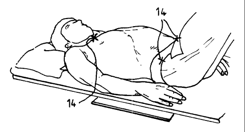
There is shown in Figure 7. a bird's-eye view of a
patient 10 lying on the table 1.2 of a x-ray system.
Figure 2 shows a perspective view of the patient. Three
retro-reflective markers 14 are: located on specific parts
of the patient's body by an operator, usually a
radiographer. The exact location of these markers
depends upon the anatomical region to be imaged. In
Figure 1 where the anterio-posterior lumbar spine is to
be imaged, so the markers are positioned on the sternal
notch and both anterior superior iliac spine. When it is
desired to obtain a lateral image of the lumbar spine,
the markers are positioned, as shown in Figure 3, on the
axilla, the anterior' superior iliac spine, and the
posterior superior iliac spine. The required positions
of the markers are readily located by an experienced
radiographer.
Each of the retro-reflective markers 14 comprise a
mirrored surface coated with gl~~ss micro-spheres
CA 02271671 1999-OS-11
WO 98/20795 PCT/GB97/03045 . .
_g..
(manufactured by 3M Corporation) which act as prisms to
reflect incident light back towards its source. The
markers reflect light received from a light source mainly .
back towards that light source and thus are highly
efficient where a Tight detector, used to detect
reflected light, is placed adjacent to the light source.
Figure 4 illustrates a combined x-ray apparatus and
positioning system, generally indicated by reference
numeral 16, which is arranged to use positional
information from the reflectin~~ markers 14 to accurately
align the x-ray tube apparatus, generally indicated by
reference numeral 1.8, to the s.=_lected anatomical region
of the patient's body. X-ray :radiation is produced by a
x-ray source 20 (shown in brokE~n outline) contained
within a lead-lined. housing 22. X-rays from the source
are collimated within a channel 24 through which the
x-ray radiation beam exits. The x-ray apparatus and
positioning system 16 is adjustably mounted on a support
26 such that the height of the apparatus 16 above the
20 patient 10 can be varied. The mounting arrangement also
allows the x-ray apparatus 16 t:o be moved in a horizontal
plane for alignment with the p~~tient.
Contained within the x-rays apparatus channel 24 is a
x-radiation translucent mirror 28, oriented at an angle
of 45° to the vertical. The mirror 28 allows x-ray
radiation to pass freely therethrough towards the patient
and the film but presents a reflecting surface to
reflected light.
Positioned inside the x-rG.y apparatus 16 is an
infra-red strobe 30 which can,c~enerate relatively short
pulses of light (i.e. 50 pulses, per second at a
wavelength of 980mm). Light from strobe 30 is reflected
from the patient 10 and from th.e reflective markers 14 is
reflected towards the x-ray apparatus 14 and the strobe
30 due to the retro-reflective nature of the markers 14.
Reflected light entering the channel 24 is reflected by
the mirror 28 towards a side-wall 32 of the channel 24.
CA 02271671 1999-OS-11
WO 98/20795 PCT/GB97/03045 . _
_g_
A charge coupled camera 33 having camera elements 34,36
are mounted in this side wall :Eor receiving the reflected
radiation. Cameras elements 3~~,36 are spaced apart in
the vertical direction such that they receive reflective
light beams 38,40 from markers 14 located horizontally
spaced apart positions on the patient 10.
Positioned beneath the table 12 is an ionisation
chamber 42 which forms part of a known type automatic
exposure device (AED). As is well known, the AED
monitors the x-ray beam transm=:tted through the area of
the patient's body under examination. Using ionisation
chamber timers, the AED terminates the exposure when a
sufficient quantity of radiation has been received to
produce a radiographic image of. the required density.
The AED exercises accurate control on the quantity of
radiation provided that the po:~itioning of the patient is
accurate in relation to the site of the ionisation
chamber. The film is placed beneath the ionisation
chamber in a mount 44 and receives the radiation which
has been transmitted through the patient and AED.
Figure 5 depicts the block: diagram which shows
schematically the image proces~~ing system, generally
indicated by reference numeral 46, which is used to
analyse the outputs of the two CCD camera elements 34,36
to determine alignment information for the x-ray
apparatus 16. The :image proce~~sing system 46 includes a
video processor 48 :Eor capturir..g visual information and a
computer for analysing the captured information. After
a predetermined time interval following activation of the
strobe 30, the video processor 48 captures an image frame
from each of the elements 34,36. The video processor 48
looks for transitions from dark. to light in these frames
and identifies any such transitions as the presence of
light reflecting markers 14. The video processor scans
each horizontal line of the captured image frames from
left to right and identifies transitions from dark to
light. Providing that transitions are located at
CA 02271671 1999-OS-11
WO 98120795 PCTIGB97/03045
-10-
. approximately the same position on two consecutive
horizontal lines, these transitions are grouped together
and are identified as the site of a reflecting marker.
In order to optimise this; search process, the
elements 34,36 are provided with respective shutters (not
shown) which open for a very short time interval during
the strobe exposura_. Typically, the shutters open for
1/1250 of a second. The outpL.t of each CCD element is
also applied to a ~;rariable gain amplifier (not shown)
within the video processor 48 the gain of which is
adjusted so that the intensity produced by the reflective
markers 14 is set <~s a 'normal' intensity. This set-up
generates image data where the reflective markers 14 show
up as bright spots while remaining areas are black. The
system may be further optimised by minimising the
background light levels in the room containing the x-ray
apparatus.
The resulting position data, which defines the
absolute position of the retro-reflective markers 14
relative to the patient 10 and the x-ray apparatus 16, is
delivered from the video processor to the computer 50.
The computer 508 contains a database of standard patient
records. The patients selected for the database cover a
range of heights arid weights, and each record contains
anatomical position data for each of a number of
anatomical regions. This anatomical data defines the
position at which t:he x-ray apparatus 16 should be
positioned for the X-ray to be centred upon the required
anatomical region, relative to the positions of the
reflecting markers 14 on the anatomical landmarks.
The radiographer uses a keyboard 52 to enter the
height, weight and identification of the patient 10 to be
imaged. The computer 50 then ,selects the appropriate
record from the stc>red database=. The radiographer then
also selects the anatomical reunion of interest and the
appropriate data is. extracted from the selected record.
From a knowledge of the actual positions of the
CA 02271671 1999-OS-11
WO 98/20795 PCT/GB97/03045
-11--
reflective markers 14, and the locating position of the
x-ray apparatus 16 relative to these markers, the
computer is able to calculate 1=he actual locating
position. This positional information can either be
displayed to the operator who cyan then manually position
the x-ray apparatus or the pos:itional information can be
fed to automatic x-ray apparatus adjustment means for
automatically repositioning them apparatus 16.
The system 46 optionally ~~ontains a display monitor
54 for displaying image data generated by one of the CCD
cameras 34,36. The. cameras 34,36 may be arranged to
generate normal continuous ima~~e data for display on the
monitor 54, the cameras only being shuttered for a
relatively short time period tc~ obtain reflective marker
position information. Once this position information has
been obtained, it can be overl;~id on the image of the
patient's body on the display. This allows the operator
to visually confirm the correct identification of the
reflective markers 14 before automatic alignment of the
x-ray apparatus occurs. Simil,~rly, the computer may be
arranged to overlay the determined locating position of
the x-ray apparatu~~ on the display.
As another example, to obtain an anterio-posterior
view of the knee joint a first marker would be placed
2.5cm above the superior aspect of the patella, a second
marker 2.5cm below the tibial tuberosity, and third and
fourth markers on t:he lateral and medial borders adjacent
to the apex of the patella. To obtain a lateral view of
the knee joint, a first marker would be placed 2.5cm
above, the upper border of the knee on the medial side,
and a second marker placed 2.5cm below the apex of the
patella on the medial side.
In addition to anatomical positioning, patient
movement can be monitored visually, thus allowing the
operator to correct patient position relative to any
movement. This function therefore provides an assessment
of the correctness of positioning before the x-ray
CA 02271671 1999-OS-11
WO 98/20795 PCT/GB97/03045
-12-
exposure has been made. Monitoring of patient movement
continues until the=_ exposure r.as been completed.
Various modifications may be made to the embodiments
hereinbefore described without departing from the scope
of the invention. The x-ray system and positioning .
apparatus can be used to image an animal body and the
light source used r_ould be replaced by an infra-red
source with an infra-red camera. The positioning system
may be used with any other suitable medical imaging
system where alignment of the source of radiation with
the part of the patient to be imaged is required.