Note: Descriptions are shown in the official language in which they were submitted.
CA 02272097 1999-OS-19
WO 98/25549 PCTIUS97/22728
-1-
ARTIFICIAL VASCULAR VALVES
Field of the Invention
The present invention relates to a tissue graft composition and method for
its preparation and use. More particularly, the present invention is directed
to non-
immunogenic submucosai tissue graft compositions prepared from warm-blooded
vertebrates and formed into vascular valves. The artificial vascular valves of
the present
invention are useful for replacing damaged or diseased valves of a warm-
blooded
vertebrate.
Background and Summary of the Invention
There are four valves in the heart that direct the flow of blood through the
two sides of the heart and out to the various organs of the body. The valves
located on
the left (systemic) side of the heart are: 1) the mitral valve, located
between the left atrium
1 S and the left ventricle, and 2) the aortic valve, located between the left
ventricle and the
aorta. These two valves direct oxygenated blood coming from the lungs, through
the left
side of the heart and into the aorta for distribution to the body. On the
right (pulmonary)
side of the heart are: 1) the tricuspid valve, located between the right
atrium and the right
ventricle, and 2) the pulmonary valve, located between the right ventricle and
the
pulmonary artery. These two valves direct deoxygenated blood coming from the
body,
through the right side of the heart, into the pulmonary artery for
distribution to the lungs,
where it again becomes re-oxygenated to being the circuit anew.
All four of these heart valves are passive structures in that they do not
themselves expend any energy and do not perform any active contractile
function. They
consist of movable "leaflets" that are designed to open and close in response
to
differential pressures on either side of the valve. The mitral and tricuspid
valves are
referred to as "atrioventricular valves" because they are located between an
atrium and a
ventricle of the heart. The mitral valve has a total of two leaflets whereas
the tricuspid
valve has three leaflets. The aortic and pulmonary valves each have three
leaflets, which
are more aptly termed "cusps".
Over 150,000 surgical procedures are performed each year to replace
diseased cardiac valves worldwide. Two out of three procedures currently
employ
CA 02272097 1999-OS-19
WO 98!25549 PCT/US97/22728
-2-
mechanical valve prostheses. Mechanical valves include caged-ball valves (such
as Starr-
Edwards valves), bi-leaflet valves (such as St. Jude valves), and titling disk
valves (such
as Medtronic-Hall or Omniscience valves). Caged ball valves typically comprise
a ball
made of a silicone rubber located inside a titanium cage, while bi-leaflet and
tilting disk
valves are made of various combinations of pyrolytic carbon and titanium. All
of these
valves are attached to a cloth (usually DacronTM) sewing ring so that the
valve prosthesis
can be sutured to the patient's native tissue to secure the implanted
artificial valve.
The main advantage of mechanical valves is their long-term durability.
However, currently available mechanical valves suffer from the disadvantage
that they are
thrombogenic and thus require lifetime anticoagulant therapy. If blood clots
form on the
valve, they may preclude the valve from opening or closing correctly or, more
importantly, the blood clots may disengage from the valve and embolize to the
brain,
causing a stroke. Anticoagulant drugs can be administered to reduce the risk
of blood
clot formation, however such drugs are expensive and potentially dangerous in
that they
may cause abnormal bleeding which, in itself, can cause a stroke if the
bleeding occurs
within the brain.
One alternative to mechanical valves are valves constructed from natural
tissues. Artificial valves constructed from natural tissues have superior
hemodynamic
characteristics, and accordingly the clinical use of tissue-based valves is
growing faster
than the overall valvular prosthesis market. Currently available tissue valves
are
constructed either by sewing the leaflets of pig aortic valves to a stmt (to
hold the leaflets
in proper position), or by constructing valve leaflets from the pericardial
sac (which
surrounds the heart) of cows or pigs and sewing them to a stmt. The stents may
be rigid
or slightly flexible and are covered with cloth (usually a synthetic material
sold under the
trademark DacronTM) and attached to a sewing ring for fixation to the
patient's native
tissue. Three tissue valves have been approved by the US FDA for implantation:
the
Carpentier-Edwards Porcine Valve, the Hancock Porcine Valve, and the
Carpentier-
Edwards Pericardial Valve.
The main advantage of tissue valves is that they do not cause blood clots
to form as readily as do the mechanical valves, and therefore, they do not
absolutely
require systemic anticoagulation. The major disadvantage of tissue valves is
that they
lack the long-term durability of mechanical valves. Currently available tissue
valves have
CA 02272097 2006-02-28
64005-656
-3-
a significant failure rate, usually appearing at approximately 8-10 years
following
implantation. In particular; currently available tissue valve prothesis
calcify after
implantation, and calcification of the valves produces stiff leaflets which
often crack.
Thus there is a need for a tissue valve construct that has long term
durability and is biocompatible with host tissues. The present invention is
directed to
artificial tissue valves formed from warm-blooded vertebrate submucosal
tissue.
Submucosal tissue, prepared in accordance with the present invention, has been
previously described as a biocompatible, non-thrombogenic gr aft material that
enhances
the repair of damaged or diseased host tissues. Numerous studies have shown
that warm-
blooded vertebrate submucosa is capable of inducing host tissue proliferation,
and
remodeling and regeneration of tissue structures following implantation in a
number of in
vivo microenvironments including lower urinary tract, body wall, tendon,
ligament, bone,
cardiovascular tissues and the central nervous system. Upon implantation,
cellular
infiltration and a rapid neovascularization are observed and the submucosa
material is
remodeled into host replacement tissue with site-specific structural and
functional
properties
Submucosal tissue can be obtained from various tissue sources, harvested
from animals raised for meat production, including, for example, pigs, cattle
and sheep or
other warm-blooded vertebrates. More particularly, the submucosa is isolated
from
warm-blooded tissues including the alimentary, respiratory, intestinal,
urinary or genital
tracts of warm-blooded vertebrates. In general submucosa is prepared from
these tissue
sources by delaminating the submucosa from both the smooth muscle layers and
the
mucosal layers. The preparation of intestinal submucosa is described and
claimed in U.S.
Patent No. 4,902,508. Urinary bladder submucosa and its preparation is
described in
U.S. Patent No. 5,554,389. Stomach submucosa has also been obtained and
characterized using similar tissue processing techniques. Such is described in
U.S.
Patent No. 6,099,567, issued on August 8, 2000. Briefly, stomach submucosa is
prepared from a segment of stomach in a procedure similar to the preparation
of
intestinal submucosa. A segment of stomach tissue is first subjected to
abrasion using
a longitudinal wiping motion to remove the outer layers (particularly
i
CA 02272097 1999-OS-19
WO 98/25549 PCT/US97122728
-4-
the smooth muscle layers) and the luminal portions of the tunics mucosa
layers. The
resulting stomach submucosa tissue has a thickness of about 100 to about 200
micrometers, and consists primarily (greater than 98%) of acelluiar,
eosinophilic staining
(H&E stain) extracellular matrix material.
Preferred submucosal tissues for use in accordance with this invention
include intestinal submucosa, stomach submucosa, urinary bladder submucosa,
and
uterine submucosa. Intestinal submucosal tissue is one preferred starting
material, and
more particularly intestinal submucosa delaminated from both the tunics
muscularis and at
least the tunics mucosa of warm-blooded vertebrate intestine.
As a tissue graft, submucosal tissue undergoes remodeling and induces the
growth of endogenous tissues upon implantation into a host. It has been used
successfully in vascular grafts, urinary bladder and hernia repair,
replacement and repair of
tendons and ligaments, and dermal grafts. The preparation and use of submucosa
as a
tissue graft composition is described in U.S. Patent Nos. 4,902,508;
5,281,422;
5,275,826; 5,554,389; and other related U.S. patents. When used in such
applications, the
graft constructs appear not only to serve as a matrix for the regrowth of the
tissues
replaced by the graft constructs, but also promote or induce such regrowth of
endogenous
tissue. Common events to this remodeling process include: widespread and very
rapid
neovascularization, proliferation of granulation mesenchymal cells;
biodegradation/resorption of implanted intestinal submucosal tissue material,
and lack of
immune rejection.
Submucosal tissue is also capable of promoting endogenous regrowth and
healing of damaged or diseased cardiac tissues, including the endocardium,
pericardium,
and myocardium. In particular, damaged or diseased myocardial tissues can be
replaced
i» vivo with a composition comprising submucosal tissue of a warm blooded
vertebrate to
enhance the formation of endogenous tissues having spontaneous contractile
properties.
The present invention is directed to the use of submucosal tissue to
prepare tissue valve constructs, and the use of those valve constructs to
replace or repair
damaged or diseased valves of the heart and the circulatory system of a warm-
blooded
vertebrate.
CA 02272097 2006-02-28
64005-656
-4a-
According to one aspect of the present invention,
there is provided a tissue graft in the form of a bicuspid
valve for replacement of a defective vascular valve, said
tissue graft comprising submucosal tissue, delaminated from
both the tunica muscularis and at least the luminal portion
of the tunica mucosa, in the form of a continuous tube
having a diameter (D) approximating that of the defective
valve said tube having first and second opposite ends and a
triple walled intermediate portion having length (L) about
1.5D to about 3.5D; said triple walled portion of the tissue
graft being formed by everting the first end of the tube to
form a tubular construct having a double walled end and a
double walled portion proximal to and extending from said
double walled end and reverting said first end over the
double walled portion and the double walled end of the
tubular construct; wherein the two walls of the double
walled portion are sutured together to form a sutured
portion having a length S and the end of the sutured portion
proximal to the double walled end is located at least a
distance 1/2D from the double walled end, and the ratio of L
to S is about 2.5 to about 3.5.
According to another aspect of the present
invention, there is provided a method of forming a synthetic
tissue vascular valve, said method comprising overlaying a
sheet of submucosal tissue onto a stmt having a plurality
of stmt posts wherein the submucosal tissue contacts the
stmt posts of the stmt; fixing the submucosal tissue to
the tips of the stmt posts; folding the sheet of submucosal
tissue to form folds that extend from the top of each stmt
post to the center of an annular base; conditioning the
tissue to retain the shape of the tissue; fixing the
submucosal tissue to the sides of the stmt posts and the
CA 02272097 2006-02-28
64005-656
-4b-
base of the stmt; and cutting the fold in the submucosal
tissue to form the commissures of the valve.
According to still another aspect of the present
invention, there is provided a synthetic tissue valve
comprising a stmt, comprising an annular base and three
stmt posts that extend vertically from said annular base,
wherein the annular base and the three stmt posts define a
central axis that extends through the center of the annular
base equidistant from each of the stmt posts; and a layer
of submucosa overlaid onto the stmt posts and fixed onto
the stmt along the perimeter of each of the stmt posts,
said submucosa being folded back upon itself along three
radial axes that extend from a point along the central axis
to the top of each of the three stmt posts to form the
submucosa layer into three concave semi-hemispheres of
submucosa, said submucosa having a slit cut along the folds
formed at the three radial axes to allow unidirectional flow
from the convex side of the submucosal tissue to the concave
side.
CA 02272097 1999-05-19
WO 98/25549 PCT/US97122728
-5-
Brief Description of the Drawings
Fig. 1 is a sectional view of a submucosal tissue wrapped mandrel wherein
the layers of submucosal tissue are subjected to vacuum pressing.
Fig. 2 is a perspective view of a mandrel wrapped with a sheet of
submucosal tissue wherein the two ends of the sheet of submucosal tissue are
overlapped
to form a tube of submucosal tissue having an overlapped region defined by the
overlap
angle 8.
Fig. 3 is a perspective view of a mandrel spirally wrapped with a narrow
sheet of submucosal tissue to form a tube of submucosal tissue.
Figs. 4a-4c are sectional views of one embodiment of a vascular valve
formed from a tube of submucosal tissue.
Figs. Sa-Se are perspective views of one embodiment of a vascular valve
formed from a tube of submucosal tissue.
Fig. 6a illustrates a stmt having an annular base and three stmt post
extending from the base.
Fig. 6b illustrates the stent of Fig. 6a covered with one or more narrow
sheets of submucosal tissue.
Fig. 7a illustrates the components used to form a tricuspid valve.
Fig. 7b illustrates the assembled construct.
Fig. 8a illustrates a heat treated submucosal tissue covered stmt shaped as
a tricuspid valve.
Fig. 8b illustrates the final tricuspid valve tissue graft construct.
Fig. 9 is a graphic representation of experimental data plotting calcium
concentration in implanted native and treated submucosal tissue, as measured
by atomic
absorption, versus length of implantation time.
Fig. 10 is a sectional view of testing apparatus for measuring forward and
reverse flow rates for tissue valve constructs.
Figs. l la and l 1b are sectional views of a tissue valve construct formed in
accordance with the present invention. Fig. l la illustrates the operation of
the valve in
the presence of a forward flow; Fig. l 1b illustrates the operation of the
valve in the
presence of a reverse flow.
i
CA 02272097 1999-OS-19
WO 98/25549 PCT/US97122728
-6-
Fig. 12 is a sectional view of a testing apparatus for measuring forward
flow resistance and leakage of the constructed tricuspid valve constructs.
Fig. 13 is a graphic representation of experimental data, plotting flow rate
versus pressure drop across various tissue valve constructs.
Detailed Description of the Invention
A variety of tissue sources have been used to fabricate and repair heart
valves, including the fascia lata, bovine pericardium and dura mater. In
addition,
researchers have studied the potential use of animal valves (such as porcine
valves) and
cadaver valves to replace human valves. Investigators working with tissue
valve
prostheses have discovered that fresh tissues have a tendency to shrink over
time,
resulting in the failure of the valves to seal completely and prevent backflow
of fluids.
Therefore investigators have used glutaraldehyde treatments to stiffen the
tissue and
prevent subsequent shrinking of those tissues. Advantageously, glutaraldehyde
treatment
1 S of the tissues also reduces the probability of the tissue implant invoking
an immune
response. However, the glutaraldehyde treatment also shortens the in vivo
lifespan of the
tissue valve.
Natural valve leaflets consist of a very pliable spongy material that
contains fibrous materials oriented such that the tissue is resistant to
stretching but not to
compression forces. This low resistance to axial compressive forces give the
natural heart
valve tissue its characteristic high pliability. When such tissue is fixed
with
glutaraldehyde, it becomes up to four times stiffer than fresh tissue. The
fixation process
induces molecular crosslinks resulting in the tissue becoming more resistant
to the axial
compression forces that accompany bending. As a result the stiffer tissue
buckles during
bending, and with each successive heartbeat the tissue tends to buckle at the
same
location, fatiguing the collagen fibers until they break. Furthermore,
glutaraldehyde
treatment of tissues appears to induce the calcification of the treated
tissues (see Example
1). Calcification of the tissues leads to further stiffening of the leaflets
aggravating the
implant's susceptibility to cracking and failure of the implanted tissue
valve.
The present tissue valve prostheses are synthesized from warm-blooded
vertebrate submucosal tissue: Submucosal tissue isolated in accordance with
the
procedures described in US Patent Nos. 4,902,508 and 5,554,389 does not induce
an
CA 02272097 1999-OS-19
WO 98/25549 PCTILTS97122728
immune response upon implantation into a host species. Therefore tissue valve
constructs
prepared from vertebrate submucosal tissue in accordance with the present
invention do
not need to be treated with glutaraldehyde prior to implantation.
Submucosal tissue can be used to repair an existing valve in vivo by
replacing a cusp of a bicuspid or tricuspid valve. Alternatively, submucosal
tissue can be
used to construct an entire valve to replace a heart valve or other
circulatory valve or duct
valve. Advantageously, the submucosal tissue of the present valve constructs
will induce
the formation of endogenous cells and tissues that infiltrate the submucosal
tissue and
ultimately replace the graft material with endogenous tissue.
The submucosal tissue graft constructs of the present invention can be
sterilized using conventional sterilization techniques including
glutaraldehyde tanning,
formaldehyde tanning at acidic pH, propylene oxide or ethylene oxide
treatment, gas
plasma sterilization, gamma radiation, electron beam radiation, peracetic acid
sterilization.
Sterilization techniques which do not adversely affect the mechanical
strength, structure,
1 S and biotropic properties of the submucosal tissue is preferred. For
instance, strong
gamma radiation may cause loss of strength in the submucosal tissue. Preferred
sterilization techniques include exposing the graft to peracetic acid, 1-4
Mrads gamma
irradiation (more preferably 1-2.5 Mrads of gamma irradiation), ethylene oxide
treatment
or gas plasma sterilization; peracetic acid sterilization is the most
preferred sterilization
method. Typically, the submucosal tissue is subjected to two or more
sterilization
processes. After the submucosal tissue is sterilized, for example by chemical
treatment,
the tissue may be wrapped in a plastic or foil wrap and sterilized again using
electron
beam or gamma irradiation sterilization techniques.
Submucosal tissue can be stored in a hydrated or dehydrated state.
Lyophilized or air dried submucosa tissue can be rehydrated and used in
accordance with
this invention without significant loss of its biotropic and mechanical
properties.
In one embodiment in accordance with the present invention, a single piece
vascular valve can be constructed from a tube of warm-blooded vertebrate
submucosa.
The tubes of submucosal tissue used to form the tissue valves of the present
invention are
formed to have fluid-tight seams and can be shaped to match the endogenous
tissue to be
replaced by the graft construct. In one preferred embodiment the vascular
valve is formed
from a tube of intestinal submucosal tissue and is configured as a duck-bill
valve.
i
CA 02272097 1999-OS-19
WO 98!25549 PCTlUS97/Z2728
_g_
Tubes of submucosal tissue can be prepared from a variety of sources
including intestinal submucosal tissue delaminated from both the tunics
muscularis and at
least the lumens! portion of the tunics mucosa as described in US Patent ~lo.
4,902,508.
In brief, a segment of vertebrate intestine, preferably harvested from
porcine, ovine or
bovine species, but not excluding other species is subjected to abrasion using
a
longitudinal wiping motion to remove the outer layers, comprising smooth
muscle tissues,
and the innermost layer, i.e., the luminal portion of the tunics mucosa.
The diameter of the prepared tube of submucosal tissue should be
approximately the same as the diameter of the recipient blood vessel. In one
embodiment
this is accomplished by manipulating the submucosal tissue to define a
cylinder having
diameter approximately the same as that of the recipient blood vessel and
suturing or
otherwise securing the submucosal tissue longitudinally to form a tube of the
appropriate
luminal diameter. Thus, for example, a vascular graft can be prepared by
selecting a
sterile glass rod having an outer diameter equal to that of the recipient
blood vessel,
inserting the glass rod into the lumen of the tube of submucosal tissue (for
example,
submucosal tissue prepared from a segment of intestinal tissue) and gathering
the
redundant tissue. The desired lumen diameter is achieved by suturing along the
length of
the graft (for example, using two continuous suture lines or a simple
interrupted suture
line) or by using other art-recognized tissue securing techniques.
The tube of submucosal tissue can also be formed from a sheet of
submucosal tissue. The term "sheet of submucosal tissue" is defined herein to
include
tissue constructs comprising multiple strips of submucosal tissue, wherein the
strips are
overlapped and compressed under dehydrating conditions to form a unitary
construct
having a surface area greater than the surface area of any one of the
individual strips used
to form said construct. The term sheet of submucosal tissue also includes a
tube of
intestinal submucosal tissue that is cut along the length of the tube and laid
flat.
In one embodiment a tube of submucosal tissue is formed from a sheet of
submucosal tissue by wrapping the tissue around a cylindrically shaped mandrel
of the
appropriate diameter. The excess tissue is removed, and the opposing ends
bound to one
another to form a tube having a lumens! diameter approximately equal to the
diameter of
the mandrel. The opposing ends of the sheet can be bound to one another by
adhesive
CA 02272097 1999-OS-19
WO 98125549 PCT/US97122728
-9-
pastes, sutures, fusion of the ends by overlapping the tissue and heating
under dehydrating
conditions or any other fixation technique known to those skilled in the art.
In one embodiment, as shown in Fig. 1, sheets of submucosal tissue 2 are
shaped into a tubular structure of any size by spirally wrapping the sheet of
submucosal
tissue 2 around a cylindrical mandrel 12 of the appropriate diameter and
compressing the
overlapped tissue under dehydrating conditions. Preferably the mandrel 12 is a
hollow
cylinder made of plastic or metal having a plurality of holes 16 formed in the
cylinder
wall. The compression of the tissue can be achieved by forming a seal 4 at one
end of the
mandrel 12 and pulling a vacuum through the lumen of the mandrel 12 (See Fig.
2).
Alternatively, the tissue can be compressed by applying an external force to
the exterior
surface of the wrapped submucosal tissue to compress the tissue against the
mandrel. In
one embodiment the final seam of the spirally wrapped tissue can be further
secured by
sutures, spot-welding with heat or treating the seam with glutaraldehyde.
In accordance with the present invention, the tube of submucosal tissue
can be formed as a multilaminate construct wherein one or more sheets of
submucosal
tissue are wrapped around a mandrel in multiple layers. The dimensions of the
individual
sheets of submucosal tissue used is not critical and the term "sheet of
submucosal tissue"
is defined herein to include submucosal tissue from one or more vertebrate
sources or
organs in a wide variety of sizes and shapes.
In one embodiment the sheet of submucosal tissue 2 has a width equal to
the desired length of the formed tube of submucosal tissue, and the tube is
formed such
that the first edge 13 of the sheet of submucosal tissue 2 is substantially
parallel to the
second opposite edge 15 of the sheet of submucosal tissue in the formed tube.
The
second opposite edge 15 extends over the first edge 13 to form an overlapped
region
defined by the overlap angle 8 {See Fig. 1). The sheet submucosal tissue 2 is
applied to
the mandrel 12 by a rolling motion with the desired number of layers
(typically two) and
an overlap region (defined by an overlap angle (8) of about 30 degrees) to
form a tube of
submucosal tissue having a longitudinally extending seam, as shown in Fig. 1.
The
wrapped submucosal tissue is compressed against said mandrel under dehydrating
conditions for a predetermined time period, and the tubular prosthesis is then
removed
from the mandrel. The resulting tubular construct has a seam extending the
length of the
construct. The seam of the tube of submucosal tissue is sealed using
techniques known to
CA 02272097 2006-02-28
64005-656
-10-
those skilled in the art including, crosslinking, suturing, binding with
adhesives or fusing
by compressing under dehdyrating conditions, to resist movement of fluids from
the
lumen through the seam to the exterior of the tube. This seam can be further
secured by
spot-welding with heat or with glutaraldehyde.
Alternatively the tube of submucosa can be formed from one or more
narrow sheets of submucosal tissue that have a width less than the desired
length of the
formed tube of submucosal tissue (See Fig. 3). In this embodiment a narrow
sheet of
submucosal tissue 18 is wound about a mandrel 14 multiple times wherein the
narrow
sheet is at least partially overlapped leaving no portion of the underlying
mandrel
exposed. In one embodiment the mandrel 14 is provided with a plurality of
holes 8. The
amount of overlap in the partially overlapped strips of submucosal tissue
ranges between
10-60% of the width of the individual strip and more preferably the overlapped
portion is
a 50% overlap. In one embodiment multiple sheets of submucosal tissue can be
overlaid
onto the mandrel, provide that at least a portion of each piece of submucosal
tissue
overlaps a portion of another piece of submucosal tissue wrapped onto the
mandrel.
Submucosal tissue can be conditioned, as described in U.S. Patent No.
5,275,826 to alter
the visco-elastic properties of the submucosal tissue. In one embodiment the
submucosal
tissue is conditioned by stretching the graft material longitudinally to a
length longer than
the length of the submucosal tissue from which the graft construct was formed.
One
method of conditioning the tissue by stretching involves application of a
given load to the
submucosa for three to five cycles. Each cycle consists of applying a load to
the graft
material for five seconds, followed by a ten second relaxation phase. Three to
five cycles
produce a stretch-conditioned graft material with reduced strain. The graft
material does
not immediately return to its original size; it remains in a "stretched"
dimension:
Optionally, the graft material can be preconditioned by stretching in the
lateral dimension.
In one embodiment the submucosai tissue is stretched using 50% of the
predicted ultimate load. The "ultimate load" is the maximum load that can be
applied to
the submucosal tissue without resulting in failure of the tissue (i.e., the
break point of the
tissue). Ultimate load can be predicted for a given strip of submucosal tissue
based on the
source and thickness of the material. Accordingly, one method of conditioning
the tissue
by stretching involves application of 50% of the predicted ultimate load to
the submucosa
CA 02272097 1999-OS-19
WO 98/25549 PCTIUS97122728
-11-
for three to ten cycles. Each cycle consists of applying a load to the graft
material for five
seconds, followed by a ten second relaxation phase. The resulting conditioned
submucosal tissue has a strain of less than 30%, more typically a strain from
about 20% to
about 28%. In one preferred embodiment conditioned the submucosal tissue has a
strain
of no more than 20%. The term strain as used herein refers to the maximum
amount of
tissue elongation before failure of the tissue, when the tissue is stretched
under an applied
load. It is expressed as a percentage of the length of the tissue before
loading. The
conditioned submucosal strips can be used to form the tubular construct or
alternatively
the tubular construct can be conditioned after its formation.
In accordance with one embodiment warm-blooded vertebrate submucosa
delaminated from the both the tunica muscularis and at least the luminal
portion of the
tunics mucosa is conditioned to have a strain of no more than 20%. The
submucosal
tissue is conditioned by stretching, chemically treating, enzymatically
treating or exposing
the tissue to other environmental factors. In one embodiment the sheets of
submucosal
tissue are conditioned by stretching in a longitudinal or lateral direction so
that the sheets
of submucosal tissue have a strain of less than 30%, more typically a strain
from about
20% to about 28%. In one preferred embodiment conditioned the submucosal
tissue has
a strain of no more than 20%.
In addition, a gentle heating treatment can be utilized to stiil'en the
submucosal tissue and to ensure the shape memory of the tissue. The heat
treatment
comprises exposing the submucosal tissue to a liquid, preferably water that
has been
heated to about 65 to about 100°C. The submucosa is exposed to the
heated liquid for a
brief time period ranging from about 10 seconds to about five minutes.
Preferably the
entire tissue graft does not equilibrate with the temperature of the liquid
medium, but only
the surface of the graft reaches the temperature of the medium.
In accordance with one embodiment, a tube of submucosal tissue is
utilized to form an artificial vascular valve for replacement of an endogenous
defective
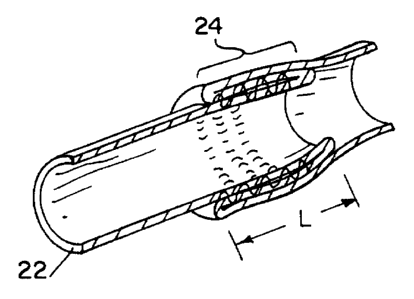
vascular valve (See Figs. 4a-4c and Figs. Sa-Se). In accordance with one
embodiment
shown in Figs. 4a-4c the tissue valve construct is in the form of a continuous
tube 30
having a diameter (D) approximating the diameter of the defective valve. The
continuous
tube 30 has a first 20 and second opposite ends 22 and a triple walled
intermediate
portion 24 having length {L) of about 1. SD to about 3 . SD. The triple walled
portion of
i
CA 02272097 1999-OS-19
w0 98125549 PCT/CTS97122728
-12-
the tissue graft is formed by evening the first end 20 of the tube to form a
tubular
construct having a double walled end 26, and a double walled portion 28
proximal to and
extending from said double walled end 26. The two walls of the double-walled
intermediate portion are sutured together over a region having a length S,
wherein the
sutured region is located at least a distance 1/2D from the double walled end
26 of the
tubular construct. Typically the suture length is about .8 to about 5cm, more
preferably
about 1 about 2 cm. The first end 20 is reverted over the sutured double-
walled portion
and the double-walled end 26 of the tubular construct, wherein the ratio of L
to S is about
2 to about 5 more preferably about 2.5 to about 3.5. Tissue valves having an
overlaplsuture ratio (ratio of L to S) in the range of 3.0 to 3.2 provide
excellent
forwardlreverse ratios of approximately 22. These valves also have been shown
to work
well over a wide range of pressures.
In another embodiment the tissue valve is in the form of a continuous tube
having a diameter (D) approximating that of the defective valve. The tube has
a first and
second opposite ends and a triple walled intermediate portion having length
(L) about
1.5D to about 3.5D. The triple walled portion of the tissue graft is formed by
evening the
first end of the tube to form a tubular construct having a double walled end,
and a double
walled portion proximal to and extending from said double walled end. The
first end is
then reverted over the double walled portion and the double walled end of the
tubular
construct, and the three walls of the triple-walled intermediate portion are
sutured
together to form a sutured portion having a length S of about 0.8 to about 5
cm, more
preferably about 1 to about 2 cm. The end of the sutured portion proximal to
the double
walled end is located at least a distance 1/2D from the double walled end of
the tubular
construct, and the ratio of L to S is about 2.0 to about 5, more preferably
about 2.5 to
about 3.5.
In another embodiment a single piece artificial valve is constructed from a
tube of submucosal tissue 32 having a first end 34 and a second end 36 in.
accordance
with the following method (See Figs. 5a-5e). The first end 34 of the tube of
submucosal
tissue 32 is evened and pulled back over the tube of submucosal tissue 32 to
form a
double walled end 38 and a double walled portion 40 proximal to and extending
from the
double walled end 38 (See Fig. 5b). The double walled portion 40 is compressed
to
flatten the tube of submucosal tissue 32 and the tube of submucosal tissue 32
is sealed
CA 02272097 1999-OS-19
WO 98125549 PCT/US97122728
-13-
along two lines extending from the lateral edge 44 of the flattened tube
towards one
another. In one embodiment a pair of diametrically opposed longitudinal suture
lines 42
are used to suture the walls of the double walled portion 40 together.
Preferably the pair
of suture lines 42 start at the lateral edge 44 of the tube and are angled
towards the center
of the flatten tube, but the suture lines 42 do not meet (See Fig. 5c). After
the double
walled portion 40 is the tube of submucosal tissue 32 of submucosa has been
sutured, the
submucosal tissue portions 46 laying outside suture lines 42 are removed
(i.e., by cutting).
The first end 34 is then reverted over the sutured double-walled portion and
the region of
the tubular construct where the suture lines 42 meet the lateral edge 44 are
sealed, for
example by sutures to prevent any leakage of the vessel contents from the
lumen to the
exterior.
Alternatively a bicuspid or tricuspid valve is constructed using an annular
shaped stent in combination with a sheet of submucosal tissue. Typically the
stmt is
constructed from a biocompatible synthetic polymer or from metal that is
coated with a
biocompatible polymer. However, other material can be used to form the stent,
provided
that the material has the requisite strength to maintain its shape when
inserted into the
host. In one embodiment the stent is formed from submucosa that has been
treated to
stiffen the material. For example the submucosal tissue can be shaped in the
form of a
stent and then crosslinked, using standard crosslinkirig agents such as
glutaraldehyde and
techniques familiar to the skilled practitioner. Alternatively the submucosal
tissue can be
formed in the shape of a stent and subjected to a heat treatment to stiffen
the graft
construct. In one embodiment the submucosa based stent is heated in a liquid
at a
temperature of about 80° to about 100° for about ten seconds to
about five minutes.
In one embodiment of the present invention (shown in Fig. 6a), a stem 48
comprises a base formed as an annular ring 50 having a plurality of stent
posts 52
extending substantially perpendicular to the plane of the annular ring. The
stent is
selected so that it has a ring diameter approximately the same as the diameter
of the vessel
that will receive the constructed valve. In one embodiment the external
surface of the
stent is covered with submucosal tissue so that upon implantation into the
host, host
tissue will contact only submucosal tissue. Therefore, when the stmt comprises
a
biocompatable synthetic polymer or comprises a biocompatable polymber covered
metal,
the surface of the stmt is optionally first covered with a layer of submucosal
tissue before
CA 02272097 2006-02-28
64005-656
-14-
formation of the tissue valve. For example, one or more sheets of submucosal
tissue 54
can be wrapped around the stent such that the entire surface of the stent is
covered with
at least one layer of submucosal tissue (See Fig. 6b). After the stent has
been wrapped
with the submucosal tissue, the tissue can be partially dried to enhance the
adherence of
S the submucosal tissue to the stent. In addition, sutures or other fixation
means known to
those skilled in the art can be used to secure the submucosal tissue to the
surface of the
stent.
Alternatively, the stent surface can be covered with submucosal tissue by
contacting the stent with fluidized submucosal tissue and then drying the
submucosa to
form a coating on the stmt. For example, a stent coated with fluidized
submucosal tissue
can be heated to 37°C for 1-2 hours to dry the fluidized tissue onto
the stem. Fluidized
submucosal tissue is prepared as described in U.S. Patent No. 5,275,826.
The bicuspid and tricuspid valves of the present invention can be formed
using a single layered sheet of submucosal tissue or a multi-laminate
submucosa
construct. Mufti-laminate submucosal tissue constructs can be formed by
overlapping
strips of submucosal tissue and binding the overlapped tissues to one another.
The
overlapped tissues can be bound together through the use of sutures,
adhesives,
crosslinking agents, heat treatments, or by compressing the tissue under
conditions
conducive to dehydration of the tissue. Advantageously, large area sheets of
submucosa
can be formed by partially overlapping strips of submucosa and compressing the
tissue
under dehydrating conditions to form a unitary heterolaminate graft construct
having a
surface area larger than any of the strips of submucosa used to form the
construct.
Alternatively, homolaminate constructs can be prepared by overlaying two or
more strips
of submucosal tissue and compressing the tissue under conditions conducive to
dehydration of the tissue, with or without the use of sutures, adhesives or
crosslinking
agents.
The submucosal tissue valve prostheses of the present invention have
excellent flow dynamics and unlike commercially available glutaraldehyde
treated porcine
valves, they do not calcify after implantation. Furthermore, the present
tissue valve
prostheses are optionally heat treated to maintain the proper form of the
valve while
avoiding/eliminating the disadvantages associated with glutaraldehyde
treatments.
CA 02272097 1999-OS-19
WO 98!25549 PCT/US97l22728
-1 S-
The preparation of a mufti-cusped vascular tissue valve construct from a
sheet of warm-blooded vertebrate submucosa is a mufti-step process. First a
vascular
stmt must be selected that has a diameter approximately the same size as the
diameter of
the vessel that will receive the tissue valve. The stmt comprises an annular
base and a
plurality of stent posts distributed equidistant from one another on the
annular base and
extending from said base. A stent having two stent posts is used to prepare a
bicuspid
valve, and a stent having three stmt posts is selected for preparation of a
tricuspid valve.
Each of the stent posts extend from the annular base at the same approximate
angle
relative to the plane defined by the circumference of the annular base. This
angle ranges
from about 50° to about 90°, more preferably from about
75° to about 90°. In one
embodiment the stmt posts extend substantially perpendicularly from' the
annular base.
The multiple stmt posts define a luminal space wherein a central axis extends
through the
center of the annular base and the luminal space equidistant from each of the
stmt posts.
A single layered sheet or multilaminate sheet is then overlaid onto the stmt
1 S posts of the stmt. Submucosal tissue has an abluminal and a luminal
surface. The luminal
surface is the submucosal surface facing the lumen of the organ source and
typically
adjacent to an inner mucosa layer in vivo, whereas the abluminal surface is
the
submucosal surface facing away from the lumen of the organ source and
typically in
contact with smooth muscle tissue in vivo. In preferred embodiments the
submucosal
tissue is overlaid onto the stent with the luminal surface up and the
abluminal surface of
the submucosa in contact with the surface of the stent. Furthermore, the sheet
of
submucosa is selected to have a length and width at least twice as large as
the diameter of
the annular base. In one embodiment the sheet of submucosal tissue is formed
as a square
piece of tissue having a length and width of 2D (twice the size of the
diameter of the stmt
annular base). The submucosa tissue is centered over the stent posts and
secured to the
top of one of the stent posts using standard fixation techniques known to
those skilled in
the art including clamps, adhesives, sutures or a combination thereof. In one
preferred
embodiment the submucosa is secured by suturing the tissue to the top of the
stent post.
The submucosa tissue is then folded back on itself to form a crease that
extends from a point above the top of the submucosa-secured stmt post to a
point along
the central axis of the stent. The tissue is then sequentially secured to the
remaining stent
posts and the tissue is folded back to form a crease at each remaining stent
post in a
i
CA 02272097 1999-OS-19
WO 98/25549 PCT/US97/22728
-16-
similar manner as for the first stent post. In accordance with one embodiment,
the folded
tissue is held in place by compressing the folded tissue between two rigid
plates. In one
embodiment the crease runs substantially parallel to the line or horizontal
plane defined by
the top of the stent posts. In an alternative embodiment, the crease is formed
at an angle
of about 1° to about 45°, more particularly about 1° to
about 20° (wherein the origin of
the angle is located at the stmt post), relative to the line or horizontal
plane defined by the
top of the stent posts. In accordance with the present invention, the
preparation of a
bicuspid valve requires the formation of two creases, one running from each of
the two
stmt posts and meeting at a point along the central axis of the stmt. The
preparation of
the tricuspid valve requires the formation of three separate creases, each of
which starts at
a point above one of the three stent posts and meets at a point along the
central axis of
the stent. Accordingly the submucosa is sequentially secured to each of the
stmt posts
and the tissue is folded to form a crease that extends from each stmt post.
After the appropriate number of creases have been prepared the folded
submucosa is optionally subjected to a heat or chemical treatment to stiffen
the
submucosa and to ensure the shape memory of the tissue. For example, the
tissue can be
treated with a dilute solution (0.1% to 1%) of a chemical crosslinking agent
such as
giutaraldehyde to stiffen the tissue. In one preferred embodiment the tissue
is stiffened by
subjecting the tissue to a heat treatment. The heat treatment in
accordance.with one
embodiment comprises heating the tissue in water at a temperature of about
80° C to
about 100° C for about ten seconds to about five minutes, more
preferably heating the
tissue at a temperature of about 88°C to about 92°C, for about
ten to about ninety
seconds.
In one embodiment the folded submucosal tissue is clamped between two
plates of rigid material, for example metal, plastic, glass or ceramic plates
or a
combination thereof, and the clamped material is subjected to a heat or
chemical
treatment to stiffen the submucosa while the tissue remains clamped. The rigid
plates are
preferably rectangular in shape with a rounded end. portion and the plates
have a width
ranging from about (2/5)D to about(1/2)D, and a length ranging from about
(1/2)D to
about D, wherein D = the diameter of the annular stmt base. In one embodiment
the
plates have a width of about(2/5)D and a length of about 3/4D.
CA 02272097 1999-05-19
WO 98125549 PCT/US97l22728
-17-
After the submucosa has been clamped and optionally treated, the clarrrps
and plates are removed and the submucosal tissue is secured along the
perimeter of the
stent posts using standard techniques know to those skilled in the art. In one
embodiment
the submucosa is secured through the use of sutures. The submucosa is then cut
along
the creases formed in the submucosa and extending from each of the stent posts
to form a
commissure. The formed tissue valve is optionally further conditioned by heat
or
chemically treating the tissue and the heat treatment can be conducted while
the tissue
valve is subjected to back pressure.
The preparation of a tricuspid valve in accordance with the present
invention is described with reference to Figs. 7a, 7b, 8a and 8b. In
accordance with one
embodiment of the present invention, a tricuspid valve is formed from
intestinal
submucosa delaminated from both the tunics muscularis and at least the luminal
portion of
the tunics mucosa by the following method: An appropriate sized stmt 56 having
an
annular base 61 of diameter D and three stent posts 60 is acquired and is
optionally
covered with a Dacron mesh or with submucosal tissue as described above. A
length of
delaminated intestinal submucosal tissue is cut approximately 2 times the
outer diameter
of the stent. The submucosal tissue segment is then cut longitudinally to form
a
rectangular submucosa sheet 58 having a length and width of approximately 2D.
The stent 56 is placed on a horizontal table with its annular base 61
contacting the table. The submucosa sheet 58 is centered over the stmt 56 with
the
luminal side 62 facing up and the submucosal tissue is laid over the stmt
posts 60 (See
Fig. 7a). One of the stent posts is selected for suturing the submucosal
tissue to the stent
post tip 65. The submucosal tissue is folded at the same stmt post where the
suture was
made and the folded line 64 of submucosal tissue is pulled above the
horizontal plane of
the stent posts. Two metal plates 66 rectangular in shape {having dimensions W
of 2/5D
and Ll of 3/4D) with a curved end portion 68 are used to sandwich the folded
submucosal
tissue between the plates 66 with the curved end portion 68 of the plate 66
facing down.
A flat head paper clip 70 is used to clamp the plates together by fitting it
around the stmt
post 60 (See Fig. 7b).
The commissure position is the only line where a fold should exist (i.e., the
submucosal tissue should not be allowed to overlap in areas that is in contact
with the
plates). Repeat the steps of forming folds of submucosa at each of the
remaining stent
l
CA 02272097 1999-OS-19
WO 98/25549 PCT/US97/22728
-18-
post locations. The valley of the cusp 72 must have a flat planar appearance
(i.e., it
should not contain wrinkles).
The excess submucosal tissue is then pinned with pins 74 at the base of the
stent 56 between each of the stmt posts 60 after the flathead clips 70 are in
place. The
valve assembly is placed into a pan of near boiling water (approximately
80°-90°C) and
the assembly is removed after about 10 to about 90 seconds. The heat treatment
will
cause the sheet of submucosal tissue to shrink. The flat head clips 70'and
plates 66 are
then removed, but the pins 74 at the annular base 61.base are not removed. The
submucosa sheet 58 now conforms closely to the top periphery of the stent 56.
The submucosa sheet 58 is then sutured along the periphery of each stent
post 60 making certain that the creases that were formed after boiling remain
in the same
position while suturing (See Fig. 8a). The spacing between the sutures should
be less
than or equal to 1.5 mm. Pins 74 are then removed from the base of the stent
and the
excess submucosal tissue around the outside area of the orifice is removed'.
The assembly
is re-clamped between the plates 66 and the flat head clips 70, in the same
manner as
above and the assembly is placed back into near boiling water (approximately
80°-90°C)
for another 10 to 90 seconds. The assembly is removed from the near boiling
water and
the clamps and plates are removed.
The folded submucosal tissue is cut in a horizontal direction from the stent
post tip 65 to the center of the luminal space defined by the stmt posts 60 to
form the
commissures (See Fig. 8b). The commissure 76 and the stmt post tips 65 should
be at
approximately the same height and the resultant leaflets must be flush with
each other.
The tricuspid synthetic tissue valve prepared in accordance with the
method described in the present invention comprises a stent and a layer of
submucosa
overlaid onto the stent posts. The stent comprises an annular base and three
stent posts
extending vertically from said annular base, wherein the annular base and the
three stent
posts define a central axis that extends through the center of the annular
base equidistant
from each of the stent posts. The submucosal tissue is fixed onto the stent
along the
perimeter of each of the scent posts, and is folded back upon itself along
three radial axes
that extend from a point along the central axis to the top of each of the
three stent posts.
The three folds in the submucosal tissue form the submucosa layer into three
concave
semi-hemispheres of submucosa. Cutting the folded submucosal tissue along the
three
CA 02272097 1999-OS-19
WO 98/25549 PCTlUS97122728
-19-
radial axes forms the commissures of the heart valve that allow unidirectional
flow from
the convex side of the submucosal tissue to the concave side. In one
embodiment, the
commisures of the radial axes of the constructed tissue valve are
perpendicular to the
central axis and are essentially co-planar with the plane defined by the tips
of the stent
posts as shown in Fig. 8b.
Ezample 1
Subcutaneous Calcification Studies on Submucosaf Tissue
Glutaraldehyde (GA) treatment of biomaterials is known to promote
calcification, poor host-tissue incorporation and ultimately mechanical
failure of
bioprotheses. To anticipate the cardiovascular applications of submucosal
tissue, the
calcification potential of submucosal tissue and the effect of GA treatment
were studied.
Experiment 1
Submucosal tissue treated with peracetic acid (PAA), a mild
glutaraldehyde (GA) exposure (0.6% for S min.), or rinsed but otherwise
untreated, along
with cusps from commercial porcine, bioprosthetic heart valves (glutaraldehyde
treated by
manufacturer) were implanted in the well-established weanling rat model. A
specimen
1 cm by 1 cm of each of the 4 tissues was implanted in surgically created
subcutaneous
pockets on the ventral abdomen of 18 rats. Six rats were sacrificed and
tissues harvested
and evaluated at 1, 2 and 4 weeks post-implantation. Histologic studies
indicated that by
2 weeks all submucosal tissue specimens, except for GA treated specimen, were
well
incorporated into the surrounding tissue and by 4 weeks all submucosal tissue
specimens
appeared similar. VonKossa's stain for mineralization indicated that no
significant
calcification occurred in the PAA or rinsed submucosal tissue specimens at any
of the
time-periods, but GA treated submucosal tissue and the porcine valve cusps
showed
significant calcium accumulation-even at the 1-week evaluation period
(P=0001).
Experiment 2
Four test samples: 1) native (cleaned, untreated) submucosal tissue, 2)
submucosal tissue disinfected with 0.1% peracetic acid (PAA), 3) submucosal
tissue
treated with 0.25% GA, and 4) commercially available GA-treated porcine
bioprosthetic
heart valve cusp segments (GPV), were each implanted subcutaneously in each of
24
weanling rats. Six rats were euthanized at 1, 2, 4 and 8 weeks post-
implantation for
i
CA 02272097 1999-OS-19
WO 98/25549 PCT/US97/22728
-20-
evaluation of calcium concentration by atomic absorption spectroscopy and
extent of
mineralization and fibrosis by light microscopy.
MATERIALS AND METHODS
Twenty-four 3-week-old, weanling Sprague-Dawley rats (60-80 g) were
allotted to 4 equal groups. One implant (one square centimeter) of each
submucosal
tissue test material (untreated, PAA-treated and GA-treated) and a segment of
commercially available porcine valve cusp was implanted subcutaneously in the
abdominal
wall of each rat. Calcification of the materials was evaluated histologically
and. by atomic
absorption spectroscopy at 1, 2, 4 and 8 weeks. Extent of calcification and
peri-implant
fibrosis was graded for comparisons.
Small Intestinal Submucosa
Preparation of submucosal tissue. Harvesting of submucosal tissue has
been previously described and will be summarized briefly. A segment of
proximal
jejunum was obtained from porcine cadavers at an abattoir and prepared as
described
below within 2 hours of donor pig euthanasia.
All mesenteric tissues were removed from the resected segment of small
intestine and the segment was evened. The superficial portions of the tunics
mucosa.
including the epithelium and lamina propria were removed by gentle abrasion
using a
longitudinal wiping motion with a scalpel handle and saline-moistened gauze. A
moderately dense layer of collagen, specifically identified as the stratum
compactum of
the basilar turuca mucosa. remained as the surface Iayer. The segment was then
returned
to original orientation (inverted) and the tunics serosa and tunics muscularis
were
removed by similar mechanical abrasion. The remaining thin (0.1 mm thick)
whitish,
translucent, acellular tube consists of the tunics submucosa with attached
stratum
compactum and muscularis mucosa of the tunics mucosa. The stratum compactum
was
the luminal lining.
The submucosal tissue was thoroughly rinsed in sterile water and frozen in
liquid nitrogen and stored at -80°C until use. At the time of
sterilization, the submucosal
tissue tube was incised longitudinally to make a sheet of submucosal tissue
which was cut
into 1 cm2 sections and treated~by 1 of 3 different protocols.
CA 02272097 1999-OS-19
WO 98/25549 PCT/US97122728
-21-
Native (untreated) submucosal tissue. The 1 cmz specimens were rinsed 3
times for 15 minutes with sterile water and placed in 5% neomycin sulfate in
saline
solution and stored at 4 ° C until the time of implantation.
Peracetic acid treated submucosal tissue. The 1 cmz specimens were
rinsed with sterile water and treated with 0.1% peracetic acid, then rinsed 3
times for 15
minutes with sterile water. The submucosal tissue was stored in sterile water
at 4 ° C until
the time of implantation.
Glutaraldehyde treated submucosal tissue. The 1 cmz specimens were
rinsed with sterile water and treated with 0.25% glutaraldehyde for 20
minutes, then
rinsed 3 times for 15 minutes with sterile water: The submucosal tissue was
stored in
sterile water at 4 ° C until the time of implantation.
Porcine valve cusp
The commercially available porcine valve cusp (Hancock porcine valve)
was processed according to proprietary methods (Medtronic Inc.). Processing,
storage
and packaging solution consisted'of 0.2% buffered isotonic glutaraldehyde and
a
bactericidal solution consisting of 1% buffered glutaraldehyde.
One square centimeter sections were cut from the valve cusps. The
specimens were rinsed 3 times for 15 minutes in sterile water and stored in
sterile water at
40 ° C until the time of implantation.
Surgical Procedure and Post-Surgical Care
Anesthesia was induced and maintained with metofane administered via
face mask. The ventral abdomen was clipped and prepared for aseptic surgery.
One 1.0
cm long longitudinal skin incision was made in each abdominal quadrant and
subcutaneous pockets were then created. One 1 cm2 test specimen was randomly
placed
within each pocket and secured in position with one S-0 polypropylene suture
to the
underlying fascia. Skin incisions were closed with a simple interrupted suture
pattern with
5-0 polypropylene. One group of animals was euthanized, after anesthesia
induction as
described above, with intracardiac potassium chloride at 1, 2, 4 and 8 weeks
post-
implantation. The test materials and associated surrounding tissues were
harvested and
divided in half. One of these specimens was processed and analyzed by standard
l
CA 02272097 1999-OS-19
WO 98/25549 PCT/US97122728
-22-
histological techniques. The submucosal tissue in the other half of each
specimen was
isolated and calcium levels determined by atomic absorption spectroscopy.
Mineral Analyses
Samples were immediately frozen in liquid nitrogen and later lyophilized.
The dry tissue weight was recorded in milligrams. Mineral analyses of 6N
nitric acid in
lanthanum chloride (LaCI) of tissue calcium (Ca) was determined by atomic
absorption
spectroscopy. Elemental concentrations are expressed throughout as micrograms
per
milligram dry tissue weight (mean ~ standard error of the mean [SEM]). In
addition, pre-
implant Ca++ analysis was performed on b samples of all implant preparations.
Morphologic Analyses
Samples were fixed in Trump's solution for 24 hours, then placed in
neutral phosphate bui~er. Specimens were embedded in paraffin and sectioned at
6 pm.
Sections were stained with hematoxylin and eosin (H&E) for overall morphology
and
with VonKossa stain to assess nuneralization.
Sections were examined by one pathologist by blinded evaluation.
Samples were semi-quantitatively scored for peri-implant fibrosis and implant
mineralization. Scores were based on a 0(absent), 1(mild), 2{moderate), and
3(severe)
grading scale.
Statistical Analyses
Mneralization scores, fibrosis scores and calcium atomic absorption in
micrograms per milligrams were tested. A General Linear Models Procedure was
used to
test calcification and fibrosis as functions of post-implantation time and
material
implanted. A Student Newman Keuls range test was used to detect differences
between
groups. Significance was determined at p < 0.05.
RESULTS
Surgery
No anesthesia deaths were encountered. No wound complications
developed and all rats recovered well.
CA 02272097 1999-OS-19
WO 98/25549 PCTILTS97122728
-23-
Mineral Analyses
The accumulated data measuring the calcium concentration in micrograms
per milligram of unimplanted and subcutaneously implanted native, PAA and GA-
treated
submucosal tissue and GPV is presented in Table 1 an in graphic form in Fig.
9. Tissue
samples having a calcium concentration significantly different than that of
the other
materials are marked in Table 1 with an asterisk (p<0.05, using the Student-
Newman-
Keuls Test). Calcium content of native and GA-treated submucosal tissue and
GPV was
not significantly different at Day 0. However, PAA-treated submucosal tissue
had a
significantly lower calcium concentration than the other 3 treatment groups at
Day 0.
Atomic absorption studies revealed that no calcification occurred in the
native or PAA-
treated submucosal tissue at any time point when compared with day 0 (pre-
implant)
calcium concentration. However, statistically significant (p < 0.0001)
calcification
occurred in the GA-created materials (submucosal tissue and GPV) at each
implant
evaluation time as compared to native and PAA-treated submucosal tissue
samples.
Histopathologic studies indicated native and PAA-treated submucosal tissue
showed no
mineralization (p < 0.0001) and little peri-implant fibrosis (p < 0.0001) and
were well-
incorporated into surrounding tissue. Calcium concentration was significantly
higher in
the GA-treated submucosal tissue and the GPV at 1, 2, 4 and 8 weeks post-
implantation
(Table 1). Time post-implantation and implant material were both statistically
significant
factors with respect to calcium concentration (p < 0.0001).
TABLE 1: Accumulation of calcium in the different groups of tissue after 7,
14, 28
and 60 days of implantation. pgCaz+/mg dry weight (mean t s.e.m.)
Day 0 7 14 28 60
Native1.5310.161.87f0.64 0.4610.11 1.330.62 0.450.10
PAA 0.46t0.24*1.2010.22 0.88f0.45 0.9510.23 0.470.14
GA 1.0010.1633.37t4.54* 48.04t3.49*72.23t11.70*71.11~11.72*
GPV 1.0010.0948.25f5.37* 80.26t4.46*83.01f4.95101.38f3.29*
i
CA 02272097 1999-OS-19
WO 98/25549 PCTlUS97/22728
-24-
Morphologic Analyses
At 1 week post-implantation, native and PAA-treated submucosal tissue
exhibited a thin zone of surrounding granulation tissue with no mineralization
present.
The implants showed evidence of incorporation into surrounding tissue by 2
weeks with
continued invasion of the implant by Granulation tissue by 4 weeks. At 8 weeks
the
implant was observed as loose connective tissue with no mineralization or peri-
implant
fibrosis.
GA-treated submucosal tissue exhibited mild fibrosis surrounding the
implant and moderate to marked mineraiization by 1 week. By 2 weeks there was
moderate invasion of the implant with granulation tissue; however, there was
surrounding
fibrosis with extensive mineralization of the implant and surrounding
connective tissue.
By 4 weeks, there was extensive invasion of the implant with granulation
tissue and
marked mineralization. GA-treated submucosal tissue showed diffuse moderate
subacute
inflammation and marked multifocal mineralization and mild adjacent fibrosis
at 3 weeks.
At 1 week the glutaraldehyde-treated porcine valve (GPV) exhibited a
mild to moderate zone of fibrosis surrounding the implant with moderate
mineralization.
At 2 weeks, there was a fibrous capsule surrounding the implant with
occasional
associated giant cells. Mineralization was mild to marked. By 4 weeks, the
fibrous
capsule persisted and mineralization was marked. At 8 weeks, the. GPV showed a
well
demarcated implant with extensive multifocal mineralization, mitd surrounding
fibrosis,
and no indication of surrounding tissue incorporation.
At all time points {1, 2, 4, and 3 weeks post-implantation), mineralization
scores were significantly higher in the GA-treated materials (submucosal
tissue and GPV)
(Table 2). Time post-implantation was not a factor in the mineralization score
(p = 0.6).
Fibrosis scores were significantly higher at weeks 2 and 4 post-implantation
in only the
GA-created submucosal tissue. However, at week 8 both GA-treated submucosal
tissue
and GPV had significantly higher fibrosis scores (Table 3). Time was a
significant factor
in the fibrosis scores {p < 0.0001). Implant material was a significant factor
in
mineralization (p < 0.0001) and fibrosis scores (p < 0.0001).
CA 02272097 1999-OS-19
WO 98/25549 PCT/US97/22728
-25-
TABLE 2: Mean Mineralization Scores
Day 7 14 28 60
Native 0 0 0 0
AAA 0.1 0 0 0
GA 2.5 3 2 2.5
GPV 1.7 2.4 2.7 2.4
TABLE 3: Mean Fibrosis Scores
Day 7 14 28 60
Native 1.3 1.3 0. 8 0
PAA 1.7 1.1 1 0
GA 1.7 2.3 2.7 0.8
GPV 1.4 1 1.3 0.8~
DISCUSSION
The mechanism of calcification secondary to glutaraldehyde fixation of
tissue is not well understood. It has been demonstrated that inter- and
intramolecular
crosslinks occur in native collagen treated with glutaraldehyde and cross-
linking appears
to be a prerequisite for mineralization of implanted bioprosthetic tissue. The
molecular
mechanisms by which these reactions permit calcification are not well defined.
Specimens
of glutaraldehyde-fixed porcine aortic valve mineralize when implanted
subcutaneously in
rats whereas fresh implants undergo inflammatory organization without
mineralization.
Calcific deposits in association with connective tissue cells in both porcine
aortic valve
and bovine pericardium precede those localized to collagen fibrils. This
suggests that
calcific deposits in bioprosthetic tissue cells and collagen occur by
independent
mechanisms.
Several studies have investigated various methods to limit calcification of
GA-treated bioprostheses. No method has been discovered to totally eliminate
i
CA 02272097 1999-05-19
WO 98125549 PCT/US97122728
-26-
calcification. Calcification has been limited by anticalcification agents, new
chemical
agents based on new methods of cross-linking, improved endothelialization of
bioprostheses by means of amino-acids and as a product of intrinsic factors
related to
composition of the tissue. Apart from these attempts to prevent the process,of
calcification, no satisfactory solution or biomaterial has yet been
formulated.
As noted above, the calcium content, as measured by atomic absorption, of
the unimplanted materials was similar in all the tissues before implantation
with the
exception of PAA-treated submucosal tissue which was significantly lower in
calcium
content than the other 3 test materials. This may be due to the treatment of
the
submucosal tissue with peracetic acid and a resultant lowering of the inherent
calcium
concentration of submucosal tissue.
After implantation native and PAA-treated submucosal tissue had
significantly lower calcium concentration at all time points when compared to
the GA-
treated materials. This was apparent with both the atomic absorption and the
histapathologic analyses. Native and PAA-treated submucosal tissue were well
incorporated into surrounding tissues by 2 weeks post-implantation. By 8 weeks
post-
implantation the submucosal tissue was a loose connective tissue with no
mineralization
or peri-implant fibrosis. The GA-treated materials (submucosal tissue and GPV)
showed
a greater peri-implant fibrotic response. GA-treated submucosal tissue had a
slower rate
of incorporation into surrounding tissue than the native and PAA-treated
submucosal
tissue. GA-treated submucosal tissue initially revealed marked peri-implant
fibrosis at 1
week post-implantation. At 8 weeks, fibrosis was less. The GPV was not
incorporated
into surrounding tissues and incited extensive peri-implant fibrosis. At 3
weeks post-
implantation, a fibrous capsule persisted and the GPV appeared as a well
demarcated
subcutaneous implant.
The native and PAA-treated submucosal tissue findings are similar to
previous autograft and xenograft studies using submucosal tissue as a vascular
graft, and
mineralization of the remodeled submucosal tissue/host tissue site has not
been found in
any previous study. Nor has rejection ever been observed in previous allograft
or
xenograft studies utilizing intestinal submucosal tissue. Submucosal tissue is
essentially
an acellular collagen and the collagen molecule is structurally conserved
between species.
CA 02272097 1999-OS-19
WO 98/25549 PCT/LTS97/22728
-27-
Results of this study suggest that implants made of native or PAA-treated
submucosal tissue have a low potential for calcification in-situ. GA-treated
submucosal
tissue and valve cusp developed significant calcification as shown in other
biomaterials
treated with GA. GA-treated materials show greater inflammatory response,
marked
mineralization, and a slower rate of incorporation. Native and PAA-treated
submucosal
tissue incorporated well into surrounding tissues and this is consistent with
findings in
other studies. For the purpose of comparison, cusps of clinically failed
porcine aortic
bioprostheses have 202-234 pglmg calcium. PAA-treated submucosal tissue at 8
weeks
had 0.33-0.61 pg/mg calcium while GA-treated submucosal tissue had a calcium
content
of 98-104 ug/mg at 3 weeks.
The apparent lack of calcification of native and PAA-treated submucosal
tissue and the previously demonstrated ability of submucosal tissue to
function as a
scaffold for host tissue ingrowth and differentiation, makes submucosal tissue
the ideal
biomaterial for construction of bioprosthetic heart valves and other
biodevices. The
submucosal tissue is ultimately replaced by endogenous tissues resulting in
the formation
of a structure that is exclusively host tissue and closely resembles the
native structure.
Example 2
Single Piece Bicuspid Optimal Design Parameters
The optimum design parameters for a piece submucosal tissue valve
construct formed from a tube of submucosa was determined by comparative
testing of
individual forwardlreverse flow ratios. The testing apparatus, as shown in
Fig. 10,
consisted of at large holding tank 78, filled with water, six feet of flexible
tubing 80, a
tube clamp 82, a large graduated flask 84 and a submucosal tissue valve
construct 88.
Small intestine submucosa was folded on top of itself and sewn, using #2
suture, in order to attain the bicuspid valve configuration shown in Fig. l la
and Fig. l 1b.
The submucosal tissue valve construct 88 can be fixed on the end of the
flexible tubing 80
in either of two orientations. The first orientation is the forward flow
orientation as
shown in Fig. 1 la that allows fluid to flow through the valve construct. The
second
orientation, the reverse flow orientation, will prevent the flow of fluids
through the valve
as shown in Fig. l 1b. After the submucosal tissue valve was constructed and
attached to
the apparatus, water was allowed to flow through the valve in the forward and
reverse
i
CA 02272097 1999-OS-19
WO 9$/25549 PCT/US97122728
-28-
directions. Water was continually added to the large container so that the
pressure could
be maintained at a constant rate. The volume of water that flowed through each
valve
over a certain time period for each given height was recorded. This
information could be
transformed into comparative data by a few manipulations. The volume flow rate
of the
water through the valve was determined and plotted versus the given height
(h). Each of
these graphs was compared via a rough estimate of itsr forward/reverse flow
ratios (See
Table 4). Although water was the liquid used to measure the pressure in the
valve, the
information was converted to mmHg so that a standard unit of measurement could
be
compared.
CA 02272097 1999-OS-19
WO 98!25549 PCT/US97122728
-29-
TABLE 4
Valve # OverIapISuture Length Forward/Reverse
Ratio Flow Ratio
1 3.5 16
2 2.333 17.5
3.0 24.5
4 2.5 11
5 2.667 6.667
6 2.0 12.0
7 2.667 1 I.5
8 2.667 10.5
3.2 23.5
10 3.0 21.0
~
11 1.6 12.5
12 1.667 2.133
Valve # Overlan/Len~th Suture Length
cm cm
1 4.445 1.27
2 4.445 1.91
3 3.87 1.27
4 3.175 1.27
5.08 1.91
6 5.08 2.54
7 5.08 1.91
8 . 2.54 0.953
4.763 1.588
' 10 5.715 1.91
11 5.08 3.175
12 3.175 1.9I
Results & Conclusion
The best forward/reverse flow ratio occurred with valve 3 (See Table 4)
with a ratio of 24.5:1. After analyzing the data, the optimum valve design
{overlap/suture) ratio appears to be in the 3.0-3.2:1 range. Overlap/suture
ratios of this
range give very good forward/reverse flow ratios of approximately 22. This
range not
i
CA 02272097 1999-OS-19
WO 98/25549 PCT/I1S97122728
-3 0-
only gives the optimum forward/reverse flow ratio, but also works very well
over a wide
range of pressures.
Eaampie 3
Tricuspid Valve Construction
The first step in constructing a submucosal tissue valve is to select a stmt
of the desired diameter (D}, as shown in Fig. 6a. The stmt is then completely
covered by
spiral wrapping with a long 1-cm wide strip of dehydrated submucosal tissue,
as shown in
Fig. 6b. This procedure ensures that only submucosal tissue will contact
tissues and
blood. A rectangular sheet of submucosal tissue of twice the diameter of the
stent is
selected and rounded metal cusp-forming plates of thin aluminum with polished
edges,
and the dimensions shown in Fig. 7a, are used to form the three cusps.
A sheet of submucosal tissue with the luminal side facing up is placed over
the stent. One of the stent posts is selected and submucosal tissue is sutured
to the post
tip. The sheet of submucosal tissue is then folded along a line running
perpendicular and
extending from the post tip of the stmt post where the suture was made. Two of
the 6
cusp-forming plates are used to sandwich the folded layers of submucosal
tissue between
the plates at the same stmt post where the suture was made. The curved edges
of the
plate face downward. The fold of the submucosal tissue is pulled above the
horizontal
plane of the stmt-posts and will form one of the commissure positions of the
finished
tissue valve. A small binder clip (or other suitable clamp) is then used to
clamp the plates
together by fitting it around the stent post. The commissure position should
be the only
line where a fold exists {i.e., no wrinkles are formed in the submucosal
tissue in contact
with the plates}. The procedure is repeated for the remaining two stem-post
locations.
The valleys of the cusps (i.e., the portion of the submucosal sheet located
between the
stmt posts} must not contain wrinkles. The excess submucosal tissue is pinned
to the
base of the stmt (or fixed to the base by some fixation device known to those
skilled in
the art) between each of the stent posts after the binder clips are in place
(See Fig. 7b).
The submucosal tissue is given a shape memory by heat treatment in water
at 90°C for 15 sec. The sheet of submucosal tissue is then sutured to
the periphery of the
stent (i.e., along all sides of each stmt post and between each stmt post},
being sure that
the creases that were formed after the heat treatment remain in the same
position while
CA 02272097 1999-OS-19
WO 98125549 PCTIUS97122728
-31-
suturing. The spacing between the sutures should be less or equal to 1.5 mm.
The clips
and plates are then removed. The pins at the stent base are removed and the
excess
submucosal tissue around the stent is removed (See Fig. 8a). The folded
submucosal
tissue is cut carefully with an iris scissor in a horizontal direction from
the stmt post tip to
the center of the orifice to form the commissures. The commissure formation
and the tip
of the stent post should be at approximately the same height (See Fig. 8b).
A final heat treatment is given by mounting the valve in a fixture and
applying hot water at 90°C with a back pressure of 100mmHg to provide a
uniform
coadaptation of the cusps. No sewing ring is needed in this embodiment because
the
original wrapping of the stent by submucosal tissue provides an adequate
structure for
suturing. Fig. 8b is an illustration of a submucosal tissue valve made
according to the
foregoing protocol.
Example 4
Tricuspid Valve Testing
Regurgitant flow (leak) and static and dynamic flow tests were performed
on the constructed submucosa tissue valves and on the commercially available
Hancock
porcine and St. Jude Medical mechanical bileaflet valves. More than 50
submucosal
tissue valves have been fabricated to date, and leakage and pressure-drop
(gradient)
studies have been performed on all.
Leakage Tests
Fig. 12 shows the apparatus and method utilized to test the valve constructs
for
forward flows resistance and leakage. In each test a 100 mmHg static back
pressure is
applied against the valve under test. The flow rate is measured with a
graduated cylinder
and stop watch. Because the valves were slightly different in area, the
leakage was
normalized to the same size (23 mm diameter). Table S presents the
experimental results
for five submucosal tricuspid tissue valves (designated by the Greek letters
alpha, beta,
delta, kappa and omega) The average regurgitant leakage (48.9 mL/min.) for the
five
tricuspid tissue valves was slightly less than that for the Hancock porcine
valve (b2.1
mL/min.) but was much less than that for the St. Jude valve (240 mL/min.).
l
CA 02272097 1999-OS-19
WO 98/25549 PCT/US97122728
-32-
Table 5
Valve leakage
mL/min
al ha 111.0
beta 5.0
delta 58.0
ka a 54.0
ome a 16.5
Mean Value for submucosal48.9
valves
Hancock Porcine 62.1
St. Jude Medical 240.0
1 S For this experiment, no special precautions were taken to align the cusps
carefully in these
first five tricuspid tissue valves. Those five valve prostheses were prepared
with a final
heat treatment, but in the absence of heating in the presence of a back
pressure as
described in Example 3. Heat treating the valves while rriaintaining ~a back
pressure
enhances cusp alignment and reduces regurgitant leakage of the final valve
product,
considerably.
Forward-flow pressure drop
The resistance to forward flow for the five tricuspid tissue valves,
(designated by the Greek letters alpha, beta, delta, kappa and omega), Hancock
and St.
Jude valves were measured in the manner shown in Fig. 12. The device for
measuring
resistance to forward flow comprises a central tube 90 having a tricuspid
tissue valve 92
fixed within the central tube 90. Ports 94 are formed on either side of the
tricuspid tissue
valve 92 and are in fluid communication with graduated cylinders 96. Clamp 98
maintains
the pressure offset between the two sides of the valve. A pressure of 100mmHg
was used
for all measurements. Because the valves were slightly different in area, the
flows were
normalized to a 23-mm diameter valve. Fig. 13 and Table 6 presents the
results. The
flow values are mL/min for a 1 mmHg pressure drop across the valve. In other
words,
CA 02272097 1999-OS-19
WO 98125549 PCTIUS97122728
-33-
the larger the flow (per mmHg), the lower the resistance to flow. The forward-
flow rate
in liters/min was plotted versus the pressure drop for each of the submucosal
tricuspid
valves Alpha (208), Beta (204), Delta (206), Kappa (202) and Omega (200) and
for the
St. Jude (210}, and Hancock (212) valves (See Fig. 13). Note that the average
flow for
the submucosal tissue valves (6.67 L/min. per mmHg) is about five times
greater than for
the Hancock and St Jude valves. In other words, the average submucosal tissue
valve has
one fifth the resistance of the Hancock and St. Jude valves.
Table 6
Summary of quality tests for submucosal tissue, St. Jude and Hancock heart
valves.
VALVE FORWARD BACKWARD
QUALITY
TYPE FLOW* FLOW * FACTOR*
Hancock 1.25 0.62 X 10'3 2,016
St. Jude 1.25 6.67 X 10⁻³ 187
submucosal
tissue Average 6.67 0.48 X 10'3 13,895
*Flow = Liters/min per mmHg.
**Quality Factor = Forward FlowBackward Flow
Quality Factor
The quality factor of a valve is defined as the ratio of forward flow to
backward flow in mL/min per mmHg. Because the quality factor is the ratio for
the
forward-flow slope to the backward-flow slope, it is clear that the quality
factor for an
ideal valve would be infinity. The quality factors for the submucosal tissue,
Hancock and
St. Jude valves were calculated and appear in Table 6. The average quality
factor for the
submucosal tissue valves was 13,895, whereas that for the Hancock valve was
2016 and
the value for the St. Jude was 187. The high quality factor for the submucosal
tissue
valve is due to its low forward pressure drop and low leakage.
Dynamic tests
The St. Jude, Hancock and submucosal tissue valves were placed in a
hydraulic cardiovascular simulator to simulate the environmental conditions
between the
i
CA 02272097 1999-OS-19
WO 98125549 PCT/US97122728
-34-
left ventricle and aorta and demonstrate dynamic valve motion. The mechanics
of the St.
Jude, Hancock and a typical submucosal tissue valve (Kappa) were measured and
compared. In all three cases the valves demonstrated rapid valve closure, with
slightly
more ringing with the submucosal tissue valve, probably because of the very
low mass of
its cusps.
The pressure drop across the submucosal tissue valves in forward flow is
much less than the two commercially available valves tested, the Hancock
porcine aortic
valve xenograft and the St. Jude Medical bileaflet mechanical valve. Leakage
of the
submucosal tissue valve under a backward pressure is almost as small as the
Hancock
valve, which leaks less than one percent of the typical forward flow, and
several times
better than the St. Jude valve. Furthermore the synthetic valves of the
present invention
have been tested and perform well in a hydraulic model of the cardiovascular
system.
Example 5
Implantation of Submucosal Tissue Heart Valves
Previous studies have indicated that implantation of an artificial valve in
sheep for 2 months is equivalent to 10 years in a human in terms of biological
processes
such as healing and calcification. Therefore sheep were selected for these
experiments.
Hand-made, pretested submucosal tissue valves will be implanted in the mitral
location of
50 juvenile sheep. Each valve will be screened by bench-top testing for
quality and proper
flow characteristics and sterilized with peracetic acid prior to implantation.
The sheep
will be divided into four groups with survival times of 1, 3, 6 or 12 months.
Twenty-five
sheep will be studied each year, and each animal will be closely monitored for
signs of
valve malfunction, failure, or infection. In vivo contrast and ultrasound
studies will be
performed to evaluate the valve function prior to explanation. The explanted
valves will
be bench-tested for flow characteristics or subjected to microscopic and
chemical analysis.
Approximately one third of the explanted valves from a given survival period
will be
reserved for the ex-vivo, bench-top flow studies, while the remaining explants
will be
reserved for the tissue-characterization studies. Bench-tests will make
comparisons to the
pre-implantation (and pre-remodeling) flow-studies possible. Light microscopy
will be
used to characterize the host response to the implant and the extent of tissue
remodeling,
while electron microscopy will be used to evaluate changes in tissue structure
CA 02272097 1999-OS-19
WO 98/25549 PCT/US97122728
-3 5-
(transmission) and extent of endothelialization (scanning). Atomic absorption
techniques
will be employed for quantitation of calcium in the explanted valves.
Submucosal Tissue Preparation
Sections of porcine small intestine are harvested following euthanasia of
the animal and placed in chilled, 0.9% saline solution immediately. Sections
are cut into
the desired lengths, and all mesenteric tissues are removed. The intestine is
first everted
and the turuca mucosa is abraded using a longitudinal wiping motion with a
scalpel handle
and moistened gauze. The specimen is then everted to its original orientation.
The serosa
and tunica muscularis are gently removed from the other surface of the
intestinal tube
using the same abrasion technique as described for the mucosal surface. The
remaining
thin (0.1 mm wall thickness), whitish, translucent tube consists of the
stratum compactum
and muscularis mucosa of the tunica mucosa and the attached submucosa. This
tube of
acellular collagen-based material is about 40 mm in diameter when removed from
a 300-
pound sow. Following preparation, the submucosal tissue is rinsed with saline
and stored
in a 10% neomycin sulfate solution in the refrigerator. Finally, the
submucosal tissue is
treated with a 0.1 % peracetic acid solution for disinfection, rinsed in
sterile water and
stored in sterile water at approximately 4°C.
Submucosal Tissue Valve Selection
The submucosal tissue valves for implantation are constructed as described
in Example 3. The submucosal tissue is screened for obvious imperfections, and
the
completed valves are bench-tested to determine forward flow resistance,
regurgitant flaw
characteristics, and quality factor. Only those valves with a quality factor
greater than
10,000 are implanted.
Surgical Procedure
Juvenile sheep, less than 6 months of age, are the recipients of the new
submucosal tissue valves. These valves will be implanted into the mitral
location ofthe
sheep heart using an aseptic surgical procedure. Anesthesia will be induced by
injection
of sodium thiopental (10 mg/kg i.v.), and a surgical plane of anesthesia is
maintained by
inhaled isoflurane. A right thoracotomy at the 4th intercostal space is
performed and
l
CA 02272097 1999-OS-19
WO 98/25549 PCT/US97122728
-36-
cardiopulmonary bypass is established. The native mitral valve is then
removed, and a 23-
mm submucosai tissue valve is implanted in the mitral position. Normal
cardiopulmonary
circulation is then restored and the thorax closed. Chest drainage is
maintained until the
animal has completely recovered from anesthesia.
Short-term anticoagulation is used in these animals. Immediately prior to
establishment of cardiac bypass 250 unitslkg of heparin is given
intravenously, and
immediately following cessation of bypass the heparin is reversed with i.v.
prolamine. For
three post-operative days, the sheep receive 1000 units of heparin, twice
daily, by
subcutaneous injection. After this period, all anticoagulation treatments
cease.
Post-Operative Care and Valve Harvesting
Each animal is monitored during the recovery and survival periods. Food
and water intake and output, temperature, blood chemistry, hemolysis and cell
counts are
observed at regular intervals for each animal. In addition, ultrasonic imaging
on the
awake sheep will be used to visualize the valve function during the growth
period.
Clinical signs of infection or cardiac decompensation secondary to valve
failure {such as
obvious hanging of the head or symptoms of pneumonia) will be cause for
euthanasia.
Infected valves are not included in calcium quantitation due to the likelihood
of calcified
vegetations.
At the end of the designated survival period, each animal undergoes a
terminal study protocol including left heart catheterization, pressure-flow
studies,
radiographic contrast evaluation, and ultrasonic imaging. Finally, the animal
is given a
lethal dose of intravenous barbiturate.
A full postmortem evaluation is performed and the valve is harvested and
examined for incorporation of the stent into the surrounding issue and pannus
encroachment on the valve leaflets. The inflow and outflow aspects of the
valve are
inspected for mature, "white" clot and fresh, "red" clot formation.
The submucosal tissue valve will undergo the post-harvest, ex-vivo flow
studies within 6 hours of euthanasia or the valve will be divided into thirds.
One of these
sections will be fixed in zinc-formaIin for light-microscopy; one will be
placed in universal
fixative for electron microscopy; and the third will be preserved for
quantitative calcium
determination by atomic absorption and VonKossa's method.
CA 02272097 1999-OS-19
WO 98125549 PCT/US97122728
-3 7-
Valvular Fiow Studies
The flow characteristics of the submucosal tissue valve will be measured
before, during and after implantation, and these studies can be divided into
two groups-in-
vivo and ex-vivo testing.
In-Vivo Measurements
Ultrasonic imaging will be performed on the awake animals prior to
surgery and at selected intervals thereafter. A terminal ultrasound along with
right-heart
catheterization for contrast angiography, pressure studies and thermal
dilution cardiac
output determinations will be performed on anesthetized animals prior to
euthanasia.
Ex-Vivo Measurements
The ex-vivo flow studies will be performed on newly-made and newly-
explanted submucosal tissue valves using an MP3 pulse-duplicator (Dynatek
Laboratories, Galena, MO). The parameters measured for each valve will include
forward
flow resistance at several flow rates, effective orifice area, closing volume,
regurgitant
volume, and overall quality factor.
The initial tests will provide baseline values for the performance
characteristics of the implanted valves as well as a method for screening the
best valves
for implantation. The follow-up tests will provide results for comparisons
with pre-
implantation values and for correlation with any clinical observations during
the
implantation period.
Explant Characterization
Histopathology
A combination of standard stains and immunohistochemical techniques are
used to evaluate the valve explants. Hematoxylin & Eosin staining will be used
to
evaluate remodeling of the submucosal tissue and incorporation into the host
tissue.
VonKossa's stain for calcium deposition will be used to visually evaluate
valve
mineralization, and trichrome-stained sections (e.g., Van Gieson's) will be
examined for
cellularity, microstructure and morphology. Factor VIII related antigen
staining (Dako
Corp. Carpinteria, CA) will be used to determine the presence of endothelial
cells in the
i~
CA 02272097 1999-OS-19
WO 98125549 PCT/LTS97/22728
-3 8-
sections, and another antibody (10C2) will be applied to evaluate the
resorption process
of the submucosal tissue valve grafts. All immunohistochemical procedures will
use the
avidin-biotin complex (ABC) method described by Hsu et al. ( 1981 ).
Chemical Analysis
Atomic absorption spectroscopy and VonKossa's histological methods will
be used to quantitate the mineralization of the submucosal tissue valves.
Calcium
deposited in the valve during the implantation period will be measured from
within one
third of each explant. This value will be determined in micrograms of calcium
per
milligram of tissue by atomic absorption or in terms of a semi-quantitative,
mineralization
score from the histological sections, and these data will be useful for
comparison to the
results for other tissue valves.
Electron Microscopy
Approximately one third of each explanted submucosal tissue valve will be
submitted for electronmicroscopic (EM) evaluation. Of particular importance
will be the
structure and fiber orientations found it the leaflet tissue and the extent of
endothelialization on the leaflets. Transmission EM will used for the
structural views, and
scanning EM will be used for the surface views. Sections from unimplanted,
submucosal
tissue valves will be studied for comparison to the remodeled tissue of the
valve explants.