Note: Descriptions are shown in the official language in which they were submitted.
CA 02272125 1999-OS-17
WO 97J49453 PCT/US97/11145
1
METHOD AND SYSTEM FOR NEURAL TISSUE MODIFICATION
FIELD OF THE INVENTION
' This invention relates generally to technological
advances in the medical field and systems and procedures for
prolonging and improving human life. More particularly,
this invention relates to a method and system for altering
or modifying neural tissue in a human body by using a
modulated radiofrequency generator coupled to a signal
applicator system that is strategically located in tissue
near a patient's neural system to relieve pain without
heating it to lethal levels.
BACKGROUND OF THE INVENTION
In the past, radiofrequency (RF) generators and
electrodes have bean applied near or in neural tissue, for
relieving pain or modifying its function. By way of one
example, a lesion generator identified by Model No. RFG-3C
RF, available from a company named Radionics, Inc., located
in Burlington, Massachusetts, has electrodes, which may be
placed near a desired neural tissue area. The desired
neural tissue area is heated by radiofrequency (RF)
resistive power dissipation of the generator power deposited
in the tissue. In some cases, thermal monitoring by a
thermo sensor in the electrode is used to control the
process. It is common to form heat lesions with tissue
temperatures ranging from 60 to 95 degrees Celcius (°C).
CA 02272125 1999-OS-17
WO 97/49453 PCT/US97/11145
2
Tissue generally dies when heated to about 45°C to 50°C,
which causes the patient to suffer severe, if not,
unbearable pain. The pain levels are so intense, that local .
or general anesthetic is often required during such a
procedure. Use of local or general anesthetic exposes a
patient to undesired risks, and the destructive nature of
and unpleasant side effects of the radiofrequency (RF) heat
lesions are limitations of this technique, which is well
known. Heat lesion generators typically use continuous wave
l0 radiofrequency {RF) generators with radiofrequency ranges
from 100 Kilo Hertz to several Mega Hertz. Heat lesion
generators are available from several companies such as
Radionics, Fisher, OWL, Elekta, Medtronic, Osypka, EPT, and
so on. The theoretical aspects and use of RF lesion
generators and electrodes for relieving pain and functional
disorders is discussed in various papers, two of which are:
(1) Cosman, et al., "Theoretical Aspects of Radiofrequency
Lesions and the Dorsal Root Entry Zone," Neurosurgery
15:945-950, 1984; and {2) Cosman ER and Cosman BJ, "Methods
of Making Nervous System Lesions," in Wilkins RH, Rengachary
SS (eds): Neurosurgery, New York, McGraw-Hill, Vol. III,
2490-2498, 1984.
Neural stimulation has also recently become a common
method for pain therapy. For neural stimulation, stimulus
generators are generally used, which typically have output
levels between 0 to 10 volts {or zero to several
milliamperes of current criteria are used). A variety of
waveforms and pulse trains in the "physiologic" frequency
CA 02272125 1999-OS-17
WO 97/49453 PCT/US97/11145
3
ranges of o to about 30o Hertz are also typically used.
This output is delivered to electrodes placed near to in
. neural tissue on a temporary basis (acute electrode
placement) of permanent basis (chronic electrode implants).
Such stimulation can relieve pain, modify neural function,
and treat movement disorders) Typically, in most cases the
stimulation must be sustained to have long-term effects.
That is, usually when the stimulus is turned off, the pain
returns or the therapeutic neural modification ceases~after
a short time (hours or days).
Thus, it is standard practice to use permanent implant
electrodes and stimulators, which may be operated on battery
power or induction driven. An example of such a
commercially available system is one manufactured by
Medtronic, Inc., located in Minneapolis, Minnesota. With
permanent implant electrodes and stimulators, the stimulus
is usually sustained or repeated on an essentially
continuous basis for years, to suppress pain or to treat
movement disorders, for example, Parkinsonism, bladder
control, spasticity, etc. Stimulators deliver regular pulse
trains or repetitive bursts of pulses in a range between 0
to 200 Hertz, which corresponds to a human body~s
physiological range of neural frequency pulse rates. This
method stimulates or inhibits neural function. It does not
seek to heat the neural tissue for destructive purposes as
in high frequency technique.
Chronologically or permanently implanted stimulators of
the type discussed above, require frequent battery changes
CA 02272125 1999-OS-17
WO 97/49453 PCT/US97/11145
4
or long-term maintenance and patient follow-up, which is
expensive and inconvenient, often requiring repeated
surgery.
Electrosurgical generators have commonly been used in
the past for cutting and coagulating tissue in surgery.
They typically comprise a high frequency, high power
generator, which is connected to an electrode that delivers
its high power output to explode tissue for purposes of
cutting, cooking, searing, or otherwise coagulating tissue
to stop bleeding. Examples of such systems are generators
available from a company named Codman, Inc., located in
Randolph, Massachusetts, or from a company named Valley
Labs, Inc., located in Boulder, Colorado, or from a company
named EMC Industries, located in Montrouge, France. Such
generators have high frequency output waveforms, which are
either continuous waves or interrupted or modulated waves.
Such generators have high power levels and duty cycles,
which when applied to the electrode, shatter and
macroscopically separate tissue (in a cutting mode) or heat
the tissue to very high temperatures, often above cell
boiling (100°C) and charring levels (in a coagulation or
cauterizing mode). It should be recognized that the purpose
of electrosurgery generators is surgical, not therapeutic.
Thus, their output controls, power ranges, duty cycles,
waveforms, and monitoring capabilities are not designed for
gentle, therapeutic, neuro-modulating, sub-lethal
temperature applications. Use of an electrosurgical unit
requires local or general anesthetic because of its violent
V
CA 02272125 1999-OS-17
WO 97149453 PCT/US97/11145
effect on tissues, whose temperature levels are raised to
very high levels.
SUMMARY OF THE INVENTION
The present invention is directed to a modulated high
5 frequency system for use with a signal applicator such as an
electrode, conductive plate, or structure, which is applied
to a patient's body to modify its neural function. The
present system advantageously relieves pain or modifies a
patient's neural system without average heating the
patient's tissue above 45°C to 50°C, without stimulating it
at frequencies in the range of 0 to about 300 Hertz, and
without burning or cauterizing it. Thus, the present system
avoids the painful effects of forming radiofrequency (RF)
lesions at high temperatures and circumvents the need for
chronic stimulation of the tissue.
In accordance with one preferred embodiment, the system
generates an RF waveform output, which is coupled to an
electrode inserted into a patient's body, near or in the
neural tissue. The system, by interrupting the RF waveform
with bursts of RF power interposed with periods of off-time,
accomplishes a pain relieving effect or other neural
modulating effect in a patient, without exceeding the tissue
temperature beyond approximately 45°C on average. With this
system, the painful heat lesions formed near the electrode,
with temperatures substantially greater than 45°C are
avoided. The modulated RF system of the present invention
may be used painlessly and easily, avoiding usual
CA 02272125 1999-OS-17
WO 97/49453 PCT/US97/11145
6
discomforts inflicted by standard RF heating procedures.
Yet relief from pain or neural disfunctions such as motor
disfunctions, spasticity, Parkinsonism, tremors, mood
disorders, incontinence, etc., are long lasting, yielding
results in many cases that are comparable to, if not
superior than, results from RF heat lesions formed at much
higher temperatures.
Some applications of the system and method in
accordance with this invention may include relief from back,
head, or facial pain, by procedures such as dorsal root
ganglion or trigeminal ganglion treatments, spinal cord
application for relief of intractable pain, spasticity, or
motor control, treatment of the basal ganglia in the brain
for relief of Parkinsonism, loss of motor control, tremors,
or intractable pain. This pain relief or control of
elimination of motor or other neural disfunction is
comparable if not more effective than relief from long-term
stimulators with implanted electrodes. Besides, the need
for permanent implants, expensive implanted devices and
circuits, battery changes, involving repeated surgery and
expense, and repeated application of stimulation energy over
long periods (months and years) is avoided.
Advantageously, unlike electrosurgical systems, the
present system accomplishes pain relief or neural
modification in patients in a non-violent, painless manner,
avoiding average tissue temperature elevations into the -
lethal range and violent macroscope tissue separations.
CA 02272125 1999-OS-17
WO 97/49453 PCT/US97/11145
7
Different embodiments of the present modulated
frequency generator and its output waveforms are disclosed
in this application. Some embodiments with temperature
monitors and thermal sensing electrodes are disclosed, which
serve to control the modulated system and its use in some
applications. For example, by using a temperature monitor
in the tissue, to which the modulated radiofrequency output
is applied, a surgeon may monitor the temperature of the
tissue and thus, avoid RF voltage or current levels, which
would raise the tissue to lethal thermal levels (which are
generally beyond 40°C-50°C).
Specific processes for implementing the modulated high
frequency neural modulation and the details of applying the
high frequency or radiofrequency (RF) voltage, current, or
power to the patient's tissue, with and without temperature
monitoring to achieve desired clinical results, are
described below.
BRIEF DESCRIPTION OF THE DRAWINGS
In the drawings, which constitute a part of the
specification, exemplary embodiments exhibiting various
forms and features hereof are set forth, specifically:
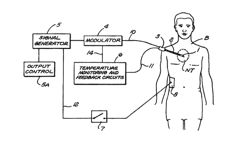
FIGURE 1 is a block diagram of the various elements and
portions of the overall system in accordance with the
present invention;
FIGURE 2 is a graphical representation of an exemplary
interrupted RF waveform output from an RF generator system
in accordance with the present invention;
CA 02272125 1999-OS-17
WO 97/49453 PCT/US97/11145
8
FIGURE 3 shows a graphical representation of a
modulated frequency waveform in accordance with the present
invention;
FIGURE 4 illustrates an irregular frequency output
waveform in accordance with the present invention;
FIGURE 5 shows repeated frequency signals with a
lowered output duty cycle;
FIGURE 6 is a block diagram of the various elements of
the system for generating modulated frequency signals;
FIGURE 7 is a flow diagram of the process in accordance
with the present invention;
FIGURE 8 is another flow diagram of the process in
accordance with the present invention;
FIGURE 9 shows a transcutaneous surface application in
accordance with the present invention;
FIGURE 10 illustrates a spinal pain relief procedure in
accordance with the present invention;
FIGURE 11 illustrates a multi-electrode dorsal column
application for pain relief in accordance with the present
invention; and
FIGURE 12 illustrates the use of intensity modulated
high frequency electrical signal applied to acupuncture
needles;
FIGURE 13 shows a schematic diagram of a percutaneously
placed electrode and differential pulsed RF versus thermal
tissue alternation zones;
CA 02272125 1999-OS-17
WO 97/49453 PCT/US97/I1145
9
FIGURE 14 shows applications of modulated high
frequency to internal and surface structures of the brain by
depth and surface electrodes; and
FIGURE 15 shows a flow diagram for possible effects of
the modulated high frequency generator output on tissue
function.
DESCRIPTION OF SOME PREFERRED EMBODIMENTS OF THE INVENTION
Referring to FIGURE 1, an illustration of the system
and the method in accordance with the present invention in
shown by a block diagram and schematic elements. An
electrode with an uninsulated conductive surface 1 (for
example, a conductive tip end) is shown in the proximity of
a region of neural tissue NT illustrated schematically
within a dashed boundary. The electrode has an insulated
shaft 2 (shown by the cross hatched lines) and connection or
hub portion 3, inside of which there may be electric
connections to the surface 1. A connector l0 electronically
connects to the surface 1 through the electrode shaft 2 and
to electronic supply units 4 and 5 (which are illustrated
outside the body, but which alternatively, may be
miniaturized and implanted inside the body). The electronic
supply unit 5 represented in block form is a signal
generator having a signal output, which may be voltage,
current, or power. The electronic supply unit 4 is a
modulator to modulate (for example the amplitude of) the
high frequency output from the signal generator. The output
from 4 and 5 is an electrical output signal such as an
CA 02272125 1999-OS-17
WO 97/49453 PCT/US97/11145
electromagnetic or other signal known to one skilled in the
art, which is connected to the electrode surface 1, and
therefore, is conductively exposed to the tissue NT.
By way of an example, the signal generator element or
5 device 5 may take the form of an RF power source with a
continuous wave output. One example of such a power source
is a generator identified by Model No. RFG-3C, which is
available from Radionics, Inc., located in Burlington,
Massachusetts. The block indicated by reference numeral 4
10 in one example represents a pulse modulation unit, which
switches the RF output from the signal generator 5 on and
off, at a designed rate and duty cycle. Use of RF output
generators or supplies and modulation circuits are known in
the use of high frequency techniques (which for example, are
described in a book entitled Radio Engineering by Frederick
E. Terman, McGraw-Hill, New York, 1947, 3rd Edition). A
temperature monitoring element or circuit 6 is also shown,
which is connected by a cable 11 to the electrode and to a
thermal sensor, which may be a thermistor or thermocouple,
disposed inside the electrode applicator or conductive tip 1
to measure the temperature of the tissue NT near the tip.
An example of such thermal sensing circuits and electrodes
is one identified by Model No. RFG-3C available from
Radionics, Inc., located in Burlington, Massachusetts.
Furthermore, Figure 1 illustrates a reference electrode 8
shown in electrical contact with the patient's body B with 9
connection wire 12 running to the generator 5 so as to
provide a circuit for return current from electrode
CA 02272125 1999-OS-17
WO 97/49453 PCT/US97/11145
11
applicator 1 through the patient B (such reference
electrodes are common with RF lesion generators; as
discussed in the research papers by Cosman, et al., entitled
"Theoretical Aspects of Radiofrequency Lesions and the
Dorsal Root Entry Zone," Neurosurgery 15:945-950, 1984; and
Cosman ER and Cosman BJ, "Methods of Making Nervous System
Lesions," in Wilkins RH, Rengachary SS (eds): Neurosurgery,
New York, McGraw-Hill, Vol. III, 2490-2498, 1984). A switch
or circuit breaker illustrated by element 7 illustrates that
20 such a return circuit could be opened to limit such direct
return current, and limit such current to inductive or
reactive current characteristic of time varying circuits
such as RF circuits.
In operation, the voltage or current output from the
signal generator 5 and the modulator 4 are impressed upon
tissue NT, which may be neural tissue (for example, spinal
nerves or roots, spinal cord, brain, etc.) or tissue near
neural tissue. In accordance with the present invention,
such an electrical output, for example, an electromagnetic
output, can cause energy deposition, electric field effects,
and/or electromagnetic field effects on the nerve cells in
the tissue NT so as to modify or destroy the function of
such nerve cells. For example, modification of neural
function may include reduction or elimination of pain
syndromes (such as spinal faces, mechanical back pain,
facial pain) in some cases, alleviating motor disorders.
Because the RF output from 5 is modulated by element 4, its
percent on-time is reduced so that sustained heating of
CA 02272125 1999-OS-17
WO 97/49453 PCT/US97/11145
12
tissue NT is reduced, yet the neural therapeutic effects of
the impressed RF voltages and currents on the neural tissue
NT are enough to produce the pain reducing result. The
signal generator 5 may have a power, voltage, or current
output control 5A (as on the Radionics Model RFG-3C RF
generator) to increase or decrease the output power
magnitude or modulated duty cycle to prevent excessive
heating of tissue NT or to grade the level of pain
interruption as clinically needed. Output control 5A may be
a knob, which raises or lowers the output in a smooth,
verniated way, or alternatively, it may be an automatic
power control with feedback circuits. In this regard, the
temperature monitor 6 provides the operator with the average
temperature of tissue NT near electrode tip 1 to
interactively prevent temperatures near tip 1 to exceed the
range of approximately 45°C (on average thermally lethal to
tissue NT), and thus, to avoid the higher temperature ranges
for the usual heat lesioning procedures described above.
For example, element or circuit 6 may include feedback
circuitry to change the modulation duty cycle (by, for
example, longer or shorter on-times) to hold the temperature
near tissue NT to below a set value (for example, 40°C to
45°C), illustrated by the feedback line 14 in FIGURE 1. In
addition, the high frequency waveform from the signal
generator 5 is free from substantial components in the 0 to
about 300 to 400 Hertz range (which is much lower than
radiofrequencies), and this avoids the stimulation effects
CA 02272125 1999-OS-17
WO 97/49453 PCT/US97/I1145
13
that are typical for stimulator system applications as
described above.
As an example of a modulated RF waveform that
accommodates the system of the present invention, FIGURE 2
shows schematically a high frequency output with a voltage
amplitude V and with a burst duration T1 between which on-
time bursts there are illustrated periods of zero voltage of
duration T2. During the on-time T1, the RF signal output is
oscillatory with time period T3 between maximum voltages V.
The reciprocal of T3 is proportional to the value of the
radiofrequency (for example, 1 Mega Hertz RF output
corresponds to T3 = 1 microsecond). This is an interrupted
or bursting type of modulated high frequency waveform.
During the high frequency on-time T1, the voltage may
oscillate between plus and minus its maximum value V.
Accordingly, an electric field is produced round the region
of the electrode applicator (as for instance the exposed
electrode tip 1 in FIGURE 1). The electric field induces a
modifying, or pain-relieving, or neural-altering effect on
the tissue near or among the nerve cells and fibers. Pain
relief and neural modification may accordingly be
accomplished by this high frequency bursting voltage and
accompanying electromagnetic field, and also accompanying
current among the neural and tissue cells. During the off
period, there is minimal or no voltage (i.e. V=0 at the
electrode applicator), and thus, no electric field or
electric currents in and among the neural tissue. During
that period, no heat deposition is present. Thus, over the
CA 02272125 1999-OS-17
WO 97149453 PCT/US97/11145
14
entire cycle, from on period T1 through off period T2, the
energy deposition, on average, may be adjusted so that there
is no excessive heating, on average, around the electrode
applicator. Thus, the usual mechanism of continuous on-time
high frequency voltage and current, as in previous heat
lesion techniques, is avoided. Therefore, the achievement
of high average temperatures near or around the applicator
tip may be eliminated by the present invention. The usual
heat lesion process in which tissue temperatures, on
average, exceed 45°C, may also be avoided. In many
instances, this avoidance of high temperature domains due to
high average heat dissipation of the radiofrequency power
prevent acute pain of the process to the patient. By having
the interrupted waveform, as in FIGURE
2, the average power is thereby reduced and the average
heating around the electrode tip or applicator is
accordingly also reduced. However, substantial voltages V
(or currents) are still sustained during the on period with
their resulting therapeutic effects on the tissue.
To give a representative example of values for
parameters in an interrupted high frequency waveform as in
FIGURE 2, the overall pattern of the waveform may have a
total period of one second, meaning that the sum of T1 + T2
- 1 second. The on period T1 may be 20 milliseconds, and
the off period T2, therefore, may be 980 milliseconds.
Voltages V in the range of 10 to 30 volts or more may be
used. It may be used to induce a pain relieving effect in
certain tissues. Average tip temperature around an
CA 02272125 1999-OS-17
WO 97/49453 PCT/US97/11145
electrode tip such as the exposed tip element 1 in FIGURE 1
may be maintained at or below 40°C, well below thermo-lethal
levels. Electrodes with diameters of 1 or 2 mm shaft (for
example, the shaft 2 of a cannula in FIGURE 1) may be
5 maintained at or below 40°C, well below thermo-lethal
levels. Electrodes with diameters of 1 or 2 mm shaft (for
example the shaft 2 of a cannula in FIGURE 1), with an
exposed tip of 1 to 10 mm (such as the tip element 1 in
FIGURE 1) may be used and the electrode may be inserted in
10 around neural structures in the brain or peripheral nerves
or peripheral nerve ganglia to accomplish pain relief or
other neurological alteration. Variations in these
parameters may be made with similar therapeutic effects, and
various geometries of conductive electrodes or applicators
15 may be effective. Illustrations of a wide variety of such
electrodes are available in the product line of Radionics,
Inc., located in Burlington, Massachusetts. Pointed or
sharpened electrodes (such as illustrated schematically by
electrode tip in FIGURE 1) are useful for penetration of the
electrode through the skin to the target neural tissue site,
and electric or current fields or higher intensity will be
present at a sharpened point for a given applied voltage
(such as V in FIGURE 2), which will be effective in altering
neural function.
FIGURE 3 shows a variation of the modulated high
frequency waveform, which accomplishes high peak voltage
swings with reduced average power deposited in tissue. The
baseline voltage may be put at zero (that is, V=0), shown by
CA 02272125 1999-OS-17
WO 97/49453 PCT/US97/11145
16
the dashed line 24. The solid line 21 represents the actual
waveform, which has rapid oscillations at the radiofrequency
and has an overall enveloped, represented by dashed line 20,
that has high points and low points with an approximate on
time T1 and a time period between the envelope of modulation
maxima T2. T1, again, may be a percentage on time of 2
percent (as described above for 20 milliseconds on time out
of 1 second total), and this on time T1 may vary
considerably while still maintaining substantial off time so
as to prevent overall average high temperature heating (as
in the usual RF heat lesion systems). Such a modulation
envelope (as shown by dashed line 20) may be achieved by
using a modulated signal generator that varies the input or
output gain of high frequency generator (for example element
5 in FIGURE 1) so as to achieve such a waveform as in FIGURE
3. In such circuitry, which is commonly used in pulse
generation techniques, low frequency filtering or selection
of modulation parameters may avoid stimulation voltage or
current components at the physiologic range of 0 to 300
Hertz so that unpleasant stimulative effects may be avoided
during the therapeutic intermittent high frequency lesion
process.
FIGURE 4 shows yet another embodiment of an interrupted
high frequency waveform in accordance with the present
invention. Here there is a non-periodic variation of the
voltage represented by the excursions of the voltage V
represented by excursions on a vertical axis. The maxima
point 25 may occur at random positions in time. The time
CA 02272125 1999-OS-17
WO 97/49453 PCT/US97/11145
17
difference between maxima may also vary in an irregular or
even random way. This waveform may have no repeating or
periodic structure, but may be analogous to high frequency
noise with random amplitudes, peaks, zero crossings, and
carrier high frequencies. Such a waveform may be generated
by random noise generators, spark gap signals, or other
noisy signals that are known in the field of signal
generation (for example, as described in Radio Engineering,
cited above). Filtering may be applied in the wave
generator and power amplifier so that lower frequencies in
the physiologic range are not present to cause undesirable
stimulation effects.
FIGURE 5 shows yet another possible high frequency
waveform of interrupted, repeated bipolar pulses with
frequency repetitive time T3 for example, the physiologic
stimulation frequency range (i.e., 0 to about 300 Hertz).
The pulse on-time may be low enough so that the power
deposition may be kept low enough to prevent heating, and
yet the peak voltage V is enough to alter the neural
function.
Variations of such waveforms are possible with the same
intermittent high frequency effect for pain or neurological
modification. For instance, a baseline V=0 may not pertain
and a slowly varying baseline of non-zero value may be used.
The time average of the signal need not be zero. The on and
off switching of a high frequency signal such as in FIGURE 2
may be done at a non-periodic or non-regular, repeating rate
so that, on average, the polarization effects in the tissue
CA 02272125 1999-OS-17
WO 97/49453 PCT/US97/11145
18
are still maintained at a low level. The average power
deposition may still be maintained at a low level with non-
periodic, interrupted high frequency waveforms. The high
frequency carrier frequency (i.e., represented by the
inverse of time T3 in FIGURE 2 and FIGURE 3) may also be
non-constant. Varying or combined or superposed high
frequency waveforms may be used as the carriers, and the
combined or composite high frequency waveforms may be
interrupted or modulated in accordance with the present
system and invention. Pulse waveforms with high frequency
carriers may be shaped in a variety of ways, for example,
with fast rising leading edges and slowly falling off or
exponential trailing edges. The signal generator waveform
may have a peak intensity, which is much higher than the
average or RMS intensity to yield a high electromagnetic
field or current density on the neural tissue while
maintaining the average power deposition in the tissue at a
sufficiently low level to prevent heating above lethal
tissue temperatures (for example 40°C to 50°C).
FIGURE 6 shows a more detailed block diagram of the
system for generating modulated high frequency signals
(similar to but more detailed than the block element of high
frequency generator 5 and modulator 4 of FIGURE 1).
A block or element 30 represents a signal generator,
which may create a high frequency signal or periodic or non-
periodic frequency. The signal generator 30 provides an
input to a filter system 31, which selectively filters out
frequencies that could cause unpleasant, undesired, or
CA 02272125 1999-OS-17
WO 97/49453 PCT/US97/11145
19
damaging physiological signals. The signal is then fed into
a waveform shaping circuit 33, which shapes the waveform
input from a block or element 32, which provides amplified
modulation and/or frequency modulation and/or phase
modulation control. Circuits of this type are described for
instance in Radio Engineering by Terman (cited above, in a
book entitled Radio Engineering by Frederick E. Terman,
McGraw-Hill, New York, 1947, 3rd Edition). Additional
waveform shaping may be done by elements 40 and 41, which
control the amplitude of waveform and/or the duty cycle of
the waveform, respectively. The resultant signal is then
fed into a power amplifier 34. This is a wide band
amplifier used to increase the signal to power levels
appropriate for clinical use. This energy is then delivered
to the patient via an electrode represented by element or
block 35.
A temperature sensor or plurality of temperature
sensors, represented by 36, may also be placed and connected
proximate the electrode so as to insure that the temperature
does not exceed desired limits. This temperature sensor
signal is fed through a filter B represented by 37, which is
a special filter module used to eliminate high frequency
components, and thus, not to contaminate the low-level
temperature signals.
The temperature signal is fed to a standard temperature
measuring unit 38 that converts the temperature signal into
a signal that may be used to display temperature and/or to
control, in a feedback manner, either the amplitude and/or
CA 02272125 1999-OS-17
WO 97149453 PCT/US97/11145
the duty cycle of the high frequency waveform. In this way,
power delivery may be regulated to maintain a given set
temperature. This flow is represented by block or element
39, which is simply a feedback control device. The dotted
5 lines from element 39 to elements 40 and 41 represent a
feedback connection that could either be electronic and/or
mechanical. Alternatively, a person may simply operate
these controls manually, based on the visual display of
temperature, as for example, on a meter or graphic display
10 readout 42.
As was explained with respect to the disclosed
embodiments, many variations of circuit design, modulated
high frequency waveforms, electrode applicators, electrode
cannulas will be appreciated by those skilled in the art.
15 For example, many electrodes or electrode applicators are
practical, including tubular shapes, square shafts, flat
electrodes, area electrodes, multiple electrodes, arrays of
electrodes, electrodes with side outlets or side-issued
tips, electrodes with broad or expandable or conformal tips,
20 electrodes that may be implanted in various portions of the
brain, spinal cord, interfacial space, interstitial or
ventricular spaces, nerve ganglia may be considered with the
system of the present invention.
The frequency range for the so-called high frequency
waveforms, as shown for instance in FIGURES 2, 3, 4, and 5
may be used over a wide range. For example, the "high
frequency" characteristic of 1/T3, which may be only one of
many high frequency components, may be above the so-called
CA 02272125 1999-OS-17
WO 97/49453 PCTIUS97/11145
21
physiological stimulation frequency range of 0 to about 300
Hertz. This high frequency may also extend up into the
radiofrequency or microwave range (for example, 50 Kilo
Hertz to many Mega Hertz).
Mixtures of frequencies may be accomplished as
discussed above. These may be mixtures of "high
frequencies" (above the physiologic stimulation) range of
(say 0 to 300 Hertz) and lower frequencies (within that
stimulation range of say 0 to 300 Hertz). Thus, one skilled
in the art may have both modulated high frequency and
stimulation frequencies for various clinical effects, such
as stimulation blockage of pain while neural modification is
being applied according to the present invention.
Referring now to FIGURE 7, the operation of the system
and method is shown with a flow diagram. Assume that an
electrode 1 is placed in contact with the patient s body and
connected to a modulated high frequency generator
(represented by blocks 5 and 4) in the manner described
above. once the electrode 1 is in place, a clinician may
decide on the desired electrode parameters and modified high
frequency parameters that should be used. This is indicated
by initialization block 100 in FIGURE 7. For example, for a
given electrode geometry or location of electrode 1 in the
patient s body, it may be decided that a certain duty cycle
of high frequency signal, voltage, current, or power level
of high frequency signal, or a mixture of high frequency
signal and stimulation signal may be desirable.
Furthermore, a choice of electrode for a given application
CA 02272125 1999-OS-17
WO 97/49453 PCT/US97/11145
22
involving a suitable electrode geometry (for example,
sharpened electrode shaft, catheter-type electrode, surface
electrodes for skin application, flattened electrodes for
cortical or spinal cord application) may be made.
Alternatively, the modified high frequency generator may
have fixed parameters, which are used universally for
certain types of procedures, in which case the
initialization block element 100 in FIGURE 7 may not be
present. This is symbolized by the dashed line between
block element 100 and block element 102.
A block or element 102 indicates the start of the high
frequency application in which an "on" button may be pulsed,
and the elevation of high frequency, voltage, current, or
power {level) is started. In a case where the temperature
sensor is disposed in or near the electrode applicator
connected to the patient's body, the temperature monitor 103
is indicated, which may sense that temperature and monitor
or read it out to the clinician. Alternatively, temperature
sensing may also be conducted away from the output
applicator. For instance, a separate temperature sensor may
be inserted at a position located at a distance from the
active RF electrode. Increasing the RF level 102 to achieve
the neural modification effect (for example, pain relief for
the patient) is accomplished by the electromagnetic,
electric, or other aspects of the high frequency field in
the presence of the neural structures. If the temperature
monitor 103 shows that the temperature of the tissue is
being elevated to lethal levels (from 40°C to 50°C, for
CA 02272125 1999-OS-17
WO 97/49453 PCT/US97/11145
23
example, then the decision block or element 104 determines
that if these levels are reached, a reduction of the RF
power (block or element 105) may be implemented so as to
reduce the temperature monitored level 103. If lethal
temperature levels have not been reached, there is the
option to continue with raising the RF level or to hold it
static at a desired, predetermined level until the proper
clinical effect has been reached. At end point of a
particular RF level or time duration for the exposure
indicated by element 106 may be utilized, and when an RF
level or time has been reached, then the unit may be shut
off, as indicated by block or element 107.
Referring to FIGURE 8, another flow diagram for cases
is shown where temperature monitoring is not conducted. In
such situations, it may be decided by block element 100a
that some target parameters for the high frequency field
(such as voltage, current, or power level) will be used in a
given anatomical region and for a given electrode 1. The RF
level is increased in step 102a, and if the level of
modulated high frequency output is reached (determined by
decision block or element 103a), then, a feedback may take
place to reduce that level as represented by block or
element 105a. Element 103a may simply be a manual control
or RF output control knob or it may be done by electronic
feedback on the RF power amplifier or signal generator.
This same type of feedback system may be, for example,
illustrated by the continuous wave radiofrequency
generators, such as one identified by Model No. RFG-3C
CA 02272125 1999-OS-17
WO 97/49453 PCT/US97/11145
24
available from Radionics, Inc., located in Burlington,
Massachusetts. If the parameter criteria for an adequate
procedure is a certain time duration, then in the decision
process, if that time is reached, element 106 may be
actuated and the system stopped when that desired time
duration has been reached. Variations of pulsed
radiofrequency signals could be applied ranging from several
seconds to several minutes or more depending on the clinical
conditions. In one clinical example, an average tip
temperature of 42°C (degrees Celcius) was maintained, and a
continuous RF signal from the radio generator 1400 (see
FIGURE 9) was applied for 120 seconds. However, it should
be recognized that depending on the clinical conditions, the
RF signal may be applied for a period ranging anywhere from
several seconds to several minutes. If time duration is not
the desired end point parameter, then possibly the
observation of a desired clinical effect such as abolition
of pain, tremor, spasticity, or other physiologic parameter
may be the desired criteria, as shown by element 108, again
to make the decision to stop the procedure, as in element
107.
Various configurations of electrodes may be used with
this modified high frequency technique for neural
modification. For example, in FIGURE 9, the patient s body
1000 may have applied to it surface electrodes 1100 and
1200, which may be connected to the high frequency generator
1400. Generator 1400 has a modified high frequency signal
such as described above. Its output may be applied via
CA 02272125 1999-OS-17
WO 97/49453 PCT/US97/11145
wires 1500 and 1600 to the surface-based applicators to
induce neural modification in nerve cells at the surface of
the body or just below the surface.
FIGURE 10 in another embodiment of the present
5 invention, which involves implanting an electrode shaft 1700
near the patient's spinal column 1800. This might be done
in the case of facet denervation, dorsal root ganglion
modification, or other neural structure modification in or
near the spine. The generator 1400 is again similar to one
10 described above with a modified high frequency signal to
cause neural modification of the spinal nerves in and around
the spinal column 1800. This may be effective in
alleviating back pain, headache pain, or other spinal
diseases. The reference electrode 1900 is applied to the
15 body as a return current source.
FIGURE 11 shows the application of the present
invention for spinal cord or dorsal stimulation where
multiple electrodes attached to a catheter or flat strip
electrode are used (such electrodes are available from
20 Medtronic, Inc., located in Minneapolis, Minnesota or
Radionics, Inc., located in Burlington, Massachusetts). In
this figure, modulated high frequency generator 1400 is
shown with multiple outputs connected to electrodes 2000,
2100, and 2200, which may be implanted or on the surface of
25 the spinal cord, as illustrated by element 2400. The
electrodes 2000, 2100, or 2200 may be greater in number, and
they may be inserted through a catheter or serial string
element, which may be tunneled near the spinal cord
CA 02272125 1999-OS-17
WO 97/49453 PCT/US97/11145
26
percutaneously. Application of the neural-generated output
from 1400 may cause pain relief, relief of spasticity,
relief of other neural disfunctions by the neural
modification as described in the previous application.
Variations of the processes and configurations of the
above figures are possible by those skilled in the art.
Variations of the steps in a high frequency neural
modification procedure may be varied from those in FIGURES 7
and 8. Automatic feedback of temperature control, for
to instance shown in FIGURE 7, may give rise to control of the
RF level in element 102 of FIGURE 7, so as to lock on a set
temperature, illustrated by element 104, whereby the system
may maintain a sub-lethal tissue temperature in the presence
of the high frequency applicator. Other variations of
electrode geometry and location in the body from those
illustrated in FIGURES 9, 10, and 11 as well as others may
be devised in the brain, spinal cord, peripheral nerves, or
other neural structures anywhere in the body. Clinical
criteria for the desired end-point parameters of the RF
generator, electrode, time duration, temperature levels, may
be applicable by those skilled in the art or to achieve a
particular clinical end result. The set temperature below
which the tissue temperature should stay is somewhat
variable in the range of normal tissue temperature (37°C) up
to or about 50°C, wherein cell structures and neural cells
die under sustained exposure to such elevation of
temperature (discussed in the papers by Cosman et al.).
CA 02272125 1999-OS-17
WO 97/49453 PCT/US97/11145
27
FIGURE 12 shows yet another embodiment of the present
invention in which multiple electrodes 2500, 2600, and 2700
are inserted into various portions of the body and connected
to a pulsed RF generator or modulated high frequency
generator 14 via the outputs 2800, which may be coincident
or sequenced. Connection 3000 is made via connector wire to
3100, which is a reference electrode, or may also be used as
an area electrode for electric field operation. The
percutaneous electrodes 2500, 2600, and 2700 may be
acupuncture electrodes or similar very fine gauge
electrodes. Acupuncture electrodes may be put into various
trigger zones within the body, and the modulated high
frequency signal from 1400 may enhance the anesthetic effect
of these electrodes or produce pain relief as described
above. Thus, the present system may be used to enhance
acupuncture type techniques.
FIGURE 13 illustrates the differential effects of the
modulated RF fields for tissue or neural tissue
modification. Electrode 3600 with insulated shaft, except
for exposed tip 3700, is inserted into the body or into an
internal organ. The tissue of the body is element 1000.
The electrode is connected via connection 3500 to a high
frequency generator 1400, which may have a reference line
1600 connected to reference electrode 1900. The dashed
portion of line 1600 illustrates that this connection may or
may not be made by an electric current-carrying wire, but it
rather may be a reactive or capacitive connection with no
wire. The generator may produce sufficient root means
CA 02272125 1999-OS-17
WO 97/49453 PCT/US97/11145
28
square (RMS) high frequency power output to produce an
isotherm contour 38, corresponding to a temperature greater
than the conventional lesion mean temperature of
approximately 45° degree Celcius. For example, the line
3800 may represent an isothermic surface of 50, 55, or 60,
or more degrees, and the tissue within the volume may be
killed by a conventional heat lesion. Nonetheless, electric
fields and current generated around the electrode tip 3700
from, for example, an electric voltage output from pulse
generator 1400 may produce electric fields that can modify
neural tissue out to a larger surface, illustrated by the
dashed line 1400. Thus, the tissue between surface 3800 and
surface 1400 may be, for example, neural tissue that is
modified by peak voltage or current intensities from the
modulated electronic output of generator 1400. That output,
for example, could be pulsed, as illustrated above. Thus,
there may be region of average thermal destruction (within
zone 38) and a region of electromagnetic, magnetic, or
electronic modification (in the shell between 3800 and 1400)
as illustrated in FIGURE 13.
If generator 1400 in Figure 13 produces a pulsed
radiofrequency signal, then the peak RF voltages,
intensities, power, and currents would be higher than for a
continuous wave radiofrequency generator that produces a
similar thermal distribution, or the same size of lethal
isotherm 3800. This difference in signal intensities and
electronic qualities of the fields for pulsed versus
continuous RF cases may produce different clinical results
CA 02272125 1999-OS-17
WO 97/49453 PCT/LTS97/11145
29
and tissue function modifications in accordance with this
invention.
A clinical experience has demonstrated such
differences. Clinical data for a group of patients (Group
A) for dorsal root ganglion lesions with a percutaneously
placed electrode (such as 3600 in Figure 13) with tip
exposure 3700 near the dorsal root ganglion, was gathered.
An average tip temperature recorded from the electrode tip
3700 of 42°C was achieved, and a continuous RF signal from
generator 1400 was applied. With the electrode tip
temperature held at 42°C for such continuous radiofrequency
wave, no appreciable pain relief was experienced by the
patients in Group A.
Clinical data for a second group of patients, Group B,
for a pulsed RF application was quite different for the same
tip temperature. An identical electrode 36 with the same
tip exposure geometry 3700 was inserted into the same region
of the basal ganglion. In Group B, generator 1400 was a
pulsed RF generator with a duty cycle of about two percent.
All other conditions and clinical pain symptoms were the
same for Group B as for Group A. The pulsed RF signal was
applied with signal intensity to achieve an average
temperature rise of 42°C at the tip 3700 (the same as for
Group A), but the result was a very significant elimination
of pain for the patients in Group B, i.e., pulsed RF
application, significant pain relief was achieved when the
average tissue temperature near the electrode tip was held
at 42°C. It is known from past experience that 42°C is
CA 02272125 1999-OS-17
WO 97/49453 PCT/US97/11145
considered less than the conventional heat lesion or
destruction temperature for tissue in such circumstances.
For Group A, the same average tip temperature of 42°C for a
continuous RF signal application did not produce significant
5 neural modification or pain relief. Previous literature by
Cosman et al. referenced above indicates that 42°C is a
"non-lethal" or "sub-lethal" lesion temperature, on average,
for continuous RF signals, i.e. 42°C is below a heat lesion
level, yet at 42°C there is significant neural modification
10 or pain relieving effect for pulsed RF signals, illustrating
the differential effects of pulsed high frequency signal and
its associated electronic fields within the tissue compared
to continuous RF fields for analogous temperatures, even
below lesion levels. Such differential effects could
15 include pain relief, motor function changes (as in
Parkinsonism), spasticity relief, epilepsy relief or
interruption, neuro-cognitive changes, mood alterations, and
so on. In the clinical example above, pain relief was
achieved without any of the usual sensory loss or other side
20 effects associated with heat lesioning at higher
temperatures, which is a major advantage of the low
temperature pulsed RF method.
Figure 14 shows another configuration with cortical C
contact electrodes 2100 and 22, which may be flat area type
25 electrodes placed on the brain surface at strategic
positions to produce neural modification within the brain.
The connection wire 4000 to generator 1400 supplies the high
frequency signal to the electrodes 2100 and 2200. Multiple
CA 02272125 1999-OS-17
WO 97/49453 PCT/US97/11145
31
wires within cable 4400 may give different signals or a
bipolar electrode configuration (see the discussion in
Cosman's paper on radiofrequency fields) across the
electrodes 2100 and 2200. Generator 1400 may also be
connected to a catheter or rod-like electrode 4500, which
would be placed deep into the brain and have electrode
contacts 4000, 4100, and 4200 to produce the electronic high
frequency field effects within the brain nearby. Again,
multiple wires may be carried back to generator 1400 through
the cable element 4600 for differential signal application
on the contacts 4000, 4100, and 4200. Application of the
pulsed RF fields in these configurations may give rise to
functional modification of the brain. Alteration of
epileptic seizures may be made by application of neuro-
modifying, pulsed RF fields in such electrodes. Electrodes
such as shown in Figure 14 are common for recording in the
study of epilepsy, as evidenced by brochures available from
Radionics, Inc. Their use for high frequency application,
however, may be applied to alter the brain function near
sites where epileptic neural foci is thought to exist.
Modification of these epileptic foci may modify or even
abolish the epileptic seizure or disease. Similar
implantation for application of deep brain or surface-type
electrodes on the brain, spinal cord, or other portions of
the body may have similar ameliorating or modifying effects
on neural structures or other organs. For example,
electrodes such as 4500 may be placed in the thalamus,
pallidum, hippocampus, etc., of the brain for alteration or
CA 02272125 1999-OS-17
WO 97J49453 PCT/US97J11145
32
modification of movement disorders such as Parkinsonism,
spasticity, epilepsy, etc. Again, these disorders may be
removed or modified by the pulsed RF application.
Figure 15 shows a schematic diagram of some ways in
which modulated high frequency signals may affect cellular
function. Modulated generator 1400 gives rise to a
modulated signal output (e.g. voltage) applied to an
applicator such as an electrode 1500. This may give rise to
modulated electric fields on cells as illustrated by block
51. Electric fields will give rise to electric force or
effects within the cells or the tissue (block or element
52). High RF fields produce alternating electric forces on
ions, cell membranes, internal cell structures such as
mitochondrion, DNA, etc., or forces of translation and
rotation on polar molecules or on membranes having polar
internal structures or charged layers. Ionic frictional
dissipation effects may occur, (discussed in the articles by
Cosman et al., cited above), producing average or
macroscopic thermal elevation (block or element 53). If
average power deposition is low enough, then the macroscopic
thermal elevations will be at non-lethal levels. If power
deposition is increased, the average temperature may exceed
45°C (heat lesion levels). Yet even at low temperatures
(for example 42°C), electric forces and currents within the
cell (block 52) may cause, nonetheless, neural modification
effects (block or element 54) as in the clinical example
above. Pulsed fields, voltages, or current may act on un-
myelinated pain-carrying fibers such as C fibers differently
CA 02272125 1999-OS-17
WO 97/49453 PCT/US97/11145
33
from other more myelinated cells such as A fibers. The
myelin sheath acts as a dielectric or capacitive protective
layer on a nerve axon. C fibers, which primarily carry pain
sensations, have minimal myelin sheath or no myelin sheath,
and thus, may be more susceptible to strong pulsed electric
fields, currents, or forces, even without significant
heating of the nerve tissue.
The action of the modulated high frequency signal on
neural tissue may eliminate pain while maintaining tactile,
sensory, and other neurological functions relatively intact
and without some of the deficits, side effects, or risks of
conventional heat lesion making. Selectivity by pulsed RF
fields may arise by selective deneravation of pain-carrying
structures or cells (such as C fibers) compared to
relatively non-destructive modification of other neural
structures related to sensation, touch, motor activity, or
higher level functions.
The selection of high frequency generator output
parameters and the selection of electrode configurations
such as size, shape, area, etc., may be interconnected to
achieve a neural modification effect without excessive
heating. At a given average power output of the generator
as applied to the electrode adapter, a very small, sharpened
electrode may give rise to high current densities in the
tissue adjacent to it, which can give rise to focal heating,
lesions, thermal cell destruction, cooking, and coagulation
of nearby tissue. If the electrode chosen is larger, then
such elevated temperature conditions may be reduced as the
CA 02272125 1999-OS-17
WO 97/49453 PCT/US97/11I45
34
current density emitting from the electrode is reduced. In
a given clinical setting, to achieve the desired neuro
modification effect without macroscopic average elevation of
neura2 tissue above, for example, the lesion temperature of
approximately 45°C (degrees Celcius), it may be necessary to
select the appropriate parameters for both the lesion
generator output such as voltage, current, power, duty
cycle, waveform, etc., in coordination with the selection of
the appropriate electrode geometry (the selection box, for
example, being indicated by element 1 of Figure 1). The
system of electronic signal generator combined with the
appropriate signal applicator to achieve a given neuro
modification may then be considered in combination and
cooperation to achieve the effect for a particular clinical
site or result.
In view of these considerations, as will be appreciated
by persons skilled in the art, implementations and systems
should be considered broadly and with reference to the
claims set forth below.