Note: Descriptions are shown in the official language in which they were submitted.
CA 02272304 1999-OS-19
WO 98/22163 PCTlUS97121267
INTEGRATED CASSETTE FOR VALVING, PUMPING AND
CONTROLLING MOVEMENT OF FLUIDS
FIELD OF THE INVENTION
The present invention relates generally to systems for controlling fluid flaw.
More particularly, the present invention relates to systems for infusing
fluids in and
withdrawing fluids from patients undergoing medical care. Still more
particularly, the present
invention relates to systems for infusing fluids in and withdrawing fluids
from medical patients
during photopheresis treatment.
BACKGROUND
Several treatments for disease require the removal of blood from a patient,
processing the one or more components of the blood and return of the processed
components
for a therapeutic effect. Those extracorporeal treatments require systems for
safely removing
blood from the patient, separating it into components, where necessary, and
returning the
blood to the patient.
Photopheresis is one treatment involving the separation of white cells from
the
blood, addition of a photoactivatable drug, and U. V. irradiation of the white
cells before re-
infusion to the patient. In known photopheresis systems, such as system 100
shown in Figure
1, blood fluids are pumped by peristaltic roller pumps 110. In system 100, a
complex tubing
set is used to couple a patient 120 to an extracorporeal blood treatment
system which includes
a cell separator 130, a white blood cell photoactivation chamber 140, a saline
bag 150x, an
anti-coagulant bag 150b and a waste bag 150c. Valves 160, bubble chambers 170,
air
detectors 180, and pressure sensors 190 are interconnected to the tubing set
for monitoring
and controlling fluid flow within the system. Complex tubing sets, such as
that shown in
Figure 1, have the potential to cause cell damage under high outlet pressure
conditions. Blood
has also been pumped with discrete pump chambers and valves which also require
complex
tubing sets. Such discrete pump chambers and valves are considered to be less
damaging to
cells under high outlet pressures.
A very real advancement in photopheresis systems would result if the size and
complexity of the tubing in systems such as that shown in Figure 1 could be
reduced, even at
the cost of a more complex blood driving system, since the blood driving
system represents
1
SUBSTITUTE SHEET (RULE 26)
CA 02272304 2005-04-05
permanent reusable equipment, whereas the tubing set must be replaced or
disposed of
after each treatment session. A similar result has been accomplished with
peritoneal
dialysis systems, where the flow of dialysate is controlled entirely with
diaphragm pumps
and valves driven by air pulses delivered to a molded cassette through a
plastic
membrane. See for instance several patents by Dean Kamen, including U.S.
Patent No.
5,178,1$2, issued January 12, 1993 and U.S. Patent No. 5,634,896 issued June
3, 1997.
The cassette contains all components of a previously complex tubing set,
except for the lines to the patient and short delivery lines from the
dialysate containers.
The air pulses delivered to the cassette are controlled by continually
analyzing the pressure
changes in the air delivered to the diaphragm pumps, processing the pressure
changes
through a computer, and making continual corrections as a result. The
resulting peritoneal
dialysis system is able to accurately measure the fluid delivered, but is
unable to provide a
fixed steadiness of flow rate. In contrast to peritoneal dialysis systems,
systems such as
photopheresis systems, which involve continuous blood cell separation, require
both a very
steady flow rate, as well as the ability to control the fluid flow rate.
Furthermore, such a
system may tend to promote clotting, hemolysis and cell lysis when pumping
blood, as
opposed to its intended fluid, dialysate which contains no cellular
components.
Summary of the Invention
The present invention overcomes these and other limitations of the prior art
to provide a system for processing blood without clotting, hemolysis or lysing
cells and
which achieves an accurate, steady flow rate during extracorporeal treatment
of disease. It
comprises an apparatus for controlling movement of fluids during a
photopheresis
treatment session. A hollow enclosure has a plurality of fluid input ports for
receiving
fluids into the enclosure and a plurality of fluid output ports for expelling
fluids from the
enclosure. A first roller pump tube is provided for pumping at least one of
the fluids
through the hollow enclosure during the photopheresis treatment session. The
roller pump
tube is coupled to the hollow enclosure by a roller pump input port and a
roller pump
output port. Internal fluid passageways disposed within the hollow enclosure
are provided
for coupling together the fluid input ports, the fluid output ports and the
roller pump input
and output ports. At least one internal valve is disposed within the hollow
enclosure for
controlling movement of the fluid within the hollow enclosure during the
photopheresis
treatment session.
2
CA 02272304 1999-OS-19
WO 98/22163 PCT/US97121267
BRIEF DESCRIPTION OF THE DRAWINGS
In order that the manner in which the above-recited and other advantages and
objects of the invention are obtained and can be appreciated, a more
particular description of
the invention briefly described above will be rendered by reference to a
specific embodiment
thereof which is illustrated in the appended drawings. Understanding that
these drawings
depict only a typical embodiment of the invention and are not therefore to be
considered
limiting of its scope, the invention and the presently understood best mode
thereof will be
described and explained with additional specificity and detail through the use
of the
accompanying drawings, wherein.
Figure 1 is a block diagram showing a prior art photopheresis system;
Figure 2 is a bottom view of an integrated disposable cassette for valuing,
pumping and controlling the movement of blood fluids during a photopheresis
treatment
session, in accordance with a preferred embodiment of the present invention;
Figure 3 is a cross-sectional view of the integrated disposable cassette shown
in
Figure 2;
Figure 4 is a top view of the integrated disposable cassette shown in Figure
2;
Figures 5 to 27 show in schematic form an alternative embodiment of an
extracorporeal blood treatment system according to the invention, including
the steps of
performing such treatment;
Figure 28 shows in schematic form a further embodiment of an extracorporeal
blood treatment system according to the invention;
Figure 29 shows in schematic form a further embodiment of an extracorporeal
blood treatment system according to the invention incorporating novel negative
pressure and
pressure relief valves;
3
SUBSTITUTE SHEET (RULE 26)
CA 02272304 1999-OS-19
WO 98/22163 PCT/US97/21267
Figure 30 is a cross sectional view through the negative pressure valve of
Figure 29;
Figure 31 is a cross sectional view through the one of the pressure relief
valves
of Figure 29;
Figure 32 is a plan view of a hematocrit detection window in a cassette
according to the invention; and
Figure 33 is a cross sectional view taken along lines 33--33 ofFigure 32.
DETAILED DESCRIPIZON OF THE INVENTTON
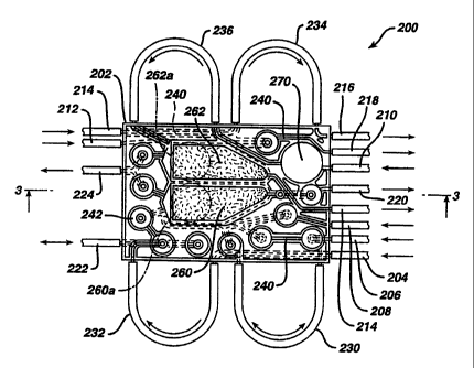
Referring now to Figure 2, there is shown a bottom (or actuator) side view of
an integrated disposable cassette Z00 for valuing, pumping and controlling the
movement of
blood fluids during a photopheresis treatment session. Cassette 200 is formed
of a hollow
injection-molded enclosure 202 having fluid input ports 204, 206, 208, 210,
212 and 214 for
receiving fluids into enclosure 202, and fluid output ports 214, 216, 218, 220
and 224 for
expelling fluids from cassette 200. Input/output port 222 is provided for both
receiving fluid
into and expelling fluid from cassette 200. As explained more fully below,
these fluid input
and output ports couple cassette 200 to a patient being treated, as well as
devices in the
photopheresis treatment system such as a cell separator 130 and a
photoactivation chamber
140 and bags, such as bags 150x, 150b and 150c, containing saline,
anticoagulation fluid, and
waste fluid, respectively. Significantly, all of the tubing, valves, sensors,
drip chambers and
pumps shown within box 195 (Figure 1) are implemented within disposable
cassette 200.
During a photopheresis treatment session, cassette 200 is snapped into a
permanent cassette actuation or driving unit (not shown), and the input and
output ports from
cassette 200 are coupled to various treatment devices and to a patient. The
details of such
couplings are explained more fully below. At the conclusion of the treatment
session, the
cassette 200 is removed from the permanent cassette actuation unit and
thereafter is discarded.
Referring still to Figure 2, ports 204, 206, 208 and 214 are provided for
coupling disposable cassette 200 to a centrifuge or cell separator. More
specifically, output
port 214 is provided for delivering whole blood from cassette 200 to the
centrifuge, and input
4
SUBSTITUTE SHEET (RULE 26)
CA 02272304 1999-OS-19
WO 98/22163 PCTIUS97121267
ports 204, 206 and 208 are respectively provided for returning plasma, white
blood cells
(WBC), and red blood cells (R.BC) to cassette 200. Ports 204, 206, 208 and 214
are
preferably coupled to the centrifuge with disposable tubing (not shown).
Similarly, ports 210,
216, 218 and 220 are provided for coupling disposable cassette 200 to a
patient. More
specifically, input port 210 is provided for delivering untreated blood from
the patient to
cassette 200, and output ports 216, 218 and 220 are respectively provided for
returning
treated blood, saline and an anti-coagulant from cassette 200 to the patient.
Ports 210, 216,
218 and 220 are preferably coupled to the patient with disposable tubing (not
shown).
Input/output port 222 is provided for delivering untreated WBC from cassette
200 to a
photoactivation chamber and for returning treated WBC from the photoactivaton
chamber to
cassette 200. Again, port 222 is preferably coupled to cassette 200 with
disposable tubing
(not shown). Finally, input ports 212 and 214 are respectively provided for
delivering saline
and anticoagulant fluid from storage bags (not shown) to cassette 200, and
output port 224 is
provided for delivery waste fluid expelled from cassette 200 to a waste
collection bag (also not
shown).
In one preferred embodiment of the present invention, four roller pumps are
used to drive the blood fluids described above through the interior of
cassette 200. The roller
pumps are part of the permanent cassette actuation or driving unit which
cassette 200 is
snapped into at the inception of each treatment session. More specifically,
roller pump tubes
230, 232, 234, and 236 engage the roller pumps in the permanent cassette
driving unit when
cassette 200 is snapped into the permanent cassette driving unit. Each roller
pump tube 230,
232, 234 and 236 is coupled to cassette 200 by two ports which respectively
receive and/or
deliver blood fluids from and to cassette 200. In the preferred embodiment,
roller pump tube
230 is provided for driving WBC through cassette 200; roller pump tube 232 is
provided for
driving plasma through cassette 200; roller pump tube 234 is provided for
driving anti-
coagulant fluid through cassette 200; and pump tube 236 is provided for
driving untreated
blood received from the patient through cassette 200.
Injection-molded enclosure 202 includes internal fluid passageways 240 which
are disposed within the interior of cassette 200. As shown in Figures 2 and 4,
interior fluid
passageways 240 function to couple together fluid ports 204, 206, 208, 210,
212, 216, 218,
220, 222, 224 and roller pump tubes 230, 232, 234 and 236 throughout the
interior of cassette
200. Passageways 240 are preferably integral with hollow-enclosure 202, and
enclosure 202
5
SUBSTITUTE SHEET (RULE 26)
CA 02272304 1999-OS-19
WO 98/22163 PCT/US97/21267
and passageways 240 are therefore preferably formed from a singular injection-
molded piece
of plastic material.
Internal diaphragm valves 242 are disposed throughout the interior of cassette
200. Valves 242 are provided for controlling the movement of the blood fluids
that travel
through internal passageways 240 during a photopheresis treatment session.
Valves 242 are
preferably formed as part of the singular injected-molded piece of plastic
material used to form
enclosure 202 and passageways 240. An elastomeric membrane 250 (shown in
Figure 3)
covers the upper and Iower surfaces of enclosure 202. During a photopheresis
treatment
session, solenoid valves disposed within the penmanent cassette driving unit
transmit
controlled air or liquid pulses to diaphragm valves 242 through membrane 250
in order to
open or close each valve 242. Alternatively, solenoid valves disposed with the
permanent
cassette driving unit could couple directly to membrane 250 and thereby
directly drive valves
242 without any intermediate driving air or liquid.
A pair of drip chambers 260, 262 are disposed within the interior of enclosure
202. As shown more clearly in Figure 3, each drip chamber is formed of
compartments 264
and 266 which are separated by a mesh 265. Mesh 265 preferably has a pore size
of about
200p,. Each compartment 264, 266 is sealed on one side by membrane 250. In
addition, each
compartment 264, 266 is connected to an internal fluid passageway 240 within
enclosure 202.
Again, the walls 268 which form compartments 260, 262 are preferably formed as
part of the
singular injected-molded piece of plastic material used to form enclosure 202,
passageways
240 and valves 242. In the preferred embodiment of the present invention, drip
chamber 260
is used for filtering treated blood before it is returned to the patient
through output port 220,
and drip chamber 262 is used for filtering whole blood before it is delivered
to a centrifuge
through output port 214. By monitoring the position of the membrane 250 used
to form drip
chambers 260, 262, the permanent cassette dri~~ng device can monitor the
pressures of the
fluids in drip chambers 260, 262. Thus, in thr ~ferred embodiment, pressure
s~:nsors are
located on the permanent cassette driving dev _ opposite locations 260a and
262b for
monitoring the pressures inside drip chambers 260 and 262. In addition, a
pressure sensor is
preferably located on the permanent cassette driving device opposite location
270 for
monitoring the pressure of untreated blood received from the patient through
input port 218.
6
SUBSTITUTE SHEET (RULE 26)
CA 02272304 1999-OS-19
WO 98!22163 PCT/US97121267
Figure 5 shows an alternative embodiment of the invention in diagrammatic
form. It employs a cassette 300 similar to that shown in Figures 2 to 4, but
employing varying
valuing and porting. A first roller pump 302 pumps an anticoagulant fluid and
a second roller
pump 304 pumps blood from a patient 306. An anticoagulant bag 308, saline bag
310,
centrifugal blood cell separator 312, plasma bag 314, recirculation bag 316
and light treatment
chamber 318 connect to the cassette 300 at ports as follows: the anticoagulant
bag 308 to an
anticoagulant solution port 320, the saline bag 310 to a saline port 322, an
inlet 324 on the cell
separator 312 to a separator inlet port 326 and an exit 328 from the cell
separator 312 to a
separator exit port 330, an inlet 332 to the plasma bag 314 to a plasma inlet
port 334, an exit
336 from the plasma bag 314 to a plasma exit port 338, an exit 340 from the
recitculation bag
316 to a recirculation exit port 342, and an inlet 344 to the treatment
chamber 3I8 to a
treatment chamber inlet port 346. Additionally, ports 348 and 350 connect to
the
anticoagulant roller pump 302, ports 352 and 354 connect to the blood roller
pump 304, port
356 connects to an anticoagulant exit line 358 and port 360 connects to the
patient 306 via a
patient access line 362. A clamp 364 in the patient access line is located
upstream of where
the anticoagulant line 358 connects to the patient access line 362. A line 368
connects an exit
370 from the treatment chamber 318 to an inlet 372 to the recirculation bag
316.
Internally of the cassette 300, passage 374 connects ports 350 and 356. A
pressure sensor 3 76, comprising an electronic pressure transducer in contact
with the
membrane (not shown) of the cassette 300, connects to the patient access line
port 360. From
the sensor 376, passage 378 leads to a first valve 380 and second valve 382.
As in Figures 2
to 4, each of the valves of cassette 300 comprise diaphragm valves with the
cassette
membrane acting to block and unblock a vertical passageway within a valve
chamber. From
the second valve 382, passage 384 leads to a third valve 386 and fifth valve
388. Passage 384
also leads to an inlet 390 of a filter 392, similar to the drip chamber filter
260 of the prior
embodiment. Passage 394 connects the third valve 386 to the saline port 322
and to an
eleventh valve 396. Passage 398 connects the fifth valve 388 to port 342.
Passage 400
connects the eleventh valve 396 to a sixth valve 402, an eighth valve 404, a
seventh valve 406
and to port 352 for the blood pump 304. Passage 408 connects the sixth valve
402 to port
326 and passage 410 connects the eighth valve 404 to port 338 and to a fourth
valve 412.
Passage 414 connects the fourth valve to port 354 for the blood pump 304 and
to port 390 of
the filter 392. Passage 416 leads from the filter 392 to the first valve 380.
Passage 418
connects the seventh valve 406 to a ninth valve 420 and to a hematocrit
detector 422
7
SUBSTITUTE SHEET (RULE 26)
CA 02272304 1999-OS-19
WO 98/22163 PCT/US97121267
comprising a light emitting diode and photodector for detecting the presence
of red blood cells
passing through the detector 422. Passage 424 connects the hematocrit detector
422 to port
346 and passage 426 connects the ninth valve 420 to a tenth valve 428 and to
port 330.
Passage 430 connects the tenth valve 428 to port 334 and, finally, passage 432
connects port
320 to port 348.
Figures 5 through 27 depicts various stages in a treatment employing the
cassette 300, with the dark lines and arrows indicating flows within the
cassette 300. Figures
5 to 12 depict the initial priming stages wherein air is displaced from the
systems and replaced
with fluid. Figure 13 shows blood collection commencing with the clamp 364
removed from
the patient Line 362. During this step plasma is being separated from the
whole blood by the
separator 312 and is passed into the plasma collection bag 314. A detector
(not shown) for
red blood within the separator 362 in connection with a timing delay sets the
cassette 300 into
the configuration of Figure 14 as the last of the plasma is leaving the
separator 362. First,
some plasma, and then the huffy coat or white blood cells pass through the
hematocrit
detector 422 and into the treatment chamber. When the hematocrit detector 422
detects the
final blood fraction, the red blood cells, it sets the cassette into the
orientation of Figure 15 so
as to empty any blood remaining in the separator 312 into the plasma
collection bag 314. The
plasma is then returned to the patient 306 as shown in Figure 16. The steps
shown in Figures
13 to 16 are typically repeated for about six times to amass sufficient white
blood cells within
the treatment chamber 318.
Figures 17 to 20 depict rinsing steps, and by the final rinsing step the
lights (not
shown) to the treatment chamber 318 are turned on to begin treating the white
blood cells
therein. Figure 21 depicts how the white blood cells are recirculated through
the treatment
chamber 318. Figures 22 and 23 depict the return of the treated cells to the
patient 306 and
Figures 24 to 26 depict the final rinsing and return to the patient of blood
from the cassette
300. Finally, saline from the saline bag 310 is supplied to the patient as
shown in Figure 27.
Figure 28 shows how a cassette 434 can be provided employing three roller
pumps, including an anticoagulant pump 436, a blood pump 438 and a
recirculation pump
440. Having the dedicated recirculation pump 440 allows a cycle to be run
whereby white
blood cells circulate through the treatment chamber even as plasma is being
returned to the
8
SUBSTITUTE SHEET (RULE 26)
CA 02272304 1999-OS-19
WO 98/22163 PCT/US97/21267
S patient. In Figures 5 to 27 the recircuiation could not begin until the
blood pump 304 was free
to be dedicated to that task.
Figure 29 depicts a cassette S00 essentially identical to cassette 300 with
the
addition of a negative presswe valve 502 into passage 378 and a pair of
pressure relief valves
504 and 506 across the ports 352 and 354 of the blood pump 304. The negative
pressure
valve 502 prevents excessive negative pressure in the passage 378 in
communication with the
patient line 362. The pressure relief valves 504 and 506 prevent overpressure
in the blood
pump 304 by recirculating flow through the pump 304 in such an event.
Figure 30 shows a sectional view through the negative pressure valve 502. Its
construction is similar to that of the other membrane valves on the cassette
500, having an
outlet passage 508 terminating in a valve inlet chamber 510 which is at least
partly defined by
a flexible membrane 512. Contact between the membrane 512 and a sealing lip
514 at an
opening 515 at the termination of the passage 508 into the chamber 510
prevents flow through
the valve 502. However, in the negative pressure valve 502, the flow is
reversed with flow
coming into to chamber 510 and exiting through the passage 508. Thus, if too
much flow is
drawn by the pump 304 creating a negative pressure at the valve chamber 510,
the membrane
will be drawn to the lip 514. The membrane 512 is biased so as to close the
valve 502 at a
predetermined negative pressure. The membrane 512 can be biased in many ways,
such as by
stretching the membrane 512, by applying a reference fluid pressure to an
opposite side 516
thereof, biasing the membrane S 1 with a spring, elastomeric member or other
known biasing
methods as will be apparent to those of skill in the art. Further, while the
valve 502 comprises
a preferred method of forming a negative pressure valve other known
expedients, such as
commercially available pressure valves, may be substituted therefor as will be
apparent to
those of skill in the art.
Figure 31 shows a sectional view through one of the pressure relief valves 504
and 506. The positive pressure relief valves are similarly structured, with an
inlet passage 518
terminating in a valve chamber 520 which is partly defined by a membrane 522.
Here, flow is
in the normal direction, but the membrane 522 normally rests against a lip 524
at the
termination of the inlet passage 518 so as to hold the valve normally closed.
Again, the
membrane is biased, such as by stretching or through application of a
reference pressure to an
opposite side 526 thereof. When pressure in the inlet passage 518 is
su>I'lcient to overcome
9
SUBSTITUTE SHEET (RULE 26)
CA 02272304 1999-OS-19
WO 98/22163 PCT/US97121267
the bias on the membrane 522 the membrane lifts away from the lip 524 allowing
flow through
the valve 504 or 506 and back through the pump 304. While valves 504 and 506
represent a
certain preferred embodiment, other biasing means and pressure relief valuing
may be
substituted therefor as will be apparent to those of skill in the art.
Figures 32 and 33 depict a preferred manner of detecting hematocrits. A
recessed area 600 is provided in a cassette 602 and membrane 604. The membrane
604 is
attached to the cassette 602 at the recessed area 600, rather than being
loose. This allows a
light emitting diode (LED) 606 or other light source to fit within the
recessed area 600 and
shine light through a passage 608 at an outside edge 610 of the cassette 602.
A photodetector
612 is positioned adjacent the cassette outside edge 610 at this point to
monitor the light
coming from the LED 606. Red blood cells absorb much more light than plasma or
white
blood cells so that as the components change in the passage 608 the decreased
light reaching
the photodector 612 indicates the presence of red blood cells. Preferably, the
passage 608
narrows and becomes taller creating an efficient window 614 through which to
shine light
from the LED 606.
Furthermore, it is to be understood that although the present invention has
been
described with reference to a preferred embodiment, various modifications,
known to those
skilled in the art, may be made to the structures and process steps presented
herein without
departing from the invention as recited in the several claims appended hereto.
SUBSTITUTE SHEET (RULE 26)