Note: Descriptions are shown in the official language in which they were submitted.
CA 02272417 2002-03-14
ADJUVANT FOR TRANSCUTANEOUS IMMUNIZATION
BACKGROUND OF THE INVENTION
The invention relates to transcutaneous
immunization, and adjuvants useful therein, to induce an
antigen-specific immune response.
Transcutaneous immunization requires both passage of
an antigen through the outer barriers of the skin, which
are normally impervious to such passage, and an immune
response to the antigen. In U.S. Patent No. 5,910,306,
use of cholera toxin as an antigen was shown to elicit a
strong antibody response that is highly reproducible; the
antigen could be applied in a saline solution to the
skin, with or without liposomes. In the present
application, we show transcutaneous immunization using
adjuvants such as, for example, bacterial exotoxins,
their subunits, and related toxins.
There is a report of transdermal immunization with
transferosomes by Paul et al. (1995) Eur J Immunol
25:3521-3524, 1995. In this publication, the
transferosomes are used as a carrier for proteins (bovine
serum albumin and gap junction proteins) against which
the complement-mediated lysis of antigen-sensitized
liposomes is directed. An immune response was not
induced when solution containing the protein was placed
on the skin; only transferosomes were able to transport
antigen across the skin and achieve immunization. As
discussed in U.S. Patent No. 5,910,306, transferosomes
are not liposomes.
Figure 1 of Paul et al. (1995) Eur J Immunol
25:3521-3524, 1995 showed that only a formulation of
antigen and transferosomes induced an immune response,
assayed by lysis of antigen-sensitized liposomes.
Formulations of antigen in solution, antigen and mixed
micelles, and antigen and liposomes (i.e., smectic
mesophases) applied to the skin did not induce an immune
CA 02272417 2002-03-14
2
response equivalent to that induced by subcutaneous
injection. Therefore, there was a positive control
(i.e., antigen and transferosomes) to validate their
negative conclusion that a formulation of antigen and
liposomes did not cause transdermal immunication.
Paul et al. (1995) Eur J Immunol 25:3521-3524, 1995
stated on page 3521 that the skin is an effective
protective barrier that is "impenetrable to substances
with a molecular mass at most 750 DA", precluding non-
invasive immunization with large immunogen through intact
skin. Therefore, the reference would teach away from
using a molecule like cholera toxin (which is 85,000
daltons) because such molecules would not be expected to
penetrate the skin and, therefore, would not be expected
to achieve immunization. Thus, skin represents a barrier
that would make penetration by an adjuvant or antigen
like cholera toxin unexpected without the disclosure of
the present invention.
Paul and Cevc (1995) Vaccine Res 3:145-164 stated on
page 145, "Large molecules normally do not get across the
intact mammalian skin. It is thus impossible to immunize
epicutaneously with simple peptide or protein solutions."
They concluded, "The dermally applied liposomal or mixed
micellar immunogens are biologically as inactive as
simple protein solutions, whether or not they are
combined with the immunoadjuvant lipid A."
Wang et al. (1996) J Immunol 154:2784-2793 placed a
solution of ovalbumin (OVA) in water on the skin of
shaved mice to induce an allergic type response as a
model for atopic dermatitis. Mice were anesthetized and
covered with an occlusive patch containing up to 10 mg of
OVA, which was placed on the skin continuously for four
days. This procedure was repeated after two weeks.
In Figure 2 of Wang et al. (1996) J Immunol
154:2784-2793, an ELISA assay done to determine the IgG2a
antibody response showed
CA 02272417 1999-OS-14
WO 98/20734 PCTlUS97121324
3
no IgG2a antibody response to OVA. However, IgE
antibodies that are associated with allergic responses
could be detected. In a further experiment, the mice
were more extensively patched with OVA in solution for
four days every two weeks. This was repeated five
times, i.e., the mice wore patches for a total of 20
days. Again, the high dose of OVA did not produce
significant IgG2a antibodies. Significant levels of
IgE antibodies were produced.
The authors stated on page 4079 that "we
established an animal model to show that epicutaneous
exposure to protein Ag, in the absence of adjuvant,
can sensitize animals and induce a dominant Th2-like
response with high levels of IgE". Extensive
epicutaneous exposure to high doses of protein antigen
could not produce significant IgG antibodies but could
induce IgE antibodies, the hallmark of an allergic
type reaction. Thus, Wang et al. (1996) teaches that
OVA exposure as described is a model for atopic
dermatitis and not a mode of immunization. Therefore,
following the teaching of the reference, one would
have expected that transcutaneous immunization with
antigen would induce high levels of IgE antibodies if
it were to pass through the skin and induce an immune
response. Instead, we have unexpectedly found that
antigen placed on the skin in a saline solution with
adjuvant induces high levels of IgG and some IgA, but
not IgE.
In contrast to the cited references, the
inventors have found that application to the skin of
antigen and adjuvant provides a transcutaneous
delivery system for antigen that can induce an
. antigen-specific immune response of IgG or IgA. The
adjuvant is preferably an ADP-ribosylating exotoxin.
Optionally, hydration, penetration enhancers, or
occlusive dressings may be used in the transcutaneous
delivery system.
CA 02272417 1999-OS-14
WO 98/20734 PCT/US97/21324
4
SUMMARY OF THE INVENTION
An object of the invention is to provide a system
for transcutaneous immunization that induces an immune
response (e.g., humoral and/or cellular effectors) in
an animal or human.
The system provides simple application to intact
skin of an organism of a formulation comprised of
antigen and adjuvant to induce a specific immune
response against the antigen.
In particular, the adjuvant may activate antigen
presenting cells of the immune system (e. g.,
Langerhans cells in the epidermis, dermal dendritic
cells, dendritic cells, macrophages, B lymphocytes)
and/or induce the antigen presenting cells to
phagocytose the antigen. The antigen presenting cells
then present the antigen to T and B cells. In the
instance of Langerhans cells, the antigen presenting
cells then may migrate from the skin to the lymph
nodes and present antigen to lymphocytes (e.g:, B
and/or T cells), thereby inducing an antigen-specific
immune response .
In addition to eliciting immune reactions leading
to generation of an antigen-specific B lymphocyte
and/or T lymphocyte, including a cytotoxic T
lymphocyte (CTL), another object of the invention is
to positively and/or negatively regulate components of
the immune system by using the transcutaneous
immunization system to affect antigen-specific helper
T lymphocytes (Thl, Th2 or both).
In a first embodiment of the invention, a
formulation containing antigen and adjuvant is applied
to intact skin of an organism, the antigen is
presented to immune cells, and an antigen-specific
immune response is induced without perforating the
skin. The formulation may include additional antigens
such that transcutaneous application of the
CA 02272417 1999-OS-14
WO 98/20734 PCTIUS97/21324
formulation induces an immune response to multiple
antigens. In such a case, the antigens may or may not
be derived from the same source, but the antigens will
have different chemical structures so as to induce
5 immune responses specific for the different antigens.
Antigen-specific lymphocytes may participate in the
immune response and, in the case of participation by B
lymphocytes, antigen-specific antibodies may be part
of the immune response.
In a second embodiment of the invention, the
above method is used to treat an organism. If the
antigen is derived from a pathogen, the treatment
vaccinates the organism against infection by the
pathogen or against its pathogenic effects such as
those caused by toxin secretion. A formulation that
includes a tumor antigen may provide a cancer
treatment, a formulation that includes an autoantigen
may provide a treatment for a disease caused by the
organism's own immune system (e. g., autoimmune
disease), and a formulation that includes an allergen
may be used in immunotherapy to treat an allergic
disease.
In a third embodiment of the invention, a patch
for use in the above methods is provided. The patch
comprises a dressing, and effective amounts of antigen
and adjuvant. The dressing may be occlusive or non-
occlusive. The patch may include additional antigens
such that application of the patch induces an immune
response to multiple antigens. In such a case, the
antigens may or may not be derived from the same
source, but the antigens will have different chemical
structures so as to induce an immune response specific
. for the different antigens. For effective treatment,
multiple patches may be applied at frequent intervals
or constantly over a period of time.
Moreover, in a fourth embodiment of the
invention, the formulation is applied to intact skin
CA 02272417 2002-03-14
6
overlying more than one draining lymph node field using
either single or multiple applications. The formulation
may include additional antigens such that application to
intact skin induces an immune response to multiple
antigens. In such a case, the antigens may or may not be
derived from the same source, but the antigens will have
different chemical structures so as to induce an immune
response specific for the different antigens.
In a broad aspect, then, the present invention
relates to a formulation for transcutaneous immunization
comprising (i) at least one antigen, (ii) at least one
adjuvant, and (iii) a dressing to form a patch; wherein
application of the patch to intact skin induces an immune
response specific for the antigen without perforating the
skin.
In another broad aspect, the present invention
relates to a formulation for transcutaneous immunization
comprising an antigen and an adjuvant; wherein
application of the formulation to intact skin induces an
immune response specific for the antigen without
perforating the skin, and an effective amount of the
antigen which is not encapsulated induces the immune
response.
The products and methods of the invention may be
used to treat existing disease, to prevent disease, or to
reduce the severity and/or duration of disease. However,
induction of allergy, atopic disease, dermatitis, or
contact hypersensitivity is not preferred.
In addition to antigen and adjuvant, the formulation
may further comprise a hydrating agent (e. g., liposomes),
a penetration enhancer, or both. For example, the
antigen-adjuvant formulation may further comprise an
emulsion made with AQUAPHOR (petrolatum, mineral oil,
mineral wax, wool wax, panthenol, bisabol, and glycerin),
emulsions (e. g., aqueous creams), oil-in-water emulsions
(e. g., oily creams), anhydrous lipids and oil-in-water
CA 02272417 2002-03-14
6a
emulsions, fats, waxes, oil, silicones, humectants (e. g.,
glycerol), a jelly (e.g., SURGILUBE, KY jelly), or a
combination thereof. The formulation may be provided as
an aqueous solution.
The formulation preferably does not include an
organic solvent. The formulation may be applied after
the skin has been swabbed with alcohol. However, removal
of the keratinocyte layer prior to application of the
formulation to the extent achieved with a depilatory
agent is not preferred.
The antigen may be derived from a pathogen that can
infect the organism (e. g., bacterium, virus,
CA 02272417 1999-OS-14
WO 98/20734 PCT/US97/21324
7
fungus, or parasite), or a cell (e.g., tumor cell or
normal cell). The antigen may be a tumor antigen or
an autoantigen. Chemically, the antigen may be a
carbohydrate, glycolipid, glycoprotein, lipid,
' 5 lipoprotein, phospholipid, polypeptide, or chemical or
recombinant conjugate of the above. The molecular
weight of the antigen may be greater than 500 daltons,
preferably greater than 800 daltons, and more
preferably greater than 1000 daltons.
Antigen may be obtained by recombinant means,
chemical synthesis, or purification from a natural
source. Preferred are proteinaceous antigen or
conjugates with polysaccharide. Antigen may be at
least partially purified in cell-free form.
Alternatively, antigen may be provided in the form of
a live virus, an attenuated live virus, or an
inactivated virus.
Inclusion of an adjuvant may allow potentiation
or modulation of the immune response. Moreover,
selection of a suitable antigen or adjuvant may allow
preferential induction of a humoral or cellular immune
response, specific antibody isotypes (e. g., IgM, IgD,
IgAl, IgA2, IgE, IgGl, IgG2, IgG3, IgG4, or a
combination thereof), and/or specific T-cell subsets
(e. g., CTL, Thl, Th2, TDTHi or a combination thereof).
Preferably, the adjuvant is an ADP-ribosylating
exotoxin or a subunit thereof. Optionally, an
activator of Langerhans cells may be used.
Optionally, antigen, adjuvant, or both may be
provided in the formulation by means of a nucleic acid
(e.g., DNA, RNA, cDNA, cRNA) encoding the antigen or
adjuvant as appropriate. This technique is called
genetic immunization.
The term "antigen" as used in the invention, is
meant to describe a substance that induces a specific
immune response when presented to immune cells of an
organism. An antigen may comprise a single
CA 02272417 1999-OS-14
WO 98/20734 PCT/US97/21324
8
immunogenic epitope, or a multiplicity of immunogenic
epitopes recognized by a B-cell receptor (i.e.,
antibody on the membrane of the B cell) or a T-cell
receptor. A molecule may be both an antigen and an
adjuvant (e.g., cholera toxin) and, thus, the
formulation may contain only one component.
The term "adjuvant" as used in the invention, is
meant to describe a substance added to the formulation
to assist in inducing an immune response to the
antigen. A substance may act as both adjuvant and
antigen by inducing both immunostimulation and a
specific antibody or T-cell response.
The term "effective amount" as used in the
invention, is meant to describe that amount of antigen
which induces an antigen-specific immune response.
Such induction of an immune response may provide a
treatment such as, for example, immunoprotection,
desensitization, immunosuppression, modulation of
autoimmune disease, potentiation of cancer
immunosurveillance, or therapeutic vaccination against
an established infectious disease.
The term "draining lymph node field" as used in
the invention means an anatomic area over which the
lymph collected is filtered through a set of defined
set of lymph nodes (e. g., cervical, axillary,
inguinal, epitrochelear, popliteal, those of the
abdomen and thorax).
BRIEF DESCRIPTION OF THE DRAWINGS
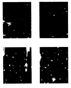
Figure 1 shows cholera toxin (CT) induces
enhanced major histocompatibility complex (MHC) class
II expression on Langerhans cells (LC), changes in LC
morphology, and loss of LCs (presumably through
migration). BALB/c mice (H-2d) were transcutaneously
immunized with 250 ~,g of cholera CT or its B subunit
(CTB) in saline solution on the ear. Previous
CA 02272417 1999-OS-14
WO 98/20734 PCT/US97/21324
9
experiments had established that mice were readily
immunized when using the skin of the ear (7000 anti-CT
. ELISA Units after a single immunization). After 16
hours, epidermal sheets were prepared and stained for
MHC class II molecules (scale bar is 50 um1_ Panai~
indicate (A) saline alone as a negative control, (B)
transcutaneous immunization with CT in saline, (C)
transcutaneous immunization with CTB in saline, and
(D) intradermal injection with tumor necrosis factor-a.
(10 ~,g) as a positive control.
DETAILED DESCRIPTION OF THE INVENTTON
A transcutaneous immunization system delivers
agents to specialized cells (e. g., antigen
presentation cell, lymphocyte) that produce an immune
response (Bos, 1997). These agents as a class are
called antigens. Antigen may be composed of chemicals
such as, for example, carbohydrate, glycolipid,
glycoprotein, lipid, lipoprotein, phospholipid,
polypeptide, protein, conjugates thereof, or any other
material known to induce an immune response. Antigen
may be provided as a whole organism such as, for
example, a bacterium or virion; antigen may be
obtained from an extract or lysate, either from whole
cells or membrane alone; or antigen may be chemically
synthesized or produced by recombinant means, or by
inactivation of a virus.
Processes for preparing a pharmaceutical
formulation are well-known in the art, whereby the
antigen and adjuvant is combined with a
pharmaceutically acceptable carrier vehicle. Suitable
vehicles and their preparation are described, for
example, in Remington's Pharmaceutical Sciences by
E.W. Martin. Such formulations will contain an
effective amount of the antigen and adjuvant together
CA 02272417 1999-OS-14
WO 98/20734 PCT/L1S97/21324
with a suitable amount of vehicle in order to prepare
pharmaceutically acceptable compositions suitable for
administration to a human or animal. The formulation
may be applied in the form of an cream, emulsion, gel,
5 lotion, ointment, paste, solution, suspension, or
other forms known in the art. In particular,
formulations that enhance skin hydration, penetration,
or both are preferred. There may also be incorporated
other pharmaceutically acceptable additives including,
10 for example, diluents, binders, stabilizers,
preservatives, and colorings.
Increasing hydration of the stratum corneum will
increase the rate of percutaneous absorbtion of a
given solute (Roberts and Walker, 1993). As used in
the present invention, "penetration enhancer" does not
include substances such as, for example: water,
physiological buffers, saline solutions, and alcohols
which would not perforate the skin.
An object of the present invention is to provide
a novel means for immunization through intact skin
without the need for perforating the skin. The
transcutaneous immunization system provides a method
whereby antigens and adjuvant can be delivered to the
immune system, especially specialized antigen
presentation cells underlying the skin such as, for
example, Langerhans cells.
Without being bound to any particular theory but
only to provide an explanation for our observations,
it is presumed that the transcutaneous immunization
delivery system carries antigen to cells of the immune
system where an immune response is induced. The
antigen may pass through the normal protective outer
layers of the skin (i.e., stratum corneum) and induce
the immune response directly, or through an antigen
presenting cell (e. g., macrophage, tissue macrophage,
Langerhans cell, dendritic cell, dermal dendritic
cell, B lymphocyte, or Kupffer cell) that presents
CA 02272417 1999-OS-14
WO 98/20734 PCT/LTS97/21324
11
processed antigen to a T lymphocyte. Optionally, the
antigen may pass through the stratum corneum via a
hair follicle or a skin organelle (e. g., sweat gland,
oil gland).
Transcutaneous immunization with bacterial ADP-
ribosylating exotoxins (bAREs) may target the
epidermal Langerhans cell, known to be among the most
efficient of the antigen presenting cells (APCs)
(Udey, 1997). We have found that bAREs activate
Langerhans cells when applied epicutaneously to the
skin in saline solution. The Langerhans cells direct
specific immune responses through phagocytosis of the
antigens, and migration to the lymph nodes where they
act as APCs to present the antigen to lymphocytes
(Udey, 1997), and thereby induce a potent antibody
response. Although the skin is generally considered a
barrier to invading organisms, the imperfection of
this barrier is attested to by the numerous Langerhans
cells distributed throughout the epidermis that are
designed to orchestrate the immune response against
organisms invading via the skin (Udey, 1997).
According to Udey (1997):
"Langerhans cells are bone-marrow
derived cells that are present in all
mammalian stratified squamous epithelia.
They comprise all of the accessory cell
activity that is present in uninflammed
epidermis, an in the current paradigm are
essential for the initiation and propagation
of immune responses directed against
epicutaneously applied antigens. Langerhans
cells are members of a family of potent
accessory cells ('dendritic cells') that are
widely distributed, but infrequently
represented, in epithelia and solid organs
as well as in lymphoid tissue . . .
"It is now recognized that Langerhans
cells (and presumably other dendritic cells)
have a life cycle with at least two distinct
stages. Langerhans cells that are located
in epidermis constitute a regular network of
antigen-trapping 'sentinel' cells.
Epidermal Langerhans cells can ingest
particulates, including microorganisms, and
CA 02272417 1999-OS-14
WO 98/20734 PCT/US97/21324
12
are efficient processors of complex
antigens. However, they express only low
levels of MHC class I and II antigens and
costimulatory molecules (ICAM-1, B7-1 and
B7-2) and are poor stimulators of unprimed T
cells. After contact with antigen, some
Langerhans cells become activated, exit the
epidermis and migrate to T-cell-dependent
regions of regional lymph nodes where they
local as mature dendritic cells. In the
course of exiting the epidermis and
migrating to lymph nodes, antigen-bearing
epidermal Langerhans cells (now the
'messengers') exhibit dramatic changes in
morphology, surface phenotype and function.
In contrast to epidermal Langerhans cells,
lymphoid dendritic cells are essentially
non-phagocytic and process protein antigens
inefficiently, but express high levels of
MHC class I and class II antigens and
various costimulatory molecules and are the
most potent stimulators of naive T cells
that have been identified."
We envision that the potent antigen presenting
capability of the epidermal Langerhans cells can be
exploited for transcutaneously delivered vaccines. A
transcutaneous immune response using the skin immune
system would require delivery of vaccine antigen only
to Langerhans cells in the stratum corneum (the
outermost layer of the skin consisting of cornified
cells and lipids) via passive diffusion and subsequent
activation of the Langerhans cells to take up antigen,
migrate to B-cell follicles and/or T-cell dependent
regions, and present the antigen to B and/or T cells
(Stingl et al., 1989). If antigens other that bAREs
(for example BSA) were to be phagocytosed by the
Langerhans cells, then these antigens could also be
taken to the lymph node for presentation to T-cells
and subsequently induce an immune response specific
for that antigen (e.g., BSA). Thus, a feature of
transcutaneous immunization is the activation of the
Langerhans cell, presumably by a bacterial ADP-
ribosylating exotoxin, ADP-ribosylating exotoxin
binding subunits (e.g., cholera toxin B subunit), or
CA 02272417 1999-OS-14
WO 98120734 PCTIUS97121324
13
other Langerhans cell activating substance.
The mechanism of transcutaneous immunization via
Langerhans cells activation, migration and antigen
presentation is clearly shown by the upregulation of
MHC class II expression in the epidermal Langerhans
cells from epidermal sheets transcutaneously immunized
with CT or CTB. In addition, the magnitude of the
antibody response induced by transcutaneous
immunization and isotype switching to predominantly
IgG is generally achieved with T-cell help stimulated
by antigen presenting cells such as Langerhans cells
or dendritic cells (Janeway and Travers, 1996), and
activation of both Thl and Th2 pathways as suggested
by the production of IgG1 and IgG2a (Paul and Seder,
1994; Seder and Paul, 1994). Additionally, T cell
proliferation to the antigen OVA is shown in mice
immunized with CT + OVA. Alternatively, a large
antibody response may be induced by a thymus-
independent antigen type 1 (TI-1) which directly
activates the B cell (Janeway and Travers, 1996).
The spectrum of more commonly known skin immune
responses is represented by contact dermatitis and
atopy. Contact dermatitis, a pathogenic manifestation
of LC activation, is directed by Langerhans cells
which phagocytose antigen, migrate to lymph nodes,
present antigen, and sensitize T cells for the intense
destructive cellular response that occurs at the
affected skin site (Dahl, 1996; Leung, 1997). Atopic
dermatitis may utilize the Langerhans cell in a
similar fashion, but is identified with Th2 cells and
is generally associated with high levels of IgE
antibody (Dahl, 1996; Leung, 1997).
Transcutaneous immunization with cholera toxin
and related bAREs on the other hand is a novel immune
response with an absence of superficial and
microscopic post-immunization skin findings (i.e.,
non-inflamed skin) shown by the absence of lymphocyte
CA 02272417 1999-OS-14
WO 98/20734 PCT/US97/21324
14
infiltration 24, 48 and 120 hours after immunization
with cholera toxin. This indicates that Langerhans
cells "comprise all of the accessory cell activity
that is present in uninflammed epidermis, and in the
current paradigm are essential for the initiation and
propagation of immune responses directed against
epicutaneously applied antigens" (Udey, 1997). The
uniqueness of the transcutaneous immune response here
is also indicated by the both high levels of antigen-
specific IgG antibody, and the type of antibody
produced (e. g., IgM, IgGl, IgG2a, IgG2b, IgG3 and IgA)
and the absence of anti-CT IgE antibody.
Thus, we have found that bacterial-derived toxins
applied to the surface of the skin can activate
Langerhans cells or other antigen presenting cells,
and induce a potent immune response manifested as high
levels of antigen-specific circulating IgG antibodies.
Such adjuvants may be used in transcutaneous
immunization to enhance the IgG antibody response to
proteins not otherwise immunogenic by themselves when
placed on the skin.
Transcutaneous targeting of Langerhans cells may
also be used to deactivate their antigen presenting
function, thereby preventing immunization or
sensitization. Techniques to deactivate Langerhans
cells include, for example, the use of interleukin-10
(Peguet-Navarro et al., 1995), monoclonal antibody to
interleukin-1(3 (Enk et al., 1993), or depletion via
superantigens such as through staphylococcal
enterotoxin-A (SEA) induced epidermal Langerhans cell
depletion (Shankar et al., 1996).
Transcutaneous immunization may be induced via
the ganglioside GMl binding activity of CT, LT or
subunits such as CTB (Craig and Cuatrecasas, 1975).
Ganglioside GM1 is a ubiquitous cell membrane
glycolipid found in all mammalian cells (Plotkin and
Mortimer, 1994). When the pentameric CT B subunit
CA 02272417 1999-OS-14
WO 98/20734 PCT/US97/21324
binds to the cell surface a hydrophilic pore is formed
which allows the A subunit to penetrate across the
lipid bilayer (Ribi et al., 1988).
We have shown that transcutaneous immunization by
5 CT or CTB may require ganglioside GM1 binding
activity. When mice were transcutaneously immunized
with CT, CTA and CTB, only CT and CTB resulted in an
immune response. CTA contains the ADP-ribosylating
exotoxin activity but only CT and CTB containing the
10 binding activity were able to induce an immune
response indicating that the B subunit was necessary
and sufficient to immunize through the skin. We
conclude that the Langerhans cell or another antigen
presenting cell may be activated by CTB binding to its
15 cell surface.
ANTIGEN
Antigen of the invention may be expressed by
recombinant means, preferably as a fusion with an
affinity or epitope tag (Summers and Smith, 1987;
Goeddel, 1990; Ausubel et al., 1996); chemical
synthesis of an oligopeptide, either free or
conjugated to carrier proteins, may be used to obtain
antigen of the invention (Bodanszky, 1993; Wisdom,
1994). Oligopeptides are considered a type of
polypeptide.
Oligopeptide lengths of 6 residues to 20 residues
are preferred. Polypeptides may also by synthesized
as branched structures such as those disclosed in U.S.
Pat. Nos. 5,229,490 and 5,390,111. Antigenic
polypeptides include, for example, synthetic or
recombinant B-cell and T-cell epitopes, universal T-
cell epitopes, and mixed T-cell epitopes from one
organism or disease and B-cell epitopes from another.
Antigen obtained through recombinant means or
peptide synthesis, as well as antigen of the invention
obtained from natural sources or extracts, may be
CA 02272417 1999-OS-14
WO 98/20734 PCT/US97/2I324
16
purified by means of the antigen's physical and
chemical characteristics, preferably by fractionation
or chromatography (Janson and Ryden, 1989; Deutscher,
1990; Scopes, 1993).
A multivalent antigen formulation may be used to
induce an immune response to more than one antigen at
the same time. Conjugates may be used to induce an
immune response to multiple antigens, to boost the
immune response, or both. Additionally, toxins may be
boosted by the use of toxoids, or toxoids boosted by
the use of toxins. Transcutaneous immunization may be
used to boost responses induced initially by other
routes of immunization such as by injection, or the
oral or intranasal routes.
Antigen includes, for example, toxins, toxoids,
subunits thereof, or combinations thereof (e. g.,
cholera toxin, tetanus toxoid).
Antigen may be solubilized in water, a solvent
such as methanol, or a buffer. Suitable buffers
include, but are not limited to, phosphate buffered
saline Ca++/Mg++ free ( PBS ) , normal saline ( 150 mM NaCl
in water), and Tris buffer. Antigen not soluble in
neutral buffer can be solubilized in 10 mM acetic acid
and then diluted to the desired volume with a neutral
buffer such as PBS. In the case of antigen soluble
only at acid pH, acetate-PBS at acid pH may be used as
a diluent after solubilization in dilute acetic acid.
Glycerol may be a suitable non-aqueous buffer for use
in the present invention.
If an antigen such as, for example, hepatitis A
virus, is not soluble per se, the antigen may be
present in the formulation in a suspension or even as
an aggregate.
Hydrophobic antigen can be solubilized in a
detergent, for example a polypeptide containing a
membrane-spanning domain. Furthermore, for
formulations containing liposomes, an antigen in a
CA 02272417 1999-OS-14
WO 98/20734 PCT/US97/21324
17
detergent solution (e.g., a cell membrane extract) may
be mixed with lipids, and liposomes then may be formed
by removal of the detergent by dilution, dialysis, or
column chromatography. Certain antigens such as, for
example, those from a virus (e. g., hepatitis A) need
not be soluble per se, but can be incorporated
directly into a liposome in the form of a virosome
(Morein and Simons, 1985).
Plotkin and Mortimer (1994) provide antigens
which can be used to vaccinate animals or humans to
induce an immune response specific for particular
pathogens, as well as methods of preparing antigen,
determining a suitable dose of antigen, assaying for
induction of an immune response, and treating
infection by a pathogen (e. g., bacterium, virus,
fungus, or parasite).
Bacteria include, for example: anthrax,
campylobacter, cholera, diphtheria, enterotoxigenic E.
coli, giardia, gonococcus, Helicobacter pylori (Lee
and Chen, 1994), Hemophilus influenza B, Hemophilus
influenza non-typable, meningococcus, pertussis,
pneumococcus, salmonella, shigella, Streptococcus B,
group A Streptococcus, tetanus, Vibrio cholerae,
yersinia, Staphylococcus, Pseudomonas species and
Clostridia species.
Viruses include, for example: adenovirus, dengue
serotypes 1 to 4 (Delenda et al., 1994; Fonseca et
al., 1994; Smucny et al., 1995), ebola (Jahrling et
al., 1996), enterovirus, hepatitis serotypes A to E
(Blum, 1995; Katkov, 1996; Lieberman and Greenberg,
1996; Mast, 1996; Shafara et al., 1995; Smedila et
al., 1994; U.S. Pat. Nos. 5,314,808 and 5,436,126),
herpes simplex virus 1 or 2, human immunodeficiency
virus (Deprez et al., 1996), influenza, Japanese
equine encephalitis, measles, Norwalk, papilloma
virus, parvovirus B19, polio, rabies, rotavirus,
rubella, rubeola, vaccinia, vaccinia constructs
CA 02272417 1999-OS-14
WO 98120734 PGT/US97I21324
18
containing genes coding for other antigens such as
malaria antigens, varicella, and yellow fever.
Parasites include, for example: Entamoeba
histolytica (Zhang et al., 1995); Plasmodium (Bathurst
et al., 1993; Chang et al., 1989, 1992, 1994; Fries et
al., 1992a, 1992b; Herrington et al., 1991; Khusmith
et al., 1991; Malik et al., 1991; Migliorini et al.,
1993; Pessi et al., 1991; Tam, 1988; Vreden et al.,
1991; White et al., 1993; Wiesmueller et al., 1991),
heishmania (Frankenburg et al., 1996), Toxoplasmosis,
and the Helminths.
Antigens may also comprise those used in
biological warfare such as ricin, for which protection
can be achieved via antibodies.
ADJUVANT
The formulation also contains an adjuvant,
although a single molecule may contain both adjuvant
and antigen properties (e. g., cholera toxin) (Elson
and Dertzbaugh, 1999). Adjuvants are substances that
are used to specifically or non-specifically
potentiate an antigen-specific immune response.
Usually, the adjuvant and the formulation are mixed
prior to presentation of the antigen but,
alternatively, they may be separately presented within
a short interval of time.
Adjuvants include, for example, an oil emulsion
(e.g., complete or incomplete Freund's adjuvant), a
chemokine (e.g., defensins 1 or 2, RANTES, MIP1-oc,
MIP-2, interleukin-8) or a cytokine (e. g.,
interleukin-lei, -2, -6, -10 or -12; y-interferon; tumor
necrosis factor-a,; or granulocyte-monocyte-colony
stimulating factor) (reviewed in Nohria and Rubin,
1994), a muramyl dipeptide derivative (e. g.,
murabutide, threonyl-MDP or muramyl tripeptide), a
heat shock protein or a derivative, a derivative of
CA 02272417 1999-OS-14
WO 98/20734 PCT/US97/21324
19
Leishmania major LeIF (Skeiky et al., 1995), cholera
toxin or cholera toxin B, a lipopolysaccharide (LPS)
derivative (e.g., lipid A or monophosphoryl lipid A),
or superantigen (Saloga et al., 1996). Also, see
Richards et al. (1995) for adjuvants useful in
immuni zation .
An adjuvant may be chosen to preferentially
induce antibody or cellular effectors, specific
antibody isotypes (e. g., IgM, IgD, IgAl, IgA2,
secretory IgA, IgE, IgGI, IgG2, IgG3, and/or IgG4), or
specific T-cell subsets (e. g., CTL, Thl, Th2 and/or
ToTH) (Glenn et al., 1995) .
Cholera toxin is a bacterial exotoxin from the
family of ADP-ribsoylating exotoxins (referred to as
bAREs). Most bAREs are organized as A:B dimer with a
binding B subunit and an A subunit containing the ADP-
ribosyltransferase. Such toxins include diphtheria
toxin, Pseudomonas exotoxin A, cholera toxin (CT), E.
coli heat-labile enterotoxin (LT), pertussis toxin, C.
botulinum toxin C2, C. botulinum toxin C3, C. limosum
exoenzyme, B. cereus exoenzyme, Pseudomonas exotoxin
S, Staphylococcus aureus EDIN, and B. sphaericus
toxin.
Cholera toxin is an example of a bARE that is
organized with A and B subunits. The B subunit is the
binding subunit and consists of a B-subunit pentamer
which is non-covalently bound to the A subunit. The
B-subunit pentamer is arranged in a symmetrical
doughnut-shaped structure that binds to GM1-ganglioside
on the target cell. The A subunit serves to ADP
ribosylate the alpha subunit of a subset of the hetero
trimeric GTP proteins (G proteins) including the Gs
protein which results in the elevated intracellular
levels of cyclic AMP. This stimulates release of ions
and fluid from intestinal cells in the case of
cholera.
Cholera toxin (CT) and its B subunit (CTB) have
CA 02272417 1999-OS-14
WO 98/20734 PCT/US97/21324
adjuvant properties when used as either an
intramuscular or oral immunogen (Elson and Dertzbaugh,
1994; Trach et al., 1997). Another antigen, heat-
labile enterotoxin from E. coli (LT) is 80o homologous
5 at the amino acid level with CT and possesses similar
binding properties; it also appears to bind the GM1-
ganglioside receptor in the gut and has similar ADP-
ribosylating exotoxin activities. Another bARE,
Pseudomonas exotoxin A (ETA), binds to the a2-
10 macroglobulin receptor-low density lipoprotein
receptor-related protein (Kounnas et al., 1992).
bAREs are reviewed by Krueger and Barbieri (1995).
The toxicity of CT by oral, nasal, and
intramuscular routes limits the dose that can be used
15 as an adjuvant. In a comparative trial of CT injected
intramuscularly, extensive swelling at the site of
injection was elicited. By contrast, equivalent or
greater doses of CT placed on the skin caused no
toxicity.
20 The examples below show that cholera toxin (CT),
its B subunit (CTB), E. coli heat-labile enterotoxin
(LT), and pertussis toxin are potent adjuvants for
transcutaneous immunization, inducing high levels of
IgG antibodies but not IgE antibodies. Also shown is
that CTB without CT can also induce high levels of IgG
antibodies. Thus, both bAREs and a derivative thereof
can effectively immunize when epicutaneouly applied to
the skin in a simple solution. Furthermore, these
examples demonstrate that CT, CTB and bAREs can act as
both adjuvant and antigen.
When an adjuvant such as CT is mixed with BSA, a
protein not usually immunogenic when applied to the
skin, anti-BSA antibodies are induced. An immune
response to diphtheria toxoid was induced using
pertussis toxin as adjuvant, but not with diphtheria
toxoid alone. Thus, bAREs can act as adjuvants for
non-immunogenic proteins in an transcutaneous
CA 02272417 1999-OS-14
WO 98/2U734 PCT/US97/21324
21
immunization system.
Other proteins may also act as both adjuvant and
antigen. For example FLUZONE (Lederle), the split
virion influenza A and B vaccine contains
neuraminidase and hemagglutinin which are highly
immunogenic, confers protection and may effectively
immunize through the skin acting as its own adjuvant
and antigen. Toxoids such as diphtheria toxoid which
has been toxoided using formalin, pertussis toxoid
which has been toxoided using hydrogen peroxide, or
mutant toxins such as cholera or heat labile
enterotoxin from E. coli which have been toxoided
using genetic techniques to destroy the ribosyl
transferase activity, may continue to harbor adjuvant
qualities and act as both antigen and adjuvant.
Protection against the life-threatening
infections diphtheria, pertussis, and tetanus (DPT)
can be achieved by inducing high levels of circulating
anti-toxin antibodies. Pertussis may be an exception
in that some investigators feel that antibodies
directed to other portions of the invading organism
are necessary for protection, although this is
controversial (see Schneerson et al., 1996) and most
new generation acellular pertussis vaccines have PT as
a component of the vaccine (Krueger and Barbieri,
1995). The pathologies in the diseases caused by DPT
are directly related to the effects of their toxins
and anti-toxin antibodies most certainly play a role
in protection (Schneerson et al., 1996).
In general, toxins can be chemically inactivated
to form toxoids which are less toxic but remain
immunogenic. We envision that the transcutaneous
immunization system using toxin-based immunogens and
adjuvants can achieve anti-toxin levels adequate for
protection against these diseases. The anti-toxin
antibodies may be induced through immunization with
the toxins, or genetically-detoxified toxoids
CA 02272417 1999-OS-14
WO 98/20734 PCT/US97/21324
22
themselves, or with toxoids and adjuvants such as CT
or by the toxoids alone. Genetically toxoided toxins
which have altered ADP-ribosylating exotoxin activity,
but not binding activity, are envisioned to be
especially useful as non-toxic activators of antigen
presenting cells used in transcutaneous immunization.
We envision that CT can also act as an adjuvant
to induce antigen-specific CTLs through transcutaneous
immunization (see Bowen et al., 1994; Porgador et al.,
1997 for the use of CT as an adjuvant in oral
immunization) .
The bARE adjuvant may be chemically conjugated to
other antigens including, for example, carbohydrates,
polypeptides, glycolipids, and glycoprotein antigens.
Chemical conjugation with toxins, their subunits, or
toxoids with these antigens would be expected to
enhance the immune response to these antigens when
applied epicutaneously.
To overcome the problem of the toxicity of the
toxins, (e.g., diphtheria toxin is known to be so
toxic that one molecule can kill a cell) and to
overcome the difficulty of working with such potent
toxins as tetanus, several workers have taken a
recombinant approach to producing genetically produced
toxoids. This is based on inactivating the catalytic
activity of the ADP-ribosyl transferase by genetic
deletion. These toxins retain the binding
capabilities, but lack the toxicity, of the natural
toxins. This approach is described by Burnette et al.
(1994), Rappuoli et al. (1995), and Rappuoli et al.
(1996). Such genetically toxoided exotoxins could be
useful for transcutaneous immunization system in that
they would not create a safety concern as the toxoids
would not be considered toxic. They may act as both
antigens and adjuvants, enhancing the immune response
to themselves or added antigens. Additionally,
several techniques exist to chemically toxoid toxins
CA 02272417 1999-OS-14
WO 98/20734 PCT/US97/21324
23
which can address the same problem (Schneerson et al.,
1996). Alternatively, fragments of the toxin or
toxoids may be used such as the C fragment of Tetanus.
These techniques could be important for certain
applications, especially pediatric applications, in
which ingested toxins (e. g., diphtheria toxin) might
possibly create adverse reactions.
Optionally, an activator of Langerhans cells may
be used as an adjuvant. Examples of such activators
include: inducers of heat shock protein; contact
sensitizers (e. g., trinitrochlorobenzene, dinitro-
fluorobenzene, nitrogen mustard, pentadecylcatechol);
toxins (e. g, Shiga toxin, Staph enterotoxin B); lipo-
polysaccharides, lipid A, or derivatives thereof;
bacterial DNA (Stacey et al., 1996); cytokines (e. g.,
tumor necrosis factor-a, interleukin-1(3, -10, -12);
and chemokines (e. g., defensins 1 or 2, RANTES, MIP-
1a, MIP-2, interleukin-8).
A combination of different adjuvants may be used
in the present invention. For example, a combination
of bacterial DNA containing CpG nucleotide sequences
and an ADP-ribosylating exotoxin could be used to
direct the T-helper response to antigens administered
transcutaneously. Thus, Th1 or Th2 like responses to
CT-adjuvanted antigens could be switched by the use of
nonmethylated CpG bacterial DNA, or other proteins
such as LeIF or calcium channel blockers.
CpGs are among a class of structures which have
patterns allowing the immune system to recognize their
pathogenic origins to stimulate the innate immune
response leading to adaptive immune responses.
(Medzhitov and Janeway, Curr. Opin. Immunol., 9:4-9,
1997). These structures are called pathogen-
associated molecular patterns (PAMPs) and include
lipopolysaccharides, teichoic acids, unmethylated CpG
motifs, double stranded RNA and mannins.
CA 02272417 1999-OS-14
WO 98120734 PCT/US97I21324
24
PAMPs induce endogenous signals that can mediate
the inflammatory response, act as costimulators of T-
cell function and control the effector function. The
ability of PAMPs to induce these responses play a role
in their potential as adjuvants and their targets are
APCs such as macrophages and dendritic cells. The
antigen presenting cells of the skin could likewise be
stimulated by PAMPs transmitted through the skin. For
example, Langerhans cells, a type of dendritic cell,
could be activated by a PAMP in solution on the skin
with a transcutaneously poorly immunogenic molecule
and be induced to migrate and present this poorly
immunogenic molecule to T-cells in the lymph node,
inducing an antibody response to the poorly
immunogenic molecule. PAMPs could also be used in
conjunction with other skin adjuvants such as cholera
toixn to induce different costimulatory molecules and
control different effector functions to guide the
immune response, for example from a Th2 to a Thl
response.
If an immunizing antigen has sufficient
Langerhans cell activating capabilities then a
separate adjuvant may not be required, as in the case
of CT which is both antigen and adjuvant. It is
envisioned that whole cell preparations, live viruses,
attenuated viruses, DNA plasmids, and bacterial DNA
could be sufficient to immunize transcutaneously. It
may be possible to use low concentrations of contact
sensitizers or other activators of Langerhans cells to
induce an immune response without inducing skin
lesions.
LIPOSOMES AND THEIR PREPARATION
Liposomes are closed vesicles surrounding an
internal aqueous space. The internal compartment is
separated from the external medium by a lipid bilayer
composed of discrete lipid molecules. In the present
CA 02272417 1999-OS-14
WO 98/20734 PCT/US97/21324
invention, antigen may be delivered through intact
skin to specialized cells of the immune system,
whereby an antigen-specific immune response is
induced. Transcutaneous immunization may be achieved
5 by using liposomes; however, as shown in the examples,
liposomes are not required to elicit an antigen-
specific immune response.
Liposomes may be prepared using a variety of
techniques and membrane lipids (reviewed in
10 Gregoriadis, 1993). Liposomes may be pre-formed and
then mixed with antigen. The antigen may be dissolved
or suspended, and then added to (a) the pre-formed
liposomes in a lyophilized state, (b) dried lipids as
a swelling solution or suspension, or (c) the solution
15 of lipids used to form liposomes. They may also be
formed from lipids extracted from the stratum corneum
including, for example, ceramide and cholesterol
derivatives (Wertz, 1992).
Chloroform is a preferred solvent for lipids, but
20 it may deteriorate upon storage. Therefore, at one-
to three-month intervals, chloroform is redistilled
prior to its use as the solvent in forming liposomes.
After distillation, 0.7% ethanol can be added as a
preservative. Ethanol and methanol are other suitable
25 solvents.
The lipid solution used to form liposomes is
placed in a round-bottomed flask. Pear-shaped boiling
flasks are preferred, particularly those flasks sold
by Lurex Scientific (Vineland, NJ, cat. no. JM-5490).
The volume of the flask should be more than ten times
greater than the volume of the anticipated aqueous
suspension of liposomes to allow for proper agitation
during liposome formation.
Using a rotary evaporator, solvent is removed at
37°C under negative pressure for 10 minutes with a
filter aspirator attached to a water faucet. The
flask is further dried under low vacuum (i.e., less
CA 02272417 1999-OS-14
WO 98!20734 PCT/US97/21324
26
than 50 mm Hg) for 1 hour in a dessicator.
To encapsulate antigen into liposomes, an aqueous
solution containing antigen may be added to
lyophilized liposome lipids in a volume that results
in a concentration of approximately 200 mM with
respect to liposome lipid, and shaken or vortexed
until all the dried liposome lipids are wet. The
liposome-antigen mixture may then be incubated for 18
hours to 72 hours at 4°C. The liposome-antigen
formulation may be used immediately or stored for
several years. It is preferred to employ such a
formulation directly in the transcutaneous
immunization system without removing unencapsulated
antigen. Techniques such as bath sonication may be
employed to decrease the size of liposomes, which may
augment transcutaneous immunization.
Liposomes may be formed as described above but
without addition of antigen to the aqueous solution.
Antigen may then be added to the pre-formed liposomes
and, therefore, antigen would be in solution and/or
associated with, but not encapsulated by, the
liposomes. This process of making a liposome-
containing formulation is preferred because of its
simplicity. Techniques such as bath sonication may be
employed to alter the size and/or lamellarity of the
liposomes to enhance immunization.
Although not required to practice the present
invention, hydration of the stratum corneum may be
enhanced by adding liposomes to the formulation.
Liposomes have been used as carriers with adjuvants to
enhance the immune response to antigens mixed with,
encapsulated in, attached to, or associated with
liposomes.
TRANSCUTANEOUS DELIVERY OF ANTIGEN
Efficient immunization can be achieved with the
present invention because transcutaneous delivery of
CA 02272417 1999-OS-14
WO 98/20734 PCT/(TS97/21324
27
antigen may target the Langerhans cell. These cells
are found in abundance in the skin and are efficient
antigen presenting cells leading to T-cell memory and
potent immune responses (Udey, 1997). Because of the
presence of large numbers of Langerhans cells in the
skin, the efficiency of transcutaneous delivery may be
related to the surface area exposed to antigen and
adjuvant. In fact, the reason that transcutaneous
immunization is so effective may be that it targets a
larger number of these efficient antigen presenting
cells than intramuscular immunization. However, even
a small number of Langerhans cells or dendritic cells
may be sufficient for immunization.
We envision the present invention will enhance
access to immunization, while inducing a potent immune
response. Because transcutaneous immunization does
not involve penetration of the skin and the
complications and difficulties thereof, the
requirements of trained personnel, sterile technique,
and sterile equipment are reduced. Furthermore, the
barriers to immunization at multiple sites or to
multiple immunizations are diminished. Immunization
by a single application of the formulation is also
envisioned.
Immunization may be achieved using epicutaneous
application of a simple solution of antigen and
adjuvant impregnated in gauze under an occlusive
patch, or by using other patch technologies; creams,
immersion, ointments and~sprays are other possible
methods of application. The immunization could be
given by untrained personnel, and is amenable to self-
application. Large-scale field immunization could
occur given the easy accessibility to immunization.
Additionally, a simple immunization procedure would
improve access to immunization by pediatric patients
and the elderly, and populations in Third World
countries.
CA 02272417 1999-OS-14
WO 98/20734 PCT/US97/21324
28
Similarly, animals could be immunized using the
present invention. Application to anatomical sites
such as the ear, underbelly, paws, conjunctiva,
intertriginous regions, or anal region, or via dipping
or immersion could be employed.
For previous vaccines, their formulations were
injected through the skin with needles. Injection of
vaccines using needles carries certain drawbacks
including the pain associated with injections, the
need for sterile needles and syringes, trained medical
personnel to administer the vaccine, discomfort from
the injection, and potential complications brought
about by puncturing the skin with the needle.
Immunization through the skin without the use of
needles (i.e., transcutaneous immunization) represents
a major advance for vaccine delivery by avoiding the
aforementioned drawbacks.
The transcutaneous delivery system of the
invention is also not concerned with penetration of
intact skin by sound or electrical energy. Such a
system that uses an electrical field to induce
dielectric breakdown of the stratum corneum is
disclosed in U.S. Pat. No. 5,469,386.
Moreover, transcutaneous immunization may be
superior to immunization using needles as more immune
cells would be targeted by the use of several
locations targeting large surface areas of skin. A
therapeutically effective amount of antigen sufficient
to induce an immune response may be delivered
transcutaneously either at.a single cutaneous
location, or over an area of intact skin covering
multiple draining lymph node fields (e. g., cervical,
axillary, inguinal, epitrochelear, popliteal, those of
the abdomen and thorax). Such locations close to
numerous different lymphatic nodes at locations all
over the body will provide a more widespread stimulus
to the immune system than when a small amount of
CA 02272417 1999-OS-14
WO 98/20734 PCT/US97/21324
29
antigen is injected at a single location by
intradermal subcutaneous or intramuscular injection.
Antigen passing through or into the skin may
encounter antigen presenting cells which process the
antigen in a way that induces an immune response.
Multiple immunization sites may recruit a greater
number of antigen presenting cells and the larger
population of antigen presenting cells that were
recruited would result in greater induction of the
immune response. Transcutaneous immunization may
allow application in close proximity to a lymph node
draining site and thereby improve efficiency or
potency of immunization. It is conceivable that
absorption through the skin may deliver antigen to
phagocytic cells of the skin such as, for example,
dermal dendritic cells, macrophages, and other skin
antigen presenting cells; antigen may also be
delivered to phagocytic cells of the liver, spleen,
and bone marrow that are known to serve as the antigen
presenting cells through the blood stream or lymphatic
system. The result would be widespread distribution
of antigen to antigen presenting cells to a degree
that is rarely, if ever achieved, by current
immunization practices.
The transcutaneous immunization system may be
applied directly to the skin and allowed to air dry;
rubbed into the skin or scalp; held in place with a
dressing, patch, or absorbent material; otherwise held
by a device such as a stocking, slipper, glove, or
shirt; or sprayed onto the skin to maximize contact
with the skin. The formulation may be applied in an
absorbant dressing or gauze. The formulation may be
covered with an occlusive dressing such as, for
example, in an emulsion of antigen solution and
AQUAPHOR (petrolatum, mineral oil, mineral wax, wool
wax, panthenol, bisabol, and glycerin from
Beiersdorf), plastic film, impregnated polymer,
CA 02272417 1999-OS-14
WO 98120734 PCT/US97/21324
COMFEEL (Coloplast) or vaseline; or a non-occlusive
dressing such as, for example, DUODERM (3M) or OPSITE
(Smith & Napheu). An occlusive dressing completely
excludes the passage of water. Alternatively, a
5 partially occlusive dressing such as TEGADERM may be
applied to provide hydration and may allow longer
application of the patch or may prevent maceration of
the skin.
The formulation may be applied to single or
10 multiple sites, to single or multiple limbs, or to
large surface areas of the skin by complete immersion.
The formulation may be applied directly to the skin.
Genetic immunization has been described in U.S.
Pat. Nos. 5,589,466 and 5,593,972. The nucleic
15 acids) contained in the formulation may encode the
antigen, the adjuvant, or both. The nucleic acid may
or may not be capable of replication; it may be non-
integrating and non-infectious. The nucleic acid may
further comprise a regulatory region (e. g., promoter,
20 enhancer, silencer, transcription initiation and
termination sites, RNA splice acceptor and donor
sites, polyadenylation signal, internal ribosome
binding site, translation initiation and termination
sites) operably linked to the sequence encoding the
25 antigen or adjuvant. The nucleic acid may be
complexed with an agent that promotes transfection
such as cationic lipid, calcium phosphate, DEAE-
dextran, polybrene-DMSO, or a combination thereof.
The nucleic acid may comprise regions derived from
30 viral genomes. Such materials and techniques are
described by Kriegler (1990) and Murray (1991).
An immune response may comprise humoral (i.e.,
antigen-specific antibody) and/or cellular (i.e.,
antigen-specific lymphocytes such as B cells, CD4+ T
cells, CD8+ T cells, CTL, Thl cells, Th2 cells, and/or
TDTH cells) effector arms. Moreover, the immune
response may comprise NK cells that mediate antibody-
CA 02272417 1999-OS-14
WO 98/20734 PCT/US97/21324
31
dependent cell-mediated cytotoxicity (ADCC).
The immune response induced by the formulation of
the invention may include the elicitation of antigen-
specific antibodies and/or cytotoxic lymphocytes (CTL,
reviewed in Alving and Wassef, 1994). Antibody can be
detected by immunoassay techniques, and the detection
of various isotypes (e. g., IgM, IgD, IgAl, IgA2,
secretory IgA, IgE, IgGl, IgG2, IgG3, or IgG9) may be
expected. An immune response can also be detected by
a neutralizing assay.
Antibodies are protective proteins produced by B
lymphocytes. They are highly specific, generally
targeting one epitope of an antigen. Often,
antibodies play a role in protection against disease
by specifically reacting with antigens derived from
the pathogens causing the disease. Immunization may
induce antibodies specific for the immunizing antigen,
such as cholera toxin. These antigen-specific
antibodies are induced when antigen is delivered
through the skin by liposomes.
CTLs are particular protective immune cells
produced to protect against infection by a pathogen.
They are also highly specific. Immunization may
induce CTLs specific for the antigen, such as a
synthetic oligopeptide based on a malaria protein, in
association with self-major histocompatibility
antigen. CTLs induced by immunization with the
transcutaneous delivery system may kill pathogen
infected cells. Immunization may also produce a
memory response as indicated by boosting responses in
antibodies and CTLs, lymphocyte proliferation by
culture of lymphocytes stimulated with the antigen,
and delayed type hypersensitivity responses to
intradermal skin challenge of the antigen alone.
It is envisioned that the T-helper response
induced by transcutaneous immunization may be
manipulated by the use of calcium channel blockers
CA 02272417 1999-OS-14
WO 98/20734 PCTlL1S97/21324
32
(e.g., nifedipine, verpamil) which suppress the
contact hypersensitivity reaction by inhibiting
antigen catabolism and subsequent presentation by
epidermal Langerhans cells. The transcutaneous
application of a calcium channel blocker would be
expected to affect surface expression of co-
stimulatory molecules (e.g., B7-related family) and
the generation of a subsequent T-helper response. It
is also envisioned that addition of the calcium
channel blocker may inhibit delayed type
hypersensitivity responses and could be used to select
an immune response that is predominantly a cellular or
a humoral response.
In a viral neutralization assay, serial dilutions
of sera are added to host cells which are then
observed for infection after challenge with infectious
virus. Alternatively, serial dilutions of sera may be
incubated with infectious titers of virus prior to
inoculation of an animal, and the inoculated animals
are then observed for signs of infection.
The transcutaneous immunization system of the
invention may be evaluated using challenge models in
either animals or humans, which evaluate the ability
of immunization with the antigen to protect the
subject from disease. Such protection would
demonstrate an antigen-specific immune response. In
lieu of challenge, achieving anti-diphtheria antibody
titers of 5 IU/ml or greater is generally assumed to
indicate optimum protection and serves as a surrogate
marker for protection (Plotkin and Mortimer, 1994).
Furthermore, the Plasmodium faciparum challenge
model may be used as to induce an antigen-specific
immune response in humans. Human volunteers may be
immunized using the transcutaneous immunization system
containing oligopeptides or proteins (polypeptides)
derived from the malaria parasite, and then exposed to
malaria experimentally or in the natural setting. The
CA 02272417 1999-OS-14
WO 98/20734 PCT/tTS97/21324
33
Plasmodium yoelii mouse malaria challenge model may be
used to evaluate protection in the mouse against
malaria (Wang et al, 1995).
Alving et al (1986) injected liposomes comprising
lipid A as an adjuvant for inducing an immune response
to cholera toxin (CT) in rabbits and to a synthetic
protein consisting of a malaria oligopeptide
containing four tetra-peptides (Asn-Ala-Asn-Pro)
conjugated to BSA. The authors found that the immune
response to cholera toxin or to the synthetic malaria
protein was markedly enhanced by encapsulating the
antigen within the liposomes containing lipid A,
compared to similar liposomes lacking lipid A.
Several antigens derived either from the
circumsporozoite protein (CSP) or from merozoite
surface proteins of Plasmodium falciparum have been
encapsulated in liposomes containing lipid A. All of
the malaria antigens that have been encapsulated in
liposomes containing lipid A have been shown to induce
humoral effectors (i.e., antigen-specific antibodies),
and some have been shown to induce cell-mediated
responses as well. Generation of an immune response
and immunoprotection in an animal vaccinated with a
malaria antigen may be assayed by immunofluorescence
to whole, fixed malaria sporozoites or CTLs killing of
target cells transfected with CSP.
Mice transcutaneously immunized with cholera
toxin can be protected against intranasal challenge
with a 20 ~,g dose of cholera toxin. Mallet et al
(personal communication) have found that C57B1/6 mice
develop a fatal hemorrhagic pneumonia in response to
intranasal challenge with CT. Alternatively, the mice
may be challenged with an intraperitoneal dose of CT
(Dragunsky et al, 1992). Cholera toxin-specific IgG
or IgA antibody may provide protection against cholera
toxin challenge (Pierce, 1978; Pierce and Reynolds,
1974).
CA 02272417 1999-OS-14
WO 98/20734 PCT/US97/21324
34
Similar protective effects could be expected in
humans immunized with LT or CT, and challenged with
LT-secreting E. coli or CT-secreting Vibrio cholerae,
respectively. Additionally, cross protection has been
demonstrated between CT and LT immune subjects and CT
and LT mediated disease.
As shown in the examples below, mucosal immunity
may be achieved via the trancutaneous route. Mucosal
IgG and IgA can be detected in mice immunized with CT
transcutaneously. This may be important for
protection in diseases where the pathology occurs at
mucosal sites such as in LT or CT mediated disease,
where entry of the pathogenic organism occurs at a
mucosal site, or where mucosal infection is important
to pathogenesis.
It would be expected that transcutaneous
immunization against diseases such as influenza could
be effective either by inducing mucosal immunity or by
systemic immunity, or by a combination of immunity
such as humoral, cellular or mucosal.
Vaccines may be effective against host effects
such as the binding of erythrocytes to vascular
endothelium in malaria by inducing anti-sequestrin
antibodies.
Protective antibodies such as anti-hepatitis A, B
or hepatitis E antibodies may be induced by the
transcutaneous route using whole inactivated virus,
virus-derived subunits or recombinant products.
Protection against tetanus, diphtheria and other
toxin mediated diseases may be conferred by
transcutaneously induced anti-toxin antibodies. A
tetanus "booster" patch could be envisioned that
contained adjuvant such as CT and toxoids such as
tetanus and diphtheria, or fragments such as the
tetanus C fragment. Boosting could be achieved
following primary immunization by injection or
transcutaneous immunization with the same or similar
CA 02272417 1999-OS-14
WO 98/20734 PCT/US97121324
antigens. For injectable immunizations that induce
immunity but have potential side effects upon
boosting, a transcutaneous boost may be preferable.
Oral or nasal immunization may conceivably be boosted
5 using the transcutaneous route. Simultaneous use of
injectable and transcutaneous immunizations could also
be used.
Vaccination has also been used as a treatment for
cancer and autoimmune disease. For example,
10 vaccination with a tumor antigen (e. g., prostate
specific antigen) may induce an immune response in the
form of antibodies, CTLs and lymphocyte proliferation
which allows the body's immune system to recognize and
kill tumor cells. Targeting dendritic cells, of which
15 Langerhans cells are a specific subset, has been shown
to be an important strategy in cancer immunotherapy.
Tumor antigens useful for vaccination have been
described for melanoma (U. S. Pat. Nos. 5,102,663,
5,141,742, and 5,262,177), prostate carcinoma (U. S.
20 Pat. No. 5,538,866), and lymphoma (U. S. Pat. Nos.
4,816,249, 5,068,177, and 5,227,159). Vaccination
with T-cell receptor oligopeptide may induce an immune
response that halts progression of autoimmune disease
(U.S. Pat. Nos. 5,612,035 and 5,614,192 Antel et al,
25 1996; Vandenbark et al, 1996). U.S. Pat. No.
5,552,300 also describes antigens suitable for
treating autoimmune disease.
The following is meant to be illustrative of the
present invention; however, the practice of the
30 invention is not limited or restricted in any way by
the examples.
EXAMPLES
Immunization Procedure
35 BALB/c mice of 6 to 8 weeks were shaved with a
#40 clipper. This shaving could be done without any
signs of trauma to the skin. The shaving was done
CA 02272417 1999-OS-14
WO 98120734 PCTIUS97/21324
36
from the mid-thorax to just below the nape of the
neck. The mice were then allowed to rest for 24
hours. Prior to this the mice had been ear-tagged for
identification, and pre-bled to obtain a sample of
pre-immune serum. Mice were also transcutaneously
immunized without shaving by applying up to 50 ~.l of
immunizing solution to each ear.
The mice were then immunized in the following
way. Mice were anesthetized with 0.03-0.06 ml of a 20
mg/ml solution of xylazine and 0.5 ml of 100 mg/ml
ketamine; mice were immobilized by this dose of
anesthesia for approximately one hour. The mice were
placed ventral side down on a warming blanket.
The immunizing solution was then placed on the
dorsal shaved skin of a mouse in the following manner:
a 1.2 cm x 1.6 cm stencil made of polystyrene was laid
gently on the back and a saline-wetted sterile gauze
was used to partially wet the skin (this allowed even
application of the immunizing solution), the
immunizing solution was then applied with a pipet to
the area circumscribed by the stencil to yield a 2 cm2
patch of immunizing solution. Alternatively, a fixed
volume of immunizing solution was evenly applied to
the shaved area or the ear. Care was used not to
scrape or rub the skin with the pipet tip. The
immunizing solution was spread around the area to be
covered with the smooth side of the pipet tip.
The immunizing solution (between about 100 ~,1 to
about 200 ~,1) was left on the back of the mouse for 60
to 180 minutes. At the end of 60 minutes, the mouse
was held gently by the nape of the neck and the tail
under a copious stream of lukewarm tap water, and
washed for 10 seconds. The mouse was then gently
patted dry with a piece of sterile gauze and a second
washing was performed for 10 seconds; the mouse was
then patted dry a second time and left in the cage.
CA 02272417 1999-OS-14
WO 98/20734 PCT/LTS97/21324
37
The mice appeared to exhibit no adverse effects from
the anesthesia, immunization, washing procedure, or
toxicity from the exotoxins. No skin irritation,
swelling or redness was seen after the immunization
and the mice appeared to thrive. Immunization using
the ear was performed as described above except that
fur was not removed prior to immunization.
Antigen
The following antigens were used for immunization
and ELISA, and were mixed using sterile PBS or normal
saline. Cholera toxin or CT (List Biologicals, Cat
#lOlB, lot #10149CB), CT B subunit (List Biologicals,
Cat #8T01, lot #CVXG-14E), CT A subunit (List
Biologicals, Cat #102A, lot #CVXA-17B), CT A subunit
(Calbiochem, Cat #608562); pertussis toxin, salt-free
(List Biologicals, lot #181120a); tetanus toxoid (List
Biologicals, lots #1913a and #1915a); Pseudomonas
exotoxin A (List Biologicals, lot #ETA25a); diphtheria
toxoid (List Biologicals, lot #15151); heat-labile
enterotoxin from E. coli (Sigma, lot #9640625); bovine
serum albumin or BSA (Sigma, Cat #3A-4503, lot #31F-
0116); and Hemophilus influenza B conjugate
(Connaught, lot#6J81401).
ELISA - IgG{H+L)
Antibodies specific for CT, LT, ETA, pertussis
toxin, diphtheria toxoid, tetanus toxoid, Hemophilus
influenza B conjugate, influenza, sequestrin, and BSA
were determined using ELISA in a technique similar to
Glenn et al (1995). All antigens were dissolved in
sterile saline at a concentration of 2 ~,g/ml. Fifty
microlilters of this solution (0.1 fig) per well was
put on IMMULON-2 polystyrene plates (Dynatech
Laboratories, Chantilly, VA) and incubated at room
temperature overnight. The plates were then blocked
with a 0.5o casein/0.05% Tween 20 blocking buffer
solution for one hour. Sera was diluted with 0.50
CA 02272417 1999-OS-14
WO 98/20734 PCT/US97/21324
38
casein/0.05% Tween 20 diluent; dilution series were
done in columns on the plate. Incubation was for 2
hours at room temperature.
The plates were then washed in a PBS-0.05% Tween
20 wash solution four times, and goat anti-mouse
IgG(H+L) horseradish peroxidase (HRP)-linked (Bio-Rad
Laboratories, Richmond, CA, Cat #170-6516) secondary
antibody was diluted in casein diluent at a dilution
of 1/500 and left on the plates for one hour at room
temperature. The plates were then washed four times
in the PBS-Tween wash solution. One hundred
microliters of 2,2'-azino-di(3-ethyl-benzthiazolone)
sulphonic acid substrate (Kirkegaard and Perry) were
added to each well and the plates were read at 405 nm
after 20-40 minutes of development. Results are
reported as the geometric mean of individual sera and
standard error of the mean of ELISA units (the serum
dilution at which the absorbance in equal to 1.0) or
as individual antibody responses in ELISA units.
ELISA - IgG(y), IgM(~) and IgA(a)
IgG(y), IgM(~,} and IgA(a) anti-CT antibody levels
were determined using ELISA with a technique similar
to Glenn et al (I995). CT was dissolved in sterile
saline at a concentration of 2 ~.g/ml. Fifty
microliters of this solution (0.1 fig) per well were
put on IMMULON-2 polystyrene plates (Dynatech
Laboratories, Chantilly, VA) and incubated at room
temperature overnight. The plates were then blocked
with a 0.5% casein-Tween 20 blocking buffer solution
for one hour. Sera was diluted and casein diluent and
serial dilutions were done on the plate. This was
incubated for two hours at room temperature.
The plates were then washed in a PBS-Tween wash
solution four times and goat anti-mouse IgG(y) HRP-
linked (Bio-Rad Laboratories, Richmond, CA, Cat #172-
CA 02272417 1999-OS-14
WO 98/20734 PCT/US97/21324
39
1038), goat anti-mouse IgM(~) HRP-linked (BioRad
Laboratories, Richmond, CA, Cat #172-1030), or goat
anti-mouse IgA HRP-linked (Sigma, St. Louis, MO, Cat
#1158985) secondary antibody was diluted in casein
diluent in a dilution of 1/1000 and left on the plates
for one hour at room temperature. The plates were
then washed four times in a PBS-Tween wash solution.
One hundred microliters of 2,2'-azino-di(3-ethyl
benzthiazolone) sulphonic acid substrate from
(Kirkegaard and Perry, Gaithersburg, MD) were added to
the wells and the plates were read at 405 nm. Results
are reported as the geometric mean of individual sera
and standard error of the mean of ELISA units (the
serum dilution at which the absorbance in equal to
1.0).
ELISA - IgG Subclass
Antigen-specific IgG (IgGl, IgG2a, IgG2b, and
IgG3) subclass antibody against CT, LT, ETA, and BSA
was performed as described by Glenn et al (1995). The
solid phase ELISA was performed in IMMULON-2
polystyrene plates (Dynatech Laboratories, Chantilly,
VA). Wells were incubated with the respective
antigens in saline overnight (0.1 ~,g/50 ~1) and
blocked with 0.5o casein-Tween 20. Individual mouse
sera diluted in 0.5~ casein were serially diluted, and
incubated at room temperature for four hours.
Secondary antibody consisted of horseradish
peroxidase-conjugated goat anti-mouse isotype-specific
antibody (IgGl, IgG2a, IgG2b, IgG3, The Binding Site,
San Diego, CA). A standard curve for each subclass
was determined using mouse myeloma IgGl, IgG2a, IgG2b,
and IgG3 (The Binding Site, San Diego, CA). Standard
wells were coated with goat anti-mouse IgG(H+L) (Bio-
Rad Laboratories, Richmond, CA, Cat #172-1054) to
capture the myeloma IgG subclass standards which were
added in serial dilutions. The myeloma IgG subclass
CA 02272417 1999-OS-14
WO 98/20734 PCTlITS97IZ1324
was also detected using the peroxidase-conjugated goat
anti-mouse subclass-specific antibody. Both the test
sera and myeloma standards were detected using 2,2'-
azino-di(3-ethyl-benzthiazolone) sulphonic acid
5 (Kirkegaard and Perry, Gaithersburg, MD) as substrate.
Absorbances were read at 405 nm. Individual antigen
specific subclasses were quantitated using the values
from the linear titration curve computed against the
myeloma standard curve and reported as ~,g/ml.
10 ELISA - IgE
Antigen-specific IgE antibody quantitation was
performed using a protocol from Pharmingen Technical
Protocols, page 541 of the Research Products Catalog,
199&-1997 (Pharmingen, San Diego, CA). Fifty
15 microliters of 2 ~g/ml purified anti-mouse IgE capture
mAb (Pharmingen, Cat# 02111D) in 0.1 M NaHC03 (pH 8.2)
were added to IMMUNO plates(Nunc, Cat #12-565-136).
Plates were incubated overnight at room temperature,
washed three times with PBS-Tween 20, blocked with 3%
20 BSA in PBS for two hours, and washed three times with
PBS-Tween. Sera were diluted in to BSA in PBS, added
at dilutions of 1/100, and diluted serially down the
columns (e. g., 1/100, 1/200, et cetera). Purified
mouse IgE standards (Pharmingen, Cat # 0312D) were
25 added with a starting dilution of 0.25 ~,g/ml and
serially diluted down the columns. Plates were
incubated for two hours and washed five times with
PBS-Tween.
Biotinylated anti-mouse IgE mAB (Pharmingen, Cat
30 #02122D) to 2 ~g/ml in to BSA in PBS, incubated for 45
minutes and washed five times with PBS-Tween. Avidin-
peroxidase (Sigma A3151, 1:400 of 1 mg/ml solution)
was added for 30 min and plates were washed six times
with PBS-Tween. Both the test sera and IgE standards
35 were detected using 2,2'-azino-di(3-ethyl-
benzthiazolone) sulphonic acid (Kirkegaard and Perry,
CA 02272417 1999-OS-14
WO 98/20734 PCT/US97/21324
41
Gaithersburg, MD) as substrate. Absorbances were read
at 405 nm. Individual antigen specific subclasses
were quantitated using the values from the linear
titration curve computed against the IgE standard
curve and reported as ~g/ml.
Liposome Preparation
Where liposomes were included in the formulation
for transcutaneous immunization, multilamellar
liposomes composed of dimyristoyl phosphatidyl
choline, dimyristoyl phosphatidyl glycerol,
cholesterol were prepared according to Alving et al
(1993). Dimyristoyl phosphatidylcholine, dimyristoyl
phosphatidylglycerol, and cholesterol were purchased
from Avanti Polar Lipids Inc. (Alabaster, AL). Stock
solutions of the lipids in chloroform were removed
from -20°C freezer where they were stored.
The lipids were mixed in a molar ratio of
0.9:0.1:0.75 dimyristoyl phosphatidyl choline,
dimyristoyl phosphatidyl glycerol, and cholesterol in
a pear shaped flask. Using a rotary evaporator, the
solvent was removed at 37°C under negative pressure
for 10 minutes. The flask was further dried under low
vacuum for two hours in a dessicator to remove
residual solvent. The liposomes were swollen at 37 mM
phospholipid using sterile water, lyophilized and
stored at -20°C. These liposomes were mixed in their
lyophilized state with normal saline (pH 7.0) to
achieve a designated phospholipid concentration in the
saline. Alternatively, the dried lipids were swollen
to make liposomes with normal saline (pH 7.0) and were
not lyophilized.
Example 1
BALB/c mice at 6 to 8 weeks of age were immunized
transcutaneously as described above for "Immunization
Procedure", in groups of five mice. The mice were
CA 02272417 1999-OS-14
WO 98/20734 PCT/US97/21324
42
immunized using 100 ~.1 of immunization solution which
was prepared as follows: liposomes prepared as
described above for "Liposome Preparation" were mixed
with saline to form the liposomes. The pre-formed
liposomes were then diluted in either saline (liposome
alone group) or with CT in saline to yield an
immunizing solution containing liposomes at 10-150 mM
phospholipid with 100 ~.g of CT per 100 ~1 of
immunizing solution. CT was mixed in saline to make
an immunizing solution containing 100 ~,g of CT per 100
~tg of solution for the group receiving CT alone.
Solutions were vortexed for 10 seconds prior to
immunization.
The mice were immunized transcutaneously at 0 and
3 weeks. Antibody levels were determined using ELISA
as described above for "ELISA IgG(H+L)" 3 weeks after
the boosting immunization, and compared against pre-
immune sera. As shown in Table l, the level of anti-
CT antibodies induced by CT without liposomes was not
different from the level of anti-CT antibodies
generated using liposomes except in the mice where 150
mM liposomes were used. CT in saline alone was able
to immunize mice against CT to produce high antibody
titers.
CA 02272417 1999-OS-14
WO 98/20734 PCT/US97/21324
43
Table 1. Anti-CT antibodies
Group ELISA Units SEM
CTalone 27,982 (16,635-48,051)
CT+ 150 mM Liposomes 4,064 *(2,845-5,072}
CT+ 100 mM Liposomes 35,055 (25,932-44,269)
CT+ 50 mM Liposomes 9,168 (4,283-12,395)
CT+ 25 mM Liposomes 18,855 (12,294-90,379)
CT+ 10 mM Liposomes 28,660 (18,208-31,498)
50mM Liposomes 0
* Significantly different from the Group CT alone
(P<0.05)
Example 2
BALB/c mice at 6 to 8 weeks of age were immunized
transcutaneously as described above for "Immunization
Procedure", in groups of five mice. The mice were
immunized at 0 and 3 weeks using 100 ~1 of
immunization solution prepared as follows: BSA was
mixed in saline to make an immunizing solution
containing 200 ~g of BSA per 100 ~,1 of saline for the
group receiving BSA alone; BSA and CT were mixed in
saline to make an immunizing solution containing 200
~g of BSA and 100 ~.g of CT per 100 ~tl of saline for
the group receiving BSA and CT. Where liposomes were
used, the liposomes were prepared as described above
for "Liposome Preparation", and were first mixed with
saline to form the liposomes. They were then diluted
in BSA or BSA and CT in saline to yield an immunizing
solution containing liposomes at 50 mM phospholipid
with 200 ~.g of BSA per 100 ~tl of immunizing solution,
or 200 ~,g BSA + 100 ~tg CT per 100 ~1 of immunizing
solution. Solutions were vortexed for 10 seconds
prior to immunization.
The antibodies were determined using ELISA as
described above for "ELISA IgG(H+L)" on sera 3 weeks
after the second immunization. The results are shown
CA 02272417 1999-OS-14
WO 98120734 PCT/US97/21324
44
in Table 2. BSA alone, with or without liposomes, was
not able to elicit an antibody response. However, the
addition of CT stimulated an immune response to BSA.
CT acted as a adjuvant for the immune response to BSA,
and anti-BSA antibodies of high titer were produced.
Table 2. Anti-BSA antibodies
Group ELISA Units SEM
BSA in saline 0
BSA + 50 mM Liposomes 0
CT + BSA in saline 8,198 (5,533-11,932)
CT + BSA + 50 mM 3,244 (128-3,242)
Example 3
BALB/c mice at 6 to 8 weeks of age were immunized
transcutaneously as described above for "Immunization
Procedure", in groups of five mice. The mice were
immunized at 0 and 3 weeks using 100 ~.1 of
immunization solution prepared as follows: LT was
mixed in saline to make an immunizing solution
containing 100 ~,g of LT per 100 ~1 of saline for the
group receiving LT alone. Where liposomes were used
the liposomes prepared as described above for
"Liposome Preparation", and were first mixed with
saline to form the liposomes. The pre-formed
liposomes were then diluted in LT in saline to yield
an immunizing solution containing liposomes at 50 mM
phospholipid with 100 ~,g of LT per 100 ~,1 of
immunizing solution. Solutions were vortexed for 10
seconds prior to immunization.
The anti-LT antibodies were determined using
ELISA as described above for "ELISA IgG(H+L)" 3 weeks
after the second immunization. The results are shown
in Table 3. LT was clearly immunogenic both with and
without liposomes, and no significant difference
CA 02272417 1999-OS-14
WO 98/20734 PCT/ITS97/21324
between the groups could be detected. LT and CT are
members of the family of bacterial ADP-ribosylating
exotoxins (bAREs). They are organized as A:B
proenzymes with the ADP-ribosyltransferase activity
5 contained in the A subunit and the target cell binding
a function of the B subunit. LT is 80% homologous
with CT at the amino acid level and has a similar non-
covalently bound subunit organization, stoichiometry
(A:B5), the same binding target, ganglioside GM1, and
10 is similar in size (MW 80,000). The similarities of
LT and CT appear to influence their immunogenicity by
the transcutaneous route as reflected by the similar
magnitude of the antibody response to both CT and LT
(Tables 1 and 3).
15 Table 3. Anti-LT antibodies
Group ELISA Units SEM
LT in saline 23,961 (20,262-27,167)
LT + 50 mM Liposomes 27,247 (19,430-38,211)
Example 4
C57B1/6 mice at 6 to 8 weeks of age were
immunized transcutaneously as described above for
20 "Immunization Procedure", in groups of five mice. The
mice were immunized once using 100 u1 of immunization
solution prepared as follows: LT was mixed in saline
to make an immunizing solution containing 100 ~,g of LT
per 100 ~1 of saline. The solution was vortexed for
25 10 seconds prior to immunization.
The anti-LT antibodies were determined using
ELISA as described above for "ELISA IgG (H+L)" 3 weeks
after the single immunization. The results are shown
in Table 4. LT was clearly immunogenic with a single
30 immunization and antibodies were produced by 3 weeks.
Rapid enhancement of antibody titers and responses to
single immunization would be a useful aspect of the
CA 02272417 1999-OS-14
WO 98/20734 PCT/I1S97/21324
46
transcutaneous immunization method. It is conceivable
that a rapid single immunization would be useful in
epidemics, for travelers, and where access to medical
care is poor.
Table 4. Anti-LT antibodies
Mouse Number ELISA Units
5141 6, 582
5192 198
5143 229
5144 6, 115
5145 17,542
Geo Mean 2,000
Example 5
C57B1/6 mice at 8 to 12 weeks of age were
immunized transcutaneously as described above for
"Immunization Procedure", in groups of five mice. The
mice were immunized once using 100 u1 of immunization
solution prepared as follows: CT was mixed in saline
to make an immunizing solution containing 100 ug of CT
per 100 u1 of saline. The solution was vortexed for
10 seconds prior to immunization.
The anti-CT antibodies were determined using
ELISA as described above for "ELISA IgG (H+L)" 3 weeks
after the single immunization. The results are shown
in Table 5. CT was highly immunogenic with a single
immunization. Rapid enhancement of antibody titers
and responses to single immunization may be a useful
aspect of the transcutaneous immunization method. It
is conceivable that a rapid single immunization would
be useful in epidemics, for travelers, and where
access to medical care is poor.
CA 02272417 1999-OS-14
WO 98/20734 PCT/US97/21324
47
Table 5. Anti-CT antibodies
Mouse Number ELISA Units
2932 18,310
2933 30,878
2934 48,691
2935 7,824
Geo Mean 21,543
Example 6
BALB/c mice at 6 to 8 weeks of age were immunized
transcutaneously as described above for "Immunization
Procedure", in groups of five mice. The mice were
immunized at 0 and 3 weeks using 100 ~,1 of
immunization solution prepared as follows: ETA was
mixed in saline to make an immunizing solution
containing 100 ~g of ETA per 100 ~,1 of saline for the
group receiving ETA alone. Where liposomes were used,
the liposomes were prepared as described above for
"Liposome Preparation", and were first mixed with
saline to form the liposomes. The pre-formed
liposomes were then diluted with ETA in saline to
yield an immunizing solution containing liposomes at
50 mM phospholipid with 100 ~,g of ETA per 100 ~1 of
immunizing solution. Solutions were vortexed for 10
seconds prior to immunization.
The antibodies were determined using ELISA as
described above for "ELISA IgG(H+Ly" on sera 3 weeks
after the second immunization. The results are shown
in Table 6. ETA was clearly immunogenic both with and
without liposomes, and no significant difference
between the groups could be detected. ETA differs
from CT and LT in that ETA is a single 613 amino acid
peptide with A and B domains on the same peptide and
binds to an entirely different receptor, the a2-
macroglobulin receptor/low density lipoprotein
CA 02272417 1999-OS-14
WO 98/20734 PCT/US97I21324
48
receptor-related protein (Kounnas et al, 1992).
Despite the dissimilarities between ETA and CT in
size, structure, and binding target, ETA also induced
a transcutaneous antibody response.
Table 6. Anti-ETA antibodies
Group ELISA Units SEM
ETA in saline 3,756 (1,926-7,326)
ETA + 50 mM Liposomes 857 (588-1,251)
Example 7
BALB/c mice at 6 to 8 weeks of age were immunized
transcutaneously as described above for "Immunization
Procedure", in groups of five mice. The mice were
immunized using 100 ~tl of immunization solution which
was prepared as follows: CT was mixed in saline to
make 100 ~.g of CT per 100 ~.1 of immunizing solution,
LT was mixed in saline to make 100 ug of LT per 100 p1
of immunizing solution, ETA was mixed in saline to
make 100 ug of ETA per 100 u1 of immunizing solution,
and CT and BSA were mixed in saline to make 100 ug of
CT per 100 u1 of immunizing solution and 200 ug of BSA
per 100 u1 of immunizing solution. Solutions were
vortexed for 10 seconds prior to immunization.
The mice were immunized transcutaneously at 0 and
3 weeks and the antibody levels were determined using
ELISA as described above for "ELISA IgG Subclass",
three weeks after the boosting immunization and
compared against the pre-immune sera. The IgG
subclass response to CT, BSA and LT had similar levels
of IgG1 and IgG2a reflecting activation of T help from
both Thl and Th2 lymphocytes (Seder and Paul, 1994),
whereas the IgG subclass response to ETA consisted of
almost exclusively IgGl and IgG3, consistent with a
Th2-like response (Table 7). Thus, it appears that
CA 02272417 1999-OS-14
WO 98120734 PCT/US97/21324
49
all IgG subclasses can be produced using
transcutaneous immunization.
Table 7. IgG subclasses of induced antibodies
Imm. Antibody IgGl IgG2a IgG2b IgG3
Antigen Specificity (ug/ul) (ug/ul) (ug/ul) (ug/~1)
CT CT 134 25 27 0
CT+BSA BSA 108 17 12 5
LT LT 155 28 10 8
ETA ETA 50 0 1 10
Example 8
BALB/c mice at 6 to 8 weeks of age were immunized
transcutaneously as described above for "Immunization
Procedure", in groups of five mice. The mice were
immunized using 100 ~.1 of immunization solution which
was prepared as follows: LT was mixed in saline to
make an immunizing solution containing 100 ~g of LT
per 100 ~1 of saline for the group receiving LT alone,
CT was mixed in saline to make an immunizing solution
containing 100 ~,g of CT per 100 ~.1 of saline for the
group receiving CT alone, ETA was mixed in saline to
make an immunizing solution containing 100 ~,g of ETA
per 100 ~1 of saline for the group receiving ETA
alone, and BSA and CT were mixed in saline to make an
immunizing solution containing 100 ~,g of BSA and 100
~.g of CT per 100 ~l of saline for the group receiving
BSA and CT.
The mice were immunized transcutaneously at 0 and
3 weeks and the antibody levels were determined using
ELISA as described above for "ELISA IgE", one week
after the boosting immunization and compared against
the pre-immune sera. As shown in Table 8, no IgE
antibodies were found although the sensitivity of
detection was 0.003 ~.g/ml. IgG antibodies were
CA 02272417 1999-OS-14
WO 98/20734 PCT/L1S97/21324
determined in the same mice using "ELISA IgG(H+L)" on
sera 3 weeks after the second immunization. The IgG
antibody response to LT, ETA, CT and BSA are shown to
indicate that the animals were successfully immunized
5 and responded with high titers of antibodies to the
respective antigens.
Table 8. IgE antibodies to LT, ETA, CT and BSA
Group Antibody IgE IgG (ELISA
Specificity (ug/ml)Units)
LT Anti-LT 0 23,961
ETA Anti-ETA 0 3,756
CT Anti-CT 0 39,828
CT + BSA Anti-BSA 0 8,198
Example 9
10 BALB/c mice at 6 to 8 weeks of age immunized
transcutaneously as described above for "Immunization
Procedure", in groups of five mice. The mice were
immunized at 0 and 3 weeks using 100 ml of
immunization solution which was prepared as follows:
15 CT was mixed in saline to make an immunizing solution
containing 100 mg of CT per 100 ml of immunizing
solution. The immunization solution was vortexed for
10 seconds prior to immunization.
The mice were immunized transcutaneously at 0 and
20 3 weeks and the antibody levels were determined using
ELISA as described above for "ELISA IgG(H+L)" and
"ELISA IgG(y)". Determinations were done at 1 and 4
weeks after the initial immunization, and compared
against the pre-immune sera. As shown in Table 9,
25 high levels of anti-CT IgG(~y) antibodies were induced
by CT in saline. Small amounts of IgM could be
detected by using IgM(~) specific secondary antibody.
By 4 weeks, the antibody response was primarily IgG.
CA 02272417 1999-OS-14
WO 98/20734 PCT/US97/21324
51
Data are reported in ELISA units.
Table 9. IgG (y) and IgM (~,)
Imm. Group Week IgG (y) IgM (~)
CT 1 72 168
CT 4 21,336 38
L()+CT 1 33 38
L()+CT 4 22,239 70
Example 10
BALB/c mice at 6 to 8 weeks of age were immunized
transcutaneously as described above for "Immunization
Procedure", in groups of five mice. The mice were
immunized once using 100 u1 of immunization solution
prepared as follows: CT was mixed in saline to make an
immunizing solution containing 100 ~Zg of CT per 100 ~1
of saline. The solution was vortexed for 10 seconds
prior to immunization. The mice were immunized
transcutaneously at 0 and 3 weeks. Antibody levels
were determined using ELISA as described above for
"ELISA IgG (H+L)" 5 weeks after the boosting
immunization, and compared against pre-immune sera.
As shown in Table 10, serum anti-CT IgA antibodies
were detected.
CA 02272417 1999-OS-14
WO 98/20734 PCT/US97/21324
52
Table 10. Anti-CT IgA antibodies
Mouse Number IgA (ng/ml)
1501 232
1502 22
1503 41
1504 16
2505 17
Example 11
BALB/c mice at 6 to 8 weeks of age were immunized
transcutaneously as described above for "Immunization
Procedure", in groups of five mice. The mice were
immunized using 100 ~1 of immunization solution which
was prepared as follows: CT was mixed in saline to
make an immunizing solution containing 100 ~,g of CT
per 100 ~,1 of immunizing solution. The immunization
solution was vortexed for 10 seconds prior to
immuni zation .
The mice were immunized with 100 u1 of immunizing
solution transcutaneously at 0 and 3 weeks and the
antibody levels were determined using ELISA as
described above for "ELISA IgG(H+L)" and "ELISA
IgG(y)". Antibody determinations were done at 8 weeks
after the initial immunization and compared against
the pre-immune sera. As shown in Table 11, high
levels of serum anti-CT antibodies were induced by CT
in saline. Lung wash IgG could be detected by ELISA
using IgG(H+L) or IgG(y) specific antibody. The
antibody found on the lung mucosal surface is diluted
by the lavage method used to collect mucosal antibody
and, thus, the exact amounts of antibody detected are
not as significant as the mere presence of detectable
antibody.
CA 02272417 1999-OS-14
WO 98/20734 PCT/US97/21324
53
Lung washes were obtained after sacrificing the
mouse. The trachea and lungs were exposed by gentle
dissection and trachea was transected above the
bifurcation. A 22 gauge polypropylene tube was
inserted and tied off on the trachea to form a tight
seal at the edges. Half a milliliter of PBS was
infused using a 1 ml syringe attached to the tubing
and the lungs were gently inflated with the fluid.
The fluid was withdrawn and reinfused for a total of 3
rounds of lavage. The lung wash was then frozen at -
20°C.
Table 11 shows the IgG(H+L) and IgG(y) antibody
response to cholera toxin in the sera and lung washes
at 8 weeks. Data are expressed in ELISA units.
Antibodies were clearly detectable for all mice in the
lung washes. The presence of antibodies in the mucosa
may be important for protection against mucosally
active diseases.
CA 02272417 1999-OS-14
WO 98/20734 PCT/US97121324
54
Table 11. Mucosal Antibody to CT
Animal# Imm. Group IgG(H+L) IgG(y) Source
1501 CT 133 34 Lungs
1502 CT 75 12 Lungs
1503 CT 162 28 Lungs
1504 CT 149 18 Lungs
1505 CT 392 56 Lungs
Geo Mean 156 26
1501 CT 34,131 13,760 Sera
1502 CT 11,131 2,928 Sera
1503 CT 21,898 10,301 Sera
1509 CT 22,025 8,876 Sera
1505 CT 34,284 10,966 Sera
Geo Mean 23,128 8,270
Example 12
BALB/c mice were immunized transcutaneously at 0
and 3 weeks as described above for "Immunization
Procedure", in groups of four mice. Liposomes were
prepared as described above for "Liposome
Preparation", and were first mixed with saline to form
the liposomes. The pre-formed liposomes were then
diluted with either CT, CTA or CTB in saline to yield
an immunizing solution containing liposomes at 50 mM
phospholipid with 50 ~.g of antigen (CT, CTA or CTB)
per 100 ~1 of immunizing solution. Solutions were
vortexed for 10 seconds prior to immunization.
The antibodies were determined using ELISA as
described above for "ELISA IgG(H+L)", one week after
the boosting immunization and compared against the
pre-immune sera. The results are shown in Table 12.
CT and CTB were clearly immunogenic whereas CTA was
not. Thus, the B subunit of CT is necessary and
sufficient to induce a strong antibody response.
CA 02272417 1999-OS-14
WO 98/20734 PCT/US97/213Z4
Table 12. Antibodies to CT, CTA and CTB
Group Anti-CT Anti-CTA Anti-CTB
CT + 50 mM Liposomes 12,636 136 7,480
CTB + 50 mM Liposomes 757 20 1,986
CTA + 50 mM Liposomes 0 0 0
Example 13
5 BALB/c mice were immunized transcutaneously as
described above for "Immunization Procedure", in
groups of five mice. Mice were immunized at 0 and 3
weeks with 100 ~g of diphtheria toxoid and 10 ~,g of
pertussis toxin per 100 ~.1 of saline solution.
10 Solutions were vortexed for 10 seconds prior to
immuni zation .
The antibodies were quantitated using ELISA as
described for "ELISA IgG(H+L)". Anti-diphtheria
toxoid antibodies were detected only in animals
15 immunized with both pertussis toxin and diphtheria
toxoid. The highest responder had anti-diphtheria
toxoid antibody ELISA units of 1,038. Thus, a small
amount of pertussis toxin acts as an adjuvant for
diphtheria toxoid antigen. The toxoid alone did not
20 induce an immune response suggesting that the
toxoiding process has affected the portion of the
molecule responsible for the adjuvant effects found in
the ADP-ribosylating exotoxin.
CA 02272417 1999-OS-14
WO 98/20734 PCT/LTS97I21324
56
Table 13. Antibody to Diphtheria
Mouse Number Immunizing Antigen IgG ELISA Units
4731 DT + PT 1,039
4732 DT + PT 1
4733 DT + PT 28
4734 DT + PT 15
4735 DT + PT' 20
4 621 DT 0
4 622 DT 0
4623 DT 0
4624 DT 0
4625 DT 0
Example 14
BALB/c mice were immunized transcutaneously as
described above for "Immunization Procedure", in
groups of five mice. Mice were immunized once at 0, 8
and 20 weeks with 50 ~g of pertussis toxin (List,
catalog # 181, lot #181-20a) per 100 u1 of saline
solution.
The antibodies were quantitated using ELISA as
described for "ELISA IgG(H+L)". Anti-pertussis toxin
antibodies were detected one week after the last boost
in animals immunized with pertussis. All five animals
had elevated levels of anti-petussis toxin antibody
after the last immunization. Thus, pertussis toxin
acts as an adjuvant for itself and induces PT-specific
PT-specific IgG antibodies. The adjuvant effect of PT
may be useful in combination vaccines such as
Diphtheria/Pertussis/Tetanus/Hib in enhancing the
antibody response to coadministered antigens as well
as to PT itself.
CA 02272417 1999-OS-14
WO 98120734 PCT/US97/21324
57
Table 14. Antibody response to Pertussis toxin
Mouse Number Antigen 2 weeks 21 weeks
5156 PT 14 256
5157 PT 22 330
5158 PT 17 303
5159 PT 33 237
5160 PT 75 418
Example 15
BALB/c mice were immunized transcutaneously as
described above for "Immunization Procedure", in
groups of five mice. Mice were immunized once at 0
weeks with 50 ~g of tetanus toxoid and 100 ~.g of
cholera toxin per 100 ~,1 of saline solution.
The antibodies were quantitated using ELISA as
described for "ELISA IgG(H+L)". Anti-tetanus toxoid
antibodies were detected at 8 weeks in animal 5173 at
443 ELISA units.
Example 16
The possibility that oral immunization occurred
through grooming after epicutaneous application and
subsequent washing of the site of application was
evaluated using l2sI-labeled CT to track the fate of
the antigen/adjuvant. Mice were anesthetized,
transcutaneously immunized as described above for
"Immunization Procedure" with 100 ~.g of lzsl_labeled CT
(150,000 cpm/~tg CT). Control mice remained
anesthetized for 6 hours to exclude grooming, and
experimental mice were anesthetized for one hour and
then allowed to groom after washing. Mice were
sacrificed at 6 hours and organs weighed and counted
for lzsl on a Packard gamma counter . A total of 2-3 ~.g
of CT was detected on the shaved skin at the site of
immunization (14,600 cpm/~.g tissue) while a maximum of
CA 02272417 1999-OS-14
WO 98/20734 PCTIUS97/21324
58
0.5 ~g of CT was detected in the stomach (661 cpm/~g
tissue) and intestine (9 cpm/~,g tissue).
Oral immunization (n=5) with 10 ~,g if CT in
saline at 0 and 3 weeks (without NaHC03) induced a 6
week mean IgG antibody response of < 1,000 ELISA units
whereas transcutaneous immunization with 100 ~.g of CT,
shown above to result in less than 5 ~tg of CT retained
in the skin after washing, resulted in an anti-CT
response of 42,178 ELISA units at 6 weeks. Induction
of an immune response to orally fed CT requires the
addition of NaHC03 to the immunizing solution (Piece,
1978; Lycke and Holmgren, 1986). Thus, oral
immunization does not significantly contribute to the
antibodies detected when CT is applied epicutaneously
to the skin.
Example 17
In vivo evidence of Langerhans cell activation
was obtained using cholera toxin (CT) in saline
applied epicutaneously to the skin, specifically the
ears of the mouse, where large populations of
Langerhans cells can be readily visualized (Enk et al,
1993; Bacci et al, 1997), and staining for major
histocompatibility complex (MHC) class II molecules
which is upregulated in activated Langerhans cells
(Shimada et al, 1987).
BALB/c mouse ears were coated on the dorsal side
with either 100 ~,g of CT in saline, 100 ~g of CTB in
saline, saline alone, or an intradermal injection of
the positive controls 100 pg LPS or 10 ~g TNFa, for
one hour while the mouse was anesthetized. The ears
were then thoroughly washed and, after 24 hours, the
ears were removed and epidermal sheets were harvested
and stained for MHC class II expression as described
by Caughman et al (1986). Epidermal sheets were
stained with MKD6 (anti-I-Ad) or negative control Y3P
CA 02272417 1999-OS-14
WO 98120734 PCT/US97/21324
59
(anti-I-Ak), and goat anti-mouse FITC F(ab)2 was used
as a second step reagent. Mice transcutaneously
immunized on the ear (as described above without
shaving) had previously been found to have anti-CT
antibodies of 7,000 ELISA units three weeks after a
single immunization.
Enhanced expression of MHC class II molecules as
detected by staining intensity, the reduced number of
Langerhans cells (especially with cholera toxin), and
changes in Langerhans cell morphology were found in
the epidermal sheets of the mice immunized with CT and
CTB comparable to controls (Fig. 1), suggesting that
the Langerhans cells were activated by the
epicutaneously applied cholera toxin (Aiba and Katz,
1990; Enk et al, 1993).
Example 18
Langerhans cells represent the epidermal
contingent of a family of potent accessory cells
termed 'dendritic cells'. Langerhans cells (and
perhaps related cells in the dermis) are thought to be
required for immune responses directed against foreign
antigens that are encountered in skin. The 'life
cycle' of the Langerhans cell is characterized by at
least two distinct stages. Langerhans cells in
epidermis (the 'sentinels') can ingest particulates
and process antigens efficiently, but are weak
stimulators of unprimed T cells. In contrast,
Langerhans cells that have been induced to migrate to
lymph nodes after contact with antigen in epidermis
(the 'messengers') are poorly phagocytic and have
limited antigen-processing capabilities, but are
potent stimulators of naive T cells. If Langerhans
cells are to fulfill both their 'sentinel' and
'messenger' roles, they must be able to persist in
epidermis, and also be able to exit epidermis in a
controlled fashion after exposure to antigen. Thus,
CA 02272417 1999-OS-14
WO 98/20734 PCT/ITS97/21324
regulation of Langerhans cell-keratinocyte adhesion
represents a key control point in Langerhans cell
trafficking and function.
Langerhans cells express E-cadherin {Blauvelt et
5 al, 1995), a homophilic adhesion molecule that is
prominently represented in epithelia. Keratinocytes
also express this adhesion molecule, and E-cadherin
clearly mediates adhesion of murine Langerhans cells
to keratinocytes in vitro. It is known that E-
10 cadherin is involved in the localization of Langerhans
cells in epidermis. See Stingl et al (1989) for a
review of the characterization and properties of
Langerhans cells and keratinocytes.
The migration of epidermal Langerhans cells (LC)
15 and their transport of antigen from the skin to
draining lymph nodes are known to be important in the
induction of cutaneous immune responses, such as
contact sensitization. While in transit to the lymph
nodes, Langerhans cells are subject to a number of
20 phenotypic changes required for their movement from
the skin and acquisition of the capacity for antigen
presentation. In addition to the upregulation of MHC
class II molecules, are alterations in the expression
of adhesion molecules that regulate interactions with
25 the surrounding tissue matrix and with T lymphocytes.
The migration of the Langerhan cell is known to be
associated with a marked reduction in the expression
of E-cadherin (Schwarzenberger and Udey, 1996, and a
parallel upregulation of ~ICAM-1 (Udey, 1997).
30 Transcutaneous immunization with bacterial ADP
ribosylating exotoxins (bARE's) target the Langerhans
cells in the epidermis. The bAREs activate the
Langerhans cell, transforming it from its sentinel
role to its messenger role. Ingested antigen is then
35 taken to the lymph node where it is presented to B and
T cells (Streilein and Grammer, 1989; Kripke et al,
1990; Tew et al, 1997). In the process, the epidermal
CA 02272417 1999-OS-14
WO 98120734 PCT/US97I21324
61
Langerhans cell matures into an antigen-presenting
dendritic cell in the lymph node (Schuler and
Steinman, 19$5); lymphocytes entering a lymph node
segregate into B-cell follicles and T-cell regions.
The activation of the Langerhans cell to become a
migratory Langerhans cell is known to be associated
with not only a marked increase in MHC class II
molecules, but also marked reduction in the expression
of E-cadherin, and upregulation of ICAM-1.
We envision that cholera toxin (CT) and its B
subunit (CTB) upregulate the expression of ICAM-1 and
downregulate the expression of E-cadherin on
Langerhans cells as well as upregulate the expression
of MHC class II molecules on the Langerhans cell. CT
or CTB acts as an adjuvant by freeing the sentinel
Langerhans cell to present antigens such as BSA or
diphtheria toxoid phagocytosed by the Langerhans cell
at the same location and time as the encounter with
the CT or CTB when they are acting as adjuvant. The
activation of a Langerhans cells to upregulate the
expression of ICAM-1 and dowregulate the expression of
E-cadherin may be mediated by cytokine release
including TNFa and IL-1~3 from the epidermal cells or
the Langerhans cells themselves.
This method of adjuvancy for transcutaneous
immunization is envisioned to work for any compound
that activates the Langerhans cell. Activation could
occur in such manner as to downregulate the E-cadherin
and upregulate ICAM-1. Langerhans cells would then
carry antigens made of mixtures of such Langerhans
cell-activating compounds and antigens (such as
diphtheria toxoid or BSA) to the lymph nodes where the
antigens are presented to T cells and evoke an immune
response. Thus, the activating substance such as a
bARE can be used as an adjuvant for an other wise
transcutaneously non-immunogenic antigen such as
Diphtheria toxoid by activating the Langerhans cell to
CA 02272417 1999-OS-14
WO 98/20734 PCT/I1S97/21324
62
phagocytose the antigen such as diphtheria toxoid,
migrate to the lymph node, mature into a dendritic
cell, and present the antigen to T cells.
The T-cell helper response to antigens used in
transcutaneous immunization may be influenced by the
application of cytokines and/or chemokines. For
example, interleukin-10 (IL-10) may skew the antibody
response towards a Th2 IgGl/IgE response whereas anti-
IL-10 may enhance the production of IgG2a
(Bellinghausen et al, 1996).
Example 19
Sequestrin is a molecule expressed on the surface
of malaria-infected erythrocytes which functions to
anchor the malaria parasitized red blood cell to
vascular endothelium. This is essential for parasite
survival and contributes directly to the pathogenesis
of P. falciparum malaria in children dying of cerebral
malaria. In cerebral malaria, the brain capillaries
become plugged with vast numbers of parasitized red
blood cells due to the specific interaction of the
sequestrin molecule with the host endothelial receptor
CD36. Ockenhouse et al identified both the host
receptor CD36 and parasite molecule (sequestrin) which
mediates this receptor-ligand interaction. Ockenhouse
et al have cloned and expressed as E. coli-produced
recombinant protein the domain of the sequestrin
molecule which interacts with the CD36 receptor. A
truncated 79 amino acid sequestrin product was used in
the example below.
Active immunization with recombinant sequestrin
or DNA encoding the gene for sequestrin should elicit
antibodies which block the adhesion of malaria
parasitized erythrocytes to host endothelial CD36 and
thereby prevent completion of parasite life cycle
leading to parasite death due to its inability to bind
to endothelium. The strategy is to develop a method
CA 02272417 1999-OS-14
WO 98/20734 PCT/US97/21324
63
of immunization which elicits high titer blocking
antibodies. One such method is the deliver the
vaccine transcutaneously. Measurement of both total
antibody titers as well as blocking activity and
opsonization forms the basis for this approach with
transcutaneous immunization. The recombinant
sequestrin protein used in the present experiments is
79 amino acids long (~18 kDa) and comprises the CD36-
binding domain of the molecule. We have also
constructed a naked DNA construct comprised of this
domain and have elicited antibodies using epidermal
gene gun delivery.
BALB/c mice (n=3) were immunized transcutaneously
as described above for "Immunization Procedure". The
mice were immunized at 0 and 8 weeks using 120 ~,1 of
immunization solution prepared as follows: a plasmid
encoded for P. falciparum sequestrin was mixed in
saline to make an immunizing solution containing 80 ~g
of plasmid, 80 ~,g of CT (List Biologicals) per 100 ~1
of saline. One hundred-twenty ~,1 was applied to the
untagged ear after gently cleansing the ear with an
alcohol swab (Triad Alcohol pad, 70~ isopropyl
alcohol). The immunizing solution was not removed by
washing.
The antibodies to sequestrin were determined
using ELISA as described above for "ELISA IgG(H+L)" on
sera collected from the tail vein at weeks 3, 4, 7 and
9 after the primary immunization. The results are
shown in Table 15.
Sequestrin DNA with CT induced a detectable
antibody response to the expressed protein after the
second boosting immunization. For immunization to
occur, the protein needs to be expressed and processed
by the immune system. Thus, CT acted as an adjuvant
for the immune response to sequestrin protein
expressed by the plasmid encoding for sequestrin.
CA 02272417 1999-OS-14
WO 98/20734 PCT/US97/21324
64
DNA vaccines have been shown to elicit
neutralizing antibodies and CTLs in non-human primates
to diseases such as malaria (Gramzinski, Vaccine,
15:913-915, 1997) and HIV (Shriver et al, Vaccine
15:884-887, 1997) and have demonstrated protection to
varying degrees in several models (McClements et al,
Vaccine, 15:857-60, 1997). DNA immunization through
the skin could be expected to elicit responses similar
to that of the gene gun which targets the skin immune
system (Prayaga et al, Vaccine, 15:1349-1352, 1997).
Table 15. Serum antibody against sequestrin (Seq) protein
in animals immunized with Seq DNA and Cholera toxin (CT)
IgG (H+L)ELISA Units
Animal # Imm. Group week 3 week week 7 week
9 9
8966 Seq DNA 58 80 33 -
CT
8967 Seq DNA/CT76 81 41 146
8968 Seq DNA/CT54 33 26 -
Geo Mean 62 60 33
pooled prebleed - 40
Example 20
BALB/c mice were immunized transcutaneously as
described above for "Immunization Procedure", in
groups of five mice, using sequestrin. The mice were
immunized at 0, 2 and 8 weeks using 100 ~,1 of
immunization solution prepared as follows: at 0 weeks
the mice were immunized with 59 ~,g of CT and 192 ~g of
sequestrin in 910 ~1 for the group receiving
sequestrin and CT, 192 ~,g in 410 ~.l for sequestrin
alone, and 120 ~,g of CTB and 250 ~g of sequestrin in
520 ~1 for the group receiving sequestrin and CTB.
Two weeks later the mice were boosted with 345 ~1 of
saline containing either 163 ~.g sequestrin for the
sequestrin alone group, 345 ~l of saline containing
163 ~g sequestrin with 60 ~,g of CT for the CT plus
sequestrin group, 345 ~1 of saline containing 163 ~,g
sequestrin and 120 ~g of CTB for the sequestrin plus
CA 02272417 1999-OS-14
WO 98120734 PCT/US97/21324
CTB group. In the second boost the mice were given
120 ~.g of sequestrin for the sequestrin alone group,
120 ~g of sequestrin and 120 ~,g of CT for the CT plus
sequestrin group and 120 ~g of sequestrin and 120 ~g
5 of CTB for the sequestrin plus CTB group.
The antibodies were determined using ELISA as
described above for "ELISA IgG(H+L)" on sera 3, 5, 7,
9, 10, 11 and 15 weeks after the first immunization.
The results are shown in Table 16. Sequestrin alone
10 induced a small but detecable antibody response.
However, the addition of CT stimulated a far stronger
immune response to sequestrin and CTB induced an
immune response that was superior to sequestrin alone.
CT and CTB acted as adjuvants for the immune response
15 to sequestrin, a recombinant protein.
CA 02272417 1999-OS-14
WO 98/20734 PCT/US97/2I324
66
0
-i ~Mr~rr ~ ~ M
~eo~o~r-'m u~ ~°ror N ~~ro~yo
N ~ ~ '~ 01 l0 N r lD O N ~ H M N ~, t1~
d, N r r v~ N N U7
3 N
-~ M M Q1 ~
.-I ~ M M a~ ~ ~ t0 01 Ll '~ 01 cT 07 O V'
x M V~ c~ H ~ u~ 01 l0 v-I ~ z '"~ N N N C r-I
v~ M OD O M H H N r N
N N H H M
3
°~ o~ Wn r v~
01 ~,.~ r N O 01 r N C7 r cr ~ N ~ r H C7 ~-I
1.r1 ~ M ~ M o H M ~ z ~ M 01 N M N ~ Z 01
E, U7 3 c~ ,W-1
~- -.-I
G
pp p m
u7 v~
.SG N ~ M N .-i V~ 01 O O OD e-I H O N '-i 00 ~ ao
.r-~ U7 ~ M ~ m ~ ri v-i l0 ~ H ~ N l9 Q' N ~ r1 r l0
x H N H r~
oa a
~w
'~ r
rr o
~ '~' .~e o ~r ao ~o ~ a~ 'n °' ~ ,-i 'n ~° r v~ ~ N ~ N
N v-I M N r N ~ ~ d' M ~ ~ ~ N ~ N 01
r-i r1 .-1
U ~ 3
t H
O O N l0 CO et'
r ~C1 ~ 01 ~ H H ~ r l0 Ul M M l0 M r O
H M r .~ M W r7
O
H M v11 ~ M r1 r u~
U .x r ~ yn o~ ~ N M o r m r ao ~ o ~o ~ 'n
N 01 r OO r-I r1 N ~-I ~ N
G $
.,1
x
o b
+~ a~
a~
b
a~ o
o ~
U
+ ~ ~ ~ G G
r0 N N
~.u aarrtrcrrrer ~ ts~trtrtr~ ~ trrr~trtr
U -.i N N N N Ul N N N N N N N 4f ~ N
N +~ V) V7 fI~ tl~ CO ~ (n Ul U1 V7 U7 ~ U7 Cn U] U7 U7
CS' ~ ~ C7 C~ C7
N
O
PO A0 a7 Oa W
~tr'CS'CJ'tTCT yyyyy UUUUU
O N O ~ N N W w
~ '~ I-I c!7 v7 Cn v1 UJ ~ U ~ ~ ~ C1' CT' CT' CT' CS'
H ~ ~ CO V1 Cf) U7 (n Cl~ U1 Cn U) U7
H
'~ r1 N M C~ ~ lO r OO 61 O .-1 N M V'In
~ ~ ~o ~o ~o ~o ~o ~o ~o ~o r r r r r r C
_~ ao ao 0o ao 0o ao ao ao m oo ao 0o m o0 0o C
C N N N N N N N N N N N N N N N G
CA 02272417 1999-OS-14
WO 98!20'734 PCT/US97/21324
67
Example 21
BALB/c mice were immunized transcutaneously as
described above for "Immunization Procedure", in
groups of five mice. The mice were immunized at 0
weeks using 100 ~l of immunization solution prepared
as follows: FLUSHIELD (Wyeth-Ayerst, purified
subvirion, 1997-98 formula, lot #U0980-35-1) was
lyophilized and was mixed in saline to make an
immunizing solution containing 90 ~,g of FLUSHIELD
subvirion per 100 ~1 of saline for the group receiving
influenza alone; influenza and CT were mixed in saline
to make an immunizing solution containing 90 ~g of
FLUSHIELD antigens and 100 ~,g of CT per 100 ~1 of
saline for the group receiving influenza and CT.
The antibodies were determined using ELISA as
described above for "ELISA IgG(H+L)" on sera 3 weeks
after the first immunization. The results are shown
in Table 17. Influenza alone did not induce an
antibody response. However, the addition of CT
stimulated a far stronger immune response which was
superior to that observed influenza alone. Thus CT
acted as an adjuvant for the immune response to
FLUSHIELD, subvirion influenza vaccine, a mixture of
virally derived antigens.
CA 02272417 1999-OS-14
WO 98/Z0734 PCT/US97/21324
68
Table 17. Serum antibody against Influenza (Inf)
types A and B in animals immunized with
Inf alone or Inf + Cholera toxin (CT)
IgG (H+L)
ELISA Units
Animal # Imm. Group week 3
8601 CT/Inf 144
8602 CT/Inf 14
8603 CT/Inf 1325
8604 CT/Inf 36
8605 CT/Inf 29
Geo Mean 77
8606 Inf 17
8607 Inf 16
8608 Inf 20
8609 Inf 23
8610 Inf 23
Geo Mean 20
Example 22
LT is in the family of ADP-ribosylating exotoxins
and is similar to CT in molecular weight, binds to
ganglioside GMl, is 80o homologous with CT and has a
similar A:BS stoichiometry. Thus, LT was also used as
an adjuvant for DT in transcutaneous immunization.
BALB/c mice (n=5) were immunized as described above at
0, 8 and 18 weeks with a saline solution containing
100 ~g of LT (Sigma, catalog #E-8015, lot 17hH12000
and 100 ~,g CT (List Biologicals, catalog #101b) in 100
~,1 of saline. LT induced a modest response to DT as
shown in Table 18.
ETA (List Biologicals, lot #ETA 25A) is in the
family of ADP-ribosylating exotoxins, but is a single
polypeptide that binds to a different receptor. One
hundred ~g of ETA was delivered in 100 ~,1 of a saline
solution containing 100 ~,g of CT to BALB/c mice on the
back as previously described at 0, 8 and 18 weeks.
ETA boosted the response to DT at 20 weeks. Thus,
other ADP-ribosylating exotoxins were able to act as
CA 02272417 1999-OS-14
WO 98/20734 PCT/US97/21324
69
adjuvants for coadministered proteins (Table 18).
Table 18. Kinetics of Diphtheria toxoid (DT) antibody
titers in animals immunized with
Pseudomonas aeruginosa exotoxin A (ETA) and DT or
E. coli heat labile enterotoxin (LT) and DT
Immunization Detecting IgG (H+L) ELISA Units
Animal Group Antigen prebleed week 20
#
5146 ETA/DT DT 31718
5147 ETA/DT DT 48815
5148 ETA/DT DT 135
5199 ETA/DT DT 34
5150 ETA/DT DT 258
Geo Mean 1129
5136 LT/DT DT 519
5137 LT/DT DT 539
5138 LT/DT DT 38
5139 LT/DT DT 531
5140 LT/DT DT 901
Geo Mean 348
pool 3
Example 23
BALB/c mice were immunized transcutaneously as
described above for "Immunization Procedure" in groups
of five mice. Mice were immunized at 0 weeks, 8 weeks
and 18 weeks with 100 u1 saline containing 100 ug
Cholera toxin (List Biologicals, catalog #lOlB, lot
#10149CB), 50 ug Tetanus toxoid (List Biologicals,
catalog # 1918, lots #1913a and 1915b) and 83 ug
Diphtheria toxoid (List Biologicals, catalog #151, lot
#15151).
The antibodies against CT, DT, and TT were
quantitated using ELISA as described for "ELISA IgG
(H+L)". Anti-CT, DT, or TT antibodies were detected
at 23 weeks following the primary immunization. Anti-
Diphtheria toxoid and Cholera toxin antibodies were
elevated in all immunized mice. The highest responder
had anti-tetanus toxoid antibody ELISA units of 342,
approximately 80 times the level of antibody detected
CA 02272417 1999-OS-14
WO 98/20734 PCT/US97/21324
in sera of unimmunized animals. Thus, a combination
of unrelated antigens (CT/TT/DT) can be used to
immunize against the individual antigens. This
demonstrates that Cholera toxin can be used as an
5 adjuvant for multivalent vaccines.
Table 19. Serum antibody in animals immunized simultaneously
with Cholera toxin, Tetanus toxoid, and Diphtheria toxoid
IgG (H+L) ELISA
Units
Animal # Imm. Group detecting antigenprebleed 23 weeks
5176 CT TT/DT CT 7636
5177 CT/TT/DT CT 73105
5179 CT/TT/DT CT 126259
5216 CT/TT/DT CT 562251
5219 CT/TT/DT CT 66266
pool <-3
Geo Mean 76535
5176 CT/TT/DT DT 64707
5177 CT/TT/DT DT 17991
5179 CT/TT/DT DT 114503
5216 CT/TT/DT DT 290964
5219 CT/TT/DT DT 125912
pool ~4
Geo Mean 86528
5176 CC/TT/DT TT 21
5177 CC/TT/DT TT 30
5179 CT/TT/DT TT 342
5216 CT/TT/DT TT 36
5219 CT/TT/DT TT 30
pool <-2
Geo Mean 47
Example 25
Transcutaneous immunization using CT induces
10 potent immune responses. The immune response to an
intramuscular injection and oral immunization was
compared to transcutaneous immunization using CT as
adjuvant and antigen. Twenty-five ~,g of CT (List
Biologicals, catalog #101b) dissolved in saline was
15 administered orally in 25 ~,1 to BALB/c mice (n=5)
using a 200 ~tl pipette tip. The mice readily
swallowed the immunization solution. Twenty-five ~.1
of 1 mg/ml CT in saline was administered on the ear as
described to the group labeled transcutaneous.
CA 02272417 1999-OS-14
WO 98/20734 PCTIUS97/21324
71
Twenty-five ~.~g of CT in saline was injected IM into
the anterior thigh in the group labeled intramuscular.
The mice injected IM with CT developed marked
swelling and tenderness at the injection site and
developed high levels of anti-CT antibodies. Mice
immunized transcutaneously had no redness or swelling
at the site of immunization and developed high levels
of ant-CT antibodies. Mice immunized orally developed
far lower levels of antibodies compared to the mice
immunized transcutaneously. This indicates that oral
immunization through grooming in the transcutaneously
immunized mice does not account for the high levels of
antibodies induced by transcutaneous immunization.
Overall, the transcutaneous route of immunization is
superior to either oral or IM immunization as high
levels of antibodies are achieved without adverse
reactions to the immunization.
Table 20. Kinetics of Cholera toxin antibody titers in animals
immunized by the transcutaneous, oral, or intramuscular route
Immunization IgG (H+L) ELISA Units
Animal # Route prebleed week 6
8962 transcutaneous 23489
8963 transcutaneous 30132
8964 transcutaneous 6918
8965 transcutaneous 20070
8825 transcutaneous 492095
pool 16
Geo Mean 39426
8951 oral 743
8952 oral 4549
8953 oral 11329
8959 oral 1672
pool 19
Geo Mean 2829
8955 intramuscular 35261
8958 intramuscular 607061
8959 intramuscular 952966
8850 intramuscular 468838
8777 intramuscular 171648
pool 12
Geo Mean 239029
CA 02272417 1999-OS-14
WO 98120734 PCTIUS97121324
72
Example 26
BALB/c mice were immunized transcutaneously as
described above for "Immunization Procedure", in
groups of five mice. The mice were immunized at 0, 8
and 20 weeks using 100 ~1 of immunization solution
prepared as follows: Hib conjugate (Connaught, lot
#6J81401, 86 ~g/ml) was lyophilized in order to
concentrate the antigen. The lyophilized product was
mixed in saline to make an immunizing solution
containing 50 ~.g of Hib conjugate per 100 ~1 of saline
for the group receiving Hib conjugate alone; Hib
conjugate and CT were mixed in saline to make an
immunizing solution containing 50 ~,g of Hib conjugate
and 100 ~g of CT per 100 ~,1 of saline for the group
receiving Hib conjugate and CT.
The antibodies were determined using ELISA as
described above for "ELISA IgG(H+L)" on sera 3 weeks
after the second immunization. The results are shown
in Table 21. Hib conjugate alone induced a small but
detectable antibody response. However, the addition
of CT stimulated a far stronger immune response to Hib
conjugate. CT acted as an adjuvant for the immune
response to Hib conjugate. This indicates that a
polysaccharide conjugate antigen can be used as a
transcutaneous antigen by the method described.
CA 02272417 1999-OS-14
WO 98/20734 PCT/US97/21324
73
Table 21. Antibody to Haemophilus influenzae b (Hib)
Animal # Imm. Group IgG (H+L)
ELISA Units
5211 Hib 57
5212 Hib 29
5213 Hib 28
5214 Hib 63
5215 Hib 31
Geo Mean 39
5201 CT/Hib 1962
5202 CT/Hib 3065
5203 CT/Hib 250
5204 CT/Hib 12
5205 CT/Hib 610
Geo Mean 406
pool prebleed - 1
Example 27
Emulsions, creams and gels may provide practical
advantages for convenient spreading of the immunizing
compound over the skin surface, over hair or body
creases. Additionally such preparations may provide
advantages such as occlusion or hydration which may
enhance the efficiency of the immunization.
Heat labile entertoxin (LT) from E. coli (Sigma,
catalog #E-8015, lot 17hH1200) was used to compare the
efficiency of transcutaneous immunization using a
simple saline solution and a commonly available
petroleum base ointment, AQUAPHOR, which "can be used
alone or in compounding virtually any ointment using
aqueous solutions or in combination with other oil
based substances and all common topical medications."
(page 507 PDR, for Non-prescriptions Drugs, 1994, 15th
Ed.). Mice were treated with a range of doses to
evaluate the relative antibody response for the
decreasing doses in the comparative vehicles.
BALB/c mice were immunized as described above
CA 02272417 1999-OS-14
WO 98/20734 PCTII1S9'7121324
74
except that the immunizing solution was applied for 3
hours on the back. Saline solutions of LT were
prepared to deliver a 50 ~1 dose of solution and
either 100 ~.g, 50 fig, 25 ~.g or 10 ~g of antigen in the
solution, using a 2 mg/ml, 1 mg/ml, 0.5 mg/ml or 0.2
mg/ml solution, respectively. After 3 hours the back
was gently wiped using wetted gauze to remove the
immunizing solution.
The water in oil preparation was performed as
follows: equal volumes of AQUAPHOR and antigen in
saline solution were mixed in 1 ml glass tuberculin
syringes with luer locks using a 15 gauge emulsifying
needle connecting the two syringes and mixing until
the mixture was homogenous. A 4 mg/ml, 2 mg/ml, 1
mg/ml or 0.5 mg/ml solution of LT in saline was used,
respectively, to mix with an equal volume of AQUAPHOR.
50 ~,1 of this mixture was applied to the shaved back
for three hours and then gently removed by wiping with
gauze. Doses of antigen for the water in oil LT
containing emulsions were weighed in order to deliver
50 ~1. The weight per volume ratio was calculated by
adding the specific gravity of saline (1.00 g/ml) and
AQUAPHOR, 0.867 gm/ml, and dividing the sum by 2 for a
final specific gravity of 0.9335 gm/ml. Approximately
97 mg of water in oil emulsion containing LT was
delivered to the mouse for immunization.
A dose-response relationship was evident for both
saline and water in oil emulsion delivered LT (Table
22). One hundred ~g induced the highest level of
antibodies and 10 ~g induced a lower but potent immune
response. Water in oil emulsified LT induced a
similar response to LT in saline and appears to offer
a convenient delivery mechanism for transcutaneous
immunization. Similarly, gels, creams or more complex
formulations such as oil-in water-in-oil could be used
to deliver antigen for transcutaneous immunization.
CA 02272417 1999-OS-14
WO 98/20734 PCT/US97111324
5
Such compositions could be used in conjunction with
patches, occlusive dressings, or reservoirs and may
allow long-term application or short term application
of the immunizing antigen and adjuvant.
Table 22. Serum antibody against E. coli heat-labile enterotoxin (LT) in
animals immunized with varying doses of LT in a saline or AQUAPHOR emulsion
Imm IgG (H+L) IgG (H+L)
Elisa Units ElisaUnits
animalpre- week animal pre- week
Group emulsionid# bleed 3 emulsionid# bleed3
LT100 ~.gsaline 8741 18439aquaphor8717 6487
LT100 t saline 8742 16320aquaphor8719 9698
~ saline 8793 19580aquaphor8774 18893
g
LT100 ~tgsaline 8749 19313aquaphor8775 18217
LT100 ~gsaline 8795 22875aquaphor8861 16230
LT100 ~g pool 32 pool 59
19190 11117
GeoMean
saline 8736 19129 aquaphor 8721 9160
LT50~xgsaline 8737 3975 aquaphor 8722 12256
LT50 saline 8738 6502 aquaphor 8725 12262
~g saline 8739 6224 aquaphor 8771 12982
LT50~g saline 8790 18499 aquaphor 8772 15246
LT50~g pool 54 pool 57
LT50~g
8929 10435
GeoMean saline 8768 3274 aquaphor 8727 3585
saline 8731 3622 aquaphor 8728 3
LT25~tgsaline 8732 557 aquaphor 8729 4206
saline 8733 626 aquaphor 8862 7353
LT25~g saline 8739 1725 aquaphor 8769 5198
LT25~g pool 56 pool 53
LT25~tg
LT25~tg
1481 1114
saline 8898 621 aquaphor 8798 1968
Geo saline 8899 475 aquaphor 8749 1935
Mean
saline 8757 858 aquaphor 8750 696
LT10t saline 8759 552 aquaphor 8747 1569
~ saline 8760 489 aquaphor 8764 1
g
LT10~g pool 43 pool 39
LT10~g
LT10~tg 585 329
LT10~xg
Geo Mean
Example 28
Mice were immunized transcutaneously as described
above for "Immunization Procedure", in groups of five
10 mice. Mice were immunized at 0, 8 and 18 weeks with
100 p1 saline containing 50 ug Tetanus toxoid (List
Biologicals, catalog #191B, lots #1913a and #1915b)
and 83 ug Diphtheria toxoid (List Biologicals, catalog
#151, lot #15151) alone or in combination with 100 ug
CA 02272417 1999-OS-14
WO 98/20734 PCT/US97/21324
76
Cholera toxin (List Biologicals, catalog #lOlB, lot
#10149CB).
Anti-Diphtheria toxoid antibodies were
quantitated using ELISA as described for "ELISA IgG
(H+L)". Elevated levels of anti-toxoid antibodies
were detected in animals given immunized with either
TT/DT or CT/TT/DT. However, the antibody titers were
far superior in animals in which CT was included as an
adjuvant. This anti-Toxoid titer was obviously
increased in both groups after each subsequent
immunization (8 and 18 weeks). Thus while DT can
induce a small but significant response against itself
the magnitude of the response can be increased by 1)
inclusion of cholera toxin as an adjuvant and 2)
boosting with the adjuvant Cholera toxin and antigen
(Diphtheria toxoid). Classic boosting responses are
dependent on T-cell memory and the boosting of the
anti-DT antibodies in this experiment indicate that T-
cells are engaged by transcutaneous immunization.
CA 02272417 1999-OS-14
WO 98120734 PCT/US97/21324
77
b
x r d' ~--~ oM ~ ,N-as °°
l0 N M M N O V~ N
N M ~ N r ~p tn OD r 01 N oW~
v N ~ r ~ N v~ M v~ r ~ O ~f1
H
H
o~ r ~o °' ~--mn
O ~ O ~ N ~ ~ M M O C" N N ~ N
O 3 N N ~ ~ r1 r M M l0 N ~ ~ O
yJ ~ ~ '~ r1 N M n-I
x r ~o ~ ~ v~ ,-mn
v o0 0~ r r .-i I ~° N r N ,-m' N
b N~ ~'-i N N OD~OIU)d1 r1
3
N
H
-~ x o w wo r
.~u ~ a~rM~orNr o~ M°~MO~M r
-~-i '.~ N .-I M ~-i N .-I N N 01 ~ N N O N
3 ~ 3 ~ ~ ~ N N N .-i
b
N a x p~ N ~ ~ lD tW f7
W N a~ l0 r 1f1 N ~, OD 01 ~ OD 01 ~ c~
N rl ~ ri N N ~ M OD ~ 61 OJ M 01
3 ~ .-a N ~r ,-a
a
H ~ x O vD ~ 01 00 to N
O 01 ~ 00 e-1 N '-I r1 N ~ M N OD
v rl d' r-I N r1 ~ M C a, M N l0 ~
M ~T M tn N
ro~
~ ro
x
HOMO ~ Or r-I v~~~'-iN O
N H N N r1 r-i n-W -i r1 '-1 -i
H 3
C
x
U7 H N ~ 0~ ~ l0 O a, O ~ r ~ 61 M 01
to U o ,--i .-r ~ ,--i ~ N
3
.'~ a x
Jt O ~ V~ N v-I O ~ O N ~ ~p M tt~ ~ 00
Q7 ~ ri N .-1 r1 N r1
3
O
x
~Nrvn~~~r r maooo~oo o~
N O
N U ,o
Ca ~ O
,.- p N
rl Q,
N
X
O
r~i N N b
H H H H E-~ ~ H H H H H
La Ca O L1 D p Ca O O D O O
N ~ N . N
U C7
GL
.,1
D
G
O ~ H f-~ H H H
Il~ H H H H H ~ p ~ D A
v1 b ~ f~ G1 C1 L1 Ll \\\\\
O ~\\~,\
'''~ Sa H H H H H H H N H H
~ C7 HHHHH \\\\\
H H H H H
G UUUUU
x
N
.-I vO r o~ o~ O ~ r 6W D ~ r1
rt o~o~o~o~o rrr~~--~ p
r-1 .~, ~--I n-i r1 e-I N r-I r-I .-r N N O
~Q ~ri N ~f'1 In Ln !f7 In tf~ N In ll~ (1,
b C
H FC
CA 02272417 1999-OS-14
WO 98/20734 PCT/US97/21324
78
Example 29
C57B1/6 mice were immunized transcutaneously with
CT (azide-free, Calbiochem) as described above on the
shaved back of the mouse. Mice were challenged using
a lethal challenge model 6 weeks after immunization
(Mallet et al, Immunoprophylactic efficacy of nontoxic
mutants of Vibrio cholerae toxin (CTK63) and
Escherichia coli heat-labile toxin (LTK63) in a mouse
cholera toxin intranasal challenge model, submitted to
Immunology Letters). In the challenge, mice were
given 20 ~g of CT (Calbiochem, azide free} dissolved
in saline intranasally via a plastic pipette tip while
anesthetized with 20 ~,1 of ketamine-rompin. In trial
#1, 12/15 immunized mice survived the challenge after
14 days and 1/9 unimmunized control mice survived.
Five control mice were lost prior to challenge due to
anesthesia. Mice in challenge #1 had anti-CT serum
antibodies of 15,000 ELISA units (geometric mean), and
five immunized mice sacrificed at the time of
challenge had lung wash IgG detected in 5/5 mice.
Lung washes were collected as described above.
The immunization and challenge was repeated with
naive C57B1/6 mice and 7/16 immunized mice survived
the challenge, while only 2/17 unimmunized mice
survived the challenge. Immunized mice in challenge
#2 had anti-CT IgG antibodies of 41,947 ELISA units
(geometric mean). Lung washes from five mice
sacrificed at the time of challenge demonstrated both
anti-CT IgG and IgA (Table 24). Stool samples from
8/9 mice demonstrated both anti-CT IgG and IgA (Table
25). Stool samples were collected fresh from animals
spontaneously defecating at the time of challenge.
The stools were frozen at -20°C. At the time of ELISA,
the stools were thawed, homogenized in 100 ~1 of PBS,
centrifuged and ELISA run on the supernatant. The
combined survival rate among immunized mice was 19/31
CA 02272417 1999-OS-14
WO 98/20734 PCT/US97/21324
79
or 61°s whereas the combined survival rate among
unimmunized mice was 3/26 or 120.
Table 24. Lung wash anti-Cholera toxin IgG and IgA
antibody titers
Sample Animal Identification Number
Dilution
8969 8970 8971 8972 8995
IgG (H+L)
anti-CT
(Optical
Density)
1:10 3.613 3.368 3.477 3.443 3.350
1:20 3.302 3.132 3.190 3.164 3.166
1:40 3.090 2.772 2.825 2.899 2.692
1:80 2.786 2.287 2.303 2.264 2.086
1:160 2.041 1.570 1.613 1.624 1.441
1:320 1.325 0.971 1.037 1.041 0.965
1:640 0.703 0.638 0.601 0.644 0.583
1:1280 0.434 0.382 0.350 0.365 0.364
IgA anti-CT (Optical
Density)
1:2 1.235 2.071 2.005 2.115 1.984
1:4 1.994 1.791 1.836 1.85 1.801
1:8 1.919 1.681 2.349 1.796 1.742
1:16 1.8 1.457 1.577 1.614 1.536
1:32 1.503 1.217 1.36 1.523 1.23
1:64 1.189 0.863 1.044 1.101 0.88
1:128 0.814 0.57 0.726 0.74 0.595
1:356 0.48 0.334 0.936 0.501 0.365
CA 02272417 1999-OS-14
WO 98/20734 PCT/US97/21324
80
M OD OO
N N
a0 .-i O
f~ O
I I 1 1 I
O I I
Op O O O
C
O N r
N
~ O
O I I I I I 1
O I I
61 O O
O
<v
r O
~
I I I I I I
O~ I I I
O
ao O
~
N ~ 01 V~ N
N
~
I I I I I I
I
+~ oo o 0 0
G
o
.>a
.,.1 m u~ '-i c-1 r
~ 01 ~ O O
N O
.1J O~ c' 01 N r d'
H '~t' N r1
N r1
U . . . . . .
. . . .
ro oo--~ .-ioooo ~IOOOo
.i
H ~
Ln N O O Wit''ri00 l0
~- dl LI~ r OD
M
N ~ N ~ N M r-1
O O O O
f~ OD ro .-I O ~ O O O
~ '-' O O O O
O
O
CTO w-I -i
~"'~'i J.~ .
~ c0
a-.~ O, U
.-. p o0 0~ -.~o ~o
-'1O m .--mr r N
W
" U r ~ ~ ~ ~ y.~O ~ N
H M ~-i .-i
O
x U O
'
p 4. ao H ~ ,--i ~-1 O
.1 o O O o
~ O 0
y .ri U
N
1~ I
_
..-i E-
~
O +~ U
~ ~
r ~ r ro C r-I .~ 01 OD
-i~ .-. N ~f7 M M
N
O r r M ~ ~ .-1 0
E-~ o o 0 o
I
U . . . . . .
. . .
U ~v ao--a 00000 ~ 0000
"~o x
+~a~ --
H
O ~ M O
.-.
'"1 ~ ~ ~ O I I
I I I I I
O U I
w o O
O -~-
+~
r M l0 M ~ ~-I
~. OO V' ~ M
OO
N 00 M N l0 ~ N .-I
H M .-i O O
p ~ . . . . . .
U . . . .
0O N ri O O O
v O O O O
O
H
r .-W l0 v~ r
.-. r1 01 M l0
r
~ ~ ~ d' .-~ l0
H N O M r-I
O
U . . . . . .
. .
m-- ~oooo .-10000
u7 r~ N N ~ c~D
.-- O O tD .-I
M
OD O ~P M v-i
H N r1 O O
O O
OO .-I O O O O
~- O O O O
O
G
O
O O O O ,
r-1 O ~ l0 N
r1 V'
-I N ~
C' OO O
r1 .. ..
'~ M
~D
y ~--W--I
rtS .-i ~-1 r-I .-i
.-i ri r-I r-1
~I
Cn
r1
A
CA 02272417 1999-OS-14
WO 98/20734 PCT/US97/21324
81
Example 30
C57B1/6 female mice were obtained from Charles
River Laboratories. The mice were immunized with 200
~g ovalbumin (OVA) (Sigma, lot #14H7035, stock
' 5 concentration of 2 mg/ml in PBS) and 50 ~g Cholera
Toxin (List Biologicals, lot #101481B, stock
concentration of 5 mg/ml). A Packard Cobra Gamma
Counter was used (serial #102389) to measure the
amount of SlCr released.
C57B1/6 mice were anesthetized with 0.03 ml of
ketamine-rompin and shaved on the dorsum with a
clipper, without traumatizing the skin, and were
rested for 24 hours. The mice were anesthetized then
immunized at 0 and 28 days with 150 ~1 of immunizing
solution placed on the shaved skin over a 2 cmz area
for 2 hours. The mice were then wiped twice with wet
gauze. The mice exhibited no adverse effects from
either anesthesia, immunization, or the washing
procedure. This procedure was repeated weekly for
three weeks.
Splenic lymphocytes were collected one week after
boosting immunization. Cells were cultured in vitro
in RPMI-1640 and loo FBS (with penicillin-
streptomycin, glutamine, non-essential amino acids,
sodium pyruvate and 2-mercaptoethanol) for 6 days with
the addition of 5% rat concanavalin A supernatant as a
source of IL-2, with or without antigen. Target cells
consist of syngeneic (H-2b) EL4 cells alone and EL4
cells pulsed with the CTL peptide SINFEKKL, allogeneic
(H-2k) L929 cells and EG7 cells. The target cells (1
x 106 cells per well) were labeled for 1 h with 0.1 mCi
per well 5lCr (NaZCr09 source, Amersham) and were added
to effector cells at ratios ranging from 3:1 to 100:1.
The cell mixtures were incubated in 96-well round
bottom tissue culture plates (Costar, catalog #3524)
in 0.2 ml complete RPMI-1640, 10°s FBS medium for 5 h
CA 02272417 1999-OS-14
WO 98/20734 PCT/LTS97/21324
82
at 37°C in a 5o C02 humidified atmosphere. At the end
of the 5 h culture, the supernatants were absorbed by
cotton wicks and processed for the determination of
5lCr release. Specific lysis was determined as:
o Specific Lysis = 100 x [(experimental release -
spontaneous release) / (maximal release - spontaneous
release)].
As shown in Table 26, part 1 CTLs were detected
against the EL4 peptide pulsed cells at an E:T ratio
of 100:1 for the group immunized with CT+OVA. CTL
assays are not positive if the percent specific lysis
is not above 10~ or clearly above the media-stimulated
effectors background percentage lysis. Similarly, as
shown in Table 26, part 2 CTLs were detected against
the EG7 (OVA transfected cells) at an E:T ratio of
100:1 for the group immunized with CT+OVA. Thus, CT
adjuvanted for the production of CTLs via the
transcutaneous route.
Table 26. OVA-specific CTL induced Transcutaneously
Part 1 - Target Cells: EL4+Peptide
2 5 Imm . Group
CT+OVA CT+OVA CT CT OVA OVA
Stimulated with
E:T Ratio Media Ova Media Ova Media Ova
100:1 9.5 13.1 11.1 12.5 23.1 21.5
30:1 6.9 6.8 5.9 8.9 14.2 10.7
10:1 4.9 3.5 3.5 8.5 7.7 5.2
Part 2 - Target Cells: EG7 (OVA Transfected)
Imm. Group
CT+OVA CT+OVA CT CT OVA OVA
Stimulated with
E:T Ratio Media Ova Media Ova Media Ova
100:1 10.6 17.6 14.5 16.8 23.8 26
30:1 4.9 9.5 8.2 10.1 13.6 10.7
10:1 6.4 4.4 4 5 7.3 4.2
Example 31
CA 02272417 1999-OS-14
WO 98/Z0734 PC"T/ITS97/21324
83
C57B1/6 mice (n=6) were immunized
transcutaneously as described above for "Immunization
Procedure". Mice were immunized at 0 and 4 weeks with
100 u1 saline containing 100 ug Cholera toxin (List
Biologicals, catalog #1018, lot #10149CB) and 250 ug
of ovalbumin protein (Sigma, albumin chicken egg,
Grade V catalog #A5503, lot #14H7035).
Single cell suspensions were prepared from
spleens harvested from animals at eight weeks after
the first immunization. Splenocytes were set up in
culture at 8 x 10⁵ cells per well in a 200 u1 volume
containing OVA protein or the irrelevant protein
Conalbumin at the concentrations indicated. Cultures
were incubated for 72 hours at 37°C in a COz incubator
at which time 0.5 uCi/well of 3H thymidine was added to
each well. Twelve hours later, proliferation was
assessed by harvesting the plates and quantitating
incorporated radiolabelled thymidine by liquid
scintillation counting. Raw values of 3H incorporation
are indicated in cpm and the fold increase (cpm
experimental / cpm media) is indicated to the left of
each sample. Fold increases greater than three are
considered significant.
Significant proliferation was only detected when
the splenocytes were stimulated with the protein,
Ovalbumin, to which the animals had been immunized
with in vivo and not with the irrelevant protein
conalbumin. Thus transcutaneous immunization with
Cholera toxin and ovalbumin protein induces antigen
specific proliferation of splenocytes in vitro
indicating that a cellular immune response is evoked
by this method.
CA 02272417 1999-OS-14
WO 98/20734 PCT/US97/21324
84
Table 27. Antigen specific proliferation of spleen
cells from animals immunized with Cholera toxin (CT)
and Ovalbumin (OVA)
Immunization Concentration media OVA protein
Conalbumin
group of in vitro stimuli
3-H incorporation fold 3-H incorporation fold
1 0 cpm increase cpm increase
CT/OVA 10 pg/m11427 13450 9.4 3353 2.3
1 pg/ml 4161 2.9 2638 1.8
0.1 ug/ml 2198 1.5 2394 1.7
0.01 pg/ml 3419 2.4 2572 1.B
The disclosures of all patents, as well as all
other printed documents, cited in this specification
are incorporated herein by reference in their
entirety. Such references are cited as indicative of
the skill in the art.
While the present invention has been described in
connection with what is presently considered to be
practical and preferred embodiments, it is understood
that the present invention is not to be limited or
restricted to the disclosed embodiments but, on the
contrary, is intended to cover various modifications
and equivalent arrangements included within the spirit
and scope of the appended claims.
Thus, it is to be understood that variations in
the described invention will be obvious to those
skilled in the art without departing from the novel
aspects of the present invention and such variations
are intended to come within the scope of the claims
below.
REFERENCES
Aiba and Katz (1990) Immunology 145:2791-2796
Alving et al (1986) Vaccine 4:166-172.
Alving and Wassef (1994) AIDS Res Hum Retro 10 (suppl.
2):S91-S94.
Alving et al (1993) In: Liposome Technology, vol. 3,
pp. 317-343.
CA 02272417 1999-OS-14
WO 98/20734 PCT/US97/21324
Antel et al (1996) Nature Medicine 2:1074-1075.
Ausubel et al (1996) Current Protocols in Molecular
Biology.
Bacci et al (1997) Eur J Immunol 27:442-448.
5 Bathurst et al (1993) Vaccine 11:449-456.
Bellinghausen et al (1996) J Invest Dermatol 107:582-
588.
Blauvelt et al (1995) J Invest Dermatol 104:293-296.
Blum (1995) Digestion 56:85-95.
10 Bodanszky (1993) Peptide Chemistry.
Bos (1997) Clin Exp Immunol 107 (suppl. 1):3-5.
Bowen et al (1994) Immunology 81:338-342.
Burnette et al (1994) In: Bioprocess Technology, pp.
185-203.
15 Caughman et al (1986) Proc Natl Acad Sci USA 83:7438-
7442.
Chang et al (1989) Proc Natl Acad Sci USA 86:6343-
6347.
Chang et al (1992) J Immunol 139:548-555.
20 Chang et al (1994) J Immunol 152:3483-3490.
Craig and Cuatrecasas (1975) Proc Natl Acad Sci USA
72:3384-2288.
Dahl (1996) In: Clinical Immunodermatology, 3rd Ed.,
pp. 345-352.
25 Delenda et al (1994) J Gen Virol 75:1569-1578.
Deprez et al (1996) Vaccine 14:375-382.
Deutscher (1990) Guide to Protein Purification.
Dragunsky et al (1992) Vaccine 10:735-736.
Elson and Dertzbaugh (1994) In: Handbook of Mucosal
30 Immunology, p. 391.
Enk et al (1993) J Immunol 150:3698-3704.
Fonseca et al (1994) Vaccine 12:279-285.
Frankenburg et al (1996) Vaccine 14:923-929.
Fries et al (1992a) Proc Natl Acad Sci USA 89:358-362.
35 Fries et al (1992b) Infect Immunity 60:1834-1839.
Glenn et al (1995) Immunol Lett 47:73-78.
Goeddel (1990) Gene Expression Technology.
CA 02272417 1999-OS-14
WO 98120734 PCT/US97/21324
86
Gregoriadis (1993) Liposome Preparation and Related
Techniques, 2nd Ed.
Herrington et al (1991) Am J Trop Med Hyg 45:695-701.
Jahrling et al (1996) Arch Virol Suppl 11:135-140.
Janeway and Travers (1996) Immunobiology.
Janson and Ryden (1989) Protein Purification.
Katkov (1996) Med Clin North Am 80:189-200.
Khusmith et al (1991) Science 252:715-718.
Kounnas et al (1992) J Biol Chem 267:12420-12423.
Kriegler (1990) Gene Transfer and, Expression.
Kripke et al (1990) J Immunol 145:2833-2838.
Krueger and Barbieri (1995) Clin Microbiol Rev 8:34-
47.
Lee and Chen (1994) Infect Immunity 62:3594-3597.
Leung (1997) Clin Exp Immunol 107 (Suppl. 1):25-30.
Lycke and Holmgren (1986) Immunology 59:301-308.
Lieberman and Greenberg (1996) Adv Pediatr Infect Dis
11:333-363.
Malik et al (1991) Proc Natl Acad Sci USA 88:3300-
3304.
Mast and Krawczynski (1996) Annu Rev Med 47:257-266.
Migliorini et al (1993) Eur J Immunol 23:582-585.
Morein and Simons (1985) Vaccine 3:83-93.
Murray (1991) Gene Transfer and Expression Protocols.
Nohria and Rubin (1994) Biotherapy 7:261-269.
Paul and Cevc (1995) Vaccine Res 3:145-164.
Paul et al (1995) Eur J Immunol 25:3521-3524, 1995.
Paul and Seder (1994) Cell 76:241-251.
Peguet-Navarro et al (1995) Adv Exp Med Biol 378:359-
361.
Pessi et al (1991) Eur J Immunol 24:2273-2276.
Pierce (1978) J Exp Med 148:195-206.
Pierce and Reynolds (1974) J Immunol 113:1017-1023.
Plotkin and Mortimer (1999) Vaccines.
Porgador et al (1997) J Immunol 158:834-841.
Rappuoli et al (1995) Int Archiv Allergy Immunol
108:327-333.
CA 02272417 1999-OS-14
WO 98/20734 PCT/US97/21324
87
Rappuoli et al (1996) Adv Exp Med Biol 397:55-60.
Ribi et al (1988) Science 239:1272-1276.
Richards et al (1995) In: Vaccine Design.
Roberts and Walker (1993) In: Pharmaceutical Skin
Penetration Enhancement.
Saloga et al (1996) Exp Dermatol 5:65-71.
Schneerson et al (1996) Lancet 348:1289-1292.
Schuler and Steinman (1985) J Exp Med 161:526-546.
Schwarzenberger and Udey (1996) J Invest Dermatol
106:553-558.
Scopes (1993) Protein Purification.
Seder and Paul (1994) Annu Rev Immunol 12:635-673.
Shafara et al (I995) Ann Intern Med 125:658-668.
Shankar et al (1996) Cell Immunol 171:240-245.
Skeiky et al (1995) J Exp Med 181:1527-1537.
Smedile et al (1994) Prog Liver Dis 12:157-175.
Smucny et al (1995) Am J Trop Med Hyg 53:432-437.
Stacey et al (1996) J Immunol 157:2116-2122.
Stingl et al (1989) Immunol Ser 46:3-42.
Streilein and Grammer (1989) J Immunol 143:3925-3933.
Summers and Smith (1987) A Manual of Methods for
Baculovirus Vectors and Insect Cell Culture Procedure.
Tam (1988) Proc Natl Acad Sci USA 85:5409-5413.
Tew et al (1997) Immunol Rev 156:39-52.
Trach et al (1997) Lancet 349:231-235.
Udey (1997) Clin Exp Immunoi 107 (Suppl. 1):6-8.
Vandenbark et al (1996) Nature Medicine 2:1109-1115.
Vreden et al (1991) Am J Trop Med Hyg 45:533-538.
Wang et al (1995) J Immunol 154:2784-2793.
Wertz (1992) In: Ziposome Dramatics, pp. 38-43.
White et al (1993) Vaccine 11:1341-1346.
Wiesmueller et al (1991) Immunology 72:109-113.
Wisdom (1994) Peptide Antigens.
Zhang et al (1995) Infect Immun 63:1349-1355.