Note: Descriptions are shown in the official language in which they were submitted.
CA 02273887 1999-06-03
WO 98/Z6731 PCT/US97/23103
-1-
MULTI-STAGE PROSTHESIS
Field of the Invention
This invention relates to a porous polytetrafluoroethylene structure that
S can be formed into an implanted prosthesis with improved physical strength
and surgical
handling for various applications. In different embodiments, the invention is
adapted to
form structures as various as endovascular liners or supports and prosthetic
vessels
wherein the entire unit has a high degree of tissue compatibility and
structural integrity.
These constructions enjoy enhanced physical properties, such as kink and
compression
resistance, ease of tunneling during surgical placement, resistance to natural
dilation and
physical strength degradation in arteriovenous applications, or alternatively,
adaptability
to mechanical dilation with a high degree of dimensional stability and
strength
thereafter. The invention also relates to methods of manufacture of the
prostheses.
This invention also relates to a lamellate polytetrafluoroethylene material
that can be formed into an implant where there is an improvement in the
surgical
handling accompanied with enhanced healing properties due to the novel
arrangement of
variable porosity regions of polytetrafluoroethylene. This invention relates
to materials
utilized in the production of devices for in vivo implantation, such as heart
valve leaflets,
sutures, vascular access devices or any related products, but more
particularly relates to
vascular grafts, for example, to porous polytetrafluoroethylene prostheses
intended for
placement or implantation to supplement or replace a segment of a natural,
biological
blood vessel. It also relates to patches or supports for tissue repair or
reinforcement. For
simplicity of exposition below, the invention will be discussed solely with
relation to an
implantable vascular graft, or a liner for a vessel which might, for example,
be delivered
intraluminally.
The present invention also relates to vessels and vascular support
structures, such as stays, stems and support rings which are used for
maintaining open a
biological passage, such as an artery.
Description of the Prior Art
Conventional vascular grafts manufactured from porous
polytetrafluoroethylene have limitations in surgical handling and healing. In
some
instances, the porous grafts are wrapped with an external reinforcing film to
increase
radial strength. Vascular grafts may also be reinforced with an external
spiral bead or
ring. The reinforcing film does not provide radial support to prevent kinking
and
collapse during placement or during access use. Furthermore, the presence of
an
external bead or ring results in interference during surgical placement
increasing trauma
CA 02273887 1999-06-03
WO 98/26731 PCT/US97123103
-2-
to the surrounding tissue. In addition, such grafts may be stiff and
noncompliant to the
natural artery.
Surgical implantation procedures require placement of the vascular graft
within the subcutaneous tissue of humans. Peripheral and angioaccess vascular
procedures require an anatomic or subcutaneous pathway commonly called
tunneling.
Tunneling is an initial surgical step in the vascular procedure which can
result in
localized injury to adjacent tissue. The tunnel diameter relative to the
implant diameter,
as well as the abrasive force exerted by the implant to the adjacent tissue
have a
significant impact on the resultant healing response.
It is advantageous in the clinical setting to minimize trauma through ease
of tunneling. One approach is to use an expensive surgical tool that often
results in
larger than required pathways influencing the healing response by creating a
fibrous
capsule that surrounds a fluid sac that does not incorporate the implant.
One problem which can arise with current PTFE arteriovenous grafts is a
lifespan limitation due to physical attrition of the graft caused by poor
dialysis access
technique identified by repeated needle punctures in concentrated areas
resulting in ever
enlarging holes or tears in the material comprising the graft wall. Maturation
of the
surrounding tissue incorporating a vascular access graft, to reduce the
adventitial space
between tunnel and implant, is a prerequisite to use of the graft for
subsequent use in
dialysis. The maturation time is necessary to prevent tunnel hematomas which
can occur
from premature graft puncture. For this reason, it is currently recommended
that one to
four weeks pass before initial needle puncture is performed.
Some known constructions incorporating PTFE as the sole or a large
portion of a vascular graft include constructions wherein an inner tube is
surrounded by
one or more other layers of tubing, foam or fiber wrapping to enhance its
mechanical
compliance and, for example, provide direct impermeability, or result in
clotting which,
after a short time, becomes impermeable. The inner tube is generally formed of
PTFE,
selected for its highly advantageous biocompatibility properties in the blood
path.
Various outer layers may consist of fibers either helically wound or
electrostatically
flocced, films of thin material, tape wrap generally also of thin material, or
coatings.
Materials used for these layers may also include impermeable polyurethane or
other
soluble polymer coatings, emulsions and also PTFE films.
These composite structures are in some ways similar to the earlier.
generation of fabric grafts made of woven or knitted Dacron or the like, and
each
represents an attempt to address or optimize some of the various constraints
encountered
in trying to replace a vessel with material which is strong, capable of long
term patency
and has some degree of tissue compatibility.
CA 02273887 1999-06-03
WO 98/26731 PCT/US97/23103
-3-
In general, however, conventional vascular grafts manufactured from
porous polytetrafluoroethylene have limitations in surgical handling and
healing.
Presently, many vascular grafts exhibit some degree of weeping or blood
. loss during implantation. A variety of factors effect this surgical
complication, one
S being prewetting of vascular grafts with heparinized saline or antibiotics
to render the
surface thrombus and infection resistant. Prewetting of the graft results in a
reduction of
the hydrophobic properties with an effective increase in permeability.
Cohesion of
platelets and adhesion of fibrin in the graft wall can initiate the
coagulation cascade
resulting in thrombus formation. The thrombi are responsible for the formation
of
emboli in tubular prosthesis with small diameters.
Native arteries and veins have a common pattern of organization made up
of three layers: an internal intima, surrounded by a media, and then an
external
adventitia. Each of these layers has a predominant structure and cell-type.
The walls of
arteries are built of elastin, collagen, a non-fibrous glucosaminoglycan-rich
matrix and
smooth muscle cells. The microscopic structure of the artery wall correlates
with the
function of the various wall-layers and components.
Several studies support the belief that there is a net transport of
macromolecules across the arterial wall. The transport process is controlled
by
diffusion, convection, and other forces. Convection is associated with the
hydraulic flux
resulting from pressure or osmotic differences across the arterial wall.
Diffusion occurs
in response to a concentration driving force.
A great many constructions for both reinforced prostheses and separately-
applied stents are known in the art, ranging from simple wire or plastic rings
and
arrangements of stiff but flexible sheets or shells, to technologically
advanced
constructions wherein a wire structure of heat memory alloy flips to an
enlarged memory
configuration, or wherein a solid tubular body is fabricated with
microscopically thin
laser-cut slots which convert the solid cylinder into an expandable body that
opens out to
form a mesh-like but reasonably stiff surface support. One commercial
embodiment of
this latter type of stmt, referred to as the Palmaz stmt after a surgeon who
popularized
this construction, is in common use now. Another common form of stent consists
of
wire crimped into a zig zag pattern which can be expanded to attain a much
larger length
or diameter. Stems of this form may be formed as individual rings, or
serpentine
windings, or as pairs of helical windings which act against each other to
counterbalance
twist while expanding radially. Numerous other constructions are known.
Many if not all of the materials used for stems involve metal or carbon
fiber materials which are highly electro-positive and are bio-active. Since
stems tend to
be used under conditions where they are counteracting disease processes,
supporting
CA 02273887 1999-06-03
WO 98126'731 PCTIITS97I23103
-4-
healing processes, or guarding against stenosis of a passage, bio-activity,
which may
encourage undesirable or poorly regulated growth processes, or lead to clot
formation,
should be avoided. Coating of the stem can keep the stmt from directly
contacting
surrounding tissue or fluids, and thus can theoretically protect against
unwanted
electrochemically induced tissue reactions.
In the field of expandable stents, however, a further problem arises due to
the fact that many effective or compact stent constructions involve
filamentous or wire-
like structures which have numerous apertures or spaces between the various
strands or
structural elements of the stent. With these constructions, tissue may grow
through the
openings of the stmt. Furthermore, the stent itself may provoke a foreign body
reaction
and be both a stimulus for and a framework supporting, proliferative tissue
growth,
resulting, for example, in scar tissue or restenosis of the very region it is
placed to
control.
One approach to this drawback is to provide a coating, liner or cover for
the stmt which prevents the healing or diseased layer of tissue from directly
contacting
the stmt or from passing through the stmt in any way. Such liners may be
formed, for
example, of porous polytetrafluoroethylene (PTFE) which allows the passage of
fluids
and vital materials while serving as a barrier to tissue growth. However, when
applying
such a construction, a further difficulty which may arise is that the layer or
sleeve of
polymer must be attached to the stmt for example, by staples or sutures at one
end, or is
prone to developing loose pockets or folds which might accumulate organic
matter or
lead to sepsis or unusual growth. Also, the necessarily thin liner material
may detach or
degrade. The risk of loose or unattached liner material is particularly great
for
constructions which utilize poorly adherent polymers, such as PTFE, or
structures which
seek to combine an expandable stmt of stiff material, which changes both its
dimension
and its shape, with a dissimilar liner or shell.
Accordingly, there remains a need for a covered support construction of
enhanced hardiness and implant compatibility.
There is also a need for an expandable vessel support which forms a
unitized and non-delaminating tissue barrier.
There is also a need for a need for a vascular liner having atraumatic
properties and haemodynamic shape.
While a number of vascular grafts, or processes for preparing the same,
provide for a stronger graft, such grafts do not generally possess a
differential
permeability effective to achieve enhanced healing and tissue ingrowth, and at
the same
time offer improved surgical handling.
r __.. _ ._ ._ __...
CA 02273887 1999-06-03
WO 98/26731 PCT/US97/23103
-5-
There is a need for an in vivo implantable material prosthesis, and in
particular vascular grafts which are formed as a lamellate structure that
mimics the
natural artery with differential cross-section permeability composed of
collagen and
elastin and is acceptable to the surrounding tissue.
There is also a need for an implantable vascular device having improved
physical strength for cannulization.
It also ,remains desirable to provide prostheses or material having
enhanced tissue compatibility or long term patency or growth compatibility
characteristics.
Summary of the Invention
Broadly speaking, the present invention provides for an implantable
multistage structure which has an integrated wall structure substantially
comprised of
porous fluoropolymer material.
In one aspect the article has an integral reinforcement within the device
wall, and has that allow for improved surgical handling at implantation,
reduced tissue
trauma to provide improved healing, and improved performance in an
arteriovenous
device, together with a method for making the same.
The implantable multistage PTFE porous structure of the invention
includes an integral circumferential support within the cross-section with one
or more
thickness zones within the cross-section having smaller than average pore
diameter than
the other sections, and in which all the zones have been bonded to the
adjacent zones
completely throughout the interfaces, free of interlaminar peeling.
The mufti-stage structure may be in the shape of any suitable medical
implantable device. However, the structure of the invention is particularly
advantageous
when in the form of an implantable tubular prosthesis, such as a vascular
graft.
One embodiment of the present invention includes in vivo implantable
structures formed with a two or more zones of different node/fibril geometry
with an
integral intrazone circumferential support. An object of this invention is to
provide
shaped products manufactured from PTFE that are biologically compatible with
surrounding tissue. Another object of the present invention is to provide an
in vivo
implantable material having improved surgical handling and implant
performance.
The biologically compatible material of the present invention has
excellent compatibility, strength, and surgical handling because of the
arrangement of
integral support and node/fibril PTFE fibrous structures. Some current
vascular
prostheses are designed with an external biaxially oriented reinforcement
wrap, spiral
bead, or ring, in direct contact with adjacent tissue, to provide additional
radial strength
CA 02273887 1999-06-03
WO 98126731 PCT/US97/23103
-6-
to a tubular product, but which results in poor surgical handling during
placement and
poor compliance. Tubes of the present invention provide improved surgical
handling
during placement which results in quick maturation and tissue incorporation
leading to
good healing. In addition, tubes of the present invention provide for greater
needle holes
per unit area without physical strength compromise in order to address the
problem of
premature physical failure due to poor cannulation technique.
The products of the present invention have a very broad application in
medical devices, such as vascular grafts, endovascular devices, and vascular
access
devices. In a preferred embodiment, each radial cross-section region of the
implant can
be distinguished from other regions by having different pore size, pore shape,
and
porosity in conjunction with an intrawall circumferential support integral to
the
structure. Indeed, the fibril-nodal microstructure throughout the matrix may
have the
internodal distance, i.e. pore size, in one section at least two to twenty
times that for its
adjacent sections. One in vivo material has two cross-section regions. The f
rst region,
for example, has an internodal distance of the pores of the PTFE luminal
surface of
about 20 or 30 microns and a specific node/fibril geometry. In the next zone
the
internodal distance of the pores is a range from about 1 to about 10 microns
and a
specific node/fibril geometry, preferably 1 to 5 microns. This pore size is
excellent for
cell growth mediator permeability, instead of undesired encapsulation. Another
embodiment of the present invention includes the luminal surface and second
and third
zones of material previously described whereby the third zone has a pore size
range of
50 to 500 microns and a specific node/fibril geometry, preferably about 50 to
100
microns which is excellent for fibroblast tissue ingrowth, as the healing
process
progresses. In a further embodiment, a circumferential support having a radius
of
diameter from 25 to 1000 microns is present within the wall structure to
provide kink
and compression resistance along with dialysis technique improvement.
As discussed above, one embodiment of the present invention includes an
in-vivo implantable material comprising the luminal, second, and third regions
in
combination with an integral circumferential support previously described.
Another
embodiment of the present invention includes the luminal, second and third
region of
material previously described with the third region or the integral support
providing a
source location for drug delivery.
In a still further preferred embodiment of this invention, a fluoropolymer
bead is wrapped around the outer surface of the composite structure under
tension. This
embodiment is particularly useful in the preparation of vascular grafts. That
is, the
multistage structure is a tubular shaped structure with maximum compression
resistance
T ~_~ _..._ _.
CA 02273887 1999-06-03
WO 98/26731 PCT/ITS97/23103
_7_
having particular utility in applications where such properties are extremely
. advantageous, (i.e., peripheral bypass surgery, endoluminal).
The above described devices do not have to be totally implanted within
y the body to be considered within the scope of the present invention and
include, among
other devices, catheters, transcutaneous tubing or artificial skin.
In accordance with another aspect of the present invention there is
provided an implantable or prosthetic material with at least first, second and
third
regions through the wall thickness extending continuously along the length and
width
thereof, and wherein material of the innermost and outermost regions has a
cellular
compatibility property such as node size or reticulation structure, while at
least one,
preferably an interior, region of the wall modulates hydraulic pressure
otherwise passing
through the porosity of the prosthesis. The first, second and third regions
join or merge
continuously together along their bounding surfaces, and form a unitary or
integrated
wall body.
The products comprising this aspect of the instant invention have a very
broad application in medical devices, such as vascular grafts, endovascular
devices,
vascular access devices, transcutaneous access devices, synthetic heart valve
leaflets,
artificial organ implants, etc. In a preferred embodiment, each cross-section
region of
the implant can be distinguished from other regions by having different pore
size, pore
shape and porosity. Indeed, the fibril-nodal microstructure throughout the
matrix may
have the internodal distance, i.e, pore size, in one section at least two to
twenty times
that for its adjacent sections. An in vivo material having three cross-section
regions, for
example, the internodal distance of the pores of luminal surface of PTFE
vascular graft
is about 20 or 30 microns with a corresponding WEP of 200 mm Hg and a specific
node/fibril geometry. The internodal distance of the poces of the next zone
comprise a
range from about 1 to about 10 microns with a corresponding WEP of 400 mm Hg
or
greater and a specific node/fibril geometry, preferably about 5 to 10 microns.
The pore
size is excellent for cell growth mediator permeability, as distinguished, for
example
from total impermeability which causes an undesirable state of encapsulation.
Another
embodiment of the present invention includes a luminal, second, and third zone
of
material as previously described whereby the third zone has a pore size range
of 50 to
500 microns with a corresponding WEP of 100 mm Hg or less and a specific
node/fibril
geometry, preferably with an internodal distance of about 50 to 100, to
effectively
promote fibroblast tissue ingrowth, as the healing process progresses. The
lamellate
structure of the present invention offers a wall architecture similar in
nature to that of a
native vessel which contains an intima, media, and adventitia.
CA 02273887 1999-06-03
WO 98126731 PCT/US97/23103
_g_
A further embodiment of the present invention includes in vivo
implantable material as described above in the form of a sheet, tube or
enclosure
comprising a luminal, a second and a third region as previously described.
Another
embodiment of the present invention includes the luminal, second and third
region of
material as previously described with the third region being filled to provide
a source
location for drug delivery.
For a vascular prosthesis, the outer wall may have a porosity or regular
structure of channels which is compatible with and serves as a
microscaffolding
structure for the growth of connective tissue. The inner face of the
prosthesis on the
other hand may have a smaller pore structure, optimized for attachment of a
neointima
for reconstituting a natural biological flow surface at the interior of the
vessel. The
modulation region serves the function of blocking the direct or immediate
transmission
of hydrostatic pressure or fluid migration through the thickness dimension of
the wall,
and prevents through-growth of tissue, allowing a stratification of tissue
layers to
redevelop over time in a more natural fashion after the prosthesis has been
implanted.
Pore structure of the modulation region may be irregular, and generally is
either small in
size, or tortuous in path. The modulation region may also have non-existent
porosity,
i.e., be a continuous solid.
The prosthesis may be constructed from plural layers or tubes of material
by radially nesting a first, second and third layer of material, either as
tubes, wound
sheets or a wrap and then coalescing the three separate bodies together into a
continuous
wall body in which each region through the thickness retains the structure of
the starting
material for that region. Preferably, the entire structure is made from PTFE,
or PTFE
with another fluoropolymer.
In accordance with yet another aspect of the present invention, a radially
expandable support body is enclosed within a solid but expandable polymer body
of
porous and expanded PTFE material that physically isolates the support body
from
surrounding blood and tissue while providing an protective surrounding that
retains its
integrity upon expansion.
In one preferred construction, the support body is a stmt that is cocooned
within a cuffed sheet. In this construction, the sheet is originally a tube of
polytetrafluoroethylene (PTFE) material, which passes through the interior of
the stmt
and is cuffed, e.g., is folded back upon itself, over the stmt, in a manner
similar to the
folding of a sock, so that the folded-back end of the tube becomes an outer
layer
smoothly extending around the end and covering the outside of the stmt. The
assembly
is then heated, causing the outer layer to shrink and coalesce with the inner
layer so that
the stmt is enclosed within a folded envelope having a continuous and seamless
end
_....._ _.._ ., T _._-__ _. _ ._. _
CA 02273887 1999-06-03
WO 98/26731 PCT/US97/23103
-9-
portion. Preferably, radial pressure is applied during the heating so that the
layers
conform tightly to the support body and fill all interstitial spaces thereof.
In other
constructions, support members lie within pockets extending in the direction
of
expansile deformation.
Preferably, the tube is porous PTFE, having a microstructure of fibrous
material interconnecting nodes of solid polymer, and the PTFE forms a soft and
pliant
surface that cushions the edges of the support body, or stmt, and blocks
direct contact
between the stmt and surrounding tissue, so that any fluids or material must
penetrate
the mat of fibrils to contact the stmt environment. By first expanding an end
portion of
the tube before folding it back over the stent, the end portion, which becomes
an exterior
surface of the finished product, may be provided with a degree of porosity
which is
greater than that of the interior surface. In a further embodiment, each end
of the central
tube is so expanded, and then folded back so that the assembly is closed over
at both
ends and has a single seam extending circumferentially around the outside
where one
end meets or overlaps a portion of the other end of the tube part way along
the body of
the assembly. Alternatively, the outer surface may be covered by a wrap, or by
a separate
polymer tube; in this case the inner tube may have a relatively short end cuff
portion,
which is preferably folded over the outer cover for a short distance.
In a preferred embodiment, the entire inner and outer portions are formed
of a single PTFE tube and are heated to both shrink the tube down into a
compact and
thin film-like cocoon, and to coalesce the inner and outer layers together at
all points
where they come in contact so that the polymer cocoon becomes unitary and non-
delaminating. Preferably, the stmt body itself is of limited axial extent,
like a ring, or a
series of spaced-apart rings, or else it possesses a number of apertures
extending entirely
through the stmt at short axial spacing, so that the remaining spaces or
apertures are
covered over or bridged by both the inner and outer polymer layers, which
coalesce into
a continuous barrier. The apertures, which may comprise five to eighty percent
or more
of the surface area of the stent, constitute a grid or network of regions or
tack points
through which the material is coalesced and continuously bonded. When the stmt
is
expanded, its changes in dimension and orientation may locally introduce shear
which
separates the stent or support body from the polymer. However, the support
body is able
to shift only within the regions where the inner and outer portions of the
tube have not
coalesced to each other, and thus it locally distributes strain to the
surrounding polymer
in a manner generally effective to prevent rupture and prevents the
development of
extended pockets or voids which could impair performance in use.
In embodiments where a two tube construction is cuffed and assembled to
arnve at a similarly unitized and seamless stmt. This is done as follows:
first a tube of
CA 02273887 1999-06-03
WO 98/26731 PCT/US97/23103
- 10-
polymer is placed through the center of the stmt and the ends of the tube are
folded back
over the stent for a short distance, or are expanded in radial extent and then
folded back.
Next, a second tube is placed over the outside of the stent covering the
folded back ends
of the first tube. As before, the assembly is then passed through an oven to
shrink the
outside and inside layers into a unitary coalesced covering enclosing the
entire stmt,
which is continuous and seamless over the end regions. One variation of this
two-tube
construction is to place the second tube over the stmt before folding back the
cuffs
formed by the inner tube. In that case, the cuffs cover the ends of the second
tube. The
second tube may be a tube having different porosity than the first tube and
may for
example, have the node size of twenty to one hundred micrometers or more, and
preferably thirty to sixty micrometers, which is suitable for ingrowth of
surrounding
tissue. This serves to better anchor the structure in the stented passage.
Alternatively,
both inside and outside polymer walls may have a relatively small pore size of
one to
five micrometers to provide a higher degree of isolation of the stmt from
surrounding
tissue, or assure that tissue does not penetrate through the continuous
boundary.
Brief Description of the Drawings
These and other features of the invention and its various aspects and
embodiments will be understood from the description herein, taken together
with
illustrative figures illustrating constructions and representative examples
thereof,
wherein:
Figure 1 A-I D is a schematic illustration of a process for manufacturing a
tubular prosthesis in accordance with the principles of this invention;
Figures 2A and 2B are microphotographs of a wall cross section of two
embodiments of an implantable prosthesis constructed in accordance with the
principles
of this invention;
Figure 3A is a cross-sectional image through a wall of a first embodiment
of multilamellate prosthesis of the invention;
Figure 3B is a cross-sectional image through a wall of a second such
embodiment of the invention;
Figure 3C is a cross-sectional image through a wall of a third such
embodiment of the invention;
Figure 4 is a cross-sectional image through a wall of a fourth embodiment
of the invention;
Figure 5 is a cross-sectional image through a wall of a fifth embodiment
of the invention;
_._....__. __.__. T _ __...._ _ _ _._ _.
CA 02273887 1999-06-03
WO 98/26731 PCT/US97/23103
-11-
Figure 6 is a cross-sectional image through a wall of a sixth embodiment
of the invention;
Figure 7 is a cross-sectional image through a wall of a seventh
embodiment of the invention;
Figure 8 is a cross-sectional image through a wall of a eighth embodiment
of the invention;
Figure 9 is a cross-sectional image through the wall of a ninth
embodiment of the invention;
Figure 10 shows a vascular prosthesis according to any of the above
embodiments of the present invention;
Figures 11 A-11 C schematically show a method of forming the enclosed
stem of the present invention;
Figures 12A-12D illustrate a two-element unitized stmt construction;
Figures 13A-33E illustrate another two element unitized construction and
the method of making it;
Figures 14A-14C show an embodiment with covered ends and a seamless
interior and a method for making it; and
Figures 15A-15C show several non-uniform expansion embodiments.
Detailed Description
Expansion of extruded PTFE material is generally known in the art. The
structure obtained is a direct result of extrusion and expansion conditions.
For example,
extrusion variables such as resin type, lubricant levels within the preform,
and reduction
ratio will have a significant effect on post extrusion processed material.
Expansion
conditions play a role whereby, in general, material expanded at lower
temperatures and
faster rates will possess a finer node/fibril structure with higher water
entry pressure
(WEP) and longitudinal tensile strength (LTS); compared to material expanded
at higher
temperatures and lower rates which has a coarser node/fibril structure
possessing lower
WEP, higher radial strength (RBT, RTS), and increased suture strength (SRT).
A PTFE porous tube which can be used in the present invention may be
initially produced by a method which is basically the same as the one
described in
United States Patents No. 5,433,909 and No. 5,474,824. The method comprises
the step
in which a mixture of unsintered PTFE powder and a liquid lubricant is
supplied into a
ram extruder to extrude in a tubular form, the tube thus obtained is then
stretched in the
longitudinal direction, while the liquid lubricant is or is not removed from
the tube;
thereafter while the stretched tube is fixed to prevent shrinkage, the
stretched tube is
sintered by heating to a sintering temperature of 327°C or more to fix
the stretched
CA 02273887 1999-06-03
WO 98/26731 PCT/US97123103
-12-
structure. The resulting PTFE porous tube provided has a microfibrous
structure
comprising nodes interconnected with fibrils. The diameter and length of the
fibrils and
the size and number of the nodes can be varied by changing the conditions of
stretching
operations, and thus the pore size and porosity of the porous tube thus
obtained can be
freely controlled. The foregoing patents describe methods of making extruded
PTFE
material having large oriented nodes, uniaxially oriented fibrils and a pore
structure of
oriented channels, and they describe methods of controlling the size and
spacing of the
node-defined radially extending through channels. They further describe
methods of
manufacturing tubes and prosthetic material such that this pore structure may
differ at
different surfaces, or vary along the thickness dimension of the material.
Each of the
aforementioned United States Patents is hereby incorporated by reference
herein in its
entirety
As illustrated in the drawing, the structure contemplated by the present
invention may be attained by the following procedures. Various porosities of
PTFE in a
tubular form having a predetermined inner diameter are radially expanded to a
size
larger than the original diameter, placed on a stainless steel forming
mandrel,
circumferentially supported with an integral support, and formed to the final
configuration, by heating to a temperature of 327°C or higher until it
acquires a multi-
stage structure. By this process, the integral support is located between both
surfaces of
the tube and within the fibrous structure of PTFE. The present invention
offers this
PTFE porous tube as a tubular prosthesis.
As described above, by appropriately controlling the temperature and
time conditions to be employed for stretching operations, along with the
arrangement of
zones within the wall cross-section, the PTFE tube can be provided with a
profile of
gradual change in its fibrous structure through the thickness of the tube wall
wherein the
porous structure of the inner surface is separated from the outside surface.
In a porous, fibrous material, that part of the total porosity which is
available to fluid flow is called the "effective porosity". The pressure
required to force a
liquid into a pore is a function of pore size and geometry, liquid surface
tension, and
solid/liquid contact angle. Surface tension opposes the entry of any
nonwetting liquid
(any liquid having a contact angle with surface of the material greater than
90°) into a
pore and this opposition may be overcome by external pressure.
In material science, there is a distinction between material porosity and
permeability. Porosity is a direct measure of the physical void volume
contained within
a boundary, whereas permeability refers to the accessibility of that void
volume.
Permeability is usually expressed as a rate of flow of liquid or gas per unit
area, as a
function of differential pressure.
I ___. _..._. _ _......._. _. ______..__._
CA 02273887 1999-06-03
WO 98/26731 PCT/U597/23103
-13-
Permeability to fluid flow can be determined by measuring the amount of
. pressure required for water to permeate the pores of the material. To
compute water
entry pressure (WEP) one subjects the material to an incrementally increasing
water
pressure until small beads of water appear on the surface. WEP is a gage which
can be
used to equate porosity to permeability.
Vascular graft porosity is a measure of the void fraction within the
prosthesis wall and is believed to give a rough prediction of the capacity of
the graft to
anchor newly formed surrounding tissue after implantation, whereas
permeability is
associated with fluid flow through the graft wall.
Vascular permeability or hydraulic conductivity is related to material
porosity. Water entry pressure (WEP) is a good measuring technique in this
application
because it closely mimics the permeation process at the blood/prosthesis
interface. WEP
is defined as the pressure value necessary to push water into the pores of a
synthetic
tubular substrate and can be classified as: High (>400 mm Hg), Medium (200-400
mm
Hg), and Low (<200 mm Hg).
It has been widely accepted since the nineteenth century that the
hydrostatic pressure difference across the arterial wall is capable of
transporting water
from the blood into the surrounding interstitial space. The filtration
coefficients of the
wall are dependent on the hydraulic conductivity of both the intima and media.
The
artery wall is a heterogeneous porous medium in which interstitial fluid can
flow
through the interstices between cells and tissue mimicking a semipermeable
membrane
. with hydrostatic and osmotic pressure components. The osmotic pressure
difference
across the vessel wall is assumed to be small compared with the hydrostatic
pressure or
hydraulic conductivity.
Expanded PTFE material is characterized by lengthwise-oriented fibrils
interrupted by transverse nodes. The pore size in microns is typically
determined by
measuring fiber length between the nodes (internodal distance). To compute
fibril
length, the material is viewed under sufficient magnification. A fibril length
is measured
from one edge of one node to the edge of an adjacent node. Fibril lengths are
measured
from the sample to compute a statistically significant mean fibril length.
Nodes and fibrils may be further characterized by their relative geometry.
That is, nodes by length, width, and height; and fibrils, by diameter and
length. It is the
relative geometry of nodes to fibrils, as well as, internodal distance that
determines
porosity and permeability of porous PTFE.
In accordance with a basic aspect of the present invention, a prosthetic
structure is formed wherein inside and outside surfaces of the structure have
controlled
characteristics while an overall physical characteristic of the device, such
as radial
CA 02273887 1999-06-03
WO 98/26731 PCT/US97/23103
-14-
strength, cannulizability, hydrostatic gradient control or other such feature
is tailored or
controlled by an inner portion. Elements of the fabrication methods involved
are best
illustrated by a first aspect of the invention wherein a winding or bead
provides elements
of strength and improved handling in a vascular graft.
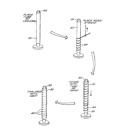
S As illustrated in Fig. 1 A through 1 C, the process may be considered in
four discrete steps. In step one (Fig. 1 A), a tube 20 formed of PTFE resin is
placed on a
tight-fitting stainless steel forming mandrel 22. The tube 20 may be formed
from PTFE
resin (Fluon CD-123 obtained from ICI Americas) which has been blended with
100
grams of "Isopar H" odorless solvent (produced by Exxon Corporation) per pound
of
PTFE, compressed into a preform billet and extruded into a 6.0 mm LD. and 6.8
mm
O.D. tube in a ram extruder having a reduction ratio of about 200:1 in cross-
sectional
area from billet to extruded tube. After removal of lubricant, the extruded
tube is
expanded and sintered, according to the method described in the aforesaid US
Patents
under various conditions to produce material with different node/fibril
structures.
In the next step (Fig. 1 B), a bead of diameter less than 1 mm., for
example, a 375 micron diameter PTFE bead 24 may be wrapped circumferentially
in a
helical manner around the tube 20. In a third step (Fig. 1 C) a PTFE outer
tube or wrap
30 covers the tube 20 with its helically wrapped beads. This tube 30 may be
formed
using PTFE resin (FLUON CD-123 obtained from ICI Americas) blended with 100
grams of "Isopar H" odorless solvent (produced by Exxon Corporation) per pound
of
PTFE, compressed into a preform billet and extruded into a 2.0 mm LD. and 2.4
mm
O.D. tube in a ram extruder having a reduction ratio of about 200:1 in cross-
sectional
area from billet to extruded tube. After removal of lubricant, the extruded
tube was
expanded and sintered, according to the method described in the aforesaid US
Patents
incorporated herein for reference, under various conditions to produce
material with
different node/fibril structures. This tube 30 is dilated to an 8 mm O.D.
prior to placing it
over the beaded tube 20.
In the final step (Fig. 1D), the outer tube 30 is restrained to prevent
longitudinal shrinkage and is then transferred to an oven at 360°C for
5 minutes to
coalesce the inner and outer tubes 20 and 30 respectively, thereby enclosing
and
smoothly covering ridges 40, to provide the final structure.
The helical bead 24 is wrapped around tube 20 with a pitch such that the
spaced apart protruding ridges 40 are spaced at a distance, such as to 1-3 mm,
which is
effective to trap a needle inserted into said space thereby preventing
longitudinal tearing
of the prosthesis when cannulized with a dialysis needle.
In an alternative method the first tube 20 is formed of PTFE resin (Fluon
CD-123 obtained from ICI Americas) blended with 100 grams of "Isopar H"
odorless
_ ... t _ _ __
CA 02273887 1999-06-03
WO 98/26731 PCT/US97/Z3103
-15-
solvent (produced by Exxon Corporation) per pound of PTFE, compressed into a
preform billet, extruded into a 4.0 mm LD. and 4.6 mm O.D. tube in a ram
extruder and
having a reduction ratio of about 200:1 in cross-sectional area from billet to
extruded
tube. After removal of lubricant, the extruded tube is expanded and sintered,
according
to the method described in the aforesaid US Patents incorporated herein for
reference,
under various conditions to produce material with different node/f bril
structures. The
PTFE bead 24 is extruded to a 250 micron diameter, and is circumferentially
wrapped in
a helical manner. Thereafter an outer tube 30 formed as in the first process
is dilated to a
6 mm O.D. and then, as in the prior process embodiment, is heated to coalesce
the tubes
to form a multistage structure.
In a third process variation the beading 24 may be formed as a metal wire
core enveloped by a PTFE jacket.
In a fourth alternate process, rather than a helical winding, discrete bead
rings at an axial spacing between one and five millimeters form a segmented
supporting
structure.
With reference now to Figs. 2A and 2B, microphotographs at a
magnification of SOX and 75X, respectively, of the cross section of a
prosthesis wall of
two embodiments of a product produced by the above described method are shown.
With reference to Fig. 2A, the inner, or luminal, surface 46 of a prosthesis
wall is formed
of a PTFE material characterized by a relatively low density, and a porosity
having
relatively large pores interconnected by fibrils. Wrapped around that surface
is a bead
46 which as above described can be formed either of a solid PTFE, or by a wire
or metal
core covered by PTFE. The next zone of the wall is a wrap cover 48 of PTFE
which has
been coalesced by heat to envelope both the inner surface 46 and the bead 42.
In some
embodiments the porosity of the cover 48 may be (as illustrated in Fig. 2A) a
different
porosity than that of the inner surface 46. Finally the outer surface of the
prosthesis wall
52 may again be formed of a relatively low porosity PTFE material.
Fig. 2B shows a similar structure wherein the porosity of the inner,
luminal zone 46 is greater than that of the wrap cover 48. In each case, the
article is
assembled and coalesced into a unitary structure such that the inner and outer
surfaces
are presented for biocontact when implanted, while the bead remains enclosed
and to
some extent cushioned and immobilized by the surrounding surface portions.
In addition to articles having an inner bead or winding, the present
invention contemplates articles wherein the inner structure forms a layer or
sheet of
substantiallly homogeneous properties in two dimensions, such as a sheet or
tube of a
defined pore structure, or a stent or series of stems, and wherein the
property of the
inner portion produces, controls or modulates the desired physical
characteristic.
CA 02273887 1999-06-03
WO 98/26731 PCT/US97/23103
-I6-
Figure 10 illustrates an implantable prosthetic member 10 according to
this aspect of the present invention, which, is shown in the figure as a
tubular member,
suitable for implantation as a vascular graft. The member 10 has an inner wall
1 and an
outer wall 2 with a thickness dimension extending therebetween. As further
illustrated
in Figure 10, there are at least three continuous regions adjacent to each
other and
extending along the entire area of the member namely, regions a, b and c,
illustratively
shown in the Figure as concentric strata from the inside to the outside. As
described in
more detail below, the successive regions a, b, c are not separate structures
but are
portions of the same wall, and are distinguished by their structural
properties as relates in
particular to aspects of porosity.
In general, each embodiment of the invention includes at least one region
having a zero or sufficiently low porosity that it effectively acts as a
barrier to fluid
penetration or a barrier which modulates transmission of hydraulic pressure
pulsation
through the thin wall of the prosthesis. This barrier region may be a
completely pore-
1 S free stratum, a stratum having small pore size, or a stratum having a high
density of
crossed, irregular, dead end, or closed cell pores such that it carries out
its modulation or
barrier function. In the latter case, even large pore material may be used,
but its water
entry pressure (WEP) is high. This stratum may exist at the region of inner
surface 1,
the region of outer surface 2, or an intermediate stratum as shown by the
position of
region b in Figure 10.
In material science, there is a distinction between material porosity and
permeability. Porosity is a direct measure of the physical void volume
contained with a
boundary, whereas permeability refers to the accessibility of that void
volume.
Permeability is usually expressed as a rate of flow of liquid or gas per unit
area, as a
function of differential pressure.
In a porous, fibrous material, that part of the total porosity which is
available to fluid flow is also called the "effective porosity." The pressure
required to
force a liquid into a pore is a function of pore size and geometry, liquid
surface tension,
and soIid/liquid contact angle. Surface tension opposes the entry of any
nonwetting
liquid into a pore, and this opposition may be overcome by external pressure.
Expanded PTFE material is characterized by lengthwise-oriented fibrils
interrupted by transverse nodes. The pore size in microns is typically
determined by
measuring fiber length between the nodes (internodal distance). To compute
fibril
length, the material is viewed under sufficient magnification. A fibril length
is measured
from one edge of one node to the edge of an adjacent node. Fibril lengths are
measured
from the sample to compute a mean fibril length.
T ___._...~.__.._. ~._
CA 02273887 1999-06-03
WO 98/26731 PCT/US97/23103
- 17-
Nodes and fibrils may be further characterized by their relative geometry.
That is, nodes by length, width, and height; and fibrils, by diameter and
length. It is the
relative geometry of nodes to fibrils, as well as, internodal distance that
determines
porosity and permeability of porous PTFE.
Permeability to fluid flow can be determined by measuring the amount of
pressure required for water to permeate the pores of the material. To compute
water
entry pressure (WEP) one subjects the material to an incrementally increasing
water
pressure until small beads of water appear on the surface. WEP is a gage which
can be
used to equate porosity to permeability.
Vascular graft porosity is a measure of the void fraction within the
prosthesis wall and is believed to give a rough prediction of the capacity of
the graft to
anchor newly formed surrounding tissue after implantation, whereas
permeability is
associated with fluid flow through the graft wall.
Vascular permeability or hydraulic conductivity is related to material
porosity. WEP is a good measuring technique to assess this trait because it
closely
mimics the permeation process at the blood/prosthesis interface. WEP is
defined as the
pressure value necessary to push water into the pores of a synthetic tubular
substrate and
can be classified as: High (>400 mm Hg), Medium (200-400 mm Hg), and Low (<200
mm Hg).
It has been widely accepted since the nineteenth century that the
hydrostatic pressure difference across the arterial wall is capable of
transporting water
from the blood into the surrounding interstitial space. The view has long been
held that
a continuous transport of material occurs across the arterial wall, from its
inner to its
outer surface. Solutes flow past the endothelium gradually passing through the
various
arterial wall layers eventually being transferred to the lymphatics or
adventitia.
The filtration coefficients of the wall are dependent on the hydraulic
conductivity of both the intima and media. The artery wall is a heterogeneous
porous
medium in which interstitial fluid can flow through the interstices between
cells and
tissue mimicking a semipermeable membrane with hydrostatic and osmotic
pressure
components. The osmotic pressure difference across the vessel wall is assumed
to be
small compared with the hydrostatic pressure or hydraulic conductivity.
More controlled healing and tissue ingrowth is achieved by providing a
specific region (outer) for cell penetration, followed by a region (harrier)
that does not
allow free cellular penetration/permeation but instead, allows the transport
of plasma
solutes such as cellular mediators (proteins, growth factors, etc.) This
barrier minimizes
the relatively large hydraulic force present in arterial transport that
retards tissue
ingrowth. Reports have shown that a negative pressure exists within the
perigraft space,
CA 02273887 1999-06-03
WO 98/26731 PCT/US97/23103
-18-
while blood components (cells, particles, etc.) are isolated to the blood side
of the
device.
A vascular graft formed from the lamellate structure of the invention
mimics the natural artery with a cross-section that offers differential
permeability
properties resulting in a healing response acceptable to the surrounding
tissue.
In a prototype embodiment of the invention, a prosthesis 10 as described
above was fabricated in a mufti-step procedure by assembling three physically
separate
bodies of material together in successive strata and then joining or
coalescing them into
a single unit.
When a vascular prosthesis is fabricated according to this method,
preferably at least one of the bodies is a tube which may, for example, be an
axially-
stretched tube having a porous structure of internodal space oriented
transverse to its
surface. Advantageously, the nodal spacing, orientation or structure of
successive strata
may be offset, non-matching or misaligned to introduce or enhance a barrier or
hydraulic
modulation effect. For example, a prosthesis may be formed by placing a first
PTFE
tube on a mandrel, wrapping a ribbon of PTFE in an overlapped or non-
overlapped spiral
winding over the tube outer surface, and then placing another PTFE tube over
the
assembly. For this construction, the outermost tube has preferably been
previously
radially expanded. Heat is then applied to the assembly, optionally with a
radial
compressive force, to shrink back the outer tube and coalesce the three
separate bodies
together into a unitary prosthesis. Although effectively "welded" together,
there are no
visible deformations, and the through-wall properties change abruptly at the
interface of
each stratum or region with the next.
EXAMPLE 1
PTFE resin (Fluon CD-123 obtained from ICI Americas) was blended
with 100 grams of "Isopar H odorless solvent (produced by Exxon Corporation)
per
pound of PTFE, compressed into a preform billet and extruded into a 3.5 mm LD.
and
4.0 mm O.D. tube in a ram extruder having a reduction ratio of about 200:1 in
cross-
sectional area from billet to extruded tube. After removal of lubricant, the
extruded tube
was expanded and sintered, according to the method described in US Patents No.
5,433,909 and 5,474,824, which patents are hereby incorporated by reference
herein in
their entirety, under various conditions to produce material with different
hydraulic
porosities. This produced three different tubes, denoted A, B and C, which
were used as
starting materials for the constructions described below.
Stretch conditions and resultant hydraulic porosities are given below in
Table 1.
T
CA 02273887 1999-06-03
WO 98/26731 PCT/US97/23103
- 19-
TABLE 1
Expansion Hydraulic Porosity
Temp(°C) Rate(in/sec) Ratio(%) wEp
(mm Hg)
(A) 320 .004 3:1 100
(B) 300 .018 3:1 200
(C) 250 7.5 2.5:1 600
Material (B) was radially expanded to a 4mm ID on a stainless steel
forming mandrel, covered with material C that had been previously dilated to a
Smm ID,
restrained to prevent longitudinal shrinkage, and transferred to an oven at
360°C for 5
minutes, to prepare a primary lamellate. The primary lamellate was removed
from the
oven and allowed to cool, covered with material (A) that had been previously
dilated to a
Smm ID, restrained to prevent longitudinal shrinkage, and placed in an oven at
360°C
for 10 minutes, to prepare a final lamellate structure (material B/C/A), a
cross-section of
which is shown in Figure 3A.
Material (B) was radially expanded to a 4mm ID on a stainless steel
forming mandrel, covered with material (A) that had been previously dilated to
a Smm
ID, restrained to prevent longitudinal shrinkage, and transferred to an oven
at 360°C for
5 minutes, to prepare a primary Iamellate. The primary lamellate was removed
from the
oven and allowed to cool, covered with material C that had been previously
dilated to a
Smm ID, restrained to prevent longitudinal shrinkage, and placed in an oven at
360°C
for 10 minutes, to prepare a final lamellate structure (material B/A/C), a
cross-section of
which is shown in Figure 3B.
Material (C) was radially expanded to a 4mm ID on a stainless steel
forming mandrel, covered with material (B) that had been previously dilated to
a Smm
ID, restrained to prevent longitudinal shrinkage, and transferred to an oven
at 360°C for
5 minutes, to prepare a primary lamellate. The primary lamellate was removed
from the
oven and allowed to cool, covered with material (A) that had been previously
dilated to a
Smm ID, restrained to prevent longitudinal shrinkage, and placed in an oven at
360°C
for 10 minutes, to prepare a final lamellate structure (material C/B/A) a
cross-section of
which is shown in Figure 3C.
CA 02273887 1999-06-03
WO 98126731 PCTIUS97l23103
-20-
Thus, the three structures of this example differ as permutations of
starting materials (A), (B), and (C) assembled into a the tubular prosthetic
device,
achieving three different articles with differing surface compatibility
properties and
through permeation profiles to affect tissue growth or biocompatibility..
EXAMPLE 2
Material (B) was radially expanded to a 4mm ID on a stainless steel
forming mandrel, biaxially wound with commercially available PTFE ribbon on a
helix
winding apparatus, and covered with material (A) that had been previously
dilated to a
Smm ID, restrained to prevent longitudinal shrinkage and placed in an oven at
360°C for
10 minutes to prepare a lamellate structure (Material BBiaxial wrap/Material
A), a
cross-section of which is shown in Figure 4.
PTFE ribbon was biaxially wound onto a stainless steel forming mandrel,
covered with material (B) that had been previously dilated to a Smm ID,
restrained to
prevent longitudinal shrinkage and placed in an oven at 360°C for 5
minutes, to prepare
a primary lamellate. The primary lamellate was removed from the oven and
allowed to
cool, covered with material (A) that had been previously dilated to a Smm ID,
restrained
to prevent longitudinal shrinkage and placed in an oven at 360°C for 10
minutes to
prepare a final lamellate structure (Biaxial ribbon/ material B/material A), a
cross-
section of which is shown in Figure 5.
Material (B) was radially expanded to a 4mm ID on a stainless steel
forming mandrel, covered with material (A) that had been previously dilated to
a Smm
ID, restrained to prevent longitudinal shrinkage, and placed in an oven at
360°C for 5
minutes, to prepare a primary lamellate. The primary lamellate was removed
from the
oven and allowed to cool, covered with a biaxial wrap of PTFE ribbon,
restrained to
prevent longitudinal shrinkage and placed in an oven at 360°C for 10
minutes to prepare
the final lamellate structure (Material B/Material A/Biaxial ribbon), a cross-
section of
which is shown in Figure 6.
EXAMPLE 3
Material (B) was radially expanded to a 4mm ID on a stainless steel
forming mandrel, longitudinally wrapped with commercially available PTFE
ribbon, and
covered with material (A) that had been previously dilated to a Smm ID,
restrained to
prevent longitudinal shrinkage, and placed in an oven at 360°C for 10
minutes to prepare
a lamellate structure (Material B/Longitudinal wrap/Material A), a cross-
section of
which is shown in Figure 7.
CA 02273887 1999-06-03
WO 98/26731 PCT/US97/23103
-21 -
PTFE ribbon was placed longitudinally around a stainless steel mandrel,
covered with material (B) that had been previously dilated to a Smm ID,
restrained to
prevent longitudinal shrinkage, and placed in an oven at 360°C for 5
minutes, to prepare
a primary lamellate. The primary lamellate was removed from the oven and
allowed to
cool, covered with material (A) that had been previously dilated to a Smm ID,
restrained
to prevent longitudinal shrinkage and placed in an oven at 360°C for 10
minutes to
prepare a final lamellate structure (Longitudinal ribbon/Material B/Material
A), a cross-
section of which is shown in Figure 8.
Material (B) was radially expanded to a 4mm ID on a stainless steel
forming mandrel, covered with material (A) that had been previously dilated to
a Smm
ID, restrained to prevent longitudinal shrinkage, and placed in an oven at
360°C for 5
minutes, to prepare a primary lamellate. The primary lamellate was removed
from the
oven and allowed to cool, covered with a longitudinal wrap of PTFE ribbon,
restrained
to prevent longitudinal shrinkage and placed in an oven at 360°C for 10
minutes to
prepare a final lamellate structure (Material B/Material A/Longitudinal
ribbon), a cross-
section of which is shown in Figure 9.
To assess the in vivo performance of prostheses prepared in this fashion,
four millimeter lamellate grafts of various configurations were implanted into
the carotid
and/or femoral arteries of dogs. Explants were taken at 14, 30, 60, and 180
days. The
presence of an intrawall low porosity, high WEP region produced enhanced
tissue
ingrowth compared to material without such a region, leading applicant to
believe that
hydraulic forces play a role in the healing process of implantable devices.
As applied to a covered or integral support prosthesis, the invention
provides advantages of strength, expandibility and enhanced tissue
compatibility. As
shown in Figures 1 lA-11 C, a method of forming an enclosed, protected support
or
reinforcing element 11 such as a stent includes the steps of (Figure 11 A)
taking a first
film-like body of liner material shown as a tube 20a, and placing it within a
stmt 3 S, and
the step (Figure 11 B) of covering the assembly with an outer film of
material, 20b. The
entire assemblage is then heated together (Figure 11 C) so as to unitize the
inside and
outside layers with the stent 35 secured between them. The foregoing
schematically
represented method is preferably carried out with a material such as expanded
polytetrafluoroethylene (PTFE) which has a porosity imparted by previous
stretching or
expansion of the material. In this case, at least the second or outer layer of
the material
is preferably a radially-expanded but unsintered layer, so that when it is
heated it shrinks
back toward its unexpanded state and presses against the inner layer. Further
radial
pressure may be provided to urge the inner and outer polymer layers together.
CA 02273887 1999-06-03
WO 98/26731 PCT/US97123103
-22-
One preferred method of fabricating the structure schematically shown in
Figures 11A-11C is illustrated in Figures 12A-12D. In accordance with this
method, a
tube of the polymer material having a diameter d is provided. One end of the
tube is
expanded radially, for example by inserting an expandable balloon inside the
tube and
inflating it to increase the diameter of the tube by a factor of five to five
hundred percent
or more. This produces a stepped tube illustrated in Figure 12A having a small
diameter
portion 14 of diameter d and a large diameter end 16 of diameter D>d, which
may in
addition have a greater degree of porosity. A stmt 35 is then placed over the
small end
14 and the large end 16 is folded back over the stent 35 (Figure 12B). This
forms a
structure that is half the original tube length, with a single cuff resulting
from the
continuous fold of material 16 over the right hand end portion, as
illustrated, of the
device. Once folded over in this fashion to form an assembly half the length
of the
original tube, heat is applied to shrink the outside down in upon the inside,
enclosing the
stmt 35 therebetween (Figure 12C). An inflatable sleeve or tightly fitting
form may
clamp around the outside to provide an inwardly-directed, radial, pressure.
Figure 12D
illustrates a cross section taken longitudinally of the resultant
construction.
Figures 13A-13E show a further practice of the method of the present
invention. In accordance with this method, a single tube of polymer material
is provided
as before, but both ends are inflated to form a first large diameter portion
16a that joins
continuously with the uninflated central portion 14, which, in turn, extends
to another
end 16b which has also been inflated and enlarged. Preferably, the portion 14
extends
for approximately half the length of the original tube while the portions 16a
and 16b are
each one quarter of the length of the tube length or slightly more. This tube
may be
placed over a mandrel (not shown) which provides a temporary rigid element to
facilitate
the process. As shown in Figure 13B, the stmt 35 is then placed around the
central
portion 14 and one end, illustrated as end 16b of the expanded tube, is folded
back along
the axial direction to cover a portion of the stmt. As illustrated, the folded
back end
portion 16b extends roughly halfway along the tube length. As best seen in
Figures 13C
and 13D, the remaining end 16a is then folded and pulled taut to the middle.
The ends
can be touching as shown in Figure 13D, or they may be overlapped to provide a
single
seam in which one end slightly extends over the already double layer of the
other end.
The assembly then remains on the mandrel and is heated for a time sufficient
to shrink
both of the turned-over ends together down over the stmt and to coalesce with
the
underlying material. With this construction, both ends of the stmt are closed
by a
3 S continuous smooth seamless cover, and both the inside and outside films
are bonded to
each other or coalesced with heat so that they form a nondelaminating and
unitized
cocoon around the stmt.
1 __. .~._.. ..._ ~.__
CA 02273887 1999-06-03
WO 98/26731 PCT/US97/23103
- 23 -
In this construction, the stmt body 35 itself may be a spiral-shaped zig-
zag wire body which lies generally in the plane of the cylindrical surface on
which it
extends, and which when radially expanded places the bends under tension and
draws
the band of zig-zags slightly narrower and straighter, thus expanding in
radius by
S elongating slightly along its spiral direction. Shear between the
surrounding polymer
layers and the wire stmt material itself will thus naturally occur, but will
be directed
along the relatively narrow band in which the stmt lies. The crimps themselves
may be
of very closely spaced zigs and zags which effectively prevent the outer film
from
contacting the inner film in the narrow band closely surrounding this area.
With such a
construction, the stmt lies in a tunnel or pocket formed between the layers.
Because the
layers fit tightly, the support effectively transfers strain to the polymer.
Thus breaking
or rupture of the film does not occur as the stmt expands. As noted above, a
preferred
material is an expanded polytetrafluoroethylene, which when heated shrinks
back and
coalesces with contacting portions of the polymer from the other side of the
stmt. The
heating is carried out to not over sinter, so this material is also capable of
restretching
without rupture. Thus that both the stent and the surrounding polymer are
expandable
and may, for example, be placed by endovascular delivery and expansion in
situ.
In addition to the foregoing methods of fabrication, the invention also
contemplates a stmt construction wherein the stmt body has a continuous and
seamless
covering over ends of the stent and along the full length of the body, but the
body
covering extends only on one side, the inside or the outside, of the
cylindrical stem.
This is achieved as shown in Figures 14A-14C. In this embodiment of the
invention, a
tube 14 is expanded at each end, as before, to form expanded end portions 16a
and 16b.
However the end portions, 16a and 16b, are each of relatively short length,
approximately one centimeter, and are folded back over the stent 35 only for a
distance
sufficient to cuff the ends and to provide a short band or margin
approximately one half
to one centimeter wide at the ends of the stmt. As before, the assembly is
then heated to
shrink down the folded over material and unitize the cuffs thus formed at each
end. In
this embodiment as in the first described schematic treatment of the method, a
tube may
then be placed over the outside to cover the folded back portions. If such an
outer tube
is provided, it is constrained so that its ends extend beyond the edges of the
folded back
cuff portions and lie in the band indicated by 1 Sa, 1 Sb in the dravi~ing.
Heat is applied
with the ends constrained so that the second tube shrinks radially but not
axially and the
cocoon structure extends and is maintained over the full length of the
prosthesis. As in
the previous embodiments, the closed ends are entirely seamless and the outer
tube
coalesces with the underlying material at a region away from the ends to form
a
continuous enclosing assembly for the stmt.
CA 02273887 1999-06-03
WO 98/26731 PCT/US97/23103
-24-
In accordance with another aspect of the present invention, a vessel
support is fabricated with a structure to assume a radially-varying extent
along its length
when expanded, such that one or both ends thereof are larger than its center
as shown in
FigurelSA. A corresponding construction may be used to form a support which
upon
expansion assumes the opposite flare or bulge, as shown in Figure 15B.
As illustrated in FigurelSA, a PTFE support liner or graft assembly 55 is
fabricated having one or more expanding rings or stents 35a, 35c at its ends
and one or
more rings 35b centrally along its length, all having an initial diameter d ~
. In this
embodiment the end rings 35a, 35c are made of heavier gauge material, or are
otherwise
dimensioned so that they either are more resistant to expansion, or else have
a limited
net expanded size, d2. The central rings 35b are either more easily expanded
(lower
resistance) or are dimensioned so that they expand to a larger diameter d3.
The result is
that during expansion the support assembly SS assumes a shape which bulges out
in the
center. This shape aids in preventing the inflation balloon from slipping out
of the
support during the inflation process, which may require several cycles of
balloon
expansion to expand successive lengths of the support tube. This contour also
enables
the support to conform more closely to a region of vessel having an aneurysm
or bulge.
Figure 15B illustrates a related construction in which the end rings have a
lesser resistance to expansion or a larger net expanded diameter than the
center rings. In
this case the intermediate and/or final expanded size is greater at the ends,
creating flared
or trumpet-shaped ends or a venturi-like profile. This profile is intended to
assure a
smooth transition from the unsupported vessel lumen to the prosthesis, without
projecting edges at the prosthesis ends. Each of the narrow rings 35 may
reside in a
pocket or band 31 such that expansion occurs relatively freely and some motion
of the
stmt may occur between the inner and outer surfaces without impairing
continuity of the
PTFE envelope material.
A related construction, applicable to either bulge- or flare-expansion
contours is shown in Figure 15C. This Figure shows a PTFE body which has been
formed with an inner region, a middle region and an outer region, as described
for
example above with refence to Figures 3-10. That construction, instead of
enclosing
stmt or rings 35, surrounds a separate tube, wrapping or body of fluoropolymer
material,
in the region between the outer and inner surface portions in a unitary
coalesced
assembly. In this case, when constructing the assembly as shown in Figure 15C
without
a stiff stmt or support member between the inner and outer surfaces, the
degree of
expansion of the assembly may be varied by providing the prosthesis with a
different
degree of sintering at different points along the length of the prosthesis.
This may be
done as described further below, by feeding the assembly end-first into a
sintering oven
_T
CA 02273887 1999-06-03
WO 98/26731 PCT/US97/23103
- 25 -
at a controlled rate to sinter each end more than the center. This provides
greater tensile
strength of the material at both ends, and results in a bulged prosthesis when
expanded,
as shown at the left of the Figure. The opposite distribution may be provided
by
reciprocating the assembly while it is centrally positioned in a short oven
such that each
end sticks out of the heat zone for a substantial portion of the sintering
cycle, to result in
flared-end expansion material at the end portions having lower tensile
strength.
Thus, the invention further provides expandable vessel liners or supports
having radial taper or curvature along their length.
Example A
The following example describes the construction of a solid tube of PTFE
having a radially expandable stmt within the tube. A starting tube, or
substrate, was a
substantially uniform length of axially-stretched tubing formed by extrusion
as
described, for example, in U.S. Patents No. 5,433,909 and 5,474,824. The PTFE
substrate was a uniaxially-oriented tube having nodes interconnected by
fibrils oriented
substantially in a single longitudinal direction. The fibrils were
approximately 30~em in
length. The PTFE substrate had an internal diameter of 2.5 mm and a wall
thickness of
approximately 0.25 mm.
This PTFE substrate material was placed on a stainless steel mandrel
having a diameter of 2.7 mm. Thus, a small amount of radial expansion was
required to
get the material on the mandrel. A Cordis peripheral stmt (part number
2969363) was
placed over PTFE layer, and a second PTFE layer was placed covering the stmt.
The
second layer consisted of the PTFE substrate material described above,
expanded to 4
mm by sliding the material over a stainless steel mandrel with a diameter of 4
mm to
radially expand it. After radial expansion, the PTFE material moved freely
over the
stmt. The PTFE/stent/PTFE combination was secured at both ends to prevent
slippage
along the mandrel, and the combination was placed in an oven and sintered at
360° C for
a 15 minute time period. During sintering, the outer layer of PTFE recovered
to its
original diameter and attached to the inner layer of PTFE. The assembly was
removed
from the oven and the excess PTFE was removed. The resulting structure
contained the
stmt entirely enveloped within a closed body of PTFE.
The PTFE stent combination was expanded with a Blue Max balloon
dilatation catheter with a balloon O.D. of 9 mm and a balloon length of 4 cm.
Using
multiple inflations the stent combination was expanded along its entire
length.
CA 02273887 1999-06-03
WO 98126731 PCT/ITS97I23103
-26-
Example B
The following example describes the construction of a solid tube of PTFE
having a radially expandable stmt within the tube, and wherein a single piece
of PTFE
tubing is used to construct the stmt combination.
The PTFE substrate was the same uniaxially-oriented tube as that of
Example A, having nodes interconnected by fibrils oriented substantially in a
single
longitudinal direction. The fibrils were approximately 30 p.m in length, and
the PTFE
substrate had an internal diameter of 2.5 mm and a wall thickness of
approximately 0.25
mm. Half the length of the PTFE tube was expanded to 6 mm by sliding the tube
material over a mandrel having a diameter of 6 mm. Figure 12A depicts what the
PTFE
tube looks like after this processing step. The small portion of the PTFE tube
was then
placed on a stainless steel mandrel having a diameter of 2.7 mm. Thus, a small
amount
of radial expansion was required to get the material on the mandrel. A Cordis
peripheral
stmt (part number 2969363) was placed over the PTFE layer. Next, the expanded
end of
the PTFE tube was folded back to cover the stmt. The PTFE material moved
freely over
the stent, and the PTFE and stent combination was secured at both ends to
prevent
slippage along the mandrel. The combination was placed in an oven and sintered
at 360°
C for a 15 minute time period. During sintering, the outer layer of the PTFE
recovered
to its original diameter and attached to the inner layer of PTFE.
The PTFE and stmt combination was removed from the oven and excess
PTFE was trimmed.
The PTFE stmt device produced in this fashion was expanded with a
Blue Max balloon dilatation catheter with a balloon O.D. of 9 mm and a balloon
length
of 4 cm. Multiple inflations were used to expand the stmt combination along
its entire
length.
Example C
The following example describes the construction of a solid tube of PTFE
having a plurality of spaced apart radially expandable stems within the tube.
The starting PTFE substrate was an uniaxially-oriented tube having nodes
interconnected by fibrils oriented substantially in a single longitudinal
direction. The
fibrils were approximately 30 p,m in length. The PTFE substrate had an
internal
diameter of 2.5 mm and a wall thickness of approximately 0.25 mm.
A layer of this PTFE material was placed on a stainless steel mandrel
having a diameter of 2.7 mm. Thus, a small amount of radial expansion was
required to
get the material on the mandrel. Two Palmaz stents (part number P394) were
placed
over the PTFE layer. The stems were spaced apart approximately 4 cm. The PTFE
T __._... _.
CA 02273887 1999-06-03
WO 98/26731 PCT/US97/23103
-27-
material described above was expanded to 4 mm by sliding the material over a
stainless
steel mandrel with a diameter of 4 mm and the expanded tube was placed over
the stems.
The radially expanded PTFE material moved freely over the stems. The PTFE and
stmt
. combination was secured at both ends to prevent slippage along the mandrel.
The
combination was placed in an oven and sintered at 360° C for a 15
minute time period.
During sintering, the outer layer of PTFE recovered to its original diameter
and attached
to the inner layer of PTFE. The PTFE and stmt combination was removed from the
oven and excess PTFE was removed.
The PTFE stmt combination was expanded with a Blue Max balloon
dilatation catheter with a balloon O.D. of 9 mm and a balloon length of 4 cm.
Multiple
inflations were used to expand the stmt combination along its entire length.
Example D
The starting PTFE substrate was made from a uniaxially-oriented tube
having nodes interconnected by fibrils oriented substantially in a single
longitudinal
direction. The PTFE substrate was 60 cm in length had an internal diameter of
6 mm,
with a wall thickness of approximately 0.6 mm and an average internodal
distance of 60
~,m on the exterior surface and 20p,m on the interior surface. The tube
material was
marked 1 cm increments along the length of the tube.
This graft material was placed on a stainless steel mandrel having a
diameter of 6 mm which allowed for the free movement of the graft along the
length of
the mandrel. The graft/mandrel combination was then fed along its long axis
into and
oven set at 360° C at a rate of 0.1 cm/sec. Consequently, the portion
of the tube entering
the oven initially is subjected to a longer sintering time than other portions
of the tube.
The tube was not restrained during sintering which permitted longitudinal
contraction,
which is also referred to as free sintering. Free sintering results in a
reduction in the
internodal distance.
Samples of the tube were taken at points 12.5, 22.5 and 52.5 centimeters
along the longitudinal axis. Thus, the samples had been subjected to different
free
sintering times. Scanning electron micrographs (SEMs) were made to observe the
effect
of sintering on the PTFE material.
Example E
An expanded PTFE tube was made with variable porosity characteristics.
The PTFE substrate was made from a uniaxially-oriented tube having nodes
interconnected by fibrils oriented substantially in a single longitudinal
direction. The
PTFE substrate was 100 cm in length had an internal diameter of 6 mm, a wall
thickness
CA 02273887 1999-06-03
WO 98126731 PCT/US97123103
-28-
of approximately 0.45 mm and an average intemodal distance of 60 p,m on the
exterior
surface and 20 ~m on the interior surface. The tube was marked in 0.5 mm
increments
along the length of the tube.
The graft was placed on a stainless steel mandrel having a diameter of 6
mm which allowed for the free movement of the graft along the length of the
mandrel.
The graft mandrel combination was then fed along its long axis into an oven
set of 320°
C at a rate of 0.05 cm/sec to the 50 cm position. Consequently, the portion of
the tube
entering the oven initially was subjected to a longer sintering time than
other portions of
the tube. The tube was not restrained during sintering, which permitted
longitudinal
contraction. This is referred to as free sintering. Free sintering results in
a reduction in
the internodal distance. The tube mandrel combination was rotated 180°.
The graft
mandrel combination was then fed along its long axis into an oven set at
320° C at a rate
of 0.05 cm/sec to the 50 cm position. This permitted the free sintering of the
opposite
section of tube material in a similar fashion to the initial section. Thus,
the center
portion of the tube was subjected to the shortest period of free sintering.
Samples of the tube material were taken at various positions along the
longitudinal axis and subjected to radial tensile strength, foreshortening,
and internodal
distance measurements, and the radial tensile strength was found to be
directly related to
the foreshortening percentage and intemodal distance.
As indicated in the above examples, the densities and porosities of the
PTFE zones maybe varied to meet the specific needs of a particular prosthesis.
The
foregoing constructions and methods provide new and useful constructions for
reinforced prostheses, prostheses having wall pore structures that vary in
diverse
physiologically important physical characteristics, and for stents and other
solid or
springy supports with a cover or surrounding of PTFE material. This
surrounding
material is permanently attached and closely conforms, providing a surface
biocompatibility and a continuous surface that is capable of expansion while
retaining its
integrity, and which essentially avoids defects and surface seams or
irregularities.
Moreover, the inner portion may have a high WEP and operate to inhibit
diffusion or
modulate biological response and growth processes occurring in or contiguous
to the
wall. As such, it provides an improved construction applicable to a broad
range of
implant and surgical protheses. The invention being thus disclosed and
described,
variations and modifications will occur to those skilled in the art, and such
variations
and modifications are considered to be within the scope of the invention, as
set forth in
the claims appended hereto.
What is claimed is: