Note: Descriptions are shown in the official language in which they were submitted.
CA 02274037 2004-06-09
75379-29
PARTICLE-MEDIATED CONIFER TRANSFORMATION
FIELD OF INVENTION
This invention relates to a method for genetically
engineering coniferous plants. In particular, this
invention relates to a particle-mediated gene transfer
method for producing and developing transgenic embryos for
plants of the genus Pinus and Pinus interspecies hybrids.
This method is well suited for producing transgenic clonal
planting stock useful for reforestation.
BACKGROUND OF THE INVENTION
The identification of gene function coupled with
the ability to precisely manipulate DNA has enabled the
construction of synthetic genes which, when properly
transferred and incorporated into a host cell, can modify
the cell's genetic makeup. This capacity to manipulate'
genes using recombinant DNA technology combined with
in vitro methods for plant propagation now permits genetic
engineering of crop species. Indeed, genetic engineering
processes have been used to successfully transfer foreign
genes into certain plant species, thereby resulting in the
recipient species acquiring a useful genetic trait (such as
resistance to herbicides or insects).
The transfer of foreign genetic material into the
chromosomes of a recipient plant is typically carried out
through the use of either an Agrobacterium-mediated or a
particle-mediated transformation process. Agrobacterium
gene transfer employs the natural ability of the soil-borne
bacterium Agrobacterium tumefaciens to transfer a portion of
DNA (known as T-DNA) from an extrachromosomal plasmid (known
as the Ti-plasmid) to a receptive plant host cell under-
specific conditions. Using suitable techniques of
1
CA 02274037 2004-06-09
75379-29
recombinant DNA manipulation, the T-DNA may be replaced with
a desired piece of DNA. This method has not proven suitable
for all plant cell types.
In particle-mediated gene transfer, the DNA of
interest is precipitated onto the surface of carrier
particles which are subsequently accelerated toward a piece
of target tissue. The carrier particles penetrate the cell
wall of the plant cell, wherein the DNA can be expressed,
and may integrate with the chromosomal DNA. Transient
expression of the transforming DNA has been reported in
conifers (Charest et al., 1993; Walter et al., 1994).
Stable expression only results if the transforming DNA
integrates with the chromosomal DNA.
In addition to Agrobacterium-mediated and
particle-mediated gene transfer, other methods of gene
transfer have been used to introduce foreign genes into
conifers, such as electroporation (Campbell et al., 1992).
However, only transient expression has been reported using
any of these other methods, and no transgenic plants have
been reported to have been generated using such methods.
This illustrates that the mere act of introducing
DNA into the host cell chromosome is, by itself, not
sufficient for the production of transgenic plants. A
tissue culture system that enables the multiplication and
subsequent development of the transformed cells is also an
important component of a successful genetic transformation
protocol.
A method known as somatic embryogenesis is
sometimes employed in the clonal propagation of certain
conifers. Propagation by somatic embryogenesis refers to
methods whereby embryos are produced in vitro from small
2
CA 02274037 2004-06-09
75379-29
pieces of plant tissue or individual cells. The embryos are
referred to as somatic because they are derived secondarily,
from somatic (vegetative) tissue, rather than directly from
the sexual process. Vegetative propagation via somatic
embryogenesis has the capability to capture the genetic gain
of highly desirable genotypes. Furthermore, these methods
may be readily amenable to automation and mechanization to
produce large numbers of plants of individual clones (e.g.
for reforestation purposes).
It was not until 1985 that somatic embryogenesis
was demonstrated in conifers and the first viable plantlets
were regenerated from conifer somatic embryos. Since 1985,
conifer tissue culture workers throughout the world have
pursued the development of somatic embryogenesis systems
capable of regenerating plants. The goal of much of this
work is to develop conifer somatic embryogenesis as an
efficient propagation system for producing clonal planting
stock en masse.
The two most economically important conifer genera
are Picea (spruce) and Pinus (pine). Those working in
conifer somatic embryogenesis have found that there is a
striking difference between Picea conifers and Pinus
conifers as to the ease with which somatic embryogenesis can
be initiated and plants regenerated. In fact, when one
measures the respective levels of achievement in the art of
conifer somatic embryogenesis among species of these two
important genera, it is clear that significantly more
success has been obtained with Picea than with Pinus.
Indeed, the recalcitrance of embryogenic cultures of Pinus
species is well documented. This is especially true for
pines commonly found in the Southeastern United States
(known in the industry as Southern yellow pines).
3
CA 02274037 2004-06-09
75379-29
Nevertheless, researchers working with Pinus
species plants have recently achieved some important
advances. For example, U.S. Patents Nos. 5,413,930,
5,491,090, 5,506,136, 5,677,185, 5,731,191, 5,731,203, and
5,731,204 disclose multi-step methods that are able to
complete the entire somatic embryogenesis regenerative
process, from explant collection to planting of somatic
embryo derived plants, for historically recalcitrant
Southern yellow pines (i.e., Pinus taeda, Pinus serotina,
Pinus palustris, and Pinus elliottii), Pinus rigida, and
hybrids thereof.
Scientists have found the dichotomy exhibited by
Picea conifers and Pinus conifers in the area of somatic
embryogenesis also exists in the field of genetic
engineering. While researchers have been able to stably
genetically transform Picea conifers, Pinus conifers - and
particularly Southern yellow pines - have proven to be
extremely resistive to such modifications. Indeed, the
relative ease of genetic transformation of Picea conifers in
comparison to Pinus conifers is evident when examining the
reports in the literature describing the success of Picea
transformation in somatic embryogenic systems and the
paucity of such reports for Southern yellow pines.
Although researchers have been able to routinely
attain stable particle-mediated genetic transformations in
Picea conifers, historically almost all of such
transformations reported in Pinus conifers, particularly
Southern yellow pines, have, upon examination, been found to
be transient transformations or transformation of tissue
which was not subsequently regenerable into whole plants.
While transient, or non-integrative, transformation can be
4
CA 02274037 2004-06-09
75379-29
achieved easily in many tissues and stages of Pinus embryo
development, the ability to achieve stable transformation in
a tissue capable of producing whole plants is the key to a
successful gene transfer system.
It has been found that a successful stable genetic
transformation protocol is heavily dependant on the
employment of an efficient tissue culture system. Moreover,
efficient tissue culture methods must be coordinated with
gene transfer at a receptive stage of embryo development in
order to achieve stable genetic transformations. The stage
of development at which transformation has been carried out
in order to attain the regeneration of transgenic plants in
Picea conifers (i.e. in embryogenic tissues initiated from
cotyledonary embryos) does not give rise to embryogenic
tissues capable of regenerating whole plants in conifers of
the genus Pinus. Furthermore, in Picea conifers embryogenic
tissues initiated from earlier stage embryos, such as pre-
stage 3 somatic embryos, have not given rise to transformed
plants. Indeed, the methods taught by Ellis in U.S. Patent
No. 5,681,730 for obtaining and genetically transforming
somatic embryos in Picea conifers have not been found
effective when applied to Pinus conifers. However, in the
genus Pinus we have found that transformation steps can be
successfully combined with a tissue culture system to derive
embryogenic cultures from pre-stage 3 somatic embryos, pre-
stage 3 zygotic embryos or somatic embryogenic tissue
containing pre-stage 3 somatic. embryos which are capable of
regenerating whole transgenic plants.
It has long been known to those skilled in the art
of transformation that a brief osmotic treatment at the time
of transformation will increase transient expression of the
transgene. In conifers, a treatment with elevated levels of
5
CA 02274037 2004-06-09
75379-29
inositol has been shown to be of benefit in transformation
of white spruce, Picea glauca (Clapham et al. 1995).
However, in Pinus taeda and P. taeda x P. rigida hybrids,
the same treatment with elevated levels of inositol (i.e.,
levels greater than about 0.2 M) is detrimental to both
growth and embryo development. In Pinus radiata, a
pretreatment with sorbitol increased transient expression of
a transgene (Walter et al. 1994), but such a pre-treatment
has not been taught for obtaining stable expression and
regeneration of transformed pine plants (Walter et al.
1997), perhaps because such treatments can also be
detrimental to the regeneration of pine plants. To address
these problems in the genus Pinus, we have developed a
variety of preparation media for use before transformation
and selection in pines. The use of the preparation media
facilitated the recovery and development of stable
genetically transformed embryos.
Therefore, an object of the present invention is
to provide a method of genetically engineering plants of the
genus Pinus and Pinus interspecies hybrids.
Another object of the present invention is to
provide a method for stably transforming embryogenic tissues
of the genus Pinus and Pinus interspecies hybrids.
A further object of the invention is to produce
stably transformed embryos of the genus Pinus and Pinus
interspecies hybrids capable of further development into
transgenic plants.
Yet another object of the present invention is to
produce genetically engineered plants of the genus Pinus and
Pinus interspecies hybrids.
6
CA 02274037 2004-06-09
75379-29
SUMMARY OF THE INVENTION
The above objectives are achieved via the use of a
particle-mediated genetic transformation method which
employs embryogenic tissues from plants of the genus Pinus
and Pinus interspecies hybrids. This method involves the
use of particle-mediated gene transfer with embryogenic
tissues which are in a particular stage of development,
namely pre-stage 3 somatic embryos, pre-stage 3 zygotic
embryos, or somatic embryogenic tissue containing pre-stage
3 somatic embryos. It is preferred to accomplish this by
employing a multi-step method which: a) prepares pre-stage
3 (i.e., pre-cotyledonary) somatic embryos, pre-stage 3
zygotic embryos, and/or somatic embryogenic tissue
containing pre-stage 3 somatic embryos as the receptive
target tissue for gene transfer via culturing the target
tissue on preparation media, b) employs particle-mediated
gene transfer to insert DNA into the target tissue, and c)
exposes the bombarded tissue to selection media in order to
identify and develop transformed embryogenic lines. Where
desired, additional steps can be utilized to both
cryopreserve such lines and to develop the transformed
embryogenic lines into plants.
This method results in recovery of transgenic
events through all stages of the transformation process
leading to the production of transgenic pine trees (even
historically recalcitrant Southern pines). This method also
allows the transfer of genetic material to embryogenic
cultures that may be used to establish clonal plantations of
pine trees that are improved economically through expression
of the transferred genetic material. This method further
permits the development of transgenic embryos from
embryogenic tissue which has been cryopreserved.
7
CA 02274037 2004-06-09
75379-29
The invention also encompasses the genetically
transformed embryos produced via the method and the
transgenic plants derived therefrom.
In one aspect, the invention provides a method for
genetically engineering conifers selected from the group
consisting of the genus Pinus and Pinus interspecies
hybrids, which comprises: (a) placing conifer target tissue
selected from the group consisting of pre-stage 3 somatic
embryos, pre-stage 3 zygotic embryos, embryogenic tissue
containing pre-stage 3 somatic embryos, and combinations
thereof, on a target surface; (b) bombarding the target
tissue by physically accelerating at the target tissue
carrier particles which are much smaller than the cells of
the target tissue, the carrier particles carrying copies of
a genetic construction including at least one gene of
interest; (c) inducing the bombarded target tissue to form
proliferative tissue which is capable of forming somatic
embryos; (d) during the step of inducing, culturing the
bombarded target tissue on a selection medium so as to
select for embryogenic tissue which is transformed by the
gene of interest; (e) inducing transformed somatic embryos
to develop from the selected embryogenic tissue; and (f)
germinating and converting the transformed somatic embryos
thus produced into clonal transgenic conifer plants.
In another aspect, the invention provides a method
for genetically engineering conifers selected from the group
consisting of the genus Pinus and Pinus interspecies
hybrids, which comprises: (a) placing a suitable conifer
explant on a culture initiation medium containing a
sufficient amount of inorganic and organic nutrients, about
0.1 to about 5.0 mg/1 of auxin, about 0.1 to about 1.0 mg/1
of cytokinin, up to about 100.0 mg/l of abscisic acid, about
5.0 to about 100.0 g/1 of sugar, and a gelling agent
8
CA 02274037 2004-06-09
75379-29
selected from the group consisting of about 2.5 to about 9.0
g/l of agar, about 0.5 to about 4.0 g/l of gellan gum, about
3.0 to about 10.0 g/l of agarose, about 1.5 to about 5.0 g/l
of AGARGELTM, and combinations thereof, for a sufficient
amount of time under suitable environmental conditions to
grow an embryogenic tissue culture containing pre-stage 3
somatic embryos; (b) placing target tissue from the
embryogenic tissue culture on a target surface, wherein the
placed target tissue is selected from the group consisting
of pre-stage 3 somatic embryos, Sembryogenic tissue
containing pre-stage 3 somatic embryos, and combinations
thereof; (c) bombarding the target tissue by physically
accelerating at the target tissue carrier particles which
are much smaller than the cells of the target tissue, the
carrier particles carrying copies of a genetic construction
including at least one gene of interest; (d) transferring
the bombarded target tissue to a selection medium so as to
select for embryogenic tissue which is transformed by the
gene of interest; (e) transferring the transformed
embryogenic tissue to an embryo development medium
containing a sufficient amount of inorganic and organic
nutrients, about 5.0 mg/l to about 300.0 mg/l of abscisic
acid with the continued maintenance of the abscisic acid
concentration, up to about 10.0 g/l of activated carbon,
about 20.0 to about 70.0 g/l of sugar, and a gelling agent
selected from the group consisting of about 6.0 to about
12.0 g/l of agar, about 1.75 to about 4.0 g/l of gellan gum,
about 6.0 to about 8.0 g/l of agarose, about 3.5 to about
6.0 g/l of AGARGELTM, and combinations thereof, for a
sufficient time under suitable environmental conditions to
develop transgenic stage 3 somatic embryos; (f) separating
the transgenic stage 3 somatic embryos from the development
medium and partially drying the embryos by exposing the
embryos to an atmosphere having a high relative humidity for
9
CA 02274037 2004-06-09
75379-29
a period of about 2 to about 5 weeks; (g) transferring the
partially dried transgenic embryos to a germination medium
containing a sufficient amount of organic and inorganic
nutrients, up to about 10.0 g/l of activated carbon, about
20.0 to about 40.0 g/l of sugar, and a gelling agent
selected from the group consisting of 6.0 to 9.0 g/l of
agar, 1.75 to 3.50 g/1 of gellan gum, 6.0 to 8.0 g/l of
agarose, 3.5 to 5.0 g/l of AGARGELTM, and combinations
thereof, for a sufficient time under suitable environmental
conditions to germinate the partially dried transgenic
embryos; (h) converting the germinated transgenic embryos
into acclimatized transgenic conifer plants; and (i) field
planting the acclimatized transgenic conifer plants.
In another aspect, the invention provides a method
for genetically engineering conifers selected from the group
consisting of the genus Pinus and Pinus interspecies
hybrids, which comprises: (a) placing conifer target tissue
selected from the group consisting of pre-stage 3 zygotic
embryos, tissues extruded from immature megagametophytes
which contain pre-stage 3 zygotic embryos, and combinations
thereof, on a target surface; (b) bombarding the target
tissue by physically accelerating at the target tissue
carrier particles which are much smaller than the cells of
the target tissue, the carrier particles carrying copies of
a genetic construction including at least one gene of
interest; (c) transferring the bombarded target tissue to a
selection medium so as to select for embryogenic tissue
which is transformed by the gene of interest; (d)
transferring the transformed embryogenic tissue to an embryo
development medium containing a sufficient amount of
inorganic and organic nutrients, about 5.0 mg/l to about
300.0 mg/l of abscisic acid with the continued maintenance
of the abscisic acid concentration, up to about 10.0 g/l of
CA 02274037 2006-05-29
75379-29
activated carbon, about 20.0 to about 70.0 g/l of sugar, and
a gelling agent selected from the group consisting of about
6.0 to about 12.0 g/l of agar, about 1.75 to about 4.0 g/l
of gellan gum, about 6.0 to about 8.0 g/1 of agarose, about
3.5 to about 6.0 g/l of AGARGELTM, and combinations thereof,
for a sufficient time under suitable environmental
conditions to develop transgenic stage 3 somatic embryos;
(e) separating the transgenic stage 3 somatic embryos from
the development medium and partially drying the embryos by
exposing the embryos to an atmosphere having a high relative
humidity for a period of about 2 to about 5 weeks; (f)
transferring the partially dried transgenic embryos to a
germination medium containing a sufficient amount of organic
and inorganic nutrients, up to about 10.0 g/l of activated
carbon, about 20.0 to about 40.0 g/l of sugar, and a gelling
agent selected from the group consisting of 6.0 to 9.0 g/l
of agar, 1.75 to 3.50 g/1 of gellan gum, 6.0 to 8.0 g/l of
agarose, 3.5 to 5.0 g/l of AGARGELT", and combinations
thereof, for a sufficient time under suitable environmental
conditions to germinate the partially dried transgenic
embryos; (g) converting the germinated transgenic embryos
into acclimatized transgenic conifer plants; and (h) field
planting the acclimatized transgenic conifer plants.
According to still another aspect of the present
invention, there is provided a plant cell transformed with a
gene of interest by the method steps as described herein.
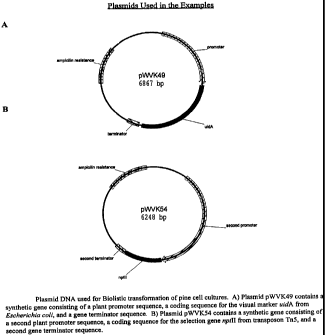
DESCRIPTION OF THE DRAWINGS
The Figures represent genetic constructions
(plasmid DNA) employed in the Examples for Biolistic
transformation of pine cell cultures.
11
CA 02274037 2006-05-29
75379-29
FIG. 1A represents the plasmid pWVK49 which
contains a synthetic gene consisting of a plant promoter
sequence, a coding sequence for the visual marker gene uidA
from Escherichia coli, and a gene terminator sequence.
11a
CA 02274037 2004-06-09
75379-29
FIG. 1B represents the plasmid pWVK54 which
contains a synthetic gene consisting of a second plant
promoter sequence, a coding sequence for the selection gene
nptll from transposon TnS, and a second gene terminator
sequence.
DESCRIPTION OF THE PREFERRED EMBODIMENT
The importance of the developmental stage of the
explant tissue used to initiate embryogenic cultures in
conifers varies considerably among different species. In
spruces, for example, cultures can be initiated from a wide
range of embryo developmental stages (i.e., immature, mature
and germinating embryos). However, pines have proven much
more restricted than spruces in terms of the responsive
embryo development stage for somatic embryogenic culture
initiation. To be successful in pines, one must use only
very immature embryos (or seeds containing such immature
embryos). The size of the developing embryo, usually
measured as length, has frequently been used to determine
the appropriate developmental stage for culture initiation
in many plant species. This has been the case with loblolly
pine where it was found that the embryogenic culture
initiation occurred most frequently when the dominant
zygotic embryo was less than about 0.5 mm in length.
Because it is difficult to measure the size of
very immature differentiated embryos, embryo staging systems
have also been used to make the determination of the
appropriate developmental stage easier. These staging
systems are based on several factors, including various
morphological characteristics of the embryo. An embryo
staging system proposed by Hakman and von Arnold (1988),
which is commonly utilized in the industry, has the
following three distinct stages. Stage 1 embryos are small
12
CA 02274037 2004-06-09
75379-29
differentiated embryos consisting of an embryonic region of
small, densely cytoplasmic region subtended by a suspensor
comprised of long, highly vacuolated cells. Stage 2 embryos
are further differentiated embryos with a prominent
embryonic region that becomes more opaque and assumes a
smooth and glossy surface. Stage 3 embryos are further
differentiated embryos which show visible cotyledonary
primordia. Thus, stage 1 and 2 embryos are at a pre-
cotyledonary stage of development, while stage 3 embryos are
cotyledonary. As used herein, the term "pre-stage 3 embryo"
means a differentiated pre-cotyledonary embryo (i.e., a
stage 1 or stage 2 embryo). Although the above three-stage
system was first used with somatic embryos of spruce, it is
generally applicable to both somatic and zygotic embryos of
all conifer species.
The present invention is a method for genetically
engineering conifers selected from the group consisting of
the genus Pinus and Pinus interspecies hybrids, which
comprises: (a) placing conifer target tissue selected from
the group consisting of pre-stage 3 somatic embryos, pre-
stage 3 zygotic embryos, embryogenic tissue containing pre-
stage 3 somatic embryos, and combinations thereof, on a
target surface; (b) bombarding the target tissue by
physically accelerating at the target tissue carrier
particles which are much smaller than the cells of the
target tissue, the carrier particles carrying copies of a
genetic construction including at least one gene of
interest; (c) inducing the bombarded target tissue to form
proliferative tissue which is capable of forming somatic
embryos; (d) during the step of inducing, culturing the
bombarded target tissue on selection medium so as to select
for embryogenic tissue which is transformed by the gene of
interest; (e) inducing transformed somatic embryos to
13
CA 02274037 2004-06-09
75379-29
develop from the selected embryogenic tissue; and (f)
germinating and converting the transformed somatic embryos
thus produced into clonal transgenic conifer plants.
It has been found that the employment of
preparation media greatly facilitates the recovery and
development of stable genetically transformed embryos. It
is, therefore, preferred that the conifer target tissue be
cultured under suitable environmental conditions on
preparation media prior to be placed on the target surface
for bombardment by the carrier particles (normally for a
period up to about 60 days). Likewise, it is preferred that
the bombarded target tissue be cultured on preparation media
following insertion of the carrier particles for a period of
time sufficient to allow tissue recovery. It is more
preferred to both: a) prepare the conifer target tissue for
carrier particle bombardment by culturing the target tissue
on preparation media prior to bombarding the tissue, and b)
culture the bombarded target tissue on preparation media
following bombardment in order to facilitate tissue
recovery.
A further preferred method for genetically
engineering conifers selected from the group consisting of
the genus Pinus and Pinus interspecies hybrids comprises:
(a) placing a suitable conifer explant on culture initiation
medium containing a sufficient amount of inorganic and
organic nutrients, about 0.1 to about 5.0 mg/l of auxin,
about 0.1 to about 1.0 mg/l of cytokinin, up to about
100.0 mg/1 of abscisic acid, about 5.0 to about 100.0 g/1 of
sugar, and a gelling agent selected from the group
consisting of about 2.5 to about 9.0 g/l of agar, about 0.5
to about 4.0 g/l of gellan gum, about 3.0 to about 10.0 g/1
of agarose, about 1.5 to about 5.0 g/l of AGARGELTM, and
combinations thereof, for a sufficient amount of time under
14
CA 02274037 2004-06-09
75379-29
suitable environmental conditions to grow an embryogenic
tissue culture containing pre-stage 3 somatic embryos; (b)
placing target tissue from the embryogenic tissue culture on
a target surface, wherein the placed target tissue is
selected from the group consisting of pre-stage 3 somatic
embryos, embryogenic tissue containing pre-stage 3 somatic
embryos, and combinations thereof; (c) bombarding the target
tissue by physically accelerating at the target tissue
carrier particles which are much smaller than the cells of
the target tissue, the carrier particles carrying copies of
a genetic construction including at least one gene of
interest; (d) transferring the bombarded target tissue to
selection medium so as to select for embryogenic tissue
which is transformed by the gene of interest; (e)
transferring the transformed embryogenic tissue to embryo
development medium containing a sufficient amount of
inorganic and organic nutrients, about 5.0 mg/l to about
300.0 mg/l of abscisic acid with the continued maintenance
of the abscisic acid concentration, up to about 10.0 g/l of
activated carbon, about 20.0 to about 70.0 g/l of sugar, and
a gelling agent selected from the group consisting of about
6.0 to about 12.0 g/1 of agar, about 1.75 to about 4.0 g/1
of gellan gum, about 6.0 to about 8.0 g/l of agarose, about
3.5 to about 6.0 g/l of AGARGEL, and combinations thereof,
for a sufficient time under suitable environmental
conditions to develop transgenic stage 3 somatic embryos;
(f) separating the transgenic stage 3 somatic embryos from
the development medium and partially drying the embryos by
exposing the embryos to an atmosphere having a high relative
humidity for a period of about 2 to about 5 weeks; (g)
transferring the partially dried transgenic embryos to
germination medium containing a sufficient amount of organic
and inorganic nutrients, up to about 10.0 g/l of activated
carbon, about 20.0 to about 40.0 g/l of sugar, and a gelling
CA 02274037 2004-06-09
75379-29
agent selected from the group consisting of 6.0 to 9.0 g/l
of agar, 1.75 to 3.50 g/1 of gellan gum, 6.0 to 8.0 g/l of
agarose, 3.5 to 5.0 g/1 of AGARGEL, and combinations
thereof, for a sufficient time under suitable environmental
conditions to germinate the partially dried transgenic
embryos; (h) converting the germinated transgenic embryos
into acclimatized transgenic conifer plants; and (i) field
planting the acclimatized transgenic conifer plants.
A further preferred method for genetically
engineering conifers selected from the group consisting of
the genus Pinus and Pinus interspecies hybrids comprises:
(a) placing conifer target tissue selected from the group
consisting of pre-stage 3 zygotic embryos, tissues extruded
from immature megagameophytes which contain pre-stage 3
zygotic embryos, and combinations thereof, on a target
surface; (b) bombarding the target tissue by physically
accelerating at the target tissue carrier particles which
are much smaller than the cells of the target tissue, the
carrier particles carrying copies of a genetic construction
including at least one gene of interest; (c) transferring
the bombarded target tissue to selection medium so as to
select for embryogenic tissue which is transformed by the
gene of interest; (d) transferring the transformed
embryogenic tissue to embryo development medium containing a
sufficient amount of inorganic and organic nutrients, about
5.0 mg/l to about 300.0 mg/l of abscisic acid with the
continued maintenance of the abscisic acid concentration, up
to about 10.0 g/l of activated carbon, about 20.0 to about
70.0 g/l of sugar, and a gelling agent selected from the
group consisting of about 6.0 to about 12.0 g/l of agar,
about 1.75 to about 4.0 g/l of gellan gum, about 6.0 to
about 8.0 g/l of agarose, about 3.5 to about 6.0 g/1 of
AGARGEL, and combinations thereof, for a sufficient time
16
CA 02274037 2004-06-09
75379-29
under suitable environmental conditions to develop
transgenic stage 3 somatic embryos; (e) separating the
transgenic stage 3 somatic embryos from the development
medium and partially drying the embryos by exposing the
embryos to an atmosphere having a high relative humidity for
a period of about 2 to about 5 weeks; (f) transferring the
partially dried transgenic embryos to germination medium
containing a sufficient amount of organic and inorganic
nutrients, up to about 10.0 g/l of activated carbon, about
20.0 to about 40.0 g/l of sugar, and a gelling agent
selected from the group consisting of 6.0 to 9.0 g/l of
agar, 1.75 to 3.50 g/l of gellan gum, 6.0 to 8.0 g/l of
agarose, 3.5 to 5.0 g/l of AGARGEL, and combinations
thereof, for a sufficient time under suitable environmental
conditions to germinate the partially dried transgenic
embryos; (g) converting the germinated transgenic embryos
into acclimatized transgenic conifer plants; and (h) field
planting the acclimatized transgenic conifer plants.
These methods are generally applicable to tissue
obtained from the Pinus species including, but not limited
to, the following: Pinus taeda (loblolly pine),
P. elliottii (slash pine), P. palustris (longleaf pine),
P. serotina (pond pine), P. echinata (shortleaf pine),
P. clausa (sand pine), P. glabra (spruce pine), P. rigida
(pitch pine), P. echinata (shortleaf pine), P. nigra
(Austrian pine), P. resinosa (red pine), P. sylvestris
(Scotch pine), P. banksiana (jack pine), P. virginiana
(Virginia pine), P. radiata (Monterey pine), P. contorta
(shore pine), P. contorta latifolia (lodgepole pine),
P. ponderosa (ponderosa pine), P. leiophylla (Chihuahua
pine), P. jeffreyi (Jeffrey pine), and P. engelmannii
(Apache pine), P. strobus (eastern white pine), P. monticola
(western white pine), P. lambertiana (sugar pine),
17
CA 02274037 2004-06-09
75379-29
P. massoniana (Masson pine), P. merkusii, P. albicaulis
(whitebark pine), P. flexilis (limber pine), P. strobiformis
(southwestern white pine), P. caribaea (Caribbean pine),
P. patula (Mexican weeping pine), P. tecunumanii (Tecun Uman
pine), P. maximinoi, P. oocarpa (Ocote Pine) and
P. chiapensis (Mexican White pine). In addition, the
current invention is specifically applicable to interspecies
hybrids of the above mentioned pines including Pinus rigida
x P. taeda, P. serotina x P. taeda, and reciprocal crosses.
It is preferred to utilize the present methods
with Southern yellow pines, Pinus rigida, Pinus radiata, and
hybrids thereof. Those skilled in the art recognize that
several species of pine indigenous to the Southeastern
United States are closely related and hybridize naturally.
Taxonomically this group of pines is referred to as
"Southern yellow pines" and includes Pinus taeda,
P. serotina, P. palustris, and P. elliottii.
Plant tissues which are suitable for use in the
present methods as target tissues for carrier particle
bombardment consist of pre-stage 3 somatic embryos, pre-
stage 3 zygotic embryos, embryogenic tissues containing pre-
stage 3 somatic embryos, and combinations thereof. Suitable
somatic embryogenic tissues contain pre-stage 3 somatic
embryos having polarity, with a prominent embryonic region
subtended by elongated suspensor cells, or pre-stage 3
somatic embryos obtained from these cultures. As used
herein, the term "pre-stage 3 somatic embryo" means a
differentiated somatic embryo which is at a pre-cotyledonary
stage of development. That is, the embryo is continuing to
differentiate, but cotyledonal primordia are not yet
outwardly visible. Likewise, the term "pre-stage 3 zygotic
embryo" means a differentiated zygotic embryo which is at
such a pre-cotyledonary stage of development.
18
CA 02274037 2004-06-09
75379-29
Where desired, zygotic embryos which are suitable
for use in the present invention can be produced by
isolating the pre-stage 3 zygotic embryos from immature
seeds. Likewise, newly extruded pre-stage 3 zygotic embryos
from recently cultured immature seeds may be employed as
target tissue for genetic transformation. Alternatively,
immature seeds which contain immature zygotic embryos at the
desired pre-stage 3 development can be used as targets for
genetic transformation. Where the conifer target tissue
consists of immature megagametophytes which contain pre-
stage 3 zygotic embryos, it is preferred to culture the
bombarded target tissue in order to encourage the extrusion
of the bombarded pre-stage 3 zygotic embryos, which are
subsequently transferred to selection medium (thereby
enabling the selection of embryogenic tissue cells which has
been transformed by the gene of interest). Media which are
suitable for culturing the bombarded target tissue include
the culture initiation media taught herein, and the like.
Appropriate somatic embryogenic tissues can be
produced by placing a suitable explant on nutrient-
containing culture media for a sufficient amount of time
under suitable environmental conditions to develop a culture
containing somatic embryogenic tissue and/or pre-stage 3
somatic embryos.
Explants which are suitable for use in the, present
methods include immature zygotic embryos, megagametophytes
containing immature zygotic embryos, and the like.
It is preferred that the somatic embryogenic
tissues be produced by first initiating tissue growth via
placing a suitable explant on culture initiation medium
containing a sufficient amount of inorganic and organic
nutrients, about 0.1 to about 5.0 mg/l of auxin, about 0.1
19
CA 02274037 2004-06-09
75379-29
to about 1.0 mg/l of cytokinin, up to about 100.0 mg/i of
abscisic acid, about 5.0 to about 100.0 g/l of sugar, and a
level of gelling agent selected from the group consisting of
about 2.5 to about 9.0 g/l of agar, about 0.5 to about
4.0 g/1 of gellan gum, about 3.0 to about 10.0 g/l of
agarose, about 1.5 to about 5.0 g/l of AGARGEL, and
combinations thereof, for a sufficient amount of time
(normally about 2 to 14 weeks) under suitable environmental
conditions to grow a somatic embryogenic culture containing
somatic embryogenic tissue and/or pre-stage 3 somatic
embryos.
Sugars which are suitable for use in the present
methods include, but are not limited to, the following:
glucose, maltose, sucrose, and combinations thereof.
Where desired, one may culture the embryogenic
tissue culture and/or pre-stage 3 somatic embryos by
transferring the somatic embryogenic culture from the
culture initiation medium to culture maintenance medium
containing a sufficient amount of inorganic and organic
nutrients, about 0.1 to about 5.0 mg/l of auxin, about 0.1
to about 1.0 mg/l of cytokinin, up to about 100.0 mg/l of
abscisic acid, up to about 10.0 g/l of activated carbon, and
about 10.0 to about 40.0 g/l of sugar, for a sufficient time
under suitable environmental conditions to grow the
embryogenic tissue culture.
Where desired, one may culture the bombarded
target tissue on culture maintenance media containing a*
sufficient amount of inorganic and organic nutrients, about
0.1 to about 5.0 mg/l of auxin, about 0.1 to about 1.0 mg/l
of cytokinin, up to about 100.0 mg/l of abscisic acid, up to
about 10.0 g/1 of activated carbon, and about 10.0 to about
40.0 g/l of sugar for a sufficient time under suitable
CA 02274037 2004-06-09
75379-29
environmental conditions to grow the bombarded target
tissue.
Where desired, one may culture the transformed
embryogenic tissue on culture maintenance media containing a
sufficient amount of inorganic and organic nutrients, about
0.1 to about 5.0 mg/l of auxin, about 0.1 to about 1.0 mg/l
of cytokinin, up to about 100.0 mg/1 of abscisic acid, up to
about 10.0 g/l of activated carbon, and about 10.0 to about
40.0 g/l of sugar for a sufficient time under suitable
environmental conditions to grow the transformed embryogenic
tissue.
Where desired, the culture maintenance media may
further contain a gelling agent selected from the group
consisting of about 6.0 to about 9.0 g/1 of agar, about 1.75
to about 4.0 g/l of gellan gum, about 6.0 to about 8.0 g/l
of agarose, about 3.5 to about 5.0 g/l of AGARGEL, and
combinations thereof.
Where desired, the embryogenic tissue culture from
the culture initiation medium may be cultured on embryo
development medium containing a sufficient amount of
inorganic and organic nutrients, about 5.0 mg/1 to about
300.0 mg/1 of abscisic acid with the continued maintenance
of the abscisic acid concentration, up to about 10.0 g/l of
activated carbon, about 20.0 to about 70.0 g/1 of sugar, and
a gelling agent selected from the group consisting of about
6.0 to about 12.0 g/l of agar, about 1.75 to about 4.0 g/1
of gellan gum, about 6.0 to about 8.0 g/l of agarose, about
3.5 to about 6.0 g/l of AGARGEL, and combinations thereof,
for a sufficient time under suitable environmental
conditions to prepare the target tissue for carrier particle
bombardment.
21
CA 02274037 2004-06-09
75379-29
It is preferred to culture the conifer target
tissue under suitable environmental conditions on
preparation media prior to the bombardment of the tissue by
the carrier particles in order to prepare the tissue for
particle insertion. It is also preferred to culture the
bombarded target tissue under suitable environmental
conditions on preparation media prior to transferring the
bombarded tissue to selection media in order to facilitate
tissue recovery from the bombardment. It is more preferred
to both culture the target tissue on preparation media prior
to bombarding the tissue, and to culture the bombarded
tissue on preparation media following the carrier particle
bombardment.
Preparation media suitable for use in the present
methods contain sufficient inorganic and organic nutrients,
up to about 5.0 mg/l of auxin, up to about 1.0 mg/l of
cytokinin, up to about 150.0 mg/l of abscisic acid, about
10.0 to about 120.0 g/l of sugar, and up to about 0.5M of
organic alcohol. When such liquid preparation media are
employed, it is necessary to disperse the target tissue on a
target surface (i.e., a solid support or a gelled medium) to
allow particles to be accelerated toward it. Normally this
dispersion occurs about 1 to 72 hours prior to particle
bombardment.
Where desired, the preparation medium may further
contain a gelling agent selected from the group consisting
of about 6.0 to about 9.0 g/l of agar, about 1.75 to about
5.0 g/l of gellan gum, about 6.0 to about 8.0 g/l of
agarose, about 3.5 to about 5.0 g/1 of AGARGEL, and
combinations thereof.
Organic alcohols which are suitable for use in the
present method include, but are not limited to, the
22
CA 02274037 2004-06-09
75379-29
following: glycerol, mannitol, sorbitol, polyethylene
glycol, and combinations thereof.
Gene transfer is carried out via particle-mediated
transfer in which a DNA genetic construction containing at
least one gene of interest is precipitated onto the surface
of a carrier particle (microparticle) and accelerated toward
the conifer target tissue. If desired, isolated pre-stage 3
somatic embryos and/or isolated pre-stage 3 zygotic embryos
can be utilized as target tissue. The common procedures for
particle-mediated transfers are well-known to those skilled
in the art of genetic engineering, as evidenced by U.S.
Patent No. 4,945,050 to Sandford et al. It is preferred to
utilize a helium-driven apparatus for the insertions. It is
also preferred to employ microparticles between 0.2 and 2.0
microns in diameter as carrier particles.
Where the DNA genetic construction that is being
transferred contains a selection gene, the target tissue may
be submitted to selective pressure to inhibit the growth of
any non-transgenic (i.e., non-transformed) cells. As used
herein, a selection gene is defined as a gene whose activity
allows cells to grow well in a selection culture medium only
if the cells have incorporated the gene, while cells which
have not incorporated the gene grow more slowly, do not
grow, or are killed. Culture selection media which are
suitable for use in the present method incorporate a toxin
to which the selection gene confers resistance, or are
composed such that a component necessary for growth of cells
is absent and must be provided by the cells that have
incorporated the selection gene, or are composed such that a
component necessary for growth is present in a form which
can only be taken up or metabolized by cells which have
incorporated the selection gene.
23
CA 02274037 2004-06-09
75379-29
A selection medium suitable for use in the present
method contains a sufficient amount of organic and inorganic
nutrients, a selection agent at a concentration which is
toxic to non-transformed cells but for which the gene of
interest confers resistance to transformed cells, up to
about 5.0 mg/i of auxin, up to about 1.0 mg/i of cytokinin,
up to about 30.0 mg/l of abscisic acid, and up to about
60.0 g/1 of sugar.
Another selection medium suitable for use in the
present method contains a sufficient amount of organic and
inorganic nutrients, up to about 5.0 mg/1 of auxin, up to
about 1.0 mg/l of cytokinin, up to about 30.0 mg/l of
abscisic acid, up to about 60.0 g/1 of sugar, and wherein
the selection medium lacks a component necessary for the
growth of non-transformed cells but for which the gene of
interest confers to transformed cells the ability to produce
the lacking component.
Another selection medium suitable for use in the.
present method contains a sufficient amount of organic and
inorganic nutrients, up to about 5.0 mg/1 of auxin, up to
about 1.0 mg/l of cytokinin, up to about 30.0 mg/1 of
abscisic acid, up to about 60.0 g/1 of sugar, and wherein
the selection medium contains a component necessary for the
growth of cells in a form which cannot be utilized by non-
transformed cells but for which the gene of interest confers
to transformed cells the ability to utilize the necessary
component.
Another selection medium suitable for use in the
present method contains a sufficient amount of organic and
inorganic nutrients, up to about 5.0 mg/l of auxin, up to
about 1.0 mg/1 of cytokinin, up to about 30.0 mg/1 of
abscisic acid, up to about 60.0 g/1 of sugar, and wherein
24
CA 02274037 2004-06-09
75379-29
the selection medium allows preferential growth of
transformed cells containing the gene of interest.
Where desired, the selection medium may further
contain a gelling agent selected from the group consisting
of about 6.0 to about 9.0 g/l of agar, about 1.75 to about
4.0 g/l of gellan gum, about 6.0 to about 8.0 g/l of
agarose, about 3.5 to about 5.0 g/l of AGARGEL, and
combinations thereof.
It is preferred to transfer the transformed
embryogenic tissue to embryo development medium containing a
sufficient amount of inorganic and organic nutrients, about
5.0 mg/l to about 300.0 mg/l of abscisic acid with the
continued maintenance of the abscisic acid concentration, up
to about 10.0 g/l of activated carbon, about 20.0 to about
70.0 g/l of sugar, and a gelling agent selected from the
group consisting of about 6.0 to about 12.0 g/l of agar,
about 1.75 to about 4.0 g/l of gellan gum, about 6.0 to
about 8.0 g/l of agarose, about 3.5 to about 6.0 g/l of.
AGARGEL,, and combinations thereof, for a sufficient time
under suitable environmental conditions to develop
transgenic stage 3 somatic embryos.
It is further preferred to add up to about
100.0 g/l of polyethylene glycol to the embryo development
medium;'and to subsequently transfer the transgenic stage 3
embryos from the embryo development medium to a second
development medium containing a sufficient amount of
inorganic and organic nutrients, about 5.0 mg/l to about
300.0 mg/l of abscisic acid with the continued maintenance
of the abscisic acid concentration, up to about 10.0 g/l of
activated carbon, up to about 100.0 g/l of polyethylene
glycol, and about 20.0 to about 70.0 g/1 of sugar, for a
sufficient time under suitable environmental conditions to
CA 02274037 2004-06-09
75379-29
further develop the transgenic stage 3 somatic embryos prior
to partially drying the embryos.
It is also preferred to add of up to about
100.0 g/1 of polyethylene glycol to the embryo development
medium; and to subsequently transfer the transgenic stage 3
embryos from the embryo development medium to a second
development medium containing a sufficient amount of
inorganic and organic nutrients, up to about 100.0 mg/l of
abscisic acid with the continued maintenance of the abscisic
acid concentration, up to about 10.0 g/l of activated
carbon, up to about 100.0 g/l of polyethylene glycol, and
about 20.0 to about 70.0 g/l of sugar, for a period of about
2 to about 12 weeks at a temperature in the range of about
0 C to about 10 C under suitable environmental conditions to
maintain the viability of the transgenic stage 3 somatic
embryos prior to partially drying the embryos.
The transgenic stage 3 somatic embryos are
subsequently separated from the development medium and are
partially dried. It is preferred that the transgenic stage
3 embryos be partially dried or dehydrated via exposure to
an atmosphere having a high relative humidity for a period
of about 2 to 5 weeks. The amount of moisture to be removed
an embryo depends upon several factors, including the
genotype of the embryo, the culture medium used, and the
storage products contained in the embryo. It is well within
the ability of a skilled artisan to determine the optimum
moisture loss necessary to prepare each embryo for
germination.
Where desired, the partially dried transgenic
somatic embryos may be transferred to germination medium.
It is preferred that the germination medium contain a
sufficient amount of organic and inorganic nutrients, up to
26
CA 02274037 2004-06-09
75379-29
about 10.0 g/l of activated carbon, about 20.0 to about
40.0 g/1 of sugar, and a gelling agent selected from the
group consisting of about 6.0 to about 9.0 g/1 of agar,
about 1.75 to about 4.00 g/1 of gellan gum, about 6.0 to
about 8.0 g/1 of agarose, about 3.5 to about 5.0 g/l of
AGARGEL, and combinations thereof, for a sufficient time
under suitable environmental conditions to germinate the
partially dried transgenic embryos. The germinated
transgenic embryos are subsequently converted into
acclimatized transgenic conifer plants and planted in soil
or similar media for glasshouse or field growth.
Where desired, the target tissues, bombarded
target tissues, and selected embryogenic tissues may be
cryopreserved (normally via the use of liquid nitrogen) in
order to maintain a bank of cultures and to insure against
loss of culture genotypes due to contamination, loss of
vigor associated with culture aging, or other deleterious
changes that may occur during long-term culture maintenance.
A number of terms are known to have differing
meanings when used in the literature. The following
definitions are believed to be the ones most generally used
in the field of botany and are consistent with the usage of
the terms in the present specification.
"Conversion" refers to the acclimatization process
that in vitro derived germinating somatic embryos undergo in
order to survive ex vitro (nonaxenic) conditions, and
subsequent continued growth under ex vitro conditions.
"Cryopreservation" is the storage of living cells
at ultra-low (cryogenic) temperatures, usually in liquid
nitrogen (-196 C) or in its vapor phase (about -150 C).
27
CA 02274037 2004-06-09
75379-29
"Embryo development" is the step in the somatic
embryogenesis process where the culture and/or environmental
conditions are changed causing the embryogenic culture to
switch from a proliferative phase of growth to a phase where
somatic embryos develop to a stage where they are ready to
harvest. In conifers the harvestable stage is typically
stage 3 embryos having cotyledons.
"(Embryo) germination" is the emergence of the
radicle or root from the embryo.
"Field planting" is the establishment of
laboratory, greenhouse, nursery, or similar grown planting
stock under field conditions.
"Initiation" is the initial cellular proliferation
or morphogenic development that eventually results in the
establishment of a culture from an explant.
A "(Liquid) Suspension Culture" is a culture
composed of cells and embryos suspended in a liquid medium,
usually agitated on a gyrotory shaker. An embryogenic
suspension culture in conifers is usually composed of both
cells and early stage somatic embryos with well-formed
suspensor cells and dense cytoplasmic head cells that float
freely in the liquid medium.
"Maintenance" is the step in which cultures are
grown and maintained in a proliferative phase by sequential
subculture to fresh medium at regular intervals after the
initiation step. Cultures are grown and maintained as
embryogenic tissue on a gelled medium or in a liquid medium
as a suspension culture.
"Pre-stage 3 embryos" are differentiated pre-
cotyledonary embryos (i.e., Stage 1 or Stage 2 embryos).
28
CA 02274037 2004-06-09
75379-29
"Selected (embryogenic) tissue" is tissue which
has been cultured on selection medium so as to select for
transgenic tissue (i.e., tissue which has been genetically
transformed).
A "selection gene" is a gene whose activity allows
cells to grow well in a selection culture medium only if the
cells have incorporated the gene, while cells which have not
incorporated the gene grow more slowly, do not grow, or are
killed.
A "selection medium" is a tissue culture medium
which: 1) incorporates a toxin to which a selection gene
confers resistance to transformed cells, 2) is composed such
that a component necessary for growth of cells is absent and
must be provided by transformed cells that have incorporated
a selection gene, 3) is composed such that a component
necessary for growth is present in a form which can only be
taken up or metabolized by transformed cells which have
incorporated a selection gene, and/or 4) is composed such to
allow preferential growth of transformed cells that have
incorporated the selection gene.
"Stage 1 embryos" are small differentiated embryos
consisting of an embryonic region of small, densely
cytoplasmic region subtended by a suspensor comprised of
long, highly vacuolated cells.
"Stage 2 embryos" are further differentiated
embryos with a prominent embryonic region that becomes more
opaque and assumes a smooth and glossy surface.
"Stage 3 embryos" are further differentiated
embryos which show visible cotyledonary primordia.
29
CA 02274037 2004-06-09
75379-29
"Target tissue" is tissue to be subjected to
bombardment by carrier particles carrying copies of a
genetic construction.
The following examples are provided to further
illustrate the present invention and are not to be construed
as limiting the invention in any manner.
EXAMPLE 1
This example teaches a method for genetically
engineering conifers. In particular, this example teaches a
method which includes the steps of pretreatment of
embryogenic cultures with preparation medium, gene transfer
via particle bombardment, recovery, and selection of
transgenic embryogenic tissues on selection agent to produce
stably transformed embryos and, subsequently, genetically
engineered hybrid pine (Pinus rigida x P. taeda) trees. The
use of preparation medium in this example was demonstrated
to greatly increase the frequency of recovery of transformed
material.
Immature seed cones were collected from several
20, different hybrid (Pinus rigida x P. taeda) pine sources
located in Westvaco's South Carolina coastal breeding
orchards near Charleston, South Carolina. The seed cones
were collected when the dominant zygotic embryo was at the
precotyledonary stage of development. Using the
classification system of von Arnold and Hakman (1988), the
dominant zygotic embryo at this stage is referred to as
being at stage 2; that is, an embryo with a prominent
embryonic region with a smooth and glossy surface, subtended
by elongated suspensor cells which are highly vacuolated.
However, zygotic embryos at an earlier stage of development
CA 02274037 2004-06-09 -
75379-29
(stage 1) may also be used effectively to initiate
embryogenic cultures.
Seed cones were harvested from selected trees,
placed in plastic bags and stored at 4 C until used for
culture initiation. Where the cones were stored for more
than two weeks at 4 C, they were aired and dried out weekly
(placed at 23 C, ambient laboratory conditions for two to
three hours) to prevent growth of fungi on the surface of
the cones and concomitant deterioration of seed quality.
For culture initiation, intact seeds removed from
seed cones were surface sterilized by treatment in a 10 to
20% commercial bleach solution (equivalent of a 0.525% to
1.050% sodium hypochlorite solution) for 15 minutes followed
by three sterile water rinses (each of five minutes
duration). Seeds were continuously stirred during the
sterilization and rinsing process.
Megagametophytes containing developing zygotic
embryos were used as the explant for culture initiation.
The seed coats of individual seeds were cracked open under a
laminar-flow hood with the use of a sterile hemostat. The
intact megagametophyte (which contains the developing
zygotic embryos) was removed from the opened seed coat with
forceps. Tissues attached to the megagametophyte, such as
the megagametophyte membrane and the nucellus, were removed
from the megagametophyte and discarded. The megagametophyte
was placed on culture medium (longitudinal axis of
megagametophyte parallel to the surface of culture medium)
with forceps. The micropyle end of the megagametophyte was
placed in contact with (but not submerged in) the culture
medium.
31
CA 02274037 2004-06-09
75379-29
The present method is not limited to any single
basal culture nutrient medium formulation. For example,
three common basal culture media formulations which are
suitable for use in the present method are listed in
Table I below. However, it should be understood that any
nutrient media commonly used in Pinus somatic embryogenesis
will be suitable for use with this method.
TABLE I
Basal Culture Media Formulations
COMPONENT DCR WV5 MSG
INORGANIC SALTS CONCENTRATION, mg/1
NH4NO3 400.00 700.00 0
KN03 340.00 259.00 100.00
Ca(N03)2'4H2O 556.00 963.00 0
MgS04'7H2O 370.00 1850.00 370.00
KH2PO4 170.00 270.00 170.00
NH4H2PO4 0 0 0
CaC12'2H20 85.00 0 440.00
KC1 0 1327.00 745.00
KI 0.83 0.83 0.83
H3BO3 6.20 31.00 6.20
MnSO4' H2O 22.30 15.16 16.90
ZnSO4'7H2O 8.60 8.60 8.60
Na2MoO4'2H20 0.25 0.25 0.25
CuSO4'5H20 0.25 0.25 0.03
COC12'6H20 0.03 0.03 0.03
NiC12'6H2O 0.03 0 0
FeSO47H2O 27.80 27.80 27.80
Na2EDTA 37.30 37.30 37.30
VITAMINS, AMINO ACIDS
Nicotinic acid 0.50 0.50 0.50
Pyridoxine'HC1 0.50 0.50 0.10
Thiamine HC1 1.00 1.00 0.10
Glycine 2.00 2.00 0
32
CA 02274037 2004-06-09
75379-29
The complete formulations of the media employed in
the Examples are listed in Table II below.
TABLE II
Initiation, Maintenance, and Preparation Media Formulations
Semi- Semi- Semi- Liquid Semi-
Solid Solid Solid Mainten- Solid
Initia- Initia- Mainten- ance Prepara-
COMPONENT tion tion ance Medium tion
Medium Medium Medium DCR2 Medium
DCR, WV51 DCRI DCR3
Basal medium a DCR WV5 DCR DCR DCR
CONCENTRATION (g/1)
Inositol 0.50 0.50 0.50 0.50 0.50
Casein hydrolysate 0.50 0.50 0.50 0.50 0.50
L-glutamine 0.25 0 0.25 0.25 0.25
Sucrose 30.00 0 30.00 30.00 0
Maltose 0 30.00 0 0 30.00
Polyethylene glycol 0 0 0 0 70.00
GELRITEb 1.5 1.5-2.0 2.00 0 2.00
Activated Carbon 0 0 0 0.5 0
CONCENTRATION (mg/1)
Auxin 3.00 1.0-3.0 3.00 3.00 3.00
Cytokinind 0.50 0.50 0.50 0.50 0.50.
Abscisic Acid 0 10.00 0 0 0
a) Refer to Table I for composition of basal
medium.
b) GELRITETM (gellan gum manufactured by Merck,
Inc.)
c) 2,4-dichlorophenoxyacetic acid (2,4-D) or
naphthalene acetic acid (NAA).
33
CA 02274037 2004-06-09
75379-29
d) N6-benzylaminopurine (BAP) or N6-benzyladenine
(BA).
The pH of the medium was adjusted to 5.8 with KOH
and HC1 prior to autoclaving at 110 kPa (16 psi) and 121 C
for 20 minutes. Aqueous stock solutions of L-glutamine were
filter sterilized and added to warm (about 60 C) medium prior
to pouring the medium into culture dishes. Approximately
20 ml of medium was poured into each 100 x 15 mm sterile
plastic petri dish.
Embryogenic tissue cultures from the hybrid pine
sources were initiated on semi-solid DCR1 initiation medium
(Table II). The dishes were incubated in the dark at a
constant temperature of 23 C. After about seven to 21 days,
embryogenic tissue extruded from the micropyle of the
megagametophyte explants. After about 28 days in culture
embryogenic tissue was removed from responsive
megagametophyte explants and moved to a new position on the
same culture dish, or the embryogenic tissue was transferred
to a new culture dish containing the same culture medium as
used for initiation. Each individual culture derived from
an individual megagametophyte explant was kept separate. and
assigned a cell line identification code.
Once cultures were extruded and subcultured, they
were maintained on DCR1. After 10-22 months on this semi
solid maintenance medium, the tissue cultures were placed in
DCR2 liquid maintenance medium containing activated carbon
(Table II). These were maintained by subculturing to fresh
DCR2 liquid medium every one to two weeks.
To prepare for gene transfer, a sterile fabric
support (here NITEXI, commercially available from Sefar
Inc.) was placed in a sterile Buchner funnel and one to five
34
CA 02274037 2004-06-09
75379-29
milliliters of embryogenic suspension was pipetted onto the
fabric support such that the embryogenic tissue was evenly
distributed over the surface. The liquid medium was
suctioned from the tissues using a mild vacuum. The fabric
support with embryogenic tissue was removed from the Buchner
funnel and placed on a GELRITE solidified DCR3 preparation
medium (Table II) in 100 X 25 mm plastic petri dishes.
Dishes were incubated in a dark growth chamber at 23 C for
about 24 hours.
DNA (genetic construction) was transferred into
the tissues and/or embryos via carrier particle
(microprojectile) bombardment technology (also known in the
industry as Biolistics) using the PDS-1000/He BIOLISTICTM
Particle Delivery System (available from Bio-Rad
Laboratories), which is a preferred method for delivery.
The DNAs of interest, here plasmids pWVK49 (Figure 1A below)
containing the visual marker gene uidA and pWVK54 (Figure 1B
below) containing the selection gene nptll, were
simultaneously precipitated onto the surface of gold
microparticles, which were subsequently accelerated toward
the pre-stage 3 embryogenic tissue described above, to
penetrate the cell walls. Once inside the cells, DNA is
released from the carrier particles and integrated randomly
into the chromosomes. The DNA used in this and subsequent
examples can be substituted with any suitable DNA sequence.
The gold microcarriers used were 0.6 to 1.6 pm in
diameter and were prepared in 50 pl aliquots of 60 mg/ml
gold suspended in sterile water, five pl of each plasmid,
pWVK49 (1 pg/pl) and pWVK54 (1 pg/pl), 50 pl 2.5 M CaCl2, and
20 pl 0.1M spermidine (free base). The mixture was
vortexed for three minutes, centrifuged at 10,000 rpm for 10
seconds and the supernatant was removed. The microcarriers
CA 02274037 2004-06-09
75379-29
were washed with 250 pl of 70% ethanol, briefly vortexed and
centrifuged. After removal of the supernatant the
microcarriers were resuspended in 65 pl 100% ethanol.
Aliquots of five pl were dispensed onto the center of the
macrocarriers and air dried.
In the PDS-1000/He Biolistic device the gap between
the rupture disk and the macrocarrier (gap distance) was
five mm and the macrocarrier travel distance was 13 mm.
The petri dishes with the fabric support and
embryonic tissues were then placed into the interior of the
PDS 1000/He Biolistic device and vacuum applied to a level
of 28 inches Hg. The gold particles carrying the DNA were
accelerated toward the embryogenic tissue following a helium
build-up and bursting regulated by a 1550 psi rupture disk.
Following DNA transfer the petri dishes containing the
fabric support and tissues were incubated in a dark growth
chamber at 23 C for about 24 hours.
The tissues and fabric support were transferred to
semi-solid maintenance medium, DCR1 (Table II) to recover
from carrier particle bombardment and incubated in a dark
growth chamber at 23 C for a period of about seven days. The
tissues and fabric support were transferred to a selection
medium, semi-solid maintenance medium DCR1 containing a level
of selection agent inhibitory to the growth of non-
transformed cells. In this and subsequent examples the
selection agent used was GENETICIN at 15 mg/l. The plates
were incubated in a dark growth chamber at 23 C for about six
to twelve weeks with the fabric supports containing the
tissues being transferred to the same fresh culture medium
every three weeks.
36
CA 02274037 2004-06-09
75379-29
Active growth on the selection medium occurred in
a number of isolated sectors on some of the petri dishes.
Such active growth in the presence of selection agent is an
indication that the growing tissues have integrated the
selection gene into their chromosomes and are stably
transformed. These areas of active growth were treated as
independent transformation events and are henceforth
referred to as sublines. The transgenic embryogenic tissue
was multiplied by transferring growing transgenic sectors to
fresh semi-solid maintenance DCR1 medium supplemented with
selection agent. Dishes were incubated in a dark growth
chamber at 23 C. The actively growing transgenic embryogenic
tissue was transferred to fresh semi-solid maintenance DCR1
medium supplemented with selection agent at three week
intervals for a period of about six to twelve weeks
depending on the rate of growth of the individual sublines
of the transgenic embryogenic tissue.
Individual sublines of the transgenic embryogenic
tissue were transferred to DCR2 liquid culture medium
(Table II) for further multiplication. The cultures were
incubated in a dark growth chamber at 23 C and maintained by
subculturing to fresh DCR2 liquid medium every one to two
weeks until sufficient multiplication of the tissue had
occurred to allow for the subsequent development step.
Using the methods described above, tissues were
transferred to a sterile fabric support and subsequently the
fabric and tissues were transferred to a MSG2 development
medium (see Table III below) containing about 125 mg/l of
ABA and no activated carbon and no polyethylene glycol. All
cultures were incubated at 23 C in the dark. It is preferred
that the cultures be incubated in the dark rather than light
condition. After three passages of about three weeks on the
37
CA 02274037 2004-06-09
75379-29
MSG2 medium, cotyledonary somatic embryos (stage 3) were
visible. Typically, multiple harvests of cotyledonary
somatic embryos were made at the end of the second and third
transfers onto MSG2 medium.
TABLE III
Development and Germination Media Formulations
Development Development Germination
Medium 1 Medium 2 Medium
MSG2 MSG3 MSG,
COMPONENT
Basal medium a MSG MSG MSG
CONCENTRATION (g/1)
Ammonium 0 0 0.80
nitrate
Inositol 0.10 0.10 0.10
L-glutamine 1.45 1.45 0
Sucrose 0 0 30.00
Maltose 60.00 60.00 0
GELRITE 2.00 2.00 2.00
Activated 0-1.25 0 5.00
carbon
Polyethylene 0-100.00 0 0
glycol
CONCENTRATION (mg/1)
ABA d 11-150 21 0
a) Refer to Table I for composition of basal
medium.
b) GELRITE (gellan gum manufactured by Merck,
Inc.).
c) Polyethylene glycol (molecular weight of 4000).
d) Abscisic acid.
38
CA 02274037 2004-06-09
75379-29
Harvested stage 3 embryos were converted into
acclimatized plants and field planted. Harvested stage 3
embryos on fabric supports were transferred to medium MSG3
(Table III), in petri plates and incubated for about four
weeks in the dark at a temperature of 4 C. Next, embryos on
their fabric supports were incubated above water in sealed
containers for about three weeks in the dark at a
temperature of 25 C. Following the above two treatments,
embryos on their fabric supports were transferred to medium
MSG4 (Table III) and incubated for about three days in the
dark at a temperature of 25 C. Embryos were then removed
from their fabric supports and placed individually onto the
surface of fresh MSG4 medium in petri plates for germination.
Germination was conducted in the light at a temperature
of 28 C. Germination plates were examined weekly, over a
period of about four weeks, and germinating embryos
transferred to MAGENTATh boxes containing 100 ml of MSG4
medium for conversion to plantlets. MAGENTA boxes
containing developing plantlets were incubated in the light
at 28 C for about eight to twelve weeks.
When the plantlets formed epicotyls (newly formed
shoots of approximately two to four cm), they were
transferred to containers filled with a potting mix [2:1:2
peat:perlite:vermiculite, containing 602 g/m3 OSMOCOTE
fertilizer (18-6-12), 340 g/m3 dolomitic lime and 78 g/m3
MICRO-MAX' micronutrient mixture (manufactured by Sierra
Chemical Co.)]. The plantlets were placed in a shaded
greenhouse and misted infrequently for a period of about two
weeks. Plantlets were then transferred to outdoor
conditions under shade for about four weeks for final
acclimatization prior to being moved to full-sun conditions.
39
CA 02274037 2004-06-09
75379-29
Stable transformation was verified through a
combination of growth on selection medium, assay for
expression of the visual marker gene at several
developmental stages including field-grown plants,
polymerase chain reaction amplification of specific segments
of the transgene DNA sequence at several developmental
stages including field-grown plants, and DNA blot
hybridization to detect the integration of the transgenes
into the genomic DNA. These techniques were carried out
using techniques well known to those skilled in the art of
molecular biology.
This method has been employed to generate over
1000 transgenic hybrid pine (Pinus rigida x P. taeda)
embryos from which more than 270 plants have been produced
and field planted.
EXAMPLE 2
This example teaches a method for genetically
engineering conifers. In particular, this example teaches a
method which includes the steps of pretreatment of
embryogenic cultures with preparation medium, gene transfer
via particle bombardment, recovery, and selection of
transgenic embryogenic tissues on selection agent to produce
stably transformed embryos and, subsequently, genetically
engineered loblolly pine (P. taeda) trees. The use of
preparation medium in this example was demonstrated to
greatly increase the frequency of recovery of transformed
material.
Immature seed cones were collected from several
different loblolly pine sources located in Westvaco's South
Carolina coastal breeding orchards near Charleston, South
Carolina. The seed cones were collected when the dominant
CA 02274037 2004-06-09
75379-29
zygotic embryo was at the precotyledonary stage of
development.
Using the methods of Example 1, cell cultures
containing pre-stage 3 embryogenic tissue were obtained.
After one to three months of culture on DCR1 semi-solid
maintenance medium, the tissue cultures were cryopreserved.
Pieces of the somatic embryogenic tissue and/or pre-stage 3
somatic embryos (about seven to 14 days since their last
subculture on culture maintenance medium) were dispersed in
liquid DCR1 medium which contained 0.4 molar sorbitol. The
amount of embryogenic tissue used was sufficient to result
in a 30% suspension. Erlenmeyer flasks containing the
suspension were incubated for 24 hours in the dark on a
gyrotory shaker (commonly at 100 rpm), and then placed on
ice. Five one milliliter aliquots of the cryoprotectant
dimethyl sulfoxide (DMSO) were added to the suspension to
bring final concentration of DMSO to 10%. One milliliter
aliquots of the cell suspension containing DMSO were then
transferred to freezing vials, placed in a programmable
freezer, and cooled to -35 C at 0.33 C per minute. The
freezing vials were subsequently immersed in liquid nitrogen
inside a cryobiological storage vessel for long-term
storage.
Frozen cultures were retrieved by removing
individual vials from the cryobiological storage vessel'and
placed in 38 C water to rapidly thaw the frozen cell
suspension. The thawed cell suspension were aseptically
poured from the cryovial onto a sterile fabric support,
which was then transferred to DCR1 maintenance medium and
incubated at 23 C for 24 hours to allow the DMSO to diffuse
into the medium. The fabric support containing embryogenic
tissue was removed from the medium and transferred to new
41
CA 02274037 2004-06-09
75379-29
maintenance medium. After growth on this medium for six to
20 weeks, the tissue cultures were placed in DCR2 liquid
maintenance medium (Table II) containing activated carbon,
and maintained by subculturing to fresh liquid medium every
one to two weeks.
To prepare for gene transfer, a sterile fabric
support was placed in a sterile Buchner funnel and one to
five milliliters of embryogenic suspension was pipetted onto
the fabric support such that the embryogenic tissue was
evenly distributed over the surface. The liquid medium was
suctioned from the tissues using a mild vacuum. The fabric
support with embryogenic tissue was removed from the Buchner
funnel and placed on a GELRITE solidified DCR3 preparation
medium (Table II) in 100 X 25 mm plastic petri dishes.
Dishes were incubated in a dark growth chamber at 23 C for
about 24 hours.
DNA was transferred into the tissues and/or
embryos via carrier particle (microprojectile) bombardment
technology (also known in the industry as Biolistics) using
the PDS-1000/He BIOLISTIC Particle Delivery System
(available from Bio-Rad Laboratories), which is a preferred
method for delivery. The DNAs of interest, here plasmids
pWVK49 (Figure IA above) containing the visual marker gene
uidA and pWVK54 (Figure 1B above) containing the selection
gene nptll, were simultaneously precipitated onto the
surface of gold microparticles, which were subsequently
accelerated toward the pre-stage 3 embryogenic tissue
described above, to penetrate the cell walls. Once inside
the cells, DNA is released from the carrier particles and
integrated randomly into the chromosomes.
The gold microcarriers used were 0.6 to 1.6 pm in
diameter and were prepared in 50 pl aliquots of 60 mg/ml
42
CA 02274037 2004-06-09
75379-29
gold suspended in sterile water, five pl of each plasmid,
pWVK49 (1 pg/pl) and pWVK54 (1 pg/pl), 50 pl 2.5 M CaC12, and
20 pl 0.1M spermidine (free base). The mixture was vortexed
for three minutes, centrifuged at 10,000 rpm for 10 seconds
and the supernatant was removed. The microcarriers were
washed with 250 p1 of 70% ethanol, briefly vortexed and
centrifuged. After removal of the supernatant the
microcarriers were resuspended in 65 pl 100% ethanol.
Aliquots of five pl were dispensed onto the center of the
macrocarriers and air dried.
In the PDS-1000/He Biolistic device the gap
between the rupture disk and the macrocarrier (gap distance)
was five mm and the macrocarrier travel distance was 13 mm.
The petri dishes with the fabric support and
embryonic tissues were then placed into the interior of the
PDS 1000/He Biolistic device and vacuum applied to a level
of 28 inches Hg. The gold particles carrying the DNA were
accelerated toward the embryogenic tissue following a helium
build-up and bursting regulated by a 1550 psi rupture disk.
Following DNA, transfer the petri dishes containing the
fabric support and tissues were incubated in a dark growth
chamber at 23 C for about 24 hours.
The tissues and fabric support were transferred to
semi-solid maintenance medium DCR1 (Table II) to recover from
Biolistics and incubated in a dark growth chamber at 23 C for
a period of about seven days. The tissues and fabric
support were transferred to a DCR1 selection medium. The
plates were incubated in a dark growth chamber at 23 C for
about six to twelve weeks with the fabric supports
containing the tissues being transferred to the same fresh
culture medium every three weeks.
43
CA 02274037 2004-06-09
75379-29
Active growth on the selection medium occurred in
a number of isolated sectors on some of the petri dishes.
Such active growth in the presence of selection agent is an
indication that the growing tissues have integrated the
selection gene into their chromosomes and are stably
transformed. These areas of active growth were treated as
independent transformation events and are henceforth
referred to as sublines. The transgenic embryogenic tissue
was multiplied by transferring growing transgenic sectors to
fresh DCR1 selection medium. Dishes were incubated in a dark
growth chamber at 23 C. The actively growing transgenic
embryogenic tissue was transferred to fresh semi-solid
maintenance DCR1 medium supplemented with selection agent at
three week intervals for a period of about six to twelve
weeks depending on the rate of growth of the individual
sublines of the transgenic embryogenic tissue.
Individual sublines of the transgenic embryogenic
tissue were transferred to DCR2 liquid culture medium
(Table II) for further multiplication. The cultures were
incubated in a dark growth chamber at 23 C and maintained by
subculturing to fresh DCR2 liquid medium every one to two
weeks until sufficient multiplication of the tissue had
occurred to allow for the subsequent development step.
Using the methods described above, tissues were
transferred to a sterile fabric support and subsequently the
fabric and tissues were transferred to a MSG2 development
medium (Table III) containing about 125 mg/l of abscisic
acid (ABA), 1.25 gm/l activated carbon, 70 gm/l polyethylene
glycol (PEG), or to a MSG3 development medium with no
activated carbon. All cultures were incubated at 23 C in the
dark. It is preferred that the cultures be incubated in the
dark rather than light condition. After three passages of
44
CA 02274037 2004-06-09
75379-29
about three weeks on the development media, cotyledonary
somatic embryos (stage 3) were visible. Typically, multiple
harvests of cotyledonary somatic embryos were made at the
end of the second and third transfers onto development
media.
Harvested stage 3 embryos were converted into
acclimatized plants and field planted. Harvested stage 3
embryos on fabric supports were transferred to medium MSG3
(Table III), in petri plates and incubated for about four
weeks in the dark at a temperature of 4 C. Next, embryos on
their fabric supports were incubated above water in sealed
containers for about three weeks in the dark at a
temperature of 25 C. Following the above two treatments,
embryos on their fabric supports were transferred to medium
MSG4 (Table III) and incubated for about three days in the
dark at a temperature of 25 C. Embryos were then removed
from their fabric supports and placed individually onto the
surface of fresh MSG4 medium in petri plates for germination.
Germination was conducted in the light at a temperature of
28 C. Germination plates were examined weekly, over a period
of about four weeks, and germinating embryos transferred to
MAGENTA boxes containing 100 ml of MSG4 medium for conversion
to plantlets. MAGENTA boxes containing developing plantlets
were incubated in the light at 28 C for about eight to twelve
weeks.
When the plantlets formed epicotyls (newly formed
shoots of approximately two to four cm), they were
transferred to containers filled with a potting mix [2:1:2
peat:perlite:vermiculite, containing 602 g/m3 OSMOCOTET"
fertilizer (18-6-12), 340 g/m3 dolomitic lime and 78 g/m3
MICRO-MAX micronutrient mixture (manufactured by Sierra
Chemical Co.)]. A number of plantlets were placed in a
CA 02274037 2004-06-09
75379-29
shaded greenhouse and misted infrequently for a period of
about two weeks. Plantlets were then transferred to outdoor
conditions under shade for about four weeks for final
acclimatization prior to being moved to full-sun conditions.
Stable transformation was verified through a
combination of growth on selection medium, assay for
expression of the visual marker gene at several
developmental stages including plants prepared for field
testing, and polymerase chain reaction amplification of
specific segments of the transgene DNA sequence at several
developmental stages including plants prepared for field
testing. These techniques were carried out using techniques
well known to those skilled in the art.
These methods have been used to produce thousands
of transgenic embryos of loblolly pine (Pinus taeda). A
subset of these transgenic embryos have been made into
plants and prepared for field planting.
EXAMPLE 3
This example illustrates the general applicability
of the invention across a broad range of cell lines of both
loblolly pine (Pinus taeda) and hybrid pine (Pinus rigida x
P. taeda).
To determine the applicability of the current
invention over a diverse range of genetic materials, a total
of 73 cell lines of loblolly pine and five cell lines of
hybrid pine somatic embryogenic cultures containing pre-
stage 3 embryos were produced and transformed using the
methods described in Examples 1 and 2 above. These included
one cell line each of loblolly pine and hybrid pine in the
procedures as control (i.e., reference) material. These
control cell lines were known to be responsive to the
46
CA 02274037 2004-06-09
75379-29
genetic transformation procedures as they were found to be
transformable using the methods of the previous examples.
The remaining 72 loblolly and four hybrid pine cell lines
had not been previously tested. The cell lines subjected to
the above procedure were microscopically examined and found
to contain pre-stage 3 embryos prior to gene transfer.
The ability to transform cell lines was assessed
by a variety of methods. The ability to grow on selection
medium in which the concentration of selection agent
inhibits the growth of non-transformed cell lines was one
criterion for transformation. Another criterion for
transformation was detection of the visual marker gene
supplied by the plasmid pWVK49. Another criterion for
transformation was the detection of either transgene as'
determined by polymerase chain reaction. These methods are
well known to those skilled in the art of molecular biology.
As shown in Table IV below, of the 72 loblolly
pine and four hybrid pine test lines subjected to the
procedure, 19 loblolly pine and two hybrid pine cell lines
produced sublines capable of growth on selection medium.
These comprise at least 26% of loblolly pine cell lines and
50% of hybrid pine cell lines tested. Eighty-seven sublines
of embryogenic tissue were recovered from the 19 loblolly
pine cell lines and three sublines were recovered from the
two hybrid pine cell lines.
47
CA 02274037 2004-06-09
75379-29
TABLE IV
Frequency of Stable Transformations for
Loblolly Pine and Hybrid Pine Cell Lines.
Loblolly Hybrid
pine pine
Number of lines tested 72 4
Number of lines giving 19 2
rise to at least one
transformed subline
Total number of 87 3
transformed sublines
recovered
To determine the frequency that transformed
sublines of pine tissue stably integrated DNA sequences from
either or both plasmids used (pWVK49 and pWVK54) during gene
transfer, DNA was isolated from a portion of the transformed
sublines prior to their transfer to MSG2 development medium
and subjected to polymerase chain reaction analysis. The
sublines subjected to analysis included the 87 loblolly pine
sublines described above in Table IV as well as sublines
obtained from the loblolly pine and hybrid pine control cell
lines described above for a total of 173 sublines. These
results are shown in Table V below.
48
CA 02274037 2004-06-09
75379-29
TABLE V
Frequency of Stable Transformation and Co-Transformation
for Loblolly Pine and Hybrid Pine Sublines
Number Percentage
Sequences detected positive /
number tested
uidA and nptll 136 / 173 79%
uidA only 1 /173 <1%
nptll only 18 /173 10%
These data show a high frequency of co-
transformation. Sequences for both transgenes, uidA and
nptll, were detected in 79 percent of the sublines tested.
Only one subline (less than one percent) contained only the
uidA visual marker gene. Ten percent of the tested sublines
contained only the nptll selection gene. In the remaining
19 sublines, no transgenes were detected by polymerase chain
reaction.
A subset of the loblolly pine sublines obtained
above were further developed and germinated according to the
methods of examples 1 and 2. Samples of developing embryos
at various stages of development as well as plants prepared
for field planting were tested for expression of the visual
marker gene and/or the selection gene. Samples were also
tested for the presence of the transgenes using polymerase
chain reaction analysis. For each of the above tests,
positive results indicative of stable transformation were
obtained. The test methods used above are well known to
those skilled in the art of molecular biology.
49
CA 02274037 2004-06-09
75379-29
EXAMPLE 4
In this example, additional preparation media
formulations and alternative selection steps were employed
to recover stably transformed embryogenic tissue of loblolly
pine (Pines taeda L.) and hybrid pine (Pinus rigida x
P. taeda).
Embryogenic cultures initiated as described in
previous examples from three different families of loblolly
pine (including one that has been recalcitrant to
transformation using the methods of the previous examples)
and three lines of hybrid pine were prepared for bombardment
by plating on DCR3 as described in previous examples, or
subjected to the same manipulations using alternative
preparation media as described in Table VI below.
Preparation media were developed using DCR or MSG basal
media as described in Tables I, II and III above. This
example demonstrates that the present invention is not
restricted to any particular basal medium formulation, but
may be based on any medium suitable for pine embryogenic
culture. Liquid preparation media were also employed. For
cultures prepared in liquid preparation media, the liquid
maintenance medium DCR2 was pipetted off the settled cells
and replaced with an equal volume of liquid preparation
medium, in which the cultures were allowed to grow until
they were plated onto the same medium to which GELRITE had
been added in order to facilitate bombardment.
CA 02274037 2004-06-09
75379-29
TABLE VI
Examples of Preparation Media Formulations
Preparation Medium Formulation
Concentration
in g/1: A B C D E F G H I J
Sucrose 0 60 30 30 30 30 30 0 30 30
Maltose 60 0 0 0 0 0 0 30 0 0
Inositol 0.5 0.5 0.5 0.5 0.5 0.5 0.5 0.5 0.5 45
Polyethylene glycol 70 0 0 0 0 0 0 0 0 0
Glycerol 0 0 0 0 0 46 46 46 0 0
Sorbitol 0 0 46 91 182 0 0 0 0 0
GELRITE 2 2 2 0 2 2 0 2 5 2
On the day following bombardment, cultures were
transferred to DCR1 for one week, and then onto selection
medium as described in the previous examples for selection
of stable transformants. However, the bombarded target
tissue was transferred onto fresh selection medium at
intervals of two weeks rather than the three week subculture
interval used in previous examples. This example serves to
show that any suitable subculture interval may be used.
Tissues were examined for actively growing sectors
at six and twelve weeks after bombardment. Actively growing
stable sublines were observed in tissue that had been
prepared on each of these media. A subset of these lines,
including both P. taeda and hybrid pine cultures, were
confirmed as transformed using assay of a visual marker and
polymerase chain reaction analysis for sequences from the
transforming DNA.
This example shows that a variety of preparation
media are effective in allowing detectable frequencies of
stable transformation. Both stable transformation and
51
CA 02274037 2004-06-09
75379-29
embryo development are necessary for the production of
stably transformed pine plants, an object of this invention.
Therefore, in order to determine the effects of the addition
of organic alcohols and higher sugar concentrations in the
preparation media on subsequent regeneration of plants,
control tissue (not subsequently bombarded or cultured on
selection media) was cultured in liquid or solid preparation
media containing added organic alcohols or higher sugar or
gelling agent concentrations (as described in Table VI
above). After 9 weeks of culture on MSG2 development medium,
during which the tissues were transferred to fresh media
every three weeks as described in the previous examples,
stage 3 embryos deemed suitable for germination were counted
and harvested. The results are listed in Table VII below.
Table VII
Effect of Preparation Media on Stage 3 Embryo Development
Formulation No. A B C D E F J
LOB Family E 14' 28 11 28 0 11 0
LOB Family K 31 95 23 1 38 74 1
LOB Family B 12 20 35 47 n.d.2 n.d. 0
P x L Family A 39 18 n.d. n.d. 9 9 n.d.
1. Results shown are the average number of high
quality harvestable stage 3 embryos produced per tissue
culture plate.
2. n.d. = not determined.
Table VII demonstrates that an elevated inositol
treatment (media formulation J) taught as being useful for
the transformation of spruce (Clapham et al. 1995) has been
52
CA 02274037 2004-06-09
75379-29
found not to be effective for the preparation of pine (as
the elevated inositol disrupted embryo development across
all the genetic backgrounds tested). In contrast, the other
preparation treatments did not show disruption of embryo
development across all tested lines.
EXAMPLE 5
In this example, the method taught by Ellis in
U.S. Patent No. 5,681,730 as being effective for
transforming conifers, specifically white spruce, was tested
and compared for effectiveness in initiating embryogenic
cultures of loblolly pine for use in genetic transformation.
It is well known by researchers that there are significant
differences between spruce and pine species in the ability
to undergo somatic embryogenesis. Protocols that are
effective with spruce may not be effective with, pine.
Furthermore, spruce is considered to be an "easy" system for
somatic embryogenesis, whereas pines are generally
considered to be much more "recalcitrant".
Immature seeds from two open pollinated families
of loblolly pine (Pinus taeda) were used in the experiment.
The seed sources were coded "A" and "J". Immature cones
containing seeds with the appropriate stage of zygotic
embryo development were collected and stored for one to two
weeks at 4 C, as described in Example 1 above, until
initiation according to the treatments described below.
Method E-1 utilized the method exactly as taught
by Ellis in U.S. Patent No. 5,681,730 as being effective for
white spruce, with the only difference being that loblolly
pine (Pinus taeda) embryos were used (Table VIII). As
taught by Ellis, immature zygotic embryos at just after
emergence of the cotyledons (stage 3) were placed
53
CA 02274037 2004-06-09
75379-29
longitudinally on the surface of the culture initiation
medium as described by Hakman and Fowke (1987) and Webb
et al. (1989). The culture initiation medium was that of
von Arnold and Eriksson (1981), supplemented with 1%
sucrose, solidified with 0.6% Difco-bacto agar, 10 pM 2,4-
dichlorophenoxyacetic acid (2,4-D), and 1 pM N6-benzyladenine
(BA). The explants were cultured for 8 weeks under
fluorescent light levels of 25 to 40 pEm-2s-1, on a 16 hr
photoperiod, at 25 C (Webb et al. 1989) . Embryogenic tissues
proliferating from these explants were placed in the dark
and subcultured on the same medium as noted above every 3
weeks.
Method E-2 used the processes and media taught by
Ellis in U.S. Patent No. 5,681,730 exactly as noted for
Method E-1 above with the sole exception that, instead of
using zygotic embryos at just after emergence of the
cotyledons (as taught by Ellis for spruce), the stage and
type of explant known to work optimally for initiation of
embryogenic tissue of pines was utilized (Becwar and Pullman
1995) -- namely immature megagametophytes containing
dominant zygotic embryos at the pre-cotyledonary
developmental stage (pre-stage 3).
Method A-3 used the same stage and type of
explants employed in Method E-2 (i.e., megagametophyte
explants containing pre-stage 3 zygotic embryos), but the
methods and media employed with these explants are those
taught in the present invention. The pre-stage 3 zygotic
embryos were cultured on WV51 initiation medium containing
3 mg/L 2,4-D and 0.5 mg/L BA, and 1.5 g/L GELRITE (see
Table II). Proliferating embryogenic cultures from
Method A-3 were maintained on the WV51 medium as above,
except that it contained 30 g/L sucrose and 2.0 g/L GELRITE.
54
CA 02274037 2004-06-09
75379-29
They were also subcultured at 3 week intervals. The
differing culture initiation methods employed are listed in
Table VIII below.
Table VIII
Culture Initiation Methods
Method Explant (Pinus taeda) Initiation Medium Light, photoperiod, etc.
E-1 Stage 3 ZE1 with cotyledons Ellis3 Ellis
E-2 MG2 with pre-stage 3 ZE Ellis As in Example 1
A-3 MG with pre-stage 3 ZE WV51 (Table II) As in Example 1
1. ZE = zygotic embryo.
2. MG = megagametophyte.
3. Ellis = As taught by Ellis in U.S. Patent
No. 5,681,730.
The number of explants with proliferating
embryogenic tissue was determined at 6 to 8 weeks after the
start of the experiment. A statistical analysis was used to
test for treatment and seed source differences in the
frequency of proliferating embryogenic tissue.
There was a significant treatment effect on
culture initiation as measured by the number of responsive
explants (Table IX). Culture initiation occurred at much
higher frequencies in Method A-3 than in either Method E-1
or Method E-2. Both seed families responded similarly,
although J was more responsive than A. This was expected,
as past experiments have shown it to be somewhat more
difficult to induce embryogeni,c cultures from seed family A
than from seed family J.
CA 02274037 2004-06-09
75379-29
Table IX
Results of Method Comparisons
Seed Method No. of No. and (%) of
Family Utilized Type of Explant (Pinus taeda) Explants Explants with
proliferating ET'
after 6-8 weeks
A E-1 Stage 3 ZE2 with cotyledons 128 1 (1%)
A E-2 MG3 with pre-stage 3 ZE 104 0 (0%)
A A-3 MG with pre-stage 3 ZE 112 32 (29%)
J E-1 Stage 3 ZE with cotyledons 120 0 (0%)
J E-2 MG with pre-stage 3 ZE 120 1 (1%)
J A-3 MG with pre-stage 3 ZE 120 92 (77%)
1. ET = embryogenic tissue.
2. ZE = zygotic embryo.
3. MG = megagametophyte.
For explants from seed family A, the method taught
in Ellis in U.S. Patent No. 5,681,730 (Method E-1) resulted
in the production of one explant which showed proliferation
of embryogenic tissue at six weeks. This one culture was
transferred to new medium, where it died and did not
proliferate further. Likewise, the method taught by Ellis
when employed with pre-stage 3 zygotic embryos (Method E-2)
resulted in no explants being produced which showed
proliferation of embryogenic tissue at six weeks.
As for explants from seed family J, the method
taught in Ellis in U.S. Patent No. 5,681,730 (Method E-1)
resulted in no explants being produced which showed
proliferation of embryogenic tissue at six weeks. Likewise,
the method taught by Ellis when employed with pre-stage 3
zygotic embryos (Method E-2) resulted in the production of
one explant which showed proliferation of embryogenic tissue
56
CA 02274037 2004-06-09
75379-29
at six weeks. This one culture was transferred to new
medium, where it died and did not proliferate further.
Thus, no embryogenic cultures of loblolly pine
were established using either Method E-1 or Method E-2.
Indeed, our comparison results indicate that one would not
obtain embryogenic cultures of pine for genetic
transformation using the protocol taught by Ellis in U.S.
Patent No. 5,681,730. It is believed that this lack of
effectiveness in pine is probably due both the employment of
an inappropriate stage of embryo explant and the use of
inappropriate tissue culture media.
In contrast, numerous vigorous embryogenic
cultures were established from both a difficult seed family
(A) and a more responsive family (J) using the methods
taught in the present invention.
EXAMPLE 6
This example describes a method of initiating
secondary embryogenic cultures of Southern pines from
individual pre-stage 3 embryos, including embryos which'have
been genetically transformed. In this example, secondary
embryogenic cultures are initiated from pre-stage 3 somatic
embryos arising from different types of cultures.
Furthermore, cultures containing visible pre-stage 3 embryos
served as the targeted tissue for genetic transformation,
and it was demonstrated that cultures lacking visible pre-
stage 3 embryos did not give rise to stable transformants.
The system of targeting individual stage 3
(cotyledonary) embryos for genetic transformation (as taught
by Ellis for white spruce in U.S. Patent No. 5,681,730) has
been found not to be effective with pines (Example 5 above)
because it has not been possible to efficiently initiate
57
CA 02274037 2004-06-09
75379-29
secondary embryogenic cultures from stage 3 embryos (somatic
or zygotic) of pine species. However, pine embryogenic
cultures can be efficiently initiated from very early stage
(pre-stage 3) zygotic embryos (Becwar and Pullman 1995).
This example teaches that they can also be initiated from
very early stage (pre-stage 3) somatic embryos.
Embryos for use in this method can be derived from
several sources, including, but not limited to: embryogenic
cultures previously initiated from immature seed explants
(megagametophytes containing immature zygotic embryos),
embryogenic cultures derived from immature zygotic embryo
explants, embryogenic cultures grown on embryo development
medium, and liquid embryogenic suspension cultures. For
this method to be successful, the explants or cultures must
contain embryos that are pre-stage 3 in development,
according to the embryo staging system of Hakman and
von Arnold (1988).
Newly initiated (7 week old) embryogenic cultures
from three genetically different parent trees of loblolly
pine (Pinus taeda) were used as a source of pre-stage 3
somatic embryos for this experiment. The cultures had been
initiated from immature seeds on WV51 initiation medium as
listed in Table II, according to the methods described in
previous examples.
To initiate secondary embryogenic cultures from
individual pre-stage 3 embryos, the extruded mass of
embryogenic tissue, containing pre-stage 3 somatic embryos,
was dissected with fine-tipped forceps to remove individual
pre-stage 3 somatic embryos with attached suspensor cells.
These isolated pre-stage 3 somatic embryos were placed on
maintenance medium DCR1r as listed in Table II except that
the medium contained 10 mg/l abscisic acid. Sixteen to 40-
58
CA 02274037 2004-06-09
75379-29
somatic embryos from the newly extruded embryogenic tissue
of each immature seed were cultured (8 per each 100 x 15 mm
plastic petri plate). Every 14 to 21 days, vigorously
proliferating secondary embryogenic tissue derived from the
isolated pre-stage 3 somatic embryos was transferred to
fresh medium of the same type (DCR1). The amount of
embryogenic tissue proliferation was quantified by measuring
the size of each pre-stage 3 somatic embryo-derived mass of
tissue with an image analysis system. Data were recorded at
38 days after beginning the experiment with 16 to 40 somatic
embryos per each seed source and DCR1 medium, and the results
are listed in Table X below.
Table X
Frequency and Mean Size of Secondary Embryogenic Tissue
Proliferating from Pre-stage 3 Somatic Embryos.
Seed source Percent proliferation Mean proliferation size (mm2)
X-1 75% 122
X-2 72% 109
X-3 97% 123
X-4 97% 145
B-1 67% 61
B-2 69% 62
B-3 88% 182
J-1 30% 6
J-2 10% 5
J-3 85% 68
J-4 35% 15
Average: 66% 82
Several secondary embryogenic cultures (sublines)
initiated from the pre-stage 3 somatic embryos were used to
regenerate stage 3 somatic embryos, by the methods described
in previous examples, and thereby verify that the cultures
59
CA 02274037 2004-06-09
75379-29
were indeed embryogenic and therefore could be used to
regenerate pine trees. Briefly, secondary cultures derived
from individual pre-stage 3 somatic embryos were used to
establish liquid suspension cultures as described in
previous examples, and aliquots of these suspensions were
plated on embryo development medium MSG2 (Table III) as.
described in previous examples. Stage 3 somatic embryos
were harvested from the embryo development medium and three
plates per individual pre-stage 3 embryo source were
counted. The results are listed in Table XI below.
Table XI
Production of Harvestable Stage 3 (Cotyledonary) Somatic
Embryos
Cell line Total number harvested
X-1 18
X-2 288
X-4 45
B-3 ill
In another experiment, pre-stage 3 somatic embryos
were obtained from embryogenic cultures growing on embryo
development medium (MSG2) Aliquots from suspension cultures
derived as described in previous examples were pipetted onto
a sterile fabric support in a.sterile Buchner funnel, such
that the embryogenic tissue was distributed over its
surface. The liquid medium was suctioned from the tissues
using a mild vacuum and the fabric bearing embryogenic
tissue was removed from the Buchner funnel and placed on MSG2
CA 02274037 2004-06-09
75379-29
development medium (see Table III above) in 100 X 25 mm
plastic petri dishes. Dishes were incubated in a dark
growth chamber at 23 C for about three to six weeks. At this
point, pre-stage 3 embryos with attached suspensor cells
could be aseptically separated from the subtending tissue
using a dissecting microscope and fine-tipped forceps as
described above. These were placed on maintenance medium
DCR1 as described above. As in the previous experiment, pre-
stage 3 somatic embryos initiated embryogenic tissue at a
high frequency and produced large masses of embryogenic
tissue.
Thus, this method was successful at initiating
embryogenic cultures from individual pre-stage 3 somatic
embryos of loblolly pine obtained from a variety of sources,
and the resulting cultures can produce somatic embryos for
use in regenerating pine trees. These results serve as the
basis for using pre-stage 3 somatic embryos of pine as
target tissue for genetic transformation.
For transformation, embryogenic cultures were.
initiated from immature seed from several different loblolly
pine and hybrid pine sources as described above. In this
example, the initiation media contained either 1.0 mg/l
naphthalene acetic acid (NAA) or 3.0 mg/i 2,4-
dichlorophenoxyacetic acid (2,4-D), and also contained
10 mg/l abscisic acid. This example is intended to show
that the present method is not limited to any single basal
culture nutrient medium formulation. It should be
understood that any nutrient media commonly used in Pinus
somatic embryogenesis may be suitable for use with this
method. Once cultures were extruded and subcultured as
described in previous examples, some of the cultures were
cryopreserved as described in previous examples. These
61
CA 02274037 2004-06-09
75379-29
tissue cultures were retrieved from cryostorage as described
above and recultured on a nutrient medium as above. Other
cultures used in this example were not cryopreserved, but
were continually grown on WV51 media. This example is
intended to show that any culture containing multicellular
pre-stage 3 embryogenic initials, derived in any manner, may
be suitable for use with this method.
Tissues to be transformed were cultured in liquid
maintenance medium DCR2 as described in previous examples
10, and then cultured on MSG2, used here as a preparation
medium, in a dark growth chamber at 23 C for about 3-8 weeks.
The tissue on MSG2 medium was bombarded using conditions
described in previous examples. Following DNA transfer,
visible pre-stage 3 embryos were dissected from the
bombarded target tissues and placed individually onto DCR1 as
described above for secondary proliferation.
Following a period of one to 14 days, when a
preponderance of pre-stage 3 embryos dissected from the
bombarded target tissue could be seen to be beginning to
proliferate secondary embryogenic cell masses, samples to be
assayed for transformation were transferred to a selection,
medium similar to that described in the previous examples
except that it contained 10 mg/l abscisic acid. This
example serves to show that the method is not limited to a
particular selection medium. Any selection media suitable
for use with pine cultures and selection genes familiar to
those skilled in the art is applicable in the present
method.
Samples of isolated pre-stage 3 embryos from each
line and the secondary tissue proliferating from them were
also cultured on DCR1 maintenance media without selection
agent to observe any effect of the bombardment treatment on
62
CA 02274037 2004-06-09
75379-29
proliferation. These cultures were transferred to fresh
maintenance media every three weeks. Proliferation of these
non-selected controls at nine weeks after dissection is
recorded in Table XII.
The pre-stage 3 embryos which had been subjected
to selection, and any secondary embryogenic tissue
proliferating on them, were transferred every three weeks to
fresh DCR1 selection media. The number of stable sublines
found to be actively growing on selection media at the end
of the selection period is listed in Table XII. Putative
transformant sublines with sufficient cell mass growing on
the selective medium were further confirmed as transformed
by use of polymerase chain reaction analysis and sequences
from the transforming DNA, via procedures well-known to
those skilled in the art.
Table XII
Proliferation of Secondary Embryogenic Cultures from
Dissected Pre-stage 3 Embryos after Bombardment.
% Secondary Sublines Growing on
Proliferation on Selection Medium, with.
Maintenance Transformation
Medium Confirmed by
Polymerase Chain
Reaction Analysis
LOB line B1 91% 1
P x L line Dl 90% 2
P x L line D2 88% 1
This example shows that sufficient preparation for
transformation was achieved by growing the cultures on MSG2,
used here as a preparation medium, prior to and during
63
CA 02274037 2004-06-09
75379-29
bombardment. That is, where the materials to be targeted
have been cultured on MSG2 medium prior to and during
bombardment, we observed that transformation frequency was
great enough to allow detection of stable transformants. It
should be noted that Family B is a superior loblolly pine
genetic stock which has been recalcitrant to transformation
by the methods used in Examples 1-3 above. For this genetic
stock, the use of MSG2 as an alternative preparation medium
is beneficial.
After removal of all visible pre-stage 3 embryos
from the bombarded target tissue, samples of the remaining
tissue, lacking any visible pre-stage 3 embryos, were
resuspended in a liquid maintenance medium similar to DCR2,
except that activated charcoal was omitted. Using the
methods described above, these suspension cultures were
plated onto maintenance medium, so that it could be
ascertained that there was still living tissue capable of
sustained growth in the cultures. Such cultures continued
to proliferate for some months after the dissection. These
results demonstrate that removing the visible pre-stage 3
embryos did not constitute removal of all tissue that was
capable of embryogenic growth.
The results demonstrate that in the mixed cultures
containing visible pre-stage 3 embryos, stage 3 embryos, and
other embryogenic and non-embryogenic tissue, the target for
transformation was specifically the visible pre-stage 3
embryos. No tissue capable of growing on selection media
was recovered from bombarded target tissues from which all
visible pre-stage 3 embryos had been removed (though this
tissue was still capable of sustained growth on maintenance
media), while sublines growing on selection media, and
confirmed as transformants using polymerase chain reaction
64
CA 02274037 2004-06-09
75379-29
analysis, were recovered from the pre-stage 3 embryos that
had been dissected from these cultures.
EXAMPLE 7
In this example, mixed cultures containing
precotyledonary embryos were dissected prior to bombardment
in order to allow pre-stage 3 embryos to serve directly as
the targeted tissue.
This example used a subset of the same embryogenic
lines used in the previous example. Initiation and
maintenance of these cultures using gelled and liquid media
was described in the previous example. For each culture to
be targeted, cultures were placed on MSG2 medium as described
in the previous example. Dishes were incubated in a dark
growth chamber at 23 C for about three to six weeks, until
many pre-stage 3 embryos were visible and could be
aseptically separated from the subtending tissue using a
dissecting microscope and fine-tipped forceps as described
in the previous example.
In this example, visible pre-stage 3 embryos were
dissected from the tissues and placed onto DCR3 preparation
medium for a period of two days prior to bombardment. The
isolated pre-stage 3 embryos were targeted for DNA transfer
by bombardment as described in previous examples, except
that acceleration pressures of 900 and 1800 psi of helium
were used. This example serves to show that multiple
variations of the Biolistic gene transfer protocol are
suitable for use within the present method.
After bombardment the pre-stage 3 embryos were
transferred to maintenance medium as described in Example 6
to grow until they could be seen to be beginning to
proliferate secondary embryogenic masses as described in
CA 02274037 2004-06-09
75379-29
Example 6 above. A sample of the dissected pre-stage 3
embryos and tissue proliferating on them were then
transferred to selection medium as described in Example 6
above. Non-selected control samples from each line
consisted of dissected pre-stage 3 embryos placed onto DCR3
preparation medium for the same periods, and bombarded at
the same times, but grown further on maintenance medium
rather than on selection medium. Additional control samples
of the dissected pre-stage 3 embryos from each line had also
been placed onto DCR3 preparation medium for the same
periods, but were not bombarded. These controls were grown
and transferred to fresh maintenance medium every three
weeks, and observed for secondary embryogenic proliferation
at three and nine weeks. The selected cultures were
transferred at three-weekly intervals to fresh selection
medium if active growth of secondary embryogenic sublines on
the selection medium could be observed.
The results showed that secondary embryogenic
cultures could be proliferated on maintenance medium at
frequencies of up to 100% from pre-stage 3 embryos that were
isolated and cultured on preparation medium, whether these
embryos had been bombarded or not. Additionally,
transformant sublines were obtained by this method from'a
superior loblolly pine genetic stock. These were confirmed
as transformed by growth on selection media and by
polymerase chain reaction analysis as described in the
previous examples. These results demonstrate that isolated
pre-stage 3 embryos of pine can serve directly as a
sufficient target for transformation. Since each subline is
derived from an individual embryo, rather than a mass of
embryogenic tissue, this method increases the probability of
having genetically homogeneous transgenic cell lines.
66
CA 02274037 2004-06-09
75379-29
Many modifications and variations of the present
invention will be apparent to one of ordinary skill in the
art in light of the above teachings. It is therefore
understood that the scope of the invention is not to be
limited by the foregoing description, but rather is to be
defined by the claims appended hereto.
67
CA 02274037 2004-06-09
75379-29
BIBLIOGRAPHY
Becwar, M. R., and G. S. Pullman. 1995. Somatic
embryogenesis in loblolly pine (Pinus taeda L.). In:
Somatic embryogenesis in woody plants, Vol. 3. Edited by
S. Jain, P. Gupta, and R. Newton. Kluwer Academic
Publishers, The Netherlands. pp. 287-301.
Campbell, M. A., C. S. Kinlaw, and D. B. Neale.
1992. Expression of luciferase and (3-glucuronidase in Pinus
radiata suspension cells using electroporation and particle
bombardment. Canadian Journal of Forest Research 22:2014-
10' 2018.
Charest, P. J., N. Calero, D. Lachance, R. S. S.
Datla, L. C. Duchesne, and E. W. T. Tsang. 1993.
Microprojectile-DNA delivery in conifer species: factors
affecting assessment of transient gene expression using the
R-glucuronidase reporter gene. Plant Cell Reports 12:
189-193.
Clapham, D., G. Manders, H. S. Yibrah and
S. von Arnold. 1995. Enhancement of short- and medium-term
expression of transgenes in embryogenic suspensions of
Picea abies (L.) Karst. Journal of Experimental Botany
46:655-662.
Hakman, I. and L. C. Fowke. 1987. Somatic
embryogenesis in Picea glauca (white spruce) and Picea
mariana (black spruce). Canadian Journal of Botany 65:656-
659.
Hakman, I. and S. von Arnold. 1988. Somatic
embryogenesis and plant regeneration from suspension
cultures of Picea glauca (White spruce). Physiologia
Plantarum 72:579-587.
68
CA 02274037 2004-06-09
75379-29
von Arnold, S. and T. Eriksson. 1981. In vitro
studies of adventitious shoot formation in Pinus contorta.
Canadian Journal of Botany 59:870-874.
von Arnold, S. and I. Hakman. 1988. Regulation
of somatic embryo development in Picea abies by abscisic
acid (ABA). Journal of Plant Physiology 132:164-169.
Walter, C., D. R. Smith, M. B. Connett, L. Grace
and D. W. R. White. 1994. A Biolistic approach for the
transfer and expression of a gusA reporter gene in
embryogenic cultures of Pinus radiata. Plant Cell Reports
14:69-74.
Walter, C., and D. R. Smith. 1997. Stable
transformation of Undifferentiated Conifer Cells.
WO 97/01641. Filed June 25, 1996. Published January 16,
1997.
Webb, D. T., F. Webster, B. S. Flinn,
D. R. Roberts, and D. D. Ellis. 1989. Factors influencing
the induction of embryogenic and caulogenic callus from
embryos of Picea glauca and P. engelmanii. Canadian Journal
of Forest Research 19:1303-1308.
69