Note: Descriptions are shown in the official language in which they were submitted.
CA 02275225 1999-06-11
WO 98/32486 PCT/US98/01362
- 1 -
CARDIAC LEAD ARRANGEMENT
BACKGROUND OF THE INVENTION
I. Field of the Invention
This invention relates to cardiac leads used in
combination with a cardiac rhythm management device, e.g.,
heart pacemakers or defibrillators, to monitor and control
the rhythm of the heart. 'this invention is more
particularly directed toward lead configurations adapted
to be implanted in the coronary 'veins on the left side of
the heart and to methods for implanting such leads.
II. Discussion of the Prioz~ Art
As explained in U.S. Patenlt 4,928,688 to Morton M.
Mower dated May 29, 1990, under normal circumstances
impulses from the SA node affect contraction of the atria
and then propagate to the AV node. The AV node then emits
a second nerve impulse which affects contraction of the
ventricles. In healthy individuals this is done in a
coordinated manner to circulate blood through the body.
However, many patients suffer from conditions which inhibit
the transfer of nerve impulses from the SA node to the AV
node and from there to the ventricles. In such cases, the
chambers of the heart do not contract in a coordinated
fashion and hemodynamic efficiency of the heart is
decreased. This has profound adverse implications for the
health and well-being of the patient. In minor cases, the
quality of life is considerably rE~duced. More severe cases
can result in death.
The Mower 4,928,688 patent: describes a method for
improving the hemodynamic efficiency of a sick heart. The
3o method proposed in that patent :is to place electrodes in
both the right and left ventricles, monitor the cardiac
signals originating in the right and left ventricles,
analyze these signals and the absence thereof in a control
circuit, and provide stimulating pulses to one or both
ventricles within a time interval designed to improve the
heart's hemodynamic efficiency.
CA 02275225 1999-06-11
WO 98/32486 PCT/LTS98/01362
- 2 -
Others have discussed the advantages of implanting
leads in both the right and left ventricles to permit a
sick heart to be more effectively defibrillated. See, for
example, U.S. Patent 4,922,407 to Williams; U.S. Patent
5,099,838 to Bardy; and U.S. Patents 5,348,021, 5,433,729,
and 5,350,404 all to Adams et al. Each of the patents
describe inserting a lead through the right atrium and
coronary sinus into one of the coronary veins. None of
these patents, however, discuss the difficulties
encountered in doing so.
Important health advantages are achieved by
positioning an electrode in a branch of the great vein of
the heart. A lead so positioned can be used to stimulate
the left ventricle. While it would be possible to position
the electrode within the left ventricle, this can increase
the potential for clot formation. If such a clot were
released to the brain, the situation could be life
threatening. However, traditional leads are not well
suited for implantation in the coronary vein. Traditional
leads tend to be too big, tend to have some type of
fixation device (such as tines or a screw) that must be
altered to advance the lead into the sinus, or tend to
require a stylet for positioning which is not flexible
enough to negotiate the coronary vessels.
An arrangement intended to address such difficulties
associated with the implantation of leads is disclosed in
U.S. Patent 5,304,218 granted to. Clifton A. Alferness on
April 19, 1994. The arrangement disclosed in this patent
includes a lead having an electrode. The electrode has a
follower means for slidably engaging a guide wire. The
electrode is implanted by feeding the guide wire along the
desired path, engaging the follower means to the guide
wire, advancing the lead along the guide wire until the
electrode resides at the implant site, and retracting the
guide wire from the follower means after the electrode is
implanted at the implant site.
CA 02275225 1999-06-11
WO 98/32486 PCT/US98/01362
- 3 -
A review of the specification and drawings of U.S.
Patent 5,304,218 and an understanding of the anatomy and
physiology of the heart demonstrates several problems with
this approach. First, the path through which the lead must
be fed is very restricted. The increased size of the
distal end of the lead, given the presence of the follower,
may make it more difficult to advance such a lead along the
desired path so as to be positioned on myocardial tissue of
the left ventricle. Second, the direction of blood flow
through the veins tends to force c;lectrodes implanted there
out of the vein. This problem is likely to be exacerbated
by the increase in the profile area of the distal end given
the presence of the follower. 'Third, the profile of the
distal end of a lead implanted in a coronary vein may need
to be made as small as possible to limit occlusion and
permit blood to flow as freely as possible through the
blood vessel when the lead is in place and to limit damage
to the vessels and/or myocardium.
SUMMARY OF THE :INVENTION
The present invention provides an improved lead for
implantation of an electrode into a coronary vein on the
left side of the heart. The lead includes an elongated,
flexible body member made of an electrically insulative
material. The body member includes a proximal end and a
distal end. A lumen extends through the body member from
the proximal end toward the distal end. The lumen may
extend all the way to the distal end so that the distal end
includes an opening. The lead also includes a conductive
member extending through the body member from the proximal
end toward the distal end. Electrically coupled to the
conductive member near its di~;tal end is an electrode.
Additional lumens, electrodes anct conductive members may be
included within and on the lead body.
Leads made in conformance 'with the present invention
can be inserted in a number of different ways. For
example, a guide catheter can be inserted and then the lead
passed through the guide catheter until it is properly
CA 02275225 2004-11-16
- 4 -
positioned. The lead can be coated with a lubricious
coating to reduce friction in the guide catheter. The
guide catheter can then be retracted. Similarly, a guide
wire can be advanced to the implant site alone or through
a guide catheter. Using the open distal lumen, the lead
can be slid over the guide wire until the electrode is
properly positioned. The guide wire or guide catheters
can then be retracted. Also, the lead can be temporarily
fixed to a guide catheter. The fixator may be designed to
be dissolved by body fluids. The lead is then inserted
along with the guide catheter. After the electrode is in
place and the fixator dissolves, the guide catheter can be
retracted.
In accordance with one aspect of the present
invention, there is provided for use with a cardiac rhythm
management device including a pacemaker for applying
pacing pulses to the heart, an intravenous lead having:
an elongated, flexible body member made of an
electrically insulative material and having a proximal end
and a distal end, said body member being of a size to
permit the distal end to be advanced through the right
atrium and coronary sinus into the coronary veins;
a lumen extending through the body member from the
proximal end toward the distal end of the body member,
said lumen having a first opening through the proximal end
and a second opening through the distal end of the body
member;
a conductive member extending through the body member
from the proximal end toward the distal end of the body
member; and
CA 02275225 2004-11-16
- 4a -
an electrode electrically coupled to said conductive
member and configured to apply pacing pulses to the left
ventricle.
Alternative embodiments of the present invention
offer other advantages and features. For example, the
wall of the lumen can be coated with a lubricious coating
or a polymer with a low coefficient of friction to reduce
friction between a guide wire and the wall of the lumen.
The lumen can also be used to deploy a separate electrode
past the distal end of the lead's body member. Additional
lumens can be provided and the cross-section of the body
member can be modified to provide a channel for a guide
wire. These features are shown in the drawings and
discussed in further detail below.
BRIEF DESCRIPTION OF THE DRAWINGS
Figure 1 is a plan view of an intravenous cardiac
lead having an electrode positioned in a coronary vein.
Figure 2 is a cross-section of a distal end portion
of the intravenous cardiac lead shown in Figure 1.
Figure 3 is a longitudinal cross-section view of a
distal end portion of an intravenous coronary lead of the
present invention with a tapered end and deployable
electrode.
Figure 4 is a longitudinal cross-section of a distal
end portion of an intravenous coronary lead inserted
within and temporarily fixed to a guide catheter.
CA 02275225 2003-12-23
WO 98132486 PCT/US98I01362
- 5 -
DETAILED DESCRIPTION OF THE INVENTION
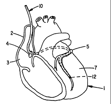
Figure 1 shows a human heart 1 with the intravenous
coronary lead 10 of the present invention passing through
the superior vena cava 2, the right atrium 3, and the
5 coronary sinus 4 into the great vein of the heart 5 so that
a surface electrode i2 on the lead 10 is implanted in a
branch of the coronary vein. When positioned as shown, the
electrode 12 can be used to sense the electrical activity
of the heart or to apply a stimulating pulse to the left
10 ventricle 7 and without the need of being in the left
ventricular chamber.
Figure 2 shows in greater detail the structure of the
intravenous coronary lead shown in Figure 1. As shown in
Figure 2, the lead 10 includes an elongated body member 14
15 having a proximal end 16 and a distal end 18. The body
member 14 is preferably made of a flexible, electrically
insulative material. The outer, surface of the body member
14 is preferably treated to prevent fibrotic attachment and
to reduce inflammation response to the lead. Such a
20 treatment could include a carbon coating, a steroid
embedded in the material, a steroid eluting collar, or the
like.
The body member 14 encapsulates a flexible
electrically conductive member 20 extending from the
25 proximal end 16 toward the distal end 18 of the lead's body
member 14. Conductive member 20 is shown as a flexible
wire coil in Figure 2. Alternatively, the conductor member
20 could be in the form of a conductive wire, a thin
ribbon, a plurality of fine wires formed as a cable, or a
30 flexible tube without deviating from the invention.
Figure 2 also shows the lead 10 as including a central
lumen 22 extending from the proximal end 16 to the distal
end 18 of the body member 14. In fact, in this embodiment,
there is an opening 24 through the distal end 18 to the
35 lumen 22. A coating of a material such as
polytetrafluoroethylene (Teflon*)preferably forms the wall
26 of the lumen 22 to increase its lubricity. The coating
*Trademark
CA 02275225 1999-06-11
WO 98/32486 PCT/US98/01362
- 6 -
material, of course, could be some other polymer having a
low coefficient of friction.
The electrode 12 shown in Figure 2 is preferably
created by removing an annular portion of the insulative
body member 14 to expose a portion of the underlying
conductive member 20. When the conductive member 20 is a
coil as shown in Figure 2, the turns of the coil can be
melt-banded such as by application of laser energy, to form
the surface electrode 12. Those skilled in the art will
recognize that a ring electrode electrically coupled to the
conductive member 20 will also suffice. Likewise, the
position of the electrode 12 along the body member 14 can
be changed. Certain advantages may be achieved, for
example, if the electrode 12 is at the tip of the lead.
The lumen 22 can be put to many uses. For example, a
surgeon can advance a guide wire through the coronary sinus
and coronary veins to the proper position for the electrode
12. The free proximal end of the guide wire can then be
inserted through the opening 24 in the distal end 18 and
the lead 10 slid over the guide wire to position the
electrode 12. The guide wire can then be retracted through
the lumen 22. The lumen 22 can also be used to insert a
small separate structure with an electrode or sensor
deployable beyond the tip of the lead. This allows
separation of the electrodes and can be used for bipolar
pacing or for a combination of pacing and defibrillation.
Likewise, the lumen could be used to inject a contrast
fluid to facilitate fluoroscopic viewing. The lumen can
also be used to deploy a fixation mechanism, deploy an
extraction mechanism, or deploy a plug to close the opening
24 and seal the lumen.
Figure 3 shows how the lead 10 can be modified to
provide a tip 40 of a reduced diameter. The body member 14
of lead 10 has a distal end 18 with an opening 24 in
communication with the lumen 22. Figure 3 shows how the
lumen 22 can be used to deploy a separate structure such as
second, miniaturized lead 42. The deployable lead 42 has
CA 02275225 1999-06-11
WO 98/32486 PCT/US98/01362
_ 7 _
a lead body 44, an electrode 46 and a conductive member
(not shown) coupled to electrode' 46 and running from the
electrode 46 to the proximal end of the lead body 44. The
lead body 44 may be designed to coil after it exits the
lumen to fix the electrode 46 in the correct position.
Figure 3 also shows a ring electrode 47 surrounding a
portion of the tip 40. The ring electrode 47, when
present, is electrically coupled to conductive member 20.
Additional electrodes and conductors can be added for
sensing, pacing or defibrillating as desired. As indicated
above, the ring electrode can also be formed by exposing
and laser bonding the coils of the conductive member 20.
The electrode 46 may be multipolar. It can be used for
defibrillating and the electrode 47 is used for pacing.
Alternatively, electrode 46 may be used for pacing and the
electrode 47 used for pacing. Electrodes 47 and 46 could
also be used for sensing electrical activity of the heart.
Electrodes 47 and 46 can also be used together for bipolar
pacing. Without limitation, t:he main portion of body
member 14 could have an outside diameter in the range of
0.020 inches to 0.100 inches. If, for example, the main
portion of the body member has an outside diameter of 0.058
inches, the diameter of the tip 40 could have an outside
diameter of approximately 0.046 inches and the deployable
lead 42 could have an outside diameter of 0.014 inches.
When used, the main lead body can be positioned first over
a guide wire. Once the lead is in place the guide wire is
removed and replaced with the deployable structure which
can be advanced beyond the tip of the larger lead body.
Figure 4 is provided to assist in explaining an
alternative method for implanting an electrode 12 in a
coronary vein. As shown in Figure 4, the lead 10 is loaded
and temporarily fixed to the inside of a guide catheter 70
designed to be placed in the coronary sinus. The fixation
means 72 may consist of a material such as mannitol which
will dissolve after short exposure to blood. Once the
guide catheter 70 is properly p~asitioned and the fixation
CA 02275225 1999-06-11
WO 98/32486 PCT/US98/01362
_ g _
means 72 is dissolved, the guide catheter 70 can be
retracted leaving the lead in place with an electrode at a
desired position. The lead can then be advanced further if
necessary using a stylet and/or guide wire as previously
described:
While not shown in any of the views, each lead will
have one or more connectors of a type known in the art at
its proximal end for mating with the pacer and/or
defibrillator pulse generator whereby depolarization
l0 signals originating in the heart can be sensed and
stimulating pulses applied in accordance with the device's
control algorithms.
The foregoing discussion is intended to illustrate
various preferred arrangements for meeting the objections
of the present invention. Modifications and variation can
be made by those skilled in the art without departing from
the invention. Accordingly, the invention is limited only
by the scope of the following claims which are intended to
cover all alternate embodiments and modifications as may
fall within the true scope of this invention.
What is claimed: