Note: Descriptions are shown in the official language in which they were submitted.
CA 02275294 2005-12-O1
79565-28
METHOD FOR IMPROVING THE RECOVERY OF TROPONIN I AND T
Field of the InvP~;tion
This invention relates to the assay of troponin I
and troponin T and complexes of these proteins, and more
specifically to the changes in conformation of these
proteins in blood, serum and plasma and to the selection
of antibodies..to the various forms of these proteins and
their use in immunoassays. In another aspect of the
invention, compositions are taught for the stabilization
and recovery of troponin I and T and their complexes in
immunoassays.
Backqg~und Art
Myocardial infarction is one of the leading causes
of death in the United States. Approximately 5 million
individuals experiencing chest pain are evaluated every
year in hospitals throughout the United States, however,
less than 30~, of these individuals are subsequently
found to have had a myocardial infarction. The accurate
and rapid diagnosis of myocardial infarction is impor-
tant both for the patient suffering a myocardial infarc-
tion and for the health care system which can minimize
CA 02275294 1999-06-16
WO 98127435 PCT/1JS97I23252
2
the costs incurred by rapidly-identifying individuals
who do need treatment.
The diagnosis of myocardial infarction is usually
performed in the emergency department of a hospital. An
individual having the symptoms of myocardial infarction
is treated in different ways depending on the obvious-
ness of the condition. Generally, an electrocardiogram
is given to assess the condition of the heart; however,
approximately 50°s of patients experiencing myocardial
infarction have a non-diagnostic electrocardiogram. The
physician is then faced with a problem of diagnosing and
treating the patient suspected of having a myocardial
infarction. Thus, diagnosis and treatment is difficult
for patients with a suspected myocardial infarction who
have non-diagnostic electrocardiograms.
The World Health Organization (WHO) has instituted
guidelines for diagnosing myocardial infarction which
state that an individual must exhibit two-of-the-three
following criteria: 1) have chest pain or a history of
cardiac disease; 2) a diagnostic electrocardiogram;
and, 3) elevated creative kinase (CK) or creative kinase
MB isoenzyme (CKMB). Thus, for the 50% of the individu-
als who are presented to hospitals for a suspected myo-
cardial infarction and who have a non-diagnostic elec-
trocardiogram, the physician must rely on symptoms of
chest pain and an elevated CK or CKMB to diagnose a
myocardial infarction.
The assay of CK or CKMB is generally performed in
hospital laboratories using sophisticated instrumenta
tion. The assays include enzyme assays and immunoassays
which detect the activity or mass of CK or CKMB present
in blood samples.
During a myocardial infarction, heart muscle cells
die and release their contents to the blood stream. The
CA 02275294 1999-06-16
WO 98/27435 PCT/US97/23252
3
CKMB is released among such cellular components. CKMB
becomes elevated above an otherwise nominal value and
can be diagnostic for myocardial infarction. The speci-
ficity of CKMB for diagnosing myocardial infarction is
not 1000 because another source of CKMB in the body is
skeletal muscle. Since the mass of skeletal muscle in
the body far exceeds the mass of cardiac muscle, through
the normal catabolic turnover of skeletal muscle cells,
the blood concentration of CKMB in healthy individuals
will vary. In general, the concentration of CKMB which
may be indicative of myocardial infarction is above 5-7
ng/ml {Circulation 87, 1542-1550 (1993), Clin. Chem. ~.9,
1725-1728 (1993)). The CKMB concentration of individu-
als who have skeletal muscle injury or who have exer-
cised has been reported to be elevated above 9 ng/ml
{Clin. Chem. ,~$, 2396-2400 (1992)). Therefore, the
problem of specificity when using CKMB as a marker for
myocardial infarction has prompted the search for other
more specific markers which are released only from dam-
aged heart muscle.
Troponin I and troponin T have recently been shown
to be more specific than CKMB for diagnosing myocardial
infarction (Circulation ,$~, 902-912 (1991), Clin. Chem.
40, 1291-1295 (1994). Although troponin T has some
disadvantages as a marker because it is elevated in
patients experiencing renal disease (Clin. Chem.
312-317 (1995)), the inventive methods herein disclose
the successful use of troponin T as a diagnostic marker.
The use of troponin I as a diagnostic marker for myocar-
dial infarction also appears to meet many of the clini-
cal requirements (Clin. Chem. ~, 1291-1295 (1994),
Clin. Chem. 41, 312-317 (1995)).
The troponin complex in muscle is comprised of
troponin I, C and T. These troponin components exist as
CA 02275294 1999-06-16
WO 98/27435 PCT/US97/23252
4 _
various tissue specific isoforms. Troponin C exists as
two isoforms, one from cardiac and slow-twitch muscle
and one from fast-twitch muscle. Troponin I and T are
expressed as different isoforms in slow-twitch, fast-
s twitch and cardiac muscle (Biochem. J. 71, 251-259
(1978), J. Biol. Chem. 2~5, 21247-21253 (1990), Hum.
Genet. 88, 101-104 (1991), Circul. Res. 6~, 1226-1233
(1991)). The unique cardiac isoforms of troponin I and
T allow them to be distinguished immunologically from
the other troponins of skeletal muscle. Therefore, the
release into the blood of troponin I and T from damaged
heart muscle has been related to cases of unstable an-
gina and myocardial infarction. The prior art, however,
has not addressed other forms of troponin I and T in
blood.
The troponin complex in muscle is tightly bound to
the contractile apparatus. Approximately 6% of the
troponin T in cardiac tissue exists as an unbound
protein in the cytoplasm and it is believed that this
pool of troponin T is released from damaged muscle (Am.
J. Cardiol. f7, 1360-1367 (1991)).
The conformations of troponin I, T and C change
upon binding when forming binary and ternary complexes
(Biochemistry 33, 12800-12806 (1994), J. Biol. Chem.
2 4, 350-355 (1979), Ann. Rev. Biophys. Biophys. Chem.
~C, 535-559 (1987)). An understanding of the
conformational changes of troponin I and troponin T and
the heterogeneity of the proteins in the blood is
critical for the development of accurate diagnostic
procedures for measuring troponin I and troponin T
concentrations. In addition, troponin I is reported to
be unstable in blood (Direction Insert for Troponin I
Immunoassay, Sanofi/ERIA Diagnostics Pasteur, Marnes la
Coquette, France), and the mechanisms responsible for
CA 02275294 2005-12-O1
.
79565-28
the instability have not been understood. This invention
addresses these problems and provides for stable troponin I
and T compositions which are useful in immunoassays.
Summary of the Invention
5 In one aspect, the invention provides a method for
facilitating the recovery of troponin I, troponin T or both
from a sample, the method comprising: contacting the sample
with a surface, to which troponin C has been added, wherein
the step of adding troponin C comprises adding a solution
that comprises troponin C to the surface and further
comprises drying the surface following addition of the
solution, whereby said troponin C reduces the adsorption of
troponin I, troponin T or both to said surface, and wherein
said surface is a blood filter.
The teachings of the instant invention provide
methods for the selection of antibodies and their use in
immunoassays for troponin I and troponin T and complexes of
these proteins. These proteins, along with troponin C,
exist in both cardiac and skeletal muscle mainly as a
ternary complex. In the muscle, the troponin complex is
bound to tropomyosin which is, in turn, bound to the actin
comprising the thin filaments. The state of troponin I and
troponin T, whether free or bound as binary or ternary
complexes, which are released from the muscle, has not been
previously investigated.
Disclosure of the Invention
Disclosed is an immunoassay system for determining
the presence or amount of a troponin form or a group of
troponin forms in a whole blood, plasma or serum sample
suspected of containing troponin from damaged heart muscle.
CA 02275294 2005-12-O1
79565-28
5a
The system comprises: a) formation of an antibody conjugate
comprising an antibody coupled to a signal generating
element, said antibody capable of specifically binding to
cardiac specific regions of a form of troponin or a group of
troponin forms; b) formation of a reaction mixture
comprising said whole blood, plasma or serum sample
incubated with said antibody conjugate; c) application of
said reaction mixture to a surface to which is bound at
least one capture antibody capable of specifically binding
to cardiac specific regions of a form of troponin or a group
of troponin forms in said antibody conjugate, said capture
antibody binding said antibody conjugate,
i
CA 02275294 1999-06-16
WO 98/27435 PCT/US97/23252
6 _
whereby the immobilized conjugate produces a detectable
signal upon formation of sandwich complexes; and, d)
relation of detectable signal to the presence or amount
of said troponin form or said group of troponin forms in
said sample.
Also disclosed are antibodies that are sensitive
and antibodies that are insensitive to the form of
troponin. An "antibody" refers to: a monoclonal
antibody, a polyclonal antibody, a binding fragment of
an antibody, a recombinant antibody, or a receptor
protein that specifically binds to a target. As used
herein, an insensitive antibody is an antibody that
yields an assay response that is less than within about
a factor of 2 (i.e., 50% of the base value); and
preferably yields an assay response that is within about
20o for each form of troponin, measured relative to
assay response for the use of that antibody in an assay
for a troponin form or group of troponin forms (the base
value). Thus, an insensitive antibody is one that will
tend to bind more than one form of troponin.
As used herein, a sensitive antibody in an
immunoassay is one that yields an assay response that is
greater by at least about a factor of 2 larger (i.e.,
200$ of the base value) and preferably a factor of 5
larger (i.e., 200°s of the base value), for one or a
group of forms of troponin (the base value), as compared
to the assay response for other forms measured. Thus, a
sensitive antibody is one that will tend to bend only a
single form of troponin.
As used herein the nine troponin forms are: 1) the
cardiac ternary complex; 2) the cardiac troponin binary
complex of I(oxidized)/T; 3)the cardiac troponin binary
complex of I(reduced)/T; 4)the cardiac troponin binary
complex of I(oxidized}/C; 5)the cardiac troponin binary
CA 02275294 1999-06-16
WO 98/27435 PCT/US97/23252
7
complex of I(reduced)/C; 6) the cardiac troponin binary
complex T/C; 7) unbound cardiac troponin I (oxidized);
8) unbound cardiac troponin I (reduced}; and, 9) unbound
cardiac troponin T.
Disclosed is a stabilized composition of troponin;
The stabilized composition can comprise a stabilized
composition of troponin I, wherein the troponin I is
oxidized, the troponin I can be unbound or the troponin
I can be in a complex. The stabilized composition can
comprise a stabilized composition of the ternary complex
of troponin I, T and C.
Disclosed is a method for improving the recovery of
troponin I or T from a surface used in immunoassays,
said method comprising: contacting with said surface at
least one strongly basic peptide, protein, or polymer
with a pI value greater than about 8; the method can
further comprise a step of washing unbound peptide,
protein or polymer from said membrane. Melittin can be
the strongly basic peptide used, protamine can be the
strongly basic protein used.
Description of Figures
Figure la illustrates the kinetics of air oxidation of
troponin I as measured by immunoassay.
Figure 1b illustrates the kinetics of oxidation by
peroxide of troponin I as measured by immunoassay.
Figure 2 illustrates the kinetics of reduction by
dithiothreitol of troponin I and reoxidation of reduced
~ troponin I by peroxide as measured by immunoassay.
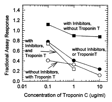
Figure 3 illustrates the effect of troponin C on the
immunoassay of troponin I in the presence or absence of
troponin T and binding inhibitors.
Figure 4 illustrates the kinetics of disruption of human
cardiac troponin ternary complex in the presence or
CA 02275294 1999-06-16
WO 98/27435 PCT/US97123252
8
absence of binding inhibitors- as measured by an
immunoassay for troponin I.
Figures 5a-5f illustrate the effect of binding
inhibitors on troponin I immunoassays from patient
samples with confirmed myocardial infarction.
Modes for Carrying Out the Invention
Definitions:
As used herein, an "antibody" or "receptor protein"
refers to a monoclonal antibody, a polyclonal antibody,
a binding fragment of an antibody, a recombinant
antibody, or a receptor protein that specifically binds
to a target. Specific binding of a substance signifies
the quality of that substance that the substance will
tend not bind to something to which it does not
specifically bind; conversely, the substance will have
greater affinity for something it specifically binds
that for a something it does not specifically bind.
As used herein, an insensitive antibody in an
immunoassay is an antibody that for each form of
troponin of interest yields an assay response value that
is the same within about a factor of 2, and preferably
the same within about 20~, as the assay response values
for the other forms of interest. Thus, an insensitive
antibody is one that will exhibit a detection of more
than one form of troponin in an immunoassay.
As used herein, a sensitive antibody in an
immunoassay is one that for one form or group of forms
of troponin yields an assay response value that is at
least about a factor of 2 larger, and preferably, about
a factor of 5 larger, than the assay response values for
other forms. Thus, a sensitive antibody is one that
will exhibit a preferential detection of one form or
group of forms of troponin in an immunoassay.
' CA 02275294 1999-06-16
WO 98/27435 PCT/US97/23252
9
As used herein the nine troponin forms are: 1) the
cardiac ternary complex; 2) the cardiac troponin binary
complex of I(oxidized)/T; 3)the cardiac troponin binary
r
complex of I(reduced)/T; 4)the cardiac troponin binary
complex of I(oxidized)/C; 5)the cardiac troponin binary
complex of I(reduced)/C; 6) the cardiac troponin binary
complex T/C; 7) unbound cardiac troponin I (oxidized);
8) unbound cardiac troponin I (reduced); and, 9) unbound
cardiac troponin T.
As used herein, a "zone" is a concept that
correlates with the ability to identify distinct
sensible signals. A zone, therefore, can correspond to
a geographic region or correspond to the ability to
separately identify distinct sensible signals. The
sensible signals can be distinct by, but not limited to,
variations between the following characteristics:
wavelength of fluorescence or optical absorbance or
reflectance; life time of, or transition energy between,
electronic states; oxidation-reduction potentials;
colorimetric characteristics; or, signal type (e. g.,
fluorescence vs. radioactivity vs. optical absorbance).
As used herein, unbound troponin is troponin that
is not in a complex. A troponin complex can be binary
or ternary.
As used herein, a "label", "signal generator" or
"signal generating element" is an entity that can embody
a number of different forms: Enzymes and their
resultant effects on a substrate, colloidal metal
particles, latex and silica particles with dye
incorporated, and dye particles are examples of signal
generators. An enzyme can react on a substrate to
produce a product that is sensible, for example, by
wavelength of fluorescence (e. g., ultraviolet, visible,
infrared), or sensible by affect on pH.
CA 02275294 1999-06-16
WO 98/27435 PCT/US97/23252
Modes:
This invention is directed to the assay of troponin
I and troponin T and complexes of these proteins in body
fluids, particularly, in human blood, serum and plasma.
5 The presence of cardiac troponin I and T in the blood,
above a nominal concentration, is diagnostic for damaged
heart muscle. The teachings of this invention show that
troponin I and T exist in various conformations in the
blood which may be the same or different than their
10 native conformations in muscle tissue. These various
conformations of the troponin molecules can react
differently with antibodies.
The ratios of the monomeric troponin I and T and
the binary and ternary complexes may be related to the
metabolic state of the heart. Based on the reactivities
of antibodies to troponin I and T and to purified
complexes of the troponins, the concentrations of
troponin I and T and their complexes can now be
elucidated in blood samples from patients suffering from
myocardial infarction.
The embodiments of this invention relate to the
conformations of troponin I and T and their complexes in
blood, serum and plasma, and to antibodies which
recognize those conformations. Specifically, antibodies
which recognize troponin I and T in the following forms
are preferred: 1) The conformations of troponin I
having intramolecularly oxidized and reduced cysteines;
2) The binary complexes of troponin I and T, of troponin
I and C, of troponin T and C; and, 3) The ternary
complex of troponin I, T and C. In addition, methods
are described for the improved recovery of troponin I
and T in immunoassays. This invention answers the
heretofore unmet need for the assays of troponin I and T
in blood.
CA 02275294 1999-06-16
WO 98/27435 PCT/US97/23252
11
The focus on troponin I and T for use as markers
for myocardial infarction has been based in part on
their molecular size: because the proteins are
relatively small, it is believed that they leak out of
damaged cells faster than the larger proteins.
Antibodies to Troponin Complexes and to Troponin I and T
The term "antibodies or receptor proteins" as used
herein refer to monoclonal and polyclonal antibodies,
binding fragments of antibodies, and receptor proteins
that specifically bend to a target. In a preferred
embodiment, receptor proteins, for example, antibodies
or binding fragments, are directed to the epitopes of
troponin I which are insensitive to the oxidation state
of the molecule. The terms sensitive any ;n~o"~;+-;«o
herein refer generally to the ability of an antibody to
recognize particular forms of free troponin or troponin
complexes. In particular, a sensitive antibody which is
useful in an immunoassay distinguishes one form or forms
of troponin from another form and an insensitive
antibody which is useful in an immunoassay does not
distinguish one form or forms of troponin from another.
The sensitivity or insensitivity of an antibody is
exhibited in an immunoassay. To determine whether an
antibody is sensitive or insensitive, the antibody is
tested with each troponin form independently to yield
the assay response of the antibody for each troponin
form. In general, a preferred antibody that is
insensitive to the troponin form will yield an assay
response that is the same, within about a factor of two
and preferably within 20%, for each form of troponin. A
preferred antibody that is sensitive to the troponin
form will yield an assay response that is at least a
factor of two, and preferably a factor of five, larger
_ CA 02275294 1999-06-16
WO 98/27435 PCT/US97/23252
12
for one form or group of forms as compared with the
other form(s). In addition, the terms "troponin I and
T" can refer to the free, uncomplexed troponins or to
the troponins in the binary or ternary complexes. Human
cardiac troponin I contains two cysteines, at positions
80 and 97 (FEBS Letters, 270, 57-61 (1990)). In the
current art, during the purification of troponin I from
tissues, the oxidation state of troponin I is directed
toward the reduced form using various reductants,
including mercaptoethanol, dithiothreitol and the like
(Can. J. Biochem. ~4, 546-553 (1976), Methods Enzymol.
85, 241-263 (1982)). After purification, the current
art also teaches to maintain troponin I in the reduced
form to prevent intermolecular disulfide formation (J.
Biol. Chem. 258, 2951-2954 (1983)).
In the development of immunoassays for a target
protein, the purified target protein acts as a standard
with which to judge the sensitivity and specificity of
the immunoassay using the antibodies that have been
selected. As disclosed herein, the cysteines in troponin
I can rapidly oxidize, intramolecularly, to alter the
conformation of the protein. The degree of oxidation of
troponin I has not previously been addressed with
respect to its effect on the immunoassay process. The
teachings described herein show that an apparent
instability in the troponin I molecule is related to the
dynamics of the intramolecular oxidation or reduction of
the troponin I molecule. In addition, the selection of
antibodies is taught for the accurate quantitation of
troponin I in blood.
Purified, reduced troponin I undergoes an
intramolecular oxidation of the cysteines, the rate of
which is not dependent on the troponin I concentration.
Special care should be exercised when preparing the
CA 02275294 1999-06-16
WO 98/27435 PCT/LTS97/23252
I3
oxidized troponin I form, especially in the presence of
various thiol reducing agents, because of the
possibility of forming mixed disulfides of the protein
and the reducing thiol reagent. The mixed disulfide
form of the protein may not behave as either the
oxidized or reduced form of the molecule, especially if
the antibodies used in the immunoassay bind to the
region of the protein surrounding cysteines 80 and 97.
Using purified preparations of oxidized and reduced
troponin I, differential effects in the immunoassays
using various antibodies raised to troponin I were
observed. With some antibody pairs, the reduced
troponin I was hardly detectable in immunoassays,
whereas with other pairs, the oxidation state had no
effect on the immunoassay process. These results showed
that selection of antibodies to the troponin I molecule,
without prior knowledge of the oxidation state of the
troponin I, can result in antibodies and an immunoassay
process which gives erroneous results. This conclusion
is exemplified by immunoassays of troponin I from
patients suffering myocardial infarction. The degree of
oxidation of troponin I in patient samples is variable
and suggests a possible basis for apparent instabilities
of troponin I assays of the current art. The free
troponin I does change its oxidation state from the
reduced to the oxidized form over time(see Example 4).
In the case when an immunoassay comprising an
insensitive antibody is to be utilized to bind to the
free and complexed troponin, a preferred antibody is one
that is insensitive with respect to the oxidized,
reduced and complex forms because the circulating
troponin in the blood can change its oxidation state and
the degree of binding to other troponin components over
time. For example, if reduced, free troponin I is
CA 02275294 1999-06-16
WO 98/27435 PCT/US97/23252
14 _
released from the heart, it can transform to the
oxidized form during circulation in the blood and until
the time troponin is measured. In addition, the free
troponin I and T in the blood can bind to each other and
to troponin C to form binary and ternary complexes. The
outcome of these transformations to the oxidized form of
troponin I or to complexes of troponin is that when an
antibody is chosen for an immunoassay that is sensitive
with respect to the oxidized and reduced forms and
complexed forms, the assay will show an apparent
increase or decrease in the troponin concentration over
time depending on which form (free, oxidized or reduced
or complexed) the antibody better recognizes, rather
than an actual change in troponin concentration. In
another example, the release of troponin complexes from
damaged heart muscle can result in the formation of free
(uncomplexed) and binary forms of troponin I and T
during circulation in the blood or until the troponin is
assayed. The variability of assay results of troponin
concentrations using immunoassays comprising a sensitive
antibody or antibodies can mislead a physician into
believing that a patient's condition is improving or
deteriorating.
In the case when an immunoassay comprising more
than one sensitive antibody is used to measure the free
and complexed troponin, each sensitive antibody is
intended to be reacted with one form or group of forms
of troponin such that the antibody exhibits a maximum
assay response for the intended forms) and a minimum
assay response for all other forms. Preferred
antibodies are ones for which the assay responses for
the intended forms are about the same, preferably within
200, for all the sensitive antibodies and said minimum
assay responses are at least a factor of 2 and
CA 02275294 1999-06-16
WO 98/27435 PCT/US97/23252
preferably a factor of 5 less-than the maximum assay
responses. For example, if two different antibodies are
used for signal antibodies in an immunoassay, and one
antibody is insensitive with respect to the oxidized and
5 reduced free troponin I (or the free troponin T) and
exhibits a maximum assay response for said free forms of
troponin and a minimum assay response for the compleXed
troponin, then the other antibody should exhibit a
maximum assay response for the complexed troponin and a
10 minimum assay response for the free troponin I and T
forms. In this way, an accurate measure of total
troponin can be determined. One skilled in the art will
also recognize that antibodies with different affinities
that is, exhibit different assay responses, for the
15 troponin forms can also be utilized in immunoassays when
each troponin form is measured alone or in discrete
zones and that the relative bias of the immunoassays can
be accounted for in the calibration of the assay. Also,
sensitive and insensitive antibodies can be attached to
solid phases to measure each troponin form in discrete
zones.
In another preferred embodiment, antibodies or
binding fragments that are directed to the epitopes of
the troponin I or T are insensitive with respect to free
troponin I or T and troponin complexes. The teachings
herein show that antibodies that are sensitive with
respect to free troponin I or T and troponin complexes
provide methods for the estimation of the extent of
binding of troponin I or T in complexes. Using purified
preparations of troponin I, T and C, and the purified
troponin complexes, the effects of troponin I or T
binding in complexes on the recognition of troponin by
antibody pairs is taught and is related to the dynamic
state of troponin I or T in blood.
CA 02275294 1999-06-16
WO 98127435 PCTIUS97/23252
16
The degree of binding of-troponin I and T to the
components of the troponin complex or to other proteins
of the contractile apparatus, including tropomyosin and
actin, in blood can also be problematic for immunoassays
depending on the degree and affinity of binding. In
their native forms, the troponin complex exists in
cardiac muscle and in slow- and fast-twitch skeletal
muscle as a ternary complex of troponin I, C and T.
Troponin I and T from skeletal muscle have different
amino acid sequences than troponin I and T from cardiac
muscle, respectively; however, troponin C from slow-
twitch muscle has the same amino acid sequence as the
cardiac muscle protein (Nature 271, 31-35 (1978), Arch.
Biochem. Biophys. 186, 411-415 (1978), FEBS Lett. 292,
5-8 (1991)). The fast-twitch skeletal muscle troponin
C, although not identical to the cardiac troponin C, can
bind to cardiac troponin I (Biochemistry 33, 8464-8471
(1994), Proc. Natl. Acad. Sci. USA ~0, 9036-9040
(1993) ) .
The release of troponin components, that is,
troponin I, C and T, or components from the contractile
apparatus, for example, tropomyosin and actin, from
skeletal muscle, due to the normal turnover of skeletal
muscle cells, may result in a significant amount of
troponin and contractile apparatus components in the
blood. Since skeletal muscle mass is much greater than
cardiac muscle mass, the troponin components present in
the blood of a normal individual may be derived largely
from skeletal muscle. The circulating troponin
components which are mainly derived from skeletal muscle
would bind to cardiac troponin I and T which are
released into the blood during a myocardial infarction
or events which lead up to creating damaged heart
muscle. As muscle damage progresses in an individual
CA 02275294 1999-06-16
WO 98/27435 PCT/US97/23252
17
the troponin components derived from heart tissue will
presumably rise in the blood. Thus, the concentration
of troponin components (bound and free) in the blood
from individuals experiencing a myocardial infarction
. 5 may be differentially derived from both cardiac and
skeletal muscle.
The form of troponin released from the heart,
whether free or as binary or ternary complexes, into the
blood may indicate a particular condition of the heart.
The assays taught herein provide for the analysis of
release patterns which may allow the physician to
diagnose a specific heart failure, for example, unstable
angina as compared to myocardial infarction or to
determine the time that an infarction occurred.
The clinical impact of an immunoassay measuring
only the free troponin I or T from a patient
experiencing a myocardial infarction can be very
significant. Since the binding of troponin I and T to
troponin components in the blood will be variable,
depending on the troponin component concentrations, and
on the time that has elapsed since the release of the
troponin components from muscle an analysis of the bound
and free form of the troponin I and T in the blood must
be considered. For example, the binding affinity of
troponin I to troponin C, in the presence of calcium
(which also is present in blood} is 1.27 x 10⁸~M-1
(Biochemistry ~, 12729-12734 (1999)). This implies
that if the troponin C concentration is 100 ng/ml and
the total (bound and free) troponin I concentration is 8
ng/ml, then the free concentration of troponin I is
- calculated to be 4.6 ng/ml. If the concentration of
troponin I which is indicative of a myocardial
infarction is 5 ng/ml or greater, then an assay which
measures only the free form of troponin I, in this case,
CA 02275294 1999-06-16
WO 98/27435 PCT/US97123252
18
4.6 ng/ml, will indicate to the physician that a
myocardial infarction has not taken place. Generally,
in hospital emergency departments which admit patients
believed to have had a myocardial infarction, a blood
sample from the individual will be obtained again in an
hour or two if the first result is negative. In this
example, the patient, having a total troponin I
concentration of 8 ng/ml in the first sample, (which is
defined as positive for a myocardial infarction), but
only a measured concentration of 4.6 ng/ml, (which would
be defined as a negative result), would not be treated
and would continue to accrue damaged heart muscle during
the time before a second sample was analyzed.
Interpretation of results of troponin T assays would
also suffer from troponin T binding to components of the
contractile apparatus in blood. Thus, immunoassays of
the current art that measure the free troponin I and T
may not correctly diagnose a myocardial infarction when
the troponin I or T concentration, respectively, is near
the decision point. In some cases, the rise or fall of
the troponin I or T concentration in a patients blood
over time as determined by analyzing blood samples drawn
at several different times might be used to diagnose the
dynamic condition of the heart, for example, to
determine whether the damaged heart is improving with
therapy or continuing to deteriorate. In these cases,
the time-course of the free troponin I or T
concentration could be different than that of the total
troponin (bound and free) concentration. One skilled in
the art will realize that increasing concentrations of
all the troponin components in the blood will result in
an increasing fraction of bound troponin I and T
relative to free troponin. The concentration of total
troponin (bound and free) may rise faster than the
CA 02275294 1999-06-16
WO 98/27435 PCT/US97/23252
19
concentration of free troponir~ I and T. An assay that
measures the total troponin concentration (bound and
free troponin I and T) would be more accurate in
assessing progression of heart damage as compared with
an assay that measures only free troponin I or T.
In a particularly preferred embodiment, antibodies
or binding fragments are directed to the cardiac
troponin complex. Specifically, antibodies are directed
to cardiac specific epitopes of troponin I and T of the
troponin complex or of the troponin I/T, I/C and T/C
interfaces in the complex. The teachings herein show
that antibodies that are raised to troponin I and T can
bind poorly to troponin I or T of the ternary complex.
Furthermore, the teachings herein show that the troponin
complex exists in the blood of patients who have
experienced myocardial infarction. Methods are also
described which teach one skilled in the art to assess
the amount of troponin complex in the blood relative to
the free troponin I and T or binary complexes of
troponin I and T, using antibodies which bind to the
free troponin molecules.
The equilibrium among the binary and ternary
complexes of troponin and free troponin I and T will be
altered during the immunoassay process because of the
binding of antibodies to the troponin components and
complexes. The change in the mole fractions of the
various species during the immunoassay may be
significant or insignificant and will be a function of
- the antibody concentrations, the affinity of the
antibodies for the troponin components and complexes,
- the association constants for the troponin components
and complexes and the time that the antibodies are
allowed to bind to the troponin. These variables can
change the perceived concentration of troponin I and T
CA 02275294 1999-06-16
WO 98/27435 PCTlUS97/23252
20 _
and lead to erroneous conclusions about the troponin
concentration. For example, if two immunoassays utilize
different antibody pairs for performing a sandwich
immunoassay and their antibody concentrations and
affinities for troponin I or T are different, and if a
proportion of the troponin I or T occurs in the sample
as binary and ternary complexes, one may expect that
each immunoassay will give a different result. In
addition, if blood samples contain varying
concentrations of troponin C, then the proportion of
troponin I and T that is bound to troponin C as a binary
complex will differentially perturb each immunoassay.
The teachings of the instant invention demonstrate
that the troponin ternary complex is more stable to
dissociation than the binary complexes of troponin.
In another preferred embodiment, antibodies or
binding fragments are directed to epitopes which are not
changed by proteolytic degradation of the N-terminal
region of troponin I. The conformation of troponin I is
also reported to be affected by
phosphorylation/dephosphorylation (Biophys. J. 63, 986-
995(1992), Biochem. 33, 12729-12734 (1994)). In another
preferred embodiment, antibodies or binding fragments
are directed to epitopes of either troponin I or
troponin I complexes, which are or are not changed by
the phosphorylation state of troponin I. Troponin I can
be phosphorylated using methods described in J. Biol.
Chem. ?~2_, 851-857 (1977). The phosphorylated and
dephosphorylated preparations of troponin I can be
utilized as immunogens for generating antibodies as well
as antigens for the selection of antibodies to the
phosphorylated and dephosphorylated troponin I. The
troponin complex can be dissociated into the component
proteins using various treatments, including high
CA 02275294 1999-06-16
WO 98/27435 PCT/US97/23252
21
concentrations of urea, low pH and metal chelating
agents which bind divalent metal rations, particularly
calcium and magnesium (Methods Enzymol. 85, 241-263
(1982)). These treatments are, in general, very harsh
and require several hours. Thus, these conditions for
dissociating the troponin complex are not practical for
immunoassays which must be performed in a matter of
minutes on samples from individuals who may be suffering
a myocardial infarction.
The generation and selection of antibodies that are
preferentially either sensitive or insensitive to the
binding of troponin I or T in binary complexes are
accomplished by first preparing binary troponin I/T, T/C
and I/C complexes from purified components (J. Biol.
Chem. ,2~, 350-355 (1979), J. Biol. Chem. 258, 2534-2542
(1983), J. Biol. Chem. ~, 2951-2954 (1983), Can. J.
Biochem. Cell Biol. 6~., 212-218 (1985), Biochemistry 33,
12729-12734 (1994), Ann. Rev. Biophys. Biophys. Chem.
_1~, 535-559 (1987)). The complexes may be stabilized,
if necessary, by chemically cross-linking the proteins
in the complex using methods familiar to those skilled
in the art. The generation and selection of antibodies
that are sensitive or insensitive to the binding of
troponin I or T in the ternary complex can be
accomplished several ways. For example, one way is to
purify the ternary complex (Methods Enzymol. ,$5, 241-263
(1983)) or to reconstitute the complex using the
purified troponin components. One skilled in the art
will recognize that various other contractile apparatus
proteins which may be associated with the binary or
ternary complexes of troponins can also be constructed
from the purified components and that the resultant
complex can be utilized to generate and select
antibodies as taught by the instant invention. The
CA 02275294 1999-06-16
WO 98/27435 PCT/L1S97/23252
22 _
complex can be stabilized with respect to dissociation
by chemically crosslinking the components. The purified
complexes are then injected, for example, into mice or
rabbits, to generate polyclonal or monoclonal
antibodies. Another way is to purify free (unbound)
troponin I or T and then inject the purified free
troponin I or T, for example, into mice or rabbits, to
generate polyclonal or monoclonal antibodies. One
skilled in the art will recognize that many procedures
are available for the production of antibodies, for
example, as described in Antibodies, A Laboratory
Mar~ual, Ed Harlow and David Lane, Cold Spring Harbor
Laboratory (1988), Cold Spring Harbor, NY. One skilled
in the art will also appreciate that binding fragments
or Fab fragments which mimic antibodies can also be
prepared from genetic information by various procedures
(Antibody Engineering: A Practical Approach
(Borrebaeck, C., ed.), 1995, Oxford University Press,
Oxford; J. Immunol. 149, 3914-3920 (1992)). In
particular, the preparation, screening and selection of
recombinant binding fragments is described in Examples
22 and 23.
The antibodies which are generated are selected by
first screening for affinity and specificity with the
purified binary or ternary complexes and comparing the
results to the affinity and specificity of the
antibodies with the purified troponin I and T molecules
for the desired properties which are defined by the
immunoassay process.
The screening procedure can involve immobilization
of the purified troponin I or T or binary or ternary
complexes or peptides to cardiac specific sequences of
the troponins in separate wells of microtiter plates.
The solution containing a potential antibody or groups
CA 02275294 1999-06-16
WO 98/27435 PCTlUS97/23252
23
of antibodies is then placed into the respective
microtiter wells and incubated for about 30 min to 2 h.
If an antibody to the protein of interest is present in
the solution, it will bind to the immobilized troponin.
In screening antibodies for binding to interfaces of
binary or ternary complexes of troponin, an antibody is
first selected which binds to the binary or ternary
complex immobilized in the microtiter well. That
antibody is then further screened for its ability to
bind to free troponin components; that is, the
potential interface antibody should not bind to free
troponin I, C or T which is immobilized in microtiter
wells. In addition, the interface antibody should never
be capable of forming a sandwich assay with binary or
ternary complexes and an antibody which is known to bind
to a specific troponin component in the complex in the
presence of binding inhibitors which are known to
disrupt the troponin complex. If this latter condition
is met, then the potential interface antibody should
also not be capable of forming a sandwich assay with the
troponin complex and an antibody to a different troponin
component than was used in the previous screen in the
presence of binding inhibitors. If this condition is
also met, then an interface antibody has been selected
for a binary complex. An extra immunoassay must be
performed for selecting an interface antibody to a
ternary complex; that is, if the previous two
conditions are met, then the potential interface
antibody should also not be capable of forming a
sandwich assay with the troponin complex and an antibody
to a different troponin component than was used in the
two previous screens. The microtiter wells are then
washed and a labeled secondary antibody (for example, an
anti-mouse antibody conjugated to alkaline phosphatase
CA 02275294 1999-06-16
WO 98/27435 PCT/US97/23252
24 _
if the raised antibodies are mouse antibodies) is added
to the wells and incubated for about 30 min and then
washed. Substrate is added to the wells and a color
reaction will appear where antibody to the troponin is
present. The antibodies which are of interest are then
further analyzed for affinity and specificity to the
cardiac specific molecules and for complementarity in
forming sandwich complexes with the antigens. Those
skilled in the art will recognize that many approaches
can be taken in producing antibodies or binding
fragments and screening and selecting for affinity and
specificity for the various troponin antigens, but these
approaches do not change the scope of the invention.
Assays for Troponin Complexes and
Uncomolexed Troponin I and T
A particularly preferred embodiment of this
invention is directed to the assay of troponin I and
troponin T, particularly immunoassays, wherein the
antibodies selected for the assay bind to cardiac
specific sequences of the ternary complex, of the binary
complexes and of the uncomplexed (free) troponin I or T
in order to measure the complexed (bound) and free
fractions of troponin I and T, respectively. The
cardiac specific sequences of troponin I and T are
described in FEBS Lett. 270, 57-61 (1990) and Genomics
21, 311-316 (1994). A synthetic peptide comprised of 14
amino acids which mimics a cardiac specific sequence of
troponin I and methods used to prepare antibodies to the
peptide are described in an International Patent
Application number PCT/US94/05468.
The immunoassay can be formulated with a cocktail
of antibodies to bind all the troponin complexes and the
free troponin I and T. Alternatively, the immunoassay
CA 02275294 2005-12-O1
79565-28
can be formulated with specific antibodies that
recognize epitopes of the troponin I and T in the
complexes and also the unbound troponin I and T. In
addition, the immunoassay can be formulated with
5 antibodies that bind epitopes at interfaces of the
component proteins in the complexes and antibodies that
bind the unbound troponin I and T.
A preferred immunoassay for troponin I or T
involves conjugation of an antibody or a cocktail of
10 antibodies to a label or a signal generator to form an
antibody conjugate(s), which are capable of binding to
cardiac specific regions of the troponin complexes of
troponin I or T and to unbound troponin I or T. One
skilled in the art will recognize that a signal
15 generator has many forms. Enzymes, colloidal metal
particles, latex and silica particles with dye
incorporated, and dye particles are examples of signal
generators. Antibodies can, be conjugated to the signal
generators in a variety of ways using heterobifunctional
20 reagents as taught in the Pierce Catalog and Handbook,
Pierce Chemical Co., Rockford, IL and in Uniform Latex
Particles by Leigh B. Bangs, Seragen Diagnostics Inc.,
Indianapolis, '
Another antibody or cocktail of antibodies is
25 immobilized on a solid phase, for example, a membrane as
taught in BioTechniques g, 272-283 (1986), and,the
membrane is placed in a device, for example, as
described in U.S. Patents, 4,727,019 and 5,458,852. The
immobilized antibody is complementary to the.antibody
conjugate. The immobilized antibody and conjugate
antibodies form sandwich complexes with the troponin I
or T complexes and also form sandwich complexes with
troponin I or T, respectively. A plasma or serum sample
suspected of containing troponin complexes or components
CA 02275294 1999-06-16
WO 98/27435 PCT/US97/23252
26
from damaged heart muscle is mixed with the antibody
conjugate to form a reaction mixture that is allowed to
incubate. The reaction mixture is then applied to the
aforementioned device. The sample flows through the
membrane and the troponin complexes and components,
bound to the antibody conjugates, bind to the
immobilized antibodies, and excess, unbound, antibody
conjugate is washed away with a wash buffer. The signal
is developed and read, either visually or
instrumentally.
A particularly preferred immunoassay for troponin I
involves conjugation of at least two antibodies to a
label or a signal generator to form an antibody
conjugate. One of the conjugate antibodies is capable
of binding to the troponin T component of the troponin
ternary complexes and the other antibody is capable of
binding to the free and binary troponin I molecules.
Another antibody or cocktail of antibodies is
immobilized on a solid phase, for example, a membrane,
and the membrane is placed in a device, as described
previously. The immobilized antibody is complementary
with the antibody conjugate antibodies to form sandwich
complexes with either troponin I bound to troponin
complexes or to the uncomplexed troponin I. A plasma or
serum sample suspected of containing troponin complexes
or components from damaged heart muscle is mixed with
the antibody conjugate to form a reaction mixture which
is allowed to incubate. The reaction mixture is then
applied to the aforementioned device. The sample flows
through the membrane and the troponin complexes and
components, bound to the antibody conjugates, bind to
the immobilized antibodies and excess, unbound antibody
conjugate is washed away with a wash buffer. The signal
is developed and read, either visually or
CA 02275294 1999-06-16
WO 98/27435 PCT/US97/23252
27
instrumentally. In this assay procedure, the antibody
conjugate binds to the troponin I in ternary complexes
through the troponin T specific antibody and all free
r
and binary troponin I molecules through the troponin I
- 5 specific antibody. The capture antibody or antibodies
on the solid phase bind antibody conjugates that are
bound to free troponin I, and to troponin ternary
complexes that contain troponin I.
A particularly preferred immunoassay for troponin I
(oxidized and reduced) involves conjugation of at least
two antibodies to a label or a signal generator to form
an antibody conjugate. One of the conjugate antibodies
is capable of binding to the oxidized troponin I and the
other antibody is capable of binding to the reduced
troponin I molecules. Another antibody or cocktail of
antibodies is immobilized on a solid phase in up to 2
discrete zones, for example, a membrane, and the
membrane is placed in a device, as described previously.
The immobilized antibody is complementary with the
antibody conjugate antibodies to form sandwich complexes
with either oxidized troponin I or reduced troponin I.
A plasma or serum sample suspected of containing
troponin components from damaged heart muscle is mixed
with the antibody conjugate to form a reaction mixture
which is allowed to incubate. The reaction mixture is
then applied to the aforementioned device. The sample
flows through the membrane and the oxidized and reduced
troponin I, bound to the antibody conjugates, bind to
. the immobilized antibodies and excess, unbound antibody
conjugate is washed away with a wash buffer. The signal
_ is developed and read, either visually or
instrumentally. In this assay procedure, the antibody
conjugates bind to the oxidized troponin I through the
oxidized troponin I specific antibody and to the reduced
CA 02275294 1999-06-16
WO 98/27435 PCT/US97/23252
28
troponin I through the reduced troponin I specific
antibody. The capture antibody or antibodies on the
solid phase only bind antibody conjugates that are
bound to oxidized and reduced troponin I. This
immunoassay may have application in the estimation of
time after an infarction has occurred.
Another particularly preferred immunoassay for
troponin I involves conjugation of an antibody or a
cocktail of antibodies to a label or a signal generator
to form an antibody conjugate. The antibody conjugate
binds to either troponin I bound to troponin complexes
or to the uncomplexed troponin I. Immobilized on a
solid phase, for example, a membrane, in 3 discrete
zones, are antibodies or cocktails of antibodies which
bind the ternary complex, the binary complexes of
troponin I and the free troponin I, and the membrane is
placed in a device, as described previously. For
example, a troponin T antibody which binds to the
troponin T of the ternary complex is immobilized in one
discrete zone, a troponin I antibody which binds to the
troponin I binary complexes (troponin I/C and I/T) is
immobilized in another discrete zone and a troponin I
antibody which binds to only the uncomplexed troponin I
is immobilized in yet another discrete zone. The
immobilized antibodies are complementary with the
antibody conjugate antibodies to form sandwich complexes
with complexed or uncomplexed troponin I, as defined by
each discrete zone. Alternatively, immobilized on a
solid phase, for example, a membrane, in 2 discrete
zones, are antibodies or cocktails of antibodies which
bind the troponin I complexes (binary and ternary) and
the free troponin I. For example, a troponin T antibody
which binds to the troponin T of the ternary complex and
a troponin I antibody which binds to the troponin I of
CA 02275294 1999-06-16
WO 98/27435 PCTIUS97/23252
29
the binary complexes are immobilized in one discrete
zone and a troponin I antibody which binds to only the
uncomplexed troponin I is immobilized in the second
discrete zone. The immobilized antibodies are
complementary with the antibody conjugate antibodies to
form sandwich complexes with complexed or uncomplexed
troponin I, as defined by each discrete zone. A further
embodiment of this invention utilizes antibodies on the
solid phase for detection of troponin I complexes which
bind to the interfaces of the binding domains of
troponin I/T and I/C. A plasma or serum sample
suspected of containing troponin complexes or components
from damaged heart muscle is mixed with the antibody
conjugate to form a reaction mixture which is allowed to
incubate. The reaction mixture is then applied to the
aforementioned device. The sample flows through the
membrane and the troponin complexes and components,
bound to the antibody conjugate(s), bind to the
respective immobilized antibodies in the discrete zones
and excess, unbound antibody conjugate is washed away
with a wash buffer. The signal is developed and read,
either visually or instrumentally. In this assay
procedure, the antibody conjugate binds to the troponin
I and the troponin I binary and ternary complexes
through the troponin I specific antibody or antibodies.
The capture antibody or antibodies in discrete zones on
the solid phase bind the antibody conjugates that are
bound to the uncomplexed troponin I or troponin
complexes containing troponin I as defined by each
discrete zone. This immunoassay allows quantification
of the fractions of troponin I, namely, the complexed
and the uncomplexed fractions. The inventive teachings
described herein show that uncomplexed and complexed
troponin exists in plasma and serum samples from
CA 02275294 1999-06-16
WO 98!27435 PCT/LTS97/23252
patients with confirmed myocardial infarction. The
determination of the complexed and uncomplexed troponin
I fractions may yield important clinical data relating
to the type and extent of muscle damage, for example,
5 from unstable angina or myocardial infarction or to the
success of thrombolytic therapy.
Another particularly preferred immunoassay measures
the cardiac troponin ternary complex, the cardiac
troponin binary complexes (troponin I/T, T/C and I/C)
10 and the free cardiac troponin I and T. This method
involves conjugation of antibodies or a cocktail of
antibodies to a label or a signal generator to form an
antibody conjugate. The antibody conjugates bind to
either troponin T and I bound to troponin complexes or
15 to the uncomplexed troponin T and I. Immobilized on a
solid phase, for example, a membrane, in 1 discrete
zone, are antibodies or cocktails of antibodies which
bind the ternary complex, the binary complexes of
troponin I and T and the free troponin I and T, and the
20 membrane is placed in a device, as described previously.
For example, a troponin I antibody which binds to the
troponin I of the ternary complex, up to 3 different
antibodies, each recognizing the interfaces of the
binding domains of troponin I/T, T/C and I/C, a troponin
25 I antibody which binds to the free troponin I and a
troponin T antibody which binds to the free troponin T
are immobilized in a discrete zone. The immobilized
antibodies are complementary with the antibody conjugate
antibodies to form sandwich complexes with complexed and
30 uncomplexed troponin T and I. A plasma or serum sample
suspected of containing troponin complexes or components
from damaged heart muscle is mixed with the antibody
conjugate to form a reaction mixture and it is allowed
to incubate. The reaction mixture is then applied to
CA 02275294 1999-06-16
WO 98/27435 PCT/US97/23252
31
the aforementioned device. The sample flows through the
membrane and the troponin complexes and components,
bound to the antibody conjugate(s), bind to the
respective immobilized antibodies in the discrete zone
and excess, unbound antibody conjugate is washed away
with a wash buffer. The signal is developed and read,
either visually or instrumentally. In this assay
procedure, the antibody conjugates bind to the free
troponin I and T, to the troponin I and T of the binary
complexes and the troponin I or T of the ternary
complex. The capture antibodies in the discrete zone on
the solid phase bind the antibody conjugates which are
specific to the free troponin I and T and to the
troponin complexes. This immunoassay allows
quantification of the ternary troponin complex, the
troponin T/C, I/T, I/C and the free troponin I and T.
One skilled in the art will recognize that the
antibodies specific to the various troponin forms can be
conjugated to one or more signal generators to form
antibody conjugates and the antibodies previously
described for the conjugates can be attached to the
solid phase in a discrete zone. The determination of
the total troponin concentration may yield a more
sensitive immunoassay for damage to the heart muscle
that will, in turn, allow a more rapid diagnosis of
unstable angina or myocardial infarction and therefore
faster administration of thrombolytic therapy.
A particularly preferred immunoassay for troponin T
involves conjugation of at least two antibodies to a
label or a signal generator to form an antibody
conjugate. One of the conjugate antibodies is capable
of binding to the troponin I component of the troponin
complexes and the other antibody is capable of binding
to the free and binary troponin T molecules. Another
CA 02275294 1999-06-16
WO 98/27435 PCT/US97/23252
32
antibody or cocktail of antibodies is immobilized on a
solid phase, for example, a membrane, and the membrane
is placed in a device, as described previously. The
immobilized antibody is complementary with the antibody
conjugate antibodies to form sandwich complexes with
either troponin T bound to troponin complexes or to the
uncomplexed troponin T. A plasma or serum sample
suspected of containing troponin complexes or components
from damaged heart muscle is mixed with the antibody
conjugate to form a reaction mixture and it is allowed
to incubate. The reaction mixture is then applied to
the aforementioned device. The sample flows through the
membrane and the troponin complexes and components,
bound to the antibody conjugates, bind to the
immobilized antibodies and excess, unbound antibody
conjugate is washed away with a wash buffer. The signal
is developed and read, either visually or
instrumentally. In this assay procedure, the antibody
conjugate binds to the troponin complexes through the
troponin I specific antibody and all free and binary
troponin T molecules through the troponin T specific
antibody. The capture antibody or antibodies on the
solid phase bind antibody conjugates that are bound to
free troponin T, and to troponin complexes containing
troponin T.
Another particularly preferred immunoassay for
troponin T involves conjugation of an antibody or a
cocktail of antibodies to a label or a signal generator
to form an antibody conjugate. The antibody conjugate
binds to either troponin T bound to troponin complexes
or to the uncomplexed troponin T. Immobilized on a
solid phase, for example, a membrane, in 3 discrete
zones, are antibodies or cocktails of antibodies which
bind the ternary complex, the binary complexes of
CA 02275294 1999-06-16
WO 98/27435 PCT/US97123252
33
troponin T and the free troponin T, and the membrane is
placed in a device, as described previously. For
example, a troponin I antibody which binds to the
troponin I of the ternary complex is immobilized in one
discrete zone, a troponin T antibody which binds to the
troponin T binary complexes (troponin I/T and C/T) is
immobilized in another discrete zone and a troponin T
antibody which binds to only the uncomplexed troponin T
is immobilized in yet another discrete zone. The
immobilized antibodies are complementary with the
antibody conjugate antibodies to form sandwich complexes
with complexed or uncomplexed troponin T, as defined by
each discrete zone. Alternatively, immobilized on a
solid phase, for example, a membrane, in 2 discrete
zones, are antibodies or cocktails of antibodies which
bind the troponin T complexes (binary and ternary) and
the free troponin T. For example, a troponin I antibody
which binds to the troponin I of the ternary complex and
a troponin T antibody which binds to the troponin T of
the binary complexes are immobilized in one discrete
zone and a troponin T antibody which binds to only the
uncomplexed troponin T is immobilized in the second
discrete zone. The immobilized antibodies are
complementary with the antibody conjugate antibodies to
form sandwich complexes with complexed or uncomplexed
troponin T, as defined by each discrete zone. A further
embodiment of this invention utilizes antibodies on the
solid phase for detection of troponin T complexes which
bind to the interfaces of the binding domains of
troponin C/T and I/T. A plasma or serum sample
suspected of containing troponin complexes or components
from damaged heart muscle is mixed with the antibody
conjugate to form a reaction mixture which is allowed to
incubate. The reaction mixture is then applied to the
CA 02275294 1999-06-16
WO 98/27435 PCTlUS97/23252
34
aforementioned device. The sample flows through the
membrane and the troponin complexes and components,
bound to the antibody conjugate(s), bind to the
respective immobilized antibodies in the discrete zones
and excess, unbound antibody conjugate is washed away
with a wash buffer. The signal is developed and read,
either visually or instrumentally. In this assay
procedure, the antibody conjugate binds to the troponin
T and the troponin T binary and ternary complexes
through the troponin T specific antibody or antibodies.
The capture antibody or antibodies in discrete zones on
the solid phase bind the antibody conjugates that are
bound to the uncomplexed troponin T or troponin
complexes containing troponin T as defined by each
discrete zone. This immunoassay allows quantification
of the fractions of troponin T, namely, the complexed
and the uncomplexed fractions. The inventive teachings
described herein show that uncomplexed and complexed
troponin exists in plasma and serum samples from
patients with confirmed myocardial infarction. The
determination of the complexed and uncomplexed troponin
T fractions may yield important clinical data relating
to the type and extent of muscle damage, for example,
from unstable angina or myocardial infarction or to the
success of thrombolytic therapy.
Another particularly preferred immunoassay
independently measures the cardiac troponin ternary
complex, the cardiac troponin binary complexes (troponin
I/T, T/C and I/C) and the free cardiac troponin I and T.
This method involves conjugation of antibodies or a
cocktail of antibodies to a label or a signal generator
to form an antibody conjugate. The antibody conjugates
bind to either troponin T and I bound to troponin
complexes or to the uncomplexed troponin T and I.
- CA 02275294 1999-06-16
WO 98/27435 PCT/LTS97/23252
Immobilized on a solid phase,-for example, a membrane,
in 6 discrete zones, are antibodies or cocktails of
antibodies which bind the ternary complex, the binary
complexes of troponin I and T and the free troponin I
5 and T, and the membrane is placed in a device, as
described previously. For example, a troponin I
antibody which binds to the troponin I of the ternary
complex is immobilized in one discrete zone, 3 different
antibodies, each recognizing the interfaces of the
10 binding domains of troponin I/T, T/C and I/C, are
immobilized in 3 discrete zones, a troponin I antibody
which binds to the free troponin I is immobilized in
another zone and a troponin T antibody which binds to
the free troponin T is immobilized in another zone. The
15 immobilized antibodies are complementary with the
antibody conjugate antibodies to form sandwich complexes
with complexed and uncomplexed troponin T and I, as
defined by each discrete zone. A plasma or serum Samn~P
suspected of containing troponin complexes or components
20 from damaged heart muscle is mixed with the antibody
conjugate to form a reaction mixture and it is allowed
to incubate. The reaction mixture is then applied to
the aforementioned device. The sample flows through the
membrane and the troponin complexes and components,
25 bound to the antibody conjugate(s), bind to the
respective immobilized antibodies in the discrete zones
and excess, unbound antibody conjugate is washed away
with a wash buffer. The signal is developed and read,
either visually or instrumentally. In this assay
30 procedure, the antibody conjugates bind to the free
troponin I and T, to the troponin I and T of the binary
complexes and the troponin I or T of the ternary
complex. The capture antibodies in discrete zones on
the solid phase bind the antibody conjugates which are
CA 02275294 1999-06-16
WO 98/27435 PCTlUS97/23252
36
specific to the free troponin-I and T and to the
troponin complexes. This immunoassay allows
quantification of the ternary troponin complex, the
troponin T/C, I/T, I/C and the free troponin I and T.
The inventive teachings described herein show that
uncomplexed and complexed troponin exists in plasma and
serum samples from patients with confirmed myocardial
infarction. The determinations of the individual
troponin ternary complex, the troponin I and T binary
complexes and uncomplexed troponin I and T fractions may
yield important clinical data relating to the type and
extent of muscle damage, for example, from unstable
angina or myocardial infarction or to the success of
thrombolytic therapy.
Another particularly preferred immunoassay
independently measures the cardiac troponin ternary
complex, the cardiac troponin binary complexes (troponin
I/T, T/C, I/C, oxidized I/T and oxidized I/C) and the
free cardiac troponin I (oxidized and reduced) and T.
This method involves conjugation of antibodies or a
cocktail of antibodies to a label or a signal generator
to form an antibody conjugate. The antibody conjugates
bind to either troponin T and I bound to troponin
complexes or to the uncomplexed troponin T and I.
Immobilized on a solid phase, for example, a membrane,
in up to 9 discrete zones, are antibodies or cocktails
of antibodies which bind the ternary complex, the binary
complexes of troponin I and T and the free troponin I
and T, and the membrane is placed in a device, as
described previously. For example, a troponin I
antibody which binds to the troponin I of the ternary
complex is immobilized in one discrete zone, 5 different
antibodies, each recognizing the interfaces of the
binding domains of troponin I/T, T/C, I/C, oxidized I/T
CA 02275294 1999-06-16
WO 98/27435 PCT/ITS97/23252
37
and oxidized I/C are immobilized in up to 5 discrete
zones, a troponin I antibody which binds to the free
troponin I (oxidized and reduced) is immobilized in up
to 2 zones and a troponin T antibody which binds to the
free troponin T is immobilized in another zone. The
immobilized antibodies are complementary with the
antibody conjugate antibodies to form sandwich complexes
with complexed and uncomplexed troponin T and I, as
defined by each discrete zone. A plasma or serum sample
suspected of containing troponin complexes or components
from damaged heart muscle is mixed with the antibody
conjugate to form a reaction mixture and it is allowed
to incubate. The reaction mixture is then applied to
the aforementioned device. The sample flows through the
membrane and the troponin complexes and components,
bound to the antibody conjugate(s), bind to the
respective immobilized antibodies in the discrete zones
and excess, unbound antibody conjugate is washed away
with a wash buffer. The signal is developed and read,
either visually or instrumentally. In this assay
procedure, the antibody conjugates bind to the free
troponin I and T, to the troponin I and T of the binary
complexes and the troponin I or T of the ternary
complex. The capture antibodies in discrete zones on
the solid phase bind the antibody conjugates which are
specific to the free troponin I and T and to the
troponin complexes. This immunoassay allows
quantification of the ternary troponin complex, the
troponin T/C, I/T, I/C, oxidized I/T and oxidized I/C
and the free troponin I (oxidized and reduced) and T.
_ The inventive teachings described herein show that
uncomplexed and complexed troponin exists in plasma and
serum samples from patients with confirmed myocardial
infarction. The determinations of the individual
CA 02275294 1999-06-16
WO 98/27435 PCT/US97/23252
38
troponin ternary complex, the-troponin I and T binary
complexes and uncomplexed troponin I and T fractions may
yield important clinical data relating to the type and
extent of muscle damage, for example, from unstable
angina or myocardial infarction or to the success of
thrombolytic therapy.
A particularly preferred immunoassay for troponin
measures the concentration of two or more forms of
troponin, for example of free and complexed troponin I
and or T, utilizing antibodies that have varying degrees
of recognition for the different forms of troponin. An
antibody or cocktail of antibodies are conjugated to a
label or signal generator to form an antibody conjugate.
The antibody conjugate has the ability to bind to each
form of troponin that is to be quantified. A preferred
antibody for the conjugate would be an "insensitive"
antibody as defined above. Immobilized on a solid phase,
for example, a membrane in a device, in discrete zones
are antibodies or cocktails of antibodies that are
complementary to the antibody conjugate antibody(sy. The
essential characteristics of the immobilized antibodies
are defined in terms of their responses in the assay,
and are, therefore, discussed below after the assay is
described. The essential features of the assay can be
discussed for the case in which two forms of troponin
are to be quantified. In the case when two forms, form 1
and 2, of troponin are to be quantified, two discrete
zones are utilized. The system is calibrated using two
sets of calibrators in a suitable matrix such as blood,
plasma or serum. Preferably one set of calibrators
contains form 1 of troponin at various concentrations
and the other contains form 2 of troponin.
Independently, each of the calibrators is mixed with the
antibody conjugate to form a reaction mixture which is
CA 02275294 1999-06-16
WO 98127435 PCT/US97123252
39
allowed to incubate. The reaction mixture is then
applied to the aforementioned device. The sample flows
through the membrane and the troponin, bound to the
antibody conjugate(s), binds to the immobilized
antibodies in the discrete zones and excess, unbound
antibody conjugate is washed away with a wash buffer.
The signal on both zones 1 and 2 is developed and
measured for each calibrator. The simplest and preferred
system would be one in which the assay signal is linear,
or can be approximated to be linear, with respect to the
troponin concentration. In this case, the calibration
procedure would yield four independent assay slopes
defined as follows:
mll = assay slope determined on zone 1 using form 1
of troponin as the calibrator (Eqn. la)
m12 = assay slope determined on zone 1 using form 2
of troponin as the calibrator (Eqn. 1b)
mzl = assay slope determined on zone 2 using form 1
of troponin as the calibrator (Eqn. lc)
m22 = assay slope determined on zone 2 using form 2
of troponin as the calibrator (Eqn. ld)
and two independent assay constants defined as follows:
cl = assay signal on zone 1 corresponding to zero
troponin concentration.
(Eqn. 2a)
c2 = assay signal on zone 2 corresponding to zero
troponin concentration. (Eqn. 2b)
CA 02275294 1999-06-16
WO 98/27435 PCTICTS97/23252
One skilled in the art will recognize that the slopes
and constants shown in Eqns. 1 and 2 could also be
determined in a calibration in which the calibrator
solutions are comprised of both forms of troponin at
5 various different ratios of concentrations. A blood,
plasma or serum sample suspected of containing forms 1
and 2 of troponin is then assayed as described above for
the calibrators. The assay yields two signal values: S1
is the signal measured in zone 1 and SZ is the signal
10 measured in zone 2. The two signals are described by two
independent linear equations:
S1 = mll[form 1] + ml2[form 2] + cl (Eqn. 3a)
Sz = mzl [form 1] + m22 [form 2] + c2 (Eqn. 3b)
where [form 1] and [form 2] are the concentrations of
15 form 1 and 2 of troponin, respectively, in the sample.
Equations 3 can be solved using standard techniques of
linear algebra to determine values for [form 1] and
[form 2] in the sample if:
mlmzz "m12m2, # 0 ( Eqn . 4 )
20 (see for example, Mathematical Methods for PhSrsicists,
Acad. Press, NY, NY). Equations 1 and 4 define,
therefore, the assay responses that are required for the
antibodies in zone 1 and zone 2 to determine the
concentrations of form 1 and form 2 of the troponin from
25 the measured signals Sland Sz. In general, the accuracy
of the determination of the concentrations of form 1 and
form 2 will increase with an increase in the difference
shown in Eqn. 4. The value of the difference required to
obtain a satisfactory result will depend on the
30 precision of the calibration and the assay, and the
accuracy required for the troponin concentration. For
CA 02275294 1999-06-16
WO 98/27435 PCT/ITS97/23252
41
most assay systems, a preferred assay is one in which
the antibody or cocktail of antibodies in at least one
of the zones is sensitive to whether the troponin is
form 1 or form 2, where "sensitive" is defined in a
previous section. For example, either the ratio mll/m12 or
the ratio m2z/mzl or both should be larger than about 2.
This procedure for determining the concentrations of two
forms of troponin can be extended to more than two
forms. If N forms are to be measured, N discrete zones
must be used. A calibration must be performed utilizing
a minimum of N+1 calibrator solutions comprised of the N
forms of.troponin. A sample suspected of containing the
N forms of troponin is assayed. The assay signal from
each of the N zones is measured; the signal S; from the
i'th zone can be expressed as follows:
N
Si =~ mid [ form j ] + ci ( Eqn . 5 )
j=1
where min is the assay slope determined on the i'th zone
using form j of the troponin as the calibrator and [form
j] is the concentration of form j of the troponin in the
sample. Eqn 5 defines a set of N linear equations that
can be solved to determine the concentrations of all N
forms of troponin using standard techniques if the
determinant of the matrix comprised of the min's is not
equal to zero. For most assay systems, a preferred assay
is one in which at least N-1 of the zones contain an
antibody or cocktail of antibodies that is sensitive to
which form the troponin is in, i. e., the ratio m;i/mi;,
where I~j, is greater than about 2. Finally, these
concepts can be extended to the case in which the assay
CA 02275294 1999-06-16
WO 98/27435 PCT/US97/23252
42
response is not linear with the troponin concentration.
In this case, the signal measured on the i'th of N zones
will be given by the relation:
N
Si =~, Fib ( Eqn . 6 )
j=1
where Fig is a function of [form j] that describes the
dose-response (Signal as a function of [form j]) of form
j on zone I and that is determined in a calibration
procedure similar to those described above. Equation 6
describes a system of N non-linear equations which may
be solved to determine the concentrations of the N forms
of troponin using, for example, approximation (computer)
methods familiar to those skilled in the art.
In another embodiment of this invention, inhibitors
which affect the affinity constants of the association
of troponin I complexes or of troponin T complexes are
added to the sample prior to or with formation of the
reaction mixture so that the free troponin I or troponin
T is measured, respectively. The binding inhibitors may
disrupt the troponin complexes or they may open up or
partially unravel the complex, such that some or all
interactions between the troponin components are broken
so that epitopes may be more easily assessable to the
antibodies for binding. Inhibitors can be selected from
the group of compounds which comprise, but is not
limited to, metal chelating agents and peptides which
compete with troponin I or troponin T for binding to
proteins of the contractile apparatus. Metal chelating
agents, particularly those which bind to calcium and
magnesium, lower the affinity constant, for example, of
troponin I and troponin C binding by about a factor of
10 as compared to the affinity in the presence of
CA 02275294 1999-06-16
WO 98/27435 PCT/IJS97/23252
43
calcium (Biochemistry ~, 12729-12739 (1994)). Peptides
which affect troponin I and troponin T binding to
proteins of the contractile apparatus include
mastoparan, melittin and peptide sequences which mimic
the troponin I and T sequences at their binding domains
with the proteins of the contractile apparatus
((Biochemistry ~, 11326-11334 (1992), J. Biol. Chem.
.2~E-7, 15715-15723(1992), Biochemistry 3~, 8233-8239
(1999)). Other peptides which are useful as inhibitors
are those which mimic the binding domains of the
troponin components. The binding domains are described,
for example, in Ann. Rev. Biophys. Biophys. Chem. l~,
535-559 (1987), and with the binding domain information,
one skilled in the art synthesizes the peptide which
mimics the peptide of the protein at the binding domain.
In another embodiment of this invention, troponin C
is added to the sample prior to or with formation of the
reaction mixture of the immunoassay so that all or
nearly all of the troponin I or T in the sample will be
bound by troponin C during the course of the assay. The
troponin C concentration in the sample should be about
0.5 ug/ml to 100 ug/ml and preferably about 1 to 10
ug/ml.
In another embodiment of this invention, troponin C
and T are added to samples prior to or with formation of
the reaction mixture of immunoassays for troponin I in
order to bind all or nearly all of the troponin I in the
form of the ternary troponin complex. The troponin C
and T concentrations in the sample should be about 0.5
to 100 ug/ml and preferably about 1 to 10 ug/ml.
In yet another embodiment of this invention,
troponin C and I are added to samples prior to or with
formation of the reaction mixture of immunoassays for
troponin T in order bind all or nearly all of the
CA 02275294 1999-06-16
WO 98/27435 PCT/US97/23252
44 _
troponin T in the form of the ternary troponin complex.
The troponin C and T concentrations in the sample should
be about 0.5 to 100 ~Zg/ml and preferably about 1 to 10
ug/ml.
These embodiments wherein troponin components are
added to the sample prior to or with formation of the
reaction mixture have several advantages.
Firstly, troponin I adsorbs tenaciously to glass
surfaces and various membranes which can result in a
lower measured troponin I concentration. Troponin T
also adsorbs to surfaces. However, when bound to
troponin C or in the ternary complex, the adsorptive
characteristics of troponin I and T may be dramatically
reduced. Thus, the recovery of the troponin I/C or T/C
complex or the ternary complex can be better than
troponin I or T. In this embodiment, antibodies that
recognize the troponin I or T complexes are used in the
immunoassays.
Secondly, if antibodies which bind only to the
complexed troponin I or T are required, then the
antibody selection process is less stringent because the
antibodies are not required to have a similar affinity
to the free troponin I or T.
One skilled in the art will appreciate the
inventive teachings described herein and will recognize
with these teachings that addition of reagents to a
device or to each other, as recited in the embodiments,
has many forms, and these forms are within the scope of
this invention.
Stabilization of Troponin I or T for Calibrator Reagents
Troponin I and T are known to be unstable in
aqueous formulations, as well as in patient samples.
The (apparent) instabilities of the proteins, as taught
CA 02275294 1999-06-16
WO 98/27435 PCT/US97/23252
herein, are related to the oxidation state of the
troponin I, the propensity of troponin I and T to form
complexes with other troponin proteins and the
adsorptive characteristics of troponin I and T onto
5 surfaces.
Stabilization of troponin I is performed by the
intramolecular oxidation of the cysteines and the
protein is stored without thiol reducing agents, such
as mercaptoethanol, dithiothreitol and the like.
10 The storage of troponin I in solutions containing
high concentrations of thiol reductants will maintain
the cysteines, overall, in the reduced form. However,
intramolecular oxidation and reduction of a protein is a
dynamic process whereby the protein will exist for some
15 time in the oxidized form even in the presence of the
reductants. In the case of reductants, such as
mercaptoethanol or N-acetylcysteine, that is, reductant
molecules with only a single thiol group, mixed
disulfides of the reductant and the protein thiol will
20 form. The half-life of this mixed disulfide will be a
function of the reductant concentration and the rate of
intramolecular oxidation; that is, the mixed disulfide
can be reduced by both the thiol reductant reagent and
the other protein cysteine, assuming that both cysteines
25 are not in the mixed disulfide form. In the case of
reducing the intramolecularly oxidized troponin I, the
reductant with a single thiol group will reduce the
intramolecular cystine to yield a cysteine and a mixed
disulfide of the protein. The mixed disulfide of the
30 protein will be reduced by either the cysteine of the
protein or the thiol reductant. This process continues
and eventually the.reductant concentration is depleted
to a level where it can no longer maintain the protein
in the reduced state. As the reductant concentration
CA 02275294 1999-06-16
WO 98127435 PCT/L1S97/2325Z
46
approaches the concentration _of thiol in the troponin I,
the protein cysteine and the thiol reductant reagent can
form mixed disulfides, which will not be reduced by the
thiol reductant. Alternatively, the protein will
oxidize, intramolecularly, and the thiol reductant is
not in sufficient concentration to reduce the cystine.
The end result, upon depletion of the thiol reductant,
will be a mixture of troponin I which is in the
intramolecularly oxidized form and protein which is in
the mixed disulfide form. Each of these forms of
troponin I has a different conformation.
In the case of utilizing thiol reductant reagents
which possess two thiol groups, for example
dithiothreitol or dithioerythritol, the end result, upon
depletion of the thiol reductant, will be only the
oxidized form of the troponin I.
Therefore, antibodies which are sensitive to the
oxidation state of the troponin I will differentially
recognize the various forms of the troponin I in the
immunoassay. The immunoassay will then measure an
inaccurate concentration of troponin I.
A preferred composition of stabilized troponin I
comprises an aqueous solution of the intramolecularly
oxidized troponin I.
A particularly preferred composition of stabilized
troponin I and T comprises a buffered solution of the
ternary complex of troponin I, T and C in the presence
or absence of calcium and magnesium salts. A preferred
range of pH of the solution is between 6 and 9 and a
range of calcium and magnesium salts concentrations, for
example calcium and magnesium chloride, of between 0.01
mM and 10 mM. A particularly preferred buffered
solution consists of up to about 100% human serum or
plasma. The ternary complex can be formed from the
- CA 02275294 1999-06-16
WO 98/27435 PCT/US97/23252
47
component troponin I, T and C, or alternatively, it can
be isolated and purified from cardiac or skeletal muscle
(Methods Enzymol. $5_, 241-263 (1982)).
Methods for Improving the Recovery of Troponin I in
Membranes
The adsorption of troponin I and T to surfaces and
to various proteins is known to occur and this
phenomenon can lower the measured troponin
concentration. In particular, when immunoassays are
performed in devices or instruments which have a large
surface area, for example, when membranes or latex
particles are incorporated into the assay process, the
surface area which is exposed to the sample can lower
the recovery of troponin. Membranes made up of nylon or
compositions of glass fibers having sizes of between 2
mm x 2 mm x 1 mm and 40 mm x 40 mm x 5 mm can influence
the recovery of troponin I and T when coupled with the
assay process. The troponin I and T molecules have a
high degree of basic amino acids. At physiological pH,
the basic amino acids are largely positively charged and
these charged groups contribute to the adsorptive
behavior of the proteins.
In a preferred embodiment of this invention,
various components are added to membranes or latex
particles to improve the recovery of troponin I and T in
the immunoassay process. Specifically, peptides or
proteins which are also strongly basic are added to
membranes or latex particles or surfaces of devices
involved in the assay process prior to or with addition
of the sample or reaction mixture. Preferred compounds
for this use include peptides, proteins and polymers
with pI values greater than about 8. Included in this
group are salmine, lysozyme, cytochromes, protamine,
CA 02275294 1999-06-16
WO 98/27435 PCT/US97/23252
48
polylysine, polyvinyl amine,-melittin and mastoparan.
Concentrations of blocking reagents which are added to
surfaces or membranes range from about 0.01 mg/ml to 100
mg/ml and typically about 0.1 mg/ml to 10 mg/ml.
Experimental Section
Example 1
Preparation of Reagents for Troponin ELISA Immunoassays
Preparation of Anti-Troponin Antibody Alkaline
Phosohatase Con~u a~i tes
Alkaline phosphatase (Calzyme, San Luis Obispo, CA)
in 50 mM potassium phosphate, 10 mM potassium borate,
150 mM sodium chloride, pH 7.0 , at 10 mg/ml was
derivatized with SMCC (succinimidyl 4-[N-
maleimidomethyl] cyclohexane-1-carboxylate, Pierce
Chemical Co., Rockford, IL) at a 15/1 molar ratio of
SMCC/enzyme. The derivatization was performed at room
temperature for 90 min and subsequently chromatographed
on a GH-25 column (Amicon Corp.,Beverly, MA)
equilibrated in 50 mM potassium phosphate, 10 mM
potassium borate, 150 mM sodium chloride, pH 7Ø
The anti-troponin antibodies in 50 mM potassium
phosphate, 10 mM potassium borate, 150 mM sodium
chloride, pH 7.0 , at 10 mg/ml were derivatized with
SPDP (N-succinimidyl-3-[2-pyridyldithio]propionate,
Pierce Chemical Co.) at a 10/1 molar ratio of
SPDP/antibody. The antibody was diluted to 2 mg/ml and
Dithiothreitol and taurine were added to the solution at
final concentrations of 1 mM and 20 mM, respectively,
and the solution was subsequently incubated at room
temperature for 30 min. The antibody-SPDP was
chromatographed on a GH-25 column~(Amicon Corp.)
equilibrated in 50 mM potassium phosphate, 10 mM
CA 02275294 2005-12-O1
79565-28
99
potassium borate, 150 mM sodium chloride, 0.1 mM
ethylenediamine tetraacetic acid, pH 7Ø
The SMCC-alkaline phosphatase (in 50 mM potassium
phosphate, 10 mM potassium borate, 150 mM sodium
chloride, 5 mM magnesium chloride, pH 7.0) and the
thiol-antibody (in 50 mM potassium phosphate, 10 mM
potassium borate, 150 mM sodium chloride, 0.1 mM
ethyienediamine tetraacetic acid pH 7.0), both diluted
to 1 mg/ml, were rapidly added to each other with
mixing in eqimolar amounts. The solution was incubated
at room temperature for 3 hours, after which N-ethyl
maleimide was added to a final concentration of 2 mM.
Prebaration of Bi otiny_l ated Tro~oni .~n Antibodies
Biotin-XX, succinimidyl ester (6-((6
((biotinoyl)amino)hexanoyl)amino)hexanoic acid,
succinimidyl ester, Molecular Probes, Eugene, OR) at 40
mM in dimethylformamide was added slowly with mixing to
an antibody solution at 2 mg/ml in 50 mM potassium
borate, 150 mM sodium chloride, pH 8.2, (BBS) to achieve
a final molar ratio of 20/1 biotin-XX/antibody. The
solution was incubated at room temperature for 2 h,
after which the solution was dialyzed at 9oC for at
least 12 h.
PrP,~paration of Avidin-HS Magne '~ Latgx
One ml of Estapor Paramagnetic latex particles
(0.94 ~z, Bangs Laboratories, Carmel, IN, at 10~ solids,
washed 9 times with deionized water) in water was added
to 9 ml of 0.55 mg/ml avidin-HS (Scripps Laboratories,
San Diego, CA) in 50 mM Tris.hydrochloride, 150 mM
sodium chloride, pH 7.5. The latex solution was
incubated at 45 C for 2 h. The latex was washed 3
CA 02275294 2005-12-O1
79565-28
times, each with 10 ml HHS, and resuspended in 10 m1
BBS.
E~amnle 2
Immunoassa~r c~f Human cardi ac TrQponin I and, Troooni_n_
5 Two immunoassay protocols are described. They were
used to detect Troponin I and Troponin T, present in
human serum, plasma or in solutions containing purified
proteins.
Protocol A
10 The sample containing troponin I or troponin T was
diluted to 1-10 ng/ml troponin I or troponin T in an
assay buffer (hereafter called assay buffer) containing
10 mM 3-(N-morpholino) propane sulfonic acid, 650.mM ....
sodium chloride, 1 mM magnesium chloride, 0.1 mM zinc
15 chloride, 1 mg/ml polvinyl alcohol (10,000 m.w.), 10
mg/ml bovine serum albumin, l mg/ml sodium azide, phi
7Ø To 25 ul of diluted sample in a microtiter plate
well was added 50 pl of assay buffer containing 2.5
ug/ml anti-troponin I or troponin T antibody conjugates
20 (Example 1) and 2.5 pg/ml biotinylated anti-troponin I
or.troponin T polyclonal antibody (Example 1) to form a
reaction mixture. After a 30 minute incubation of the
reaction mixture at room temperature, 25 ul of avidin-HS
coated magnetic latex (Example 1; 0.5$ latex in assay
25 buffer) was added to the microtiter plate well, followed
by a 5 minute incubation at room temperature. The
magnetic latex was pelleted using a microtiter plate
magnet (Perceptive Diagnostics, Cambridge, MA) and
M.
washed twice in BBS-Tween (20 mM borate, 150 mM sodium
30 chloride, 0.1 mg/ml sodium azide, 0.02$ Polyoxyethylene-
TM
20-Sorbitan Monolaurate (Tween-20), pH 8.2) and once in
TBS (40 mM Tris, 150 mM sodium chloride, pH 7.5) The
CA 02275294 1999-06-16
WO 98/27435 PCT/US97/23252
51
pellet was resuspended in ELhSA amplification reagents
(Gibco BRL, Gaithersburg, MD) according to the
manufacturer's instructions. After the amplification
was complete, the magnetic latex was pelleted and 80 ul
of the colored supernatant was transferred to a fresh
microtiter plate. The absorbance at 490 nm (OD490) was
measured using a microtiter plate reader (Molecular
Devices, Palo Alto, CA).
Protocol B
The sample containing troponin I or troponin T was
diluted into assay buffer as described in protocol A.
To 80 ul of diluted sample in a microtiter plate well
was added 40 ul of assay buffer also containing 30 ug/ml
anti-troponin I or troponin T monoclonal antibody and
7.5 ug/ml biotinylated anti-troponin I or troponin T
polyclonal antibody (Example 1) that was complimentary
to the monoclonal antibody to form the reaction mixture.
Aliquots ( 25 ul) were removed at various times (2
minutes to 24 hours) and were added to microtiter plate
wells containing 25 ul of avidin-HS coated magnetic
latex (0.5% latex solids in assay buffer), followed by a
5 minute incubation. The magnetic latex was pelleted
and washed once in BBS-Tween and once in assay buffer.
The pellet was resuspended in 25 ul of assay buffer also
containing 5 ug/ml of goat anti mouse kappa antibody
conjugated to alkaline phosphatase (Southern
BioTechnology Associates, Inc., Birmingham, AL) followed
by a 15 minute incubation. The magnetic latex was
pelleted and the remainder of the assay was performed as
indicated in Protocol A.
CA 02275294 1999-06-16
WO 98127435 PCT/US97/23252
52
Example 3 _
Election of Anti-Troponin I Antibodies that Bind the
Oxidized, the Reduced or Both the Oxidized and Reduced
Forms of Human Cardiac Troponin I
Anti-troponin I antibody conjugates (Example 1) and
complimentary biotinylated troponin I polyclonal
antibodies (Example 1) were tested for recognition of
intramolecularly oxidized or reduced troponin I. The
anti troponin I monoclonal antibodies tested were: clone
2D5 and clone 1A12 (BiosPacific, Emeryville, CA), clone
110 and 111 (Research Diagnostics, Inc., Flanders, N.J.
and clone TRI-7 F81 (DAKO Corporation, Carpinteria,
CA). The biotinylated anti-troponin I antibodies tested
were affinity-purified goat polyclonals, specified as
peptide 1, peptide 2, peptide 3 or peptide 4 specific
(BiosPacific, Emeryville, CA). Human cardiac troponin
I,(P. Cummins, University of Birmingham, Birmingham, UK)
was air oxidized at 1.0 ug/ml as described in example 4
to form the intramolecular disulfide. The oxidized
troponin I was diluted to 1-10 ng/ml in assay buffer
either without (oxidized sample) or with (reduced
sample) dithiothreitol (DTT) at a final concentration of
3mM, followed by a 3 hour incubation at room temperature
to allow reduction of the disulfide by DTT. The
oxidized and reduced samples were assayed without
further dilution using Protocol A of Example 2. The
results are shown in Table 1 and are expressed in terms
of a ratio of the assay slope for oxidized troponin I
(TnI) divided by the assay slope for reduced troponin
I. The assay slope increases with increasing
recognition of troponin I by the antibody pair.
The data show that antibodies can be selected that
either preferentially bind oxidized or reduced troponin
I or bind oxidized and reduced troponin I approximately
CA 02275294 1999-06-16
WO 98/27435 PCT/US97/23252
53
the same. Selection of the antibodies without regard to
the oxidation-reduction state of troponin I can lead to
a substantial error in the quantification of the
troponin I concentration.
Table 1
Anti troponin Anti troponin Ratio of assay
I monoclonal I polyclonal slopes (oxized
antibody antibody TnI/reduced
TnI)
Clone 2D5 peptide 1 8.3
specific
Clone 2D5 peptide 3 10
specific
Clone 1A12 peptide 1 0.6
specific
Clone 1A12 peptide 3 1.3
specific
Clone 1A12 peptide 4 1.2
specific
Clone TRI-7 peptide 1 0.5
F81 specific
Clone TRI-7 peptide 2 0.5
F81 specific
Clone TRI-7 peptide 3 0.5
F81 specific
Clone TRI-7 peptide 4 0.5
F81 specific
Clone 110 peptide 4 0.8
specific
Clone 111 peptide 3 1.0
specific
Clone 111 peptide 4 ~ 0.9
specific
CA 02275294 1999-06-16
WO 98/27435 PCT/US97/23252
54
Example 4 -
Oxidation-Reduction of Purified Human Cardiac Troeonin I
The kinetics of intramolecular oxidation and
reduction of purified troponin I (P. Cummins, University
of Birmingham, UK) was measured with an immunoassay
(Protocol A, Example 2) using a clone 2D5 antibody
conjugate (Example 1) and biotinylated goat anti
troponin I peptide 1 polyclonal antibody (Example 1).
This antibody pair binds strongly to oxidized troponin I
and weakly to reduced troponin I as described in Example
3. The results of the assay are expressed in terms of an
assay slope [OD490 per ng/ml total (oxidized + reduced)
troponin I] in the linear range of the assay. The assay
slope increases with the fraction of oxidized troponin
I.
Air Oxidation of Reduced Troponin I
The rate of air oxidation of troponin I at two
troponin I concentrations was measured. Reduced troponin
I at 0.27 mg/ml in a buffer containing 20 mM Tris-HC1,
0.5 M sodium chloride, 60 mM 2-mercaptoethanol, pH 7.5,
was diluted to either 1300 ng/ml or 10 ng/ml in assay
buffer containing either no or 25 mM 2-mercaptoethanol.
The solutions were incubated at room temperature.
Aliquots were taken after various incubation times, as
indicated in Figure 1a, diluted to 4 and 8 ng/ml
troponin I in assay buffer, and assayed immediately.
The results are shown in Figure la, wherein the error
bars represent 1 standard deviation (SD).
Peroxide Oxidation of Reduced Troponin I
Peroxide (Fisher, unstabilized) was added (final
concentration of peroxide 2 mM or 20 mM) to an aliquot
of the 1300 ng/ml solution of reduced troponin I (see
this example, air oxidation) immediately after the
troponin I was diluted from the 0.27 mg/ml stock
CA 02275294 1999-06-16
WO 98/27435 PCT/US97/23252
solution. The solutions were-incubated at room
temperature. Aliquots were taken after various
incubation times, as indicated in Figure lb, treated
with catalase (Calbiochem, La Jolla, CA; 0.01 mg/ml
5 final concentration for 5 minutes) to remove the
peroxide, diluted to 4 and 8 ng/ml troponin I, and
assayed immediately. The results are shown in Figure 1b,
wherein the error bars represent 1 SD.
DTT Reduction of Oxidized Troponin I
10 Troponin I that was incubated (air oxidized) at
1000 ng/ml in assay buffer for 15 hours at room
temperature was diluted to 4 and 8 ng/ml in assay
buffer. DTT was added to a final concentration of 0, 1.5
and 3.0 mM followed by incubation at room temperature
15 for the times indicated in Figure 2. The aliquots were
then assayed for troponin I. After steady state was
reached (approximately 6 hours), aliquots (100 ul) from
the three DTT concentration samples were reoxidized with
20 mM peroxide for 15 minutes, treated with catalase for
20 5 minutes and assayed. The results are shown in Figure
2, wherein the error bars represent 1 SD.
The data show that the results of an immunoassay
can vary over time if the oxidation-reduction state of
the troponin I is allowed to change. The oxidation-
25 reduction state of troponin I, and thus the immunoassay
results, can be reversibly changed and greatly.
stabilized over time by the use of oxidants and
reductants.
Exam lp a 5
30 A k5rlation of Reduced Troponin I
Troponin I was rapidly alkylated using various
alkylating reagents. The stock reduced troponin I
(University of Birmingham) was at 0.27 mg/ml in 20 mM
CA 02275294 1999-06-16
WO 98127435 PCT/US97/23252
56
Tris hydrochloride, 0.5 M sodium chloride, 50 mM 2-
mercaptoethanol. Three alkylation reactions (#1-3) were
performed and a control was prepared (#9):
1. 20 ul of stock troponin I was added to 20 ul 0.5 M
potassium borate, 0.2 mM ethylenediamine tetraacetic
acid, pH 8.0 and subsequently, 10 ul 398 mM
iodoacetamide was added.
2. 20 ul of stock troponin I was added to 20 ul 0.5 M
potassium borate, 0.2 mM ethylenediamine tetraacetic
acid, pH 8.0 and subsequently, 10 ul 398 mM iodoacetic
acid was added.
3. 20 ul of stock troponin I was added to 20 ul 0.5 M
potassium borate, 0.2 mM ethylenediamine tetraacetic
acid, pH 8.0 and subsequently, 12.5 ul 319 mM N-
ethylmaleimide was added.
4. 20 ul of stock troponin I was added to 20 ul 0.5 M
potassium borate, 0.2 mM ethylenediamine tetraacetic
acid, pH 8.0 and subsequently, 10 ul 0.5 M potassium
borate, 0.2 mM ethylenediamine tetraacetic acid, pH 8.0
was added.
The reactions were incubated at room temperature
for 1 h 25 min. During this incubation, the stock
troponin I was kept on ice. Aliquots (24 ul) of each
solution (1-4) were added to 0.9 ul of 2.9 M
mercaptoethanol and were incubated at room temperature
for 15 min, after which the samples were frozen in
liquid nitrogen. The remaining aliquots of each
solution (1-4) were also frozen in liquid nitrogen with
no further treatment.
Example 6
Immunoassa~r of AlkSrlated Troponin I
Freshly thawed Troponin I alkylated (Example 5)
with N-ethyl maleimide (NEM), iodoacetic acid (IHAC),
CA 02275294 1999-06-16
WO 98/27435 PCT/US97/23252
57
iodoacetamide (IAM), or not a.lkylated (control sample,
Example 5) was diluted to 1-10 ng/ml in assay buffer. A
freshly thawed aliquot of the reduced stock troponin I
(Example 5) was diluted (standard sample) into assay
buffer containing either 0 or 3 mM DTT. Aliquots (25 ul)
of all dilutions were taken after either a 0.5 hour or
5.5 hour incubation at room temperature and assayed
(Protocol A, Example 2) using a clone 2D5 anti troponin
I monoclonal antibody conjugate and biotinylated goat
anti troponin I peptide 1 specific polyclonal antibody
(Example 1). This antibody pair binds strongly to
oxidized troponin I and weakly to reduced troponin I
(Examples 3 and 4). Troponin I will remain substantially
reduced during a 0.5 hour incubation but will be almost
completely oxidized (by air) after a 5.5 hour incubation
unless DTT is present to stabilize the reduced form (see
Example 4). The results are shown in Table 2 and are
given in terms of assay slope (ODq90 Per ng/ml total
troponin I) in the linear range. A larger assay slope
indicates a stronger binding interaction between the
antibodies and the troponin I.
The data show that the antibody pair binds
alkylated troponin I similarly to reduced troponin I,
that is, weakly in comparison with oxidized troponin I.
Furthermore, alkylation stabilizes the immunoassay
result with respect to time, similarly to the effect
observed by the use of oxidants or reductants to
stabilize the oxidation-reduction state of troponin I
(Example 4). The lower and more stable assay slope of
the control sample as compared with the standard sample
is explained by the presence of mixed disulfides formed
between the two cysteine residues~of the control sample
troponin I and 2-mercaptoethanol during the room
CA 02275294 1999-06-16
WO 98/27435 PCTIUS97I23252
58
temperature incubation of the control sample at pH 8
(see Example 5).
Table 2
Sample Assay slope Assay slope Ratio of
(0.5 hour (5.5 hour assay
incubation) incubation) slopes (5.5
hour/0.5
hour)
reduced 0.030 0.030 1.0
TnI
standard
(+DTT)
reduced 0.058 0.21 3.6
TnI
standard
(-DTT)
TnI 0.037 0.078 2.1
Control
TnI 0.023 0.026 1.1
alkylated
with NEM
TnI 0.013 0.013 1.0
alkylated
with IAM
TnI x0.01 __<0.01
alkylated
with IHAC
CA 02275294 1999-06-16
WO 98/27435 PCT/US97/23252
59
xample 7 _
Effect of Peroxide on Immunoas av of Cardiac Tro~onin I
from Patients with Confirmed M~~ocardial Infarct'on
Frozen Human serum or plasma, drawn in heparin
tubes from patients with confirmed myocardial
infarction, was obtained from local hospitals. The serum
or plasma was thawed at room temperature and immediately
split into two aliquots. One aliquot was oxidized at
room temperature by the addition of peroxide at a final
concentration of 20 mM. The second aliquot was
untreated. The oxidation reaction was stopped after 20
minutes by the addition of catalase at a final
concentration of 0.01 mg/ml. Ten minutes after the
catalase was added both the oxidized and the untreated
aliquots were diluted serially by factors of four in
assay buffer and assayed immediately for cardiac
troponin I using the 2D5 anti troponin I conjugate and
biotinylated anti troponin I peptide 3 specific
antibodies (Example 2, Protocol A). This complimentary
antibody pair binds oxidized troponin I strongly and
reduced troponin I weakly (Example 3). Air oxidized
(example 4) purified troponin I (P. Cummins, University
of Birmingham), diluted to 2, 4, and 8 ng/ml in assay
buffer, was assayed with the same antibody reagents to
construct a standard curve. The concentration of
troponin I in the neat oxidized or untreated serum or
plasma sample (Table 3) was calculated from this
standard curve using the ODqgO measurements that fell
within the linear range of the assay.
The data show that oxidation of serum or plasma
. samples from patients with confirmed myocardial
infarction can have a substantial effect on the
concentration of cardiac troponin I determined by
immunoassay. Immunoassay of troponin I in serum or
CA 02275294 1999-06-16
WO 98/27435 PCT/US97/23252
plasma without regard to the oxidation state of the
troponin I could lead to a serious underestimation of
the troponin I concentration and result in the non-
diagnosis of a myocardial infarction. ***
CA 02275294 1999-06-16
WO 98127435 PCT/US97123252
61
Table 3
Sample Time between Troponin I Troponin I
sample concentration concentration
collection by assay of by assay of
and freezing untreated Peroxide
(hours) sample (ng/ml) oxidized
sample
(ng/ml)
1 Plasma 2 6.3 9.4
2 Plasma 6.5 0.8 1.0
3 Serum 9.3 6.6 8,3
4 Serum 6.5 31.9 46.5
5 Plasma 6.5 31.7 49.7
6 Plasma 9.5 0.6 1.0
7 Serum 11.5 0.4 0.4
8 Plasma 5.0 4.5 5.4
9 Serum 10.5 1.6 2.3
10 unknown 13.6 13.2
Plasma
or
Serum
Example 8
Effect of Peroxide on Immunoassa~r of Car ~a~ Tro~onin I
in Human Plasma after two Free /Thaw CSrcles
Plasma sample number 5 (Table 3, Example 7) was
stored untreated on ice for three hours after it was
initially thawed and then refrozen and stored at -70 C
for several days. The plasma was thawed at room
temperature and split into two aliquots; one was
oxidized with peroxide and the other was left untreated
as described in Example 7. The concentration of troponin
I in the oxidized and untreated aliquots was determined
immediately by the immunoassay described in example 7
CA 02275294 1999-06-16
WO 98/27435 PCT/US97/23252
62 _
and was found to be 53.9 ng/ml in the untreated aliquot
and 56.4 ng/ml in the oxidized aliquot.
The data show that oxidation of the plasma after
the second thaw did not have a substantial effect on the
concentration of cardiac troponin I determined by
immunoassay.
Example 9
Immunoassay of Cardiac Troponin I in Oxidized and
Reduced Plasma From a Patient With Myocardial
Infarction
Frozen Human plasma drawn in heparin tubes from a
patient with a confirmed myocardial infarction was
obtained from a local hospital. The plasma was thawed at
room temperature and immediately split into two
aliquots. One aliquot was oxidized with peroxide as
described in Example 7. The other aliquot was reduced by
addition of DTT to a final concentration of lOmM,
followed by a 3 hour incubation at room temperature.
The oxidized aliquot was then diluted serially by
factors of 2 into assay buffer and the reduced aliquot
was diluted serially by factors of 2 into assay buffer
containing 3 mM DTT. The diluted aliquots were assayed
for troponin I immediately (Protocol A, Example 2)
either with the complementary antibody pair clone 2D5
anti troponin I conjugate and biotinylated anti troponin
I peptide 3 polyclonal antibody or with the
complementary antibody pair clone TRI-7 F81 anti
troponin I conjugate and biotinylated anti troponin I
peptide 3 polyclonal antibody. Purified troponin I
(University of Birmingham) which was air oxidized
(example 4) was diluted to 2, 9, and 8 ng/ml in assay
buffer and was used to construct the standard curve from
which the concentration of troponin I in the neat
CA 02275294 1999-06-16
WO 98/27435 PCT/US97/23252
63
oxidized or reduced plasma sample was determined. The
results are shown in Table 4.
The data show that chemical oxidation and reduction
of cardiac troponin I in the plasma sample affects the
recognition of the tested antibody pairs for the
troponin I in a manner similar to that observed for
purified troponin I (Example 3).
Table 4
MonoclonalAssayed Assayed Ratio of troponin
1 antibody troponin I troponin I I concentrations
0
conjugate concentration concentration (oxidized
(ng/ml) in (ng/ml) in plasma/reduced
oxidized reduced plasmaplasma)
plasma
Clone 2D5 82 < 1 > 82
Clone TRI-52.8 77.5 0.68
7 F81
Example 10
Selection of Anti Troponin I Antibodies Tha Are Either
Sensitive or Insensitive To The Bindina of Troponin C to
Tr ~onin I
Monoclonal anti troponin I conjugates and
complimentary biotinylated anti troponin I polyclonal
antibodies (Example 1) were tested for their recognition
of free troponin I and troponin I bound to troponin C in
a binary complex. Four types of troponin I samples were
prepared at room temperature and assayed for troponin I;
they are: oxidized troponin I with and without added
troponin C and reduced troponin I with and without added
troponin C. Oxidized (by air, see Example 4) Human
cardiac troponin I (P. Cummins, University of
Birmingham) was diluted to 2, 9, and 8 ng/ml in assay
buffer containing 2 mM calcium chloride. One aliquot of
CA 02275294 1999-06-16
WO 98127435 PCT/US97/23252
64
each concentration of troponin I was either untreated or
reduced by the addition of DTT to a final concentration
of 3 mM from a 30 mM DTT stock solution in assay buffer
to form a reduction reaction. Three hours after the
reduction reaction was started, each oxidized and
reduced troponin I aliquot was split into two aliquots;
to one aliquot was added human cardiac troponin C (Bio-
Tech International Inc., Seattle, WA) to a final
concentration of 0.1 mg/ml from a 1 mg/ml stock solution
in 20 mM potassium phosphate, 4 mM potassium borate, 150
mM sodium chloride, pH 7.0 to form a binding reaction
mixture, and to the other aliquot was added the same
volume of the above buffer without troponin C. One hour
after the troponin C was added, all the aliquots were
assayed for troponin I (Protocol A, Example 2) using the
antibody pairs listed in Table 5. The results in Table
5 are expressed as a fractional assay response which was
determined by dividing the assay slope in the presence
of troponin C by the assay slope in the absence of
troponin C.
The results in Table 5 show that some antibody
pairs recognize free troponin I and troponin I bound to
troponin C equally well, while other antibody pairs
recognize only free troponin I. An immunoassay with
antibodies that do not recognize troponin I bound to
troponin C will underestimate the total troponin I
concentration when some of the troponin I is present as
the troponin I/C binary complex.
CA 02275294 1999-06-16
WO 98/27435 PCT/US97/23252
Tabl-a 5
Anti troponinBiotinylated Fractional
I antibody anti troponinassay j
i
conjugate I polyclonal response
antibody
- Oxidized Reduced
troponin troponin I
I
5 Clone 2D5 Peptide 3 0.81 0.60
specific
Clone 111 Peptide 1 0.83 Not determined
specific
Clone 111 Peptide 3 0.47 0.52
specific
Clone 111 Peptide 4 0.59 0.19
specific
Clone 110 Peptide 9 0.96 0.48
specific
1 0 Clone lAl2 Peptide 1 < 0.05 < 0.05
specific
Clone 1A12 Peptide 3 < 0.05 < 0.05
specific
Clone 1A12 Peptide 4 < 0.05 < 0.05
specific
Clone TR7 Peptide 1 0.74 0,79
F81
specific
Clone TR7 Peptide 2 0.92 1.04
F81
specific
15 Clone TR7 Peptide 3 0.94 0.97
F81
specific
Clone TR7 Peptide 4 0.70 0.79
F81
specific
Example 11
Effect of Troponin T EDTA MPl;ttin and Mast paran on a
Troponin I Immunoassa_5r With Troponin C Presen in larae
20 Excess Over Troponin I
Ethylenediamine tetraacetic acid (EDTA) lowers the
binding affinity of troponin I for troponin T and
troponin C by chelating calcium and magnesium ions.
Melittin lowers the affinity of troponin I for troponin
CA 02275294 1999-06-16
WO 98/27435 PCT/US97/23252
66
C by binding to troponin C. T-he effectiveness of EDTA
and Melittin (hereafter referred to as binding
inhibitors) in breaking up the binary complex of
troponin I and troponin C in the presence and absence of
troponin T was assessed. Oxidized Human cardiac troponin
I (P. Cummins, University of Birmingham) at 1.0 ug/ml in
assay buffer containing 2 mM calcium chloride was
reduced with dithiothreitol at a final concentration of
3 mM for three hours at room temperature. The reduced
troponin I was diluted to 2 and 4 ng/ml in assay buffer
containing 2 mM calcium chloride and 3 mM
dithiothreitol. Each concentration was split into four
aliquots to which were added human cardiac troponin C
(Bio-tech International, Inc.) to final concentrations
of 0, 0.1, 1.0, and 10.0 ug/ml from 100-fold excess
stock solutions in 20 mM potassium phosphate, 4 mM
potassium borate, 150 mM sodium chloride, pH 7Ø Each
of the resulting aliquots were further split into two
aliquots to which was added human troponin T (Scripps
Labs) to a final concentration of either 0.0 or 0.1
ug/ml from a 100-fold excess stock solution in deionized
water. The aliquots were incubated at room temperature
for one hour after the addition of troponin T, then
assayed for troponin I (Protocol B, Example 2). The
antibody solution added to the microtiter plate wells
contained 30 ug/ml clone lAl2 anti troponin I and 7.5
ug/ml biotinylated anti troponin I peptide 4 specific
antibodies (example 1) either without or with binding
inhibitors (30 mM EDTA and 0.15 mM Melittin (Sigma
Chemical,Co., St. Louis., MO)). Aliquots of the reaction
mixtures formed by the addition of antibodies to the
troponin I samples were removed after 0.5 h and were
further treated as described in Protocol B, Example 2.
The assay results for the samples containing no troponin
CA 02275294 1999-06-16
WO 98/27435 PCT/US97/23252
67
C or T and no binding inhibitors were used to construct
a standard dose-response curve. The effect on the
standard curve of addition of the binding inhibitors to
the assay was tested and found to be negligible. The
fractional assay response (shown in Figure 3) for
samples containing inhibitors and troponin components
was determined by dividing the assay slope for each
sample by the slope of the standard curve.
The data show that in the presence of troponin C,
the troponin I concentration is largely underestimated.
The binding inhibitors almost completely reverse the
effect of troponin C. The presence of troponin T in the
absence of troponin C has no effect on the troponin I
immunoassay. (Data not shown in Figure 3). In the
presence of troponin C and T, the measured
concentration of troponin I is dramatically reduced. The
binding inhibitors appear to be less effective at
opening up or partially unraveling the troponin complex
when the complex is ternary than when the complex is
binary. Mastoparan or Meli.ttin at 0.1 mM was also
tested as a binding inhibitor to dissociate the I/C
complex for a troponin C concentration of 10 ug/ml (Data
not shown). The melittin was as effective as the
melittin/EDTA (Figure 3) at increasing the fractional
assay response, while the mastoparan was about one third
as effective as the melittin at the concentrations
tested.
Example 22
Effect of Bindina Inhibitors on an Immunoassa5r of
Troponin I in the Presence of Troponin C or Trononin C
and T
Solutions containing 1.0 ug/ml purified human
cardiac troponin I (reduced by DTT, Example 4) and
CA 02275294 1999-06-16
WO 98/27435 PCT/US97/23252
68
either 1.2 ug/ml human cardiac troponin C (Bio-tech
International, Inc.) or 1.2 ug/ml troponin C and 3.1
ug/ml human cardiac troponin T (Scripps Labs) were
incubated for 2 hours at room temperature in assay
buffer containing 3 mM DTT. Troponin C was added to
troponin I prior to addition of troponin T. The
troponin solutions were diluted to 2, 4 and 8 ng/ml in
terms of troponin I concentration in assay buffer
containing 2 mM calcium chloride and 0.5 mM DTT and
assayed immediately with and without binding inhibitors
as described in example 11. Aliquots of the reaction
mixtures of antibodies and troponin components were
removed 0.5 h and 2.2 h after the antibodies were added.
These aliquots were further treated as described in
Protocol B, Example 2. Reduced troponin I without added
troponin C and T and without binding inhibitors was
assayed to produce a standard curve. The effect on the
standard curve of addition of the binding inhibitors to
the assay was tested and found to be negligible. The
results are expressed in Table 6 as a fraction assay
response which was determined by dividing the assay
slope for each sample by the slope of the standard
curve.
The data show that troponin T and C present in
approximately a two fold molar excess above the troponin
I concentration substantially lowers the amount of
troponin I measured in the immunoassay. Troponin C
alone has a smaller effect on the measured troponin I
concentration at the antibody concentrations used in
this assay. The binding inhibitors partially reverse
the effect of troponin C and. T at 0.5 h incubation and
completely at 2.2 h.
CA 02275294 1999-06-16
WO 98/27435 PCTIUS97/23252
69
Table 6
Time afterFractional
assay
response
antibodies
added
(hours)
With trobonin With trononin
C C and T
without without
with with
binding binding
binding binding
inhibitors inhibitors
inhibitors inhibitors
0.5 0.88 0.99 0.55 0.75
2.2 1.15 1.02 0.99 1.02
Example 13
Assav of Purified Human Cardia Ternary Troponin Complex
for Troponin I
Purified human cardiac ternary troponin complex
(Bio-tech International, Inc., 3 mg/ml stock solution in
a buffer containing 20 mM sodium citrate, pH6) was
diluted to concentrations ranging from 1 to 15 ng/ml
total troponin I in assay buffer containing 2 mM calcium
chloride but without the 0.1 mM zinc chloride or the 1
mM magnesium chloride (hereafter called metal-free assay
buffer). The diluted solutions were assayed for troponin
I with and without binding inhibitors present as
described in Example 11. Aliquots of the reaction
mixtures of the antibodies with the troponin complex
were taken at the times indicated in Figure 4 and were
further treated as described in Protocol B, Example 2.
Purified troponin I (Bio-tech International, Inc.) was
assayed and used to construct a standard curve. The
effect on the standard curve of addition of the binding
inhibitors to the assay was tested and found to be
negligible. The fractional assay response shown in
Figure 4 was determined by dividing the assay slope for
CA 02275294 1999-06-16
WO 98/27435 PCT/US97/23252
the complex (OD4g0 as a function of total troponin I
concentration) by the slope of the standard curve.
The results show that troponin I that is bound in
the ternary complex with troponin C and T is not
5 recognized very well by the antibodies at the antibody
and troponin I concentrations used in this assay, even
after a very long incubation time. In particular, the 30
minute time point had no detectable troponin I, with or
without inhibitors. The binding inhibitors slowly open
10 up or partially unravel the complex and thus slowly
increase the fraction of troponin I recognized by the
antibodies.
Example 14
Effect of Binding Inhibitors on a Troponin I Immunoassay
15 of Plasma and Serum from Patients with Confirmed
Myocardial Infarction
Frozen serum and plasma drawn in EDTA and Heparin
tubes from patients with a confirmed myocardial
infarction were obtained from local hospitals and thawed
20 at room temperature. Calcium chloride was added to a
final concentration of 6 mM (to bind all the EDTA)and
the resultant solution was incubated for two hours at
room temperature and then overnight at 4 C. The
incubated samples were diluted by factors of two to a
25 maximum dilution factor of 256 in metal-free assay
buffer. The diluted samples were immediately assayed for
troponin I with and without binding inhibitors as
described in Example 11, with the exception that the
polyclonal antibody was goat anti-troponin I peptide 3
30 specific. Aliquots of the reaction mixture formed by the
addition of antibodies to the diluted samples were taken
at the times indicated in Figures 5a-f and further
treated as described in Example 2, Protocol B. Oxidized
CA 02275294 1999-06-16
WO 98/27435 PCT/U897/Z3252
71
troponin I (University of Birmingham) at 2,4 and 8 ng/ml
in metal-free assay buffer was assayed to produce a
standard curve. The effect on the standard curve of
addition of the binding inhibitors to the assay was
tested and found to be negligible. The OD490 values
measured for the diluted serum or plasma samples were
plotted as a function of the inverse of the dilution
factor. The slope in the linear region of the resultant
curve (typically at OD490 < 2, which corresponds to a
troponin I concentration of less than 8 ng/ml) was
divided by the slope of the standard curve to obtain the
concentration of troponin I in the~neat serum or plasma
sample shown in Figures 5a-f. Each Figure 5a through 5f
reflects immunoassays on serum or plasma from different
patients.
The data show that the measured concentration of
troponin I in all of the serum and plasma samples
tested was increased by the addition of binding
inhibitors. Importantly, the time profile of the
measured concentration of troponin I was in some cases
biphasic (Figures 5a-c and 5e). The fast phase was
complete within the first assay time of 0.5 h. The slow
phase continued to rise for 6-29 hours depending on the
sample. A slow phase was also observed for the purified
ternary troponin complex (Example 13). The slow phase
observed for the diluted serum and plasma samples, may,
therefore, be associated with the opening up or partial
unraveling of a ternary complex by the inhibitors and
antibodies. The fast phase indicates a bound complex of
troponin I that is more easily opened up or partially
unraveled than the ternary complex, since the fast phase
is absent for the purified ternary complex (Example 13).
Thus, the fast phase could be associated with the
CA 02275294 1999-06-16
WO 98127435 PCT/US97/23252
72
opening up or partial unraveling of binary complexes of
troponin I.
Example 15
Immunoassays That are Sensitive To Free Troponin I,
Troponin I Bound in a Ternary Complex, and Both Free and
Bound Troponin I
Three sets of antibodies were evaluated for their
ability to recognize free troponin I and troponin I
bound in the ternary complex. Three antibody stock
solutions described below as #1-3 were prepared in
metal-free assay buffer either with or without binding
inhibitors (30 mM EDTA and 0.15 mM Melittin) and the
following antibodies:
1. 30 ug/ml 1A12 anti troponin I and 7.5 ug/ml
biotinylated anti troponin I peptide 4 specific;
2. 30 ug/ml 1A12 anti troponin I, 30 ug/ml 9B1 anti
troponin T monoclonal (Biospacific), 5 ug/ml each of
biotinylated anti troponin I peptide 1, 2, 3 and 4
specific;
3. 30 ug/ml 9B1 anti troponin T and 5 ug/ml each of
biotinylated anti troponin I peptide 1, 2, 3 and 4
specific.
Human cardiac troponin ternary complex (Bio-tech
International, Inc.) was diluted to 1-15 ng/ml troponin
I equivalents in metal free assay buffer and purified
troponin I (Bio-tech International, Inc.) was diluted to
2,4 and 8 ng/ml in the same buffer. The dilutions of the
troponin complex and troponin I were assayed immediately
using Protocol B, Example 2 with antibody solutions #1-
3. Aliquots of the reaction mixtures formed by the
addition of the antibodies to the troponin complex and
troponin I samples were taken at 0.5 h and 2.5 h after
the antibodies were added and were further treated as
CA 02275294 1999-06-16
WO 98/27435 PCT/US97/23252
73
described in Example 2, Protocol B. The results are
shown in Table 7 and are expressed in terms of an assay
slope with units of ODq90 per ng/ml total troponin I in
the linear range of the assay. A higher slope indicates
better binding of the antibodies to the troponin
components. Antibody solution #2 was tested and found
to be negative for cross reactivity with purified human
cardiac troponin T (Scripps Labs) at 1-6 ng/ml using the
assay protocol described herein (data not shown).
The data show that the immunoassay using the
antibodies in solution #1 recognizes free troponin I but
not troponin I in the ternary complex. The immunoassays
using the antibodies in solution #2 recognizes free
troponin I and troponin I in the ternary complex almost
equally well. Thus, antibody solution #2 is superior to
solution #1 for the assay of total troponin I when a
fraction of the troponin I is present as the ternary
complex. The immunoassay using the antibodies in
solution #3 recognizes the ternary complex well but
recognizes free troponin I poorly. This poor
recognition of free troponin I causes the assay slope
for antibody solution #3 to decrease over time in the
presence of binding inhibitors. By using all three
antibody solutions in immunoassays, one can estimate
independently the concentrations of free troponin I
(solution #1), total troponin I (solution #2) and bound
troponin I (solution #3).
- CA 02275294 1999-06-16
WO 98/27435 PCT/LJS97/23252
74
Table 7
AntibodyTime afterAssay Slope
(OD490
Per ng/ml
total
solutionantibodiestroponin
I)
# added
(hours) Free Troponin Traponin
Troponin ternary ternary
I
complex complex
(without (with
binding binding
inhibitors)inhibitors
#1 0.5 0.14 < 0.006 < 0.006
#1 2.5 0.19 0.003 0.021
#2 0.5 0.12 0:18 0.14
#2 2.5 0.22 0.17 0.12
#3 0.5 0.008 0.18 0.17
#3 2.5 0.008 0.17 0.08
Examgle 16
Estimation of Free Troponin I, Bound Troponin I and
Total Troponin I in Plasma From a Patient with a
Myocardial Infarction
The three antibody solutions described in Example
15 were used in immunoassays to measure the troponin I
concentration in plasma from a patient with a confirmed
myocardial infarction. The frozen plasma was treated and
diluted into metal-free assay buffer as described in
Example 14. The diluted plasma was assayed for troponin
I immediately using Protocol B, Example 2 with antibody
solutions #1-3 (Example 15). Aliquots were taken 0.5 h
and 2.5 h after the antibodies were added to the
samples to form the reaction mixtures and were further
treated as described in Example 2, Protocol B. Either
free troponin I at 1-9 ng/ml or the troponin complex
(Bio-tech International, Inc.) at 1-8 ng/ml troponin I
in metal-free assay buffer was assayed and used to
construct a standard curve of OD4g0 as a function of
CA 02275294 1999-06-16
WO 98/27435 PCT/US97/23252
total troponin I concentration for each antibody
solution. The concentration of troponin I in the neat
plasma sample, as shown in Table 8, measured by each
antibody solution was determined using either the
5 standard curve for free troponin I or for troponin I in
the ternary complex in the absence of binding inhibitors
as indicated in Table 8 and as described in Example 14.
The ratio determined by dividing the assayed troponin I
concentration with binding inhibitors by the
10 concentration without inhibitors is also shown in Table
8.
The data show that the concentration of troponin I
determined by immunoassay using antibody solution #1 is
much more sensitive to the presence of binding
15 inhibitors and thereby to the opening up or partial
unraveling of the troponin complex than that determined
using solution #2 or #3. The data of Example 15 suggest
that antibody solution #1 measures mainly free troponin
I, solution #2 measures both free troponin I and
20 troponin I bound in the ternary complex and solution #3
measures mainly troponin I bound in the ternary complex.
Thus, the conclusions from Example 15 taken together
with the data in Table 8, indicate that substantial
amounts of both free and bound troponin I are present in
25 the diluted plasma sample. Among the three antibody
solutions used in the immunoassays, solution #2 gives
the largest assayed troponin I concentration, as
expected, because the immunoassay using antibody
solution #2 measures both free and bound troponin I.
30 Thus, antibody solution.#2 appears to provide the most
comprehensive measure of troponin I in the plasma
sample. Antibody solution #2 standardized with the
purified troponin complex gave the most stable assay
with respect to inhibitor addition and assay incubation
CA 02275294 1999-06-16
WO 98/2?435 PCT/US97/23252
76
time. The decrease of troponi.n I concentration at 2.5 h
measured with antibody solution #2 standardized with
free Troponin I is due to an increase at 2.5 h of the
slope of the standard curve.
Table 8
AntibodyTime Standard Troponin Ratio
used I
solutionafter to determineconcentration
in
antibodietroponin plasma,
I (ng/ml)
s added, concentration
(hours)
Without With
binding binding
inhibitorinhibitors
s
#1 0.5 Troponin 91 186 2.0
I
1 #1 2.5 Troponin 83 251 3.0
0 I
#3 0.5 Ternary 139 108 0.8
Troponin
Complex
#3 2.5 Ternary 87 65 0.74
Troponin
Complex
#2 0.5 Troponin 280 360 1.3
I
#2 2.5 Troponin 172 220 1.3
I
1 #2 0.5 Ternary 229 299 1.3
5
Troponin
Complex
#2 2.5 Ternary 223 286 1.3
Troponin
Complex
Example 17
Immunoassa5r of Free Human Cardiac Troponin T and
Troponin T in the Human Cardiac Ternary Complex
20 Two antibody stock solutions (#1 and 2) were
prepared as described below in metal-free assay buffer
either with or without binding inhibitors (30 mM EDTA
and 0.15 mM Melittin) and with the following antibodies:
CA 02275294 1999-06-16
WO 98/27435 PCT/US97/23252
77
1. 30 ug/ml 1A12 anti troponin I, 30 ug/ml 9B1 anti
troponin T and 7.5 ug/ml biotinylated anti troponin T
peptide 3 specific (Biospacific).
2. 30 ug/ml 9B1 anti troponin T and 7.5 ug/ml
biotinylated anti troponin T peptide 3 specific.
Human cardiac troponin ternary complex (Bio-tech
International, Inc.) was diluted to 1-20 ng/ml troponin
T in metal free assay buffer and purified Human cardiac
troponin T (Scripps Labs) was diluted to 1.5, 3.0 and
6.0 ng/ml in the same buffer. The dilutions of the
troponin complex and troponin T were assayed immediately
using Protocol B, Example 2 with antibody solutions #1
and #2. Aliquots of the reaction mixtures formed by the
addition of the antibodies to the troponin complex and
troponin T samples were taken at 0.5 h and 3.0 h after
the antibodies were added and were further treated as
described in Example 2, Protocol B. The results are
shown in Table 9 and are expressed in terms of an assay
slope with units of OD4~0 per ng/ml total troponin T in
the linear range of the assay. A higher slope indicates
better binding of the antibodies to the troponin
components.
The data show that the antibodies in solution #1
recognize both free troponin T and troponin T bound in
the ternary complex equally well. The antibodies in
solution #2 recognize free troponin T well but
recognizes troponin T in the ternary complex poorly.
Thus, antibody solution #1 is expected to provide the
most comprehensive and accurate measure of the total
concentration of troponin T in human blood samples in
which a substantial amount of the ternary complex is
present.
CA 02275294 1999-06-16
WO 9812?435 PCT/US97/23252
78
Table 9
ntibodyTime Assay slope
after (OD490
per ng/ml
troponin
T}
solutionantibodies
added
to
troponin
sample
(hours)
Free troponinFree Troponin Troponin
T without troponin complex complex
T
finding ith bindingithout ith
inhibitors inhibitorsfinding finding
inhibitors inhibitor
#1 0.5 0.069 0.078 0.061 0.070
#2 0.5 0.078 0.085 0.013 0.012
#2 3.0 >0.08 >0.08 0.018 1 0.023
Example 18
Use of Troponin C to Prevent Non-specific Bindina of
Troponin I to Filter Membranes
Oxidized cardiac troponin I (air oxidized, Example
4, University of Birmingham) at 100 ng/ml final
concentration in human serum (Hybritech, Inc., San
Diego) was incubated either with or without 100 ug/ml
human cardiac troponin C (Bio-tech International, Inc.)
at room temperature for 30 minutes. Two filter
membranes, a CytoSep filter (Ahlstrom Filtration, Mount
Holly Springs, PA) and a glass fiber filter (GB-1008,
Micro Filtration Systems, Dublin, CA) were cut into
rectangles measuring 1.5 cm by 3.0 cm and were secured
to a transparency film (catalog #pp2500, 3M, Austin, TX)
by a piece of tape across the filters. The troponin I
solutions with and without troponin C (300 ul) were
applied slowly to the top of the filters at one end and
the solution migrated through the filter to the far end
by wicking action. About 15 ul of solution was collected
from the far end with a plastic pipet tip. The collected
solutions were diluted by factors of 20, 40 and 80 in
CA 02275294 1999-06-16
WO 98/27435 PCT/US97/23252
79
assay buffer and assayed usin-g Protocol A, Example 2
with TRI-7 F81 anti troponin I conjugate and
biotinylated anti troponin I peptide 3 specific
antibodies. Aliquots of the troponin I solutions with
and without troponin C that had not been passed through
the filters were also assayed and used to construct a
standard curve from which the concentration of troponin
I in the solution that had passed through the membrane
was determined. The calculated concentration was divided
by 100 ng/ml to obtain the fraction of recovered
troponin I shown in Table 10. The experimental errors
given in Table 10 represent one standard deviation.
The data show that the presence of troponin C helps
to lower the non-specific binding of troponin I to the
filter membranes.
Table 10
Filter Fraction of
troponin I recovered
Without troponin C With troponin
C
CytoSep 0.030.03 0.150.04
Mass Fiber 0.000.03 0.090.04
Example 19
Use of Proteins of High Isoe~ectric Point to Prevent
Non-specific BindinQ~ of Troponin I to Filter M mbrane~
A blood filter (CytoSep 1.5 cm x 3.0 cm) was soaked
for 16 hours at room temperature in solutions of
deionized water containing 1 mg/ml of the proteins
listed in Table 11. The filters were rinsed once with
deionized water and dried for 2 hours at 35 C. Oxidized
human cardiac troponin I (Bio-tech International, Inc.)
at 100 ng/ml in human serum (Hybritech, Inc., San Diego,
CA) also containing 2 mM added calcium chloride was
passed through the filters as described in Example 18.
CA 02275294 1999-06-16
WO 98/27435 PCT/US97/23252
s0
The amount of troponin I recovered from the filters was
determined by assay (Protocol B, Example 2) using TRI-7
F81 anti-troponin I and biotinylated anti troponin I
peptide 4 specific antibodies with added binding
inhibitors (Example 11). The data in Table 11 are
expressed as the fraction of troponin I recovered, which
was determined as described in Example 18.
The results show that Melittin and protamine
sulfate substantially reduces the non-specific binding
of troponin I to the blood filter, whereas casein and
non-fat dried milk had little effect at the
concentrations tested.
Table 11
Protein Fraction of troponin I
recovered
No addition 0.16
Protamine Sulfate 0.77
Casein 0.16
Melittin 0.72
Non-fat dried milk 0.08
Example. 20
Immunoassay of Ternar5r Troponin Complex usina TRI-7 F81
anti Trobonin I and Biotin5rlated anti Troponin I
jntide 4 Antibodies
The purified ternary troponin complex (Bio-tech
International, Inc.) was assayed for troponin I as
described in Example 13, except that the title antibody
pair was used in the immunoassay and the aliquot of the
reaction mixture of the antibodies with the troponin
sample was taken three hours~after the antibodies were
added to the troponin. The fractional assay response was
0.16 in the absence of binding inhibitors and 0.49 in
the presence of binding inhibitors.
CA 02275294 1999-06-16
WO 98/27435 PCT/US97/23252
81
The data show that the title antibody pair
recognizes troponin I in the ternary complex poorly. In
example 10, it was shown that the presence of troponin C
without Troponin T had little effect on the recognition
of the title antibody pair for troponin I. Thus, the
title antibody pair can bind to troponin I present in
the binary complex with troponin C better than it can
bind to Troponin I present in the ternary complex.
Example 21
Immunoassa3r of Troponin I in Plasma From a Patien Wiyh
Confirmed MSrocardial Infarction Using TRI-7 F81 Anti
Troponin I Conjugate and Biotinylated Anti Troponin I
Pgp ide 1 Specific Antibodies
Frozen plasma from a patient with a confirmed
myocardial infarction was thawed and diluted in human
serum (Hybritech Inc., San Diego, CA} also containing
0.5 M added sodium chloride and assayed for troponin I
with'the title antibody pair using Protocol A, Example
2. Oxidized purified human cardiac troponin I
(University of Birmingham} was assayed and used to
construct a standard curve from which the troponin I in
the plasma was determined. The neat plasma sample was
refrozen in a -70 C freezer after being on ice for
several hours. The frozen plasma was rethawed at room
temperature and was assayed using the same protocol as
described above except the plasma and troponin I
standards were diluted into assay buffer. The standards
exhibited almost identical assay slopes when diluted in
serum (first assay) or assay buffer (second assay}. The
neat plasma sample was further incubated at 4 C for the
times indicated in Table 12 and reassayed in assay
buffer.
CA 02275294 1999-06-16
WO 98/27435 PCT/US97/23252
82
The data show a substantial increase in the assayed
troponin I concentration after a freeze/thaw cycle and
after incubation at 4 C. This assay instability may be
associated with the opening up or partial unraveling of
the ternary troponin complex by the freeze/thaw cycle.
Table 12
Time of assay Assayed concentration
of
troponin I
fter first thaw of 284 ng/ml
plasma
2 hours after second > 800 ng/ml
thaw of plasma
19 hours after second 1760 ng/ml
thaw of plasma
90 hours after second 2300 ng/ml
thaw of plasma
Example 22
Expression, Screenina and Selection of Recombinant
Antibodies (Binding Fragments or Fab Fragments)
Immunization of mice
Mice were immunized by the following method. Each
of ten mice were immunized intraperitoneally using 50 ug
protein antigen (troponin I and troponin ternary
complex) emulsified in Freund's complete adjuvant on day
0, and day 28. Test bleeds of mice were obtained
through puncture of the retro-orbital sinus. When the
titers were determined to be high for biotinylated
antigen, the mice were boosted with 50 ug of protein on
day 70, 71, and 72, with subsequent sacrifice and
splenectomy on day 77. If titers of antibody were not
satisfactory, mice were boosted with 50 ug antigen on
day 56 and a test bleed was obtained on day 63. If
CA 02275294 1999-06-16
WO 98/27435 PCT/LTS97/23252
83
satisfactory titers were obtained the animals were
boosted with 50 ug of antigen on day 98, 99, and 100 and
the spleens extracted on day 105.
Antibody phage display libraries
Mice having high titers of antibodies to the
desired protein antigen (troponin I or troponin ternary
complex) were sacrificed, and total RNA was purified
from the spleen cells (Anal. Biochem. 162:156-159
(1987)). The total RNA (50 ug) from the spleen cells
was used directly as template for SuperscriptT"' II
reverse transcriptase (Gibco BRL, Gaithersburg, MD) with
oligo (dT)12 as the primer to make cDNA for PCR. A total
of 32 PCR reactions was used to amplify the heavy chain
variable regions using Taq DNA polymerase (Boehringer
Mannheim, Indianapolis, IN), 3 uL cDNA per reaction, 32
different 5'oligonucleotides and a single 3'
oligonucleotide. The kappa chain variable regions were
amplified similarly using 29 different 5'
oligonucleotides and a single 3' oligonucleotide. The
oligonucleotides were previously designed to ensure that
nearly all antibody variable regions present in the
spleen would be amplified with no more than 2 changes in
the actual amino acid sequence caused by mismatches of
the oligonucleotide with the cDNA based on a compilation
of mouse antibody sequences. (Sequences of proteins of
immunological interest. Vol. 2., 5th edition, 1991,
Cambridge, MA.) An aliquot of each double stranded PCR
product was amplified a second time using only the 3'
oligonucleotide to produce single stranded DNA (ss-DNA).
The ss-DNA products from all of the kappa chain
amplification reactions were pooled, and the ss-DNA
products from all of the heavy chain reactions were
pooled separately. The ss-DNA from each pooled sample
CA 02275294 2005-12-O1
79565-28
84
was purified using high perfo-rmance liquid
'TM
chromatography (HPLC) and a Genpak Fax HPLC column
(Waters Inc, Milford, MA). The purified ss-DNA was used
to perform mutagenesis of an M13 uracil template
following standard oligonucleotide directed mutagenesis
techniques (J: Immunol. 149:3903-3913. ~, 92). The M13
template contains DNA sequences complementary to the 5'
and 3' ends of the ss-DNA for both the heavy chain and
the kappa chain variable regions. As a result, when the
heavy chain and kappa chain variable regions were
annealed to the M13 vector, a mixture of different
antibody sequences was created. The resulting
antibodies were expressed as Fab antibody fragments
comprising the entire kappa chain and the variable
region of the heavy chain linked to the first constant
region of the heavy chain. Electrocompetent cells
(DH12S Gibco BRL., Gaithersburg, MD) were transformed by
electroporation with the resultant M13 DNA. A typical
electroporation of 500 ng M13 vector into 40 uL DH12S
cells yielded 10' to 108 different antibody clones. The
clones were further amplified to produce an antibody
phage display library. The M13 vector was designed so
that the heavy chain was expressed as a fusion protein
with the major coat protein of M13, the gene VIII
protein. There are approximately 2700 copies of the
gene VIII protein on every phage particle. After the
heavy chain/gene VIII fusion protein was secreted to the
periplasm of E. coli, the secreted kappa chain binds to
the heavy chain to form a functional antibody Fab
fragment. This leads to the production of phage having
CA 02275294 1999-06-16
WO 98/27435 PCT/LTS97/23252
functional antibodies on the-surface of the phage with
the DNA encoding the antibody packaged inside the phage.
Screening of antibody phage display libraries
The antibody phage display library obtained from
5 electroporating the M13 mutagenesis DNA into E. coli
consists of many different antibodies displayed on phage
particles (antibody phage). The number of high affinity
antibodies to the protein antigen is low. The following
screening procedure was developed to isolate antibodies
10 specific to troponin I and the troponin ternary complex.
Antibody phage was incubated with biotinylated, oxidized
troponin I(10-9M, 10 biotins/protein) or biotinylated
troponin ternary complex (10-9M, 11 biotins/protein) in
solution to equilibrium. The antibody phage binding to
15 biotinylated protein antigen was isolated by incubation
with magnetic latex coated with avidin. The nonspecific
antibody phage were washed away, and antibody phage
binding to latex was eluted and amplified by growth in
XL1 blue E. coli cells (Stratagene, La Jolla, CA). The
20 amplified antibody phage were then subjected to a second
round of selection. Since the incubation of
biotinylated protein antigen and antibody phage was
performed in solution, the concentration of biotinylated
protein antigen was adjusted to select only for high
25 affinity antibodies. The process described above was
repeated until the antibody phage consisted of a high
percentage of phage encoding troponin antibodies. The
antibody sequences were then ready for subcloning.
Antibod5r subclonina
30 The entire antibody DNA sequence was amplified
using a 5' oligonucleotide binding to the signal
sequence of the kappa chain, which is on the 5' side of
the heavy chain, and a 3' oligonucleotide binding to the
CA 02275294 1999-06-16
WO 98/27435 PCTIUS97123252
86
end of the heavy chain constant region sequence. After
amplification, the DNA was purified using agarose gel
electrophoresis, then annealed and ligated to a pBR322
expression vector. Transformation of DH10B cells by
electroporation was accomplished using this DNA, and the
cells were grown on tetracycline plates to select clones
containing the inserted DNA. Colonies were transferred
into 3 mL 2YT media with 10 ug/mL tetracycline, and
cultures were grown overnight at 37;C. Overnight
cultures of cells were diluted 1:100 into 50mL 2YT media
with 1o glycerol and lOmg/mL tetracycline in 250 mL
baffled shake flasks to obtain sufficient antibody for
testing. Cultures were grown at 37;C until the
absorbance of the culture at 600 nm was between 2 and 4.
At that point, cultures were induced and grown overnight
at 23;C. The cells were disrupted in a high pressure
homogenizer, and the antibody was purified.
Antibodx characterization by epitope ma~pina
The antibodies produced were characterized with
regard to their ability to perform in a double antibody,
solid-phase, noncompetitive enzyme immunoassay (sandwich
assay). Antibody Fab fragments (Fab) were selected to
bind distinctly different epitopes on the protein
antigen so that the binding of one Fab did not
sterically hinder the binding of the second Fab. The
protein antigen was labeled with biotin by using biotin-
XX-NHS ester (Molecular Probes, Portland, OR). The
biotinylation of the protein antigen was sufficiently
low in molar ratio of biotin to protein (on average, 4
biotin/protein) so that the probability of an epitope
being substantially affected with.regard to the native
structure of the antibody binding site was minimal. The
first Fab fragment was incubated in microtiter plate
CA 02275294 1999-06-16
WO 98/27435 PCT/US97/23252
87
wells which contained adsorbed, affinity-purified goat-
anti-mouse Fab. The wells were washed and a blocking
solution containing a non-specific mouse Fab at high
concentrations was placed in each well to saturate
remaining anti-Fab sites. In separate microtiter plate
wells, the second Fab fragment was incubated to
equilibrium with the biotinylated protein antigen. The
second Fab was present in substantial molar excess over
the biotinylated protein antigen. The mixture
containing the second Fab and the biotinylated protein
was added to the microtiter plate wells that were coated
with the first Fab and incubated. A conjugate of
streptavidin and alkaline phosphatase was added to the
mixture to bind to the biotinylated protein to detect
the presence of the protein antigen. The microtiter
wells were washed and the presence of bound alkaline
phosphatase was detected by adding phenolphthalein
monophosphate, and the rate of product formation at 560
nm was determined using a spectrophotometric microtiter
plate reader. The excess non-specific mouse Fab in the
mixture prevented the second Fab from binding to anti-
Fab adsorbed to the wells of the microtiter plate.
Controls were performed using the same Fab as the first
and second Fab to show that when the epitope bound by
the second Fab is the same as the first, very little
binding of the alkaline phosphatase to the microtiter
plate well was observed. If the two different Fab
fragments being tested can bind to different epitopes on
the same protein, then the amount of alkaline
phosphatase activity bound to the well was substantially
greater than that of the control. The advantage of this
assay procedure is that the antibodies were unlabeled so
that many antibodies can be rapidly assayed to determine
if they bind distinctly different epitopes. The number
CA 02275294 1999-06-16
WO 98/27435 PCT/US97123252
88
of distinct epitopes was determined and antibodies were
grouped according to their epitope specificity.
Example 23
Characterization and Selection of Complementary Antibody
Pairs For Recognition of Oxidized and Reduced Free
tror~onin I and Trgponin I bound in the ternar~r complex
Antibodies directed to troponin I were tested in a
sandwich immunoassay in pairs consisting of an antibody
conjugate antibody and a complementary biotinylated
antibody (example 1). The antibodies tested were anti
troponin I recombinant antibodies that were originally
raised to free troponin I but were selected to bind both
free troponin I and troponin I in the ternary complex
(example 22). Goat anti troponin I peptide 3 specific
polyclonal antibody was also tested.
Purified human cardiac ternary troponin complex
(Bio-Tech International) was diluted into assay buffer
and assayed (protocol A, Example 2) with the
complementary antibody pairs shown in Table 13. The
results are expressed as an assay slope with units of
OD49o per ng/ml total troponin I in the linear range of
the assay. A higher slope indicates better binding of
the antibodies to the troponin I.
Troponin I was prepared by dissociating the
components of the ternary troponin complex under
oxidizing (hereafter called dissociated/oxidized sample)
and reducing (hereafter called dissociated/reduced
sample) conditions. Purified human cardiac ternary
troponin complex was incubated at 4 ug/ml for 4 hours at
room temperature in a dissociation buffer consisting of
50 mM 3-(N-morpholino) propane sulfonic acid, 150 mM
sodium chloride, 6 M urea, 10 mM EDTA and 0.05 mM
Melittin, pH 7.0, to form the dissociated/oxidized
CA 02275294 1999-06-16
WO 98127435 PCT/US97/23252
89
sample. The dissociated/oxidized sample was diluted to
1-30 ng/ml (total protein concentration) in assay buffer
and assayed (protocol A, Example 2) with the
complementary antibody pairs shown in Table 13. The same
procedure was used to form and assay the
dissociated/reduced sample except that the dissociation
and assay buffers contained also 1.5 mM DTT to reduce
the intramolecular disulfide of troponin I.
To show that the dissociation procedure did not
harm free troponin I with respect to assays of free
troponin I, purified oxidized troponin I (Bio-Tech
International) was treated using the procedure to form
the dissociated/oxidized sample and was then assayed
with ten different complementary antibody pairs (pairs
19-28 in Table 13). The assay results (not shown) were
compared with those obtained on purified oxidized
troponin I that was not treated with the dissociation
procedure. No significant difference between the assay
results for the treated and the untreated purified
troponin I was observed for any of the ten tested
antibody pairs.
The antibody pair (#29, Table 13) consisting of
biotinylated 9B1 anti troponin T monoclonal and Goat
anti troponin I peptide 3 specific polyclonal conjugate
antibodies does not recognize troponin I that is
dissociated from the ternary complex (Example 15), which
is consistent with the zero assay slope obtained for
this antibody pair with the dissociated/oxidized and
dissociated/reduced samples.
The data (Table 13) show that antibodies directed
to troponin I can be generated and selected to form an
immunoassay that gives essentially the same assay
response (slope) for troponin I in the ternary complex,
the dissociated/oxidized and the dissociated/reduced
CA 02275294 1999-06-16
WO 98/27435 PCT/US97/Z3252
samples(for example, antibody pair #17). Thus, an
antibody pair such as #17 could be used to measure the
total concentration of troponin I in a sample containing
all of the forms of troponin I that were tested. The
5 procedure used to generate and select the antibodies
produces antibodies that are good candidates for use in
an assay that measures oxidized and reduced free
troponin I and troponin I in the ternary complex in the
blood of patients who have suffered a myocardial
10 infarction.
CA 02275294 1999-06-16
WO 98/27435 PCT/LTS97/23252
91
v -..
~b
ra
v
U
U ~
O 'b
v M M r M N o0 61 61 of l0 O
H ~ ~ N ~ r O ~ 01 lfl CO
-.'j ~ i~ J
" ~ .
Ll r-1 O O 1-I O 'O d O O O O
O
,~
v v -.
o
a,~ .~v
o rt
.~ v
r-1 -rl
N
cn U -.-I
,-..~
O
U7 N u~ r r .-I c-I lO l0 N o0 00
-rl
(d fn r tf7 r l0 ~ lD l0 OJ .-1 M t!7
~ x
O
U1 Ll O O O O O O O O O O O
v
~1
v
~,
x
v
o~ r r ao M o o~ u~ ,-m r ao
L s~ r r o m r r oo m N m r
~
O v O
H U o o ~ o O o 0 0 0 o O
't~
O
.L7
-.-i M l~
-.-~-.-I-r~i-ri -r-i-r-I-~-I-r-I-r-I-r-I-r-I
M M
1~ 1-~ ~1 ~ a.J -t-~~.1 +.~ 1~ -IJ a.-1
\ \
~ ~ ~ ~ ~ ~ ~ ~ ~ ~
l0 C31 OJ ~1 O rl M l0 r al
td td td rt1 b rt1 r~ rd rd r(i
.-I rl N ~ u7 ~ ~ N N N
N ~ ~ .4~ ~ +~ .J--~~ ~-1 -IJ
J-1 ~ ~ ~ ~ ~ ~ ~ ~ C'.,~ ~
H H H H H H H H H H H
(d N r0 td rti rti t~ ft3 ra rd rti (~f
H
't -r-I-rl -~-i-n-I-.-~-ri -r-I-ri -r-I-.~-Irl
r-I -rl -ri -.-1'ml -.~-I-.-I-rl -~--I-~ -r-1
.fl .~ .R .~ ~ ~ -i~
-rl ~ C ~ ~ ~ ~ ~ ~ ~
O ~ ~ ~ ~ O
O O O O
O ~ ~ O O
O O
O O O O O O O O O O O
i1 i~ fl~ C1, ~2, p., C1, p., p, ~
O U U U U U U U U U U U
O O O O O O O O O O O
-ri N N N N N v v N N N N
~-1 b-1 S-I ~-I ~ f~-1~I ~-i ~1 ~-1 1-~
f~ PG fx AG C~ GL fx fx C~ fx Ix lx
1~ +~ J--~+~ ~ +~ .1-~~ .1-~1~
H H H H H H H H H H H
J~ G ~ ~ ~ ~ ~ ~ G ~ ~ G
U U U U U U U U U U U
'D -r1 -'-i-'-I''~ -'-~-,~ -~1 -,-I-r-I-ri -.i
-'-I-'~ -'~ -~ '~ -,~ -,~ -,-1-rl -r-Ir-I
O ~ ~ ~ ~ ~ ~ ~ ~ ~ ~ G
4-1 4-I 4-1 4-I 4-1 4-I 4-I 4-I 4-I 4-1 4-I
.C~ O O O O O O O O O O O
-.-1w-1 -rl -~-I-.--1.-1 -rl -.-1-.-1-rl -rl
-rl L1 p, p., p, , C1, (2, p., p, L1, Cl.
U U U U U U U U U U U
+~ o o o o o o o o o o o
v v v v v v v v v v v
s~ ~ s~ s~ s~ ~ a s~ ~ s~ s~
a, a, a a, a, a ~, a, a, c~
FC ~ -a .wn r~ ~ +~ .~ ~ wn .~ .~
cn cn va tn cn cn cn cn v~
v -ri -ri -rl -r-I-r-I-r-I-.-~-r1 r-I -ri -ri
M M M M M M M M M M M
M .~ 1-~ .1-~
ri b ~ G ~ G ~ ~ ~ ~ ~ ~ ~
N v N N N v N v v v N
tT t~ rd rti tC1 b ~t3 t~ b c~S N rd
'~ 'b 'Lf 'L3 -Ll 'L1 'LS '~ '~ 'C3 'Z5
GI ~ -r-I-r-I-r1 -.~-I-r-I-ri -~ -r-I-r-I-r-I-r-I
-I-1 +~ +~ -IJ .~J 1-~ .~-)+-~ aJ +.~ .yJ ~
.1.-~.y-~~J +~ J-~ -t~ ~1 .t~ +.) yJ .1J
G c~ ttS c0 r~ rt t0 rte rti rd rti rti
~1.,~.L P.~ f-1~Q, O.~ fy C~ Q~ O.,
0 o o o o o o o o o o o
v v v v v v v v v v v
H U C7 Ch C7 t7 C7 C9 C7 c~ c-~ C7 C7
w w w w w w w w w r~ w
N
o ,-,
W rl N M Wit't!7 l0 r OO 01 rf r-1
O
H
CA 02275294 1999-06-16
WO 98/27435 PCT/LTS97/23252
92
v' ao W ~ u7 ao W W o W M o, o
lD tn V' V' O O a1 O O1 N lfl l0 N
O O O O r-I w-I O c--IO r-I (~ tf~ ~--I
N v' ~' N O1 ao O N '-I N rl N M
l0 if7 t17 61 O OW E ~ .~ M t!7
O O O O O ~-1 O O O r-i M N O
O ~ M M O1 N al OJ .-I 00 N O M
l0 I~ N N lO O M ~' N r-1 C N M
O O O O O r-1 O O O v-I N ~-I ~-I
H
G
U
l0 -r-I to
-r1
-~-I-r-I-r-l-r-I -r~ -ri .-I -ri -~-IC.,"-n-I-r-I-~-i
M 4-I M
1~ 1J .1.).4J .1-l.~J -1-!.t-)a--~O ~ .1.~J-)
\ -rl \
~ ~ ~ ~ u~ ~ C ~ ~ ~ 11, ~ ~ ~
'-I N ~ ~ l~ ao 00 U ao to
b rd rt1 rd rtf td rd td tp O t~ rt1 ttf
M M ~ ~ iI7 ~ tn C N N ~r N r-1
~ sa ~
fl..
J-~ 1.J ~7 -N ~..~J-~ E~ .IJ
c!~
H ~ ~ ~ H ~ C ~ ~ ~ ~ ~ ~
H H H H H H H H H H
td td rd rd r0 ~ rtf N b -ri ~ r~ rt1
M
-~-I-~-i-r-I-~-I -r-I-r-I-r-I-rl r-I ~ -ri -r-I-r-I
-.-I-~-i-~-i-~-1 -r-I-r-I-rl -r~ -r-IN -r-Ir-I -rl
O ~ ~ ~ O ~ ~ ~ ~ ~ ~ ~ ~ ~
O O O O O O O ~ O O O
O O O O C2~ O O O O O 1~ O O O
Cl, ~ G~ ~ t1, OJ (1..Oa J-~ ~., Lh
U U U U O U U U U U ~ U U U
O O O O O O O O p., O O O
N N N O s-a N N N N N O ~ UJ O
~ f-t N ~-I S-I ~-1 S-I ~I N ~-I ~-I 1-I
fx f~ P~ L~ !.L ~ ~ G~ fx C~ fx U.' fx
~ ~ J-~ .~-~ 1~ ~ a-~ .1.~+-~ W +~ ~ -t-i
H H H H H H H
U ~ ~ ~ U ~ ~ ~
U U U U U
~".,~".,("..~'., ~.,"'L.'~'.,-rl -ri -ri -1-1-r-I-~-I
4-I 4--IW 4-a 4-! 4-1 4~
O O O O -.-IO O O d-~ ~ +W .-~ 1.~
-rl -rl -rl -~-1-rl rl
C1. ~1 Cl, , U p, p, C1. G ~ ~ ~ G ~
U U U U U U ~ ~ o~ .-1 r-I O
O O O O ~ O O O t~ c0 r~ r~ rd rt3
N N ~ N N N ~ ~r ~r ~ ~r7 ~t1
s-~ sa ~ Sa ~ s~ S~
C1. C~ C~, Aa ~. Q.
~ h J-~ J-~ ~1-~1-> .7-~1~ +~ .7-~.7-1J~ ~J
U7 U7 fn V7 C~ Cn Cn
H ~ ~ ~ ~ ~
H H H H H
-rl -r-I-r~ -rl -ri -rl -rl (Lf rd (~3 tL5 (d
M M M M M M M
~-1 +J +J +J .!, ~ .1..1~ .~-~~ ~
~ ~' ~ .~,
C: C." G' ~".. C~' C. .C".-f-I-r-I-r-I-r-i-r-I-rl
N N N O N N 4J -r-I-r-I-r-I-r-I-r-I-r-I
b N N ~ 'Z3 rb rtJ b ,~ .L~ .~ ..c~
'~ 'L3 'L3 T3 -d 'b ~ ~ ~ ~
'~ -~ -~ -~ -~ -~ -~ ~ ~ ~ ~ ~ ~
O O O O O O
J~ +.~ ~ .1~ 1~ l~ ~ O O O O O O
~ +~ ~ .1.~ +-~ J-~ .i~ i~.,C2. Oa C~ Q.,
td t0 rd rt1 b r0 td U U U U U U
O., G.. Q, fly C2, ~7.,O-~ O O O O O O
O O O O 4J O O O N N N N N ~
~ N N N 4J N f-I ~-1 S-a S-I 1,-i7-1
C7 Ch CJ C~ C7 th C7 n: OG LL fx t~ CL
0.~ W W CL G4 0.i W ~t-~~ ~ .~ 1~ .t~
N M Wit'~ l0 l~ 00 61 O ,-1 N M
H H H H H H H H N N N N N
O
r-f
CA 02275294 1999-06-16
WO 98/27435 PCT/US97123252
93
N O1 ao O
01 '-1 Wit'N O
~-I N M ~' O
N M tn O
00 t!7 M f~ O
O ~-1 M M O
M N 00 O f~
a1 Q1 ~ l0 V~
H ~-I v-1 v-i O
H
E~
U
M rl al ~'.,
~-I
.-i ~".,r-t ri ri
M 4--I t~
.1J O ~ .1-~
\ --1 \
G 0., W ~ O
~ U O M
to O rtS b fl,
r-i ~ v--IM
~ f-I ~ ~ O
!3~
+~ E1 1W .r
cn
H ~ ~ .1J
H H r-I
-rl td
M
~ +~ C ~
G
-r-IL: -.-Ir-I 1-~
-r-1N -ri -rl O
O ~ ~ ~ to
-~ O O U
O +~ O O O
C), +~ CL ~
U ~ U U r--~
O f3~ O O G
U O N ~ pq
s-a N s-I s-a O
LY. U' 04 CY O~
.1~ W .1-~.t-~
H
U
.,-I
-,
.a
.N +~ O
-~-I
O ~ ~ ~ ,
O M M U
rtS of rti tWn O
~ ~r~ u'7
~ N
1~ +~ E1
v~
G ~ ~ ~
H H H H
(f5 r~ fd f~ -rl
M
~ +~
-r-I-rl -r-I-ri L."
-'-I-r1 -r1 -rl N
O ~ ~ ~ ~
O O O -~
O O O O .1.~
Cl~ p., O.. Q., 1
U U U U ~
O O O O Q.,
N N N N O
S-1 ?-I S-I ~-I
U4 C~ OG !~ U'
+.~ .1-~.l-~~ W
N N N N N
CA 02275294 1999-06-16
WO 98/27435 PCT/L1S97/23252
94
Example 24 _
Effect of Protamine and Melittin on the Non-Sbecific
Absorption of Trooonin to Latex Particles
Avidin-HS coated magnetic latex (Example 1) was
washed and resuspended to 1o solids in three different
diluents: serum (Hybritech Inc., San Diego, CA), serum
containing 0.2 mg/ml protamine chloride and serum
containing 0.1 mM melittin. A 125 ul volume of each
resuspended latex solution was mixed with a 125 ul
volume of serum containing either 6 ng/ml of oxidized
purified troponin I (Bio-Tech International, Inc) or 18
ng/ml of purified ternary troponin complex to form a
solution of troponin and latex. Also, a 125 ul volume of
serum, serum containing 0.2 mg/ml protamine chloride or
serum containing 0.1 mM Melittin was mixed with a 125 ul
volume of serum containing either 6 ng/ml of oxidized
purified troponin I or 18 ng/ml of purified ternary
complex to form a solution of troponin without latex.
The solutions were incubated for 30 minutes at room
temperature. The latex was pelleted and the supernatants
collected. The supernatants from the solutions of
troponin and latex and the solutions of troponin without
latex were assayed for troponin (Protocol A, Example 2,
except serum was used in place of assay buffer) using
antibody pair #17 (Table 13) to determine the
concentration of troponin. For each diluent, the
fraction of troponin recovered in the supernatants of
the solutions of troponin and latex (Table 14) was
determined by dividing the measured concentration of
troponin in the supernatant from the solution of
troponin and latex by the measured concentration of
troponin in the solution of troponin without latex.
The data show that both melittin and protamine
chloride increase the recovery of troponin, indicating
CA 02275294 1999-06-16
WO 98/27435 PCT/IJS97/23252
that melittin and protamine chloride reduce the non-
specific absorption of troponin to the latex.
Table 14
Fraction
of Troponin
Recovered
in
the Supernatant
Troponin No + protamine
5 Form Addition Chloride + Melittin
Free
Oxidized 0.49 0.73 0.80
Troponin I
Troponin
10 Complex 0.58 0.87 0.77
Example 25
Effect of Protamine and Melittin on the Assa~r Response
of a Sandwich Immunoassa5r For Troponin That Utilize
latex Particles
15 Avidin-HS magnetic latex (Example 1) was washed and
resuspended to 1~ solids in two different diluents:
assay buffer and assay buffer containing 0.1 mM
melittin. In the wells of a microtiter plate, 25 ul of
each resuspended latex solution was mixed with 25 ul of
20 assay buffer containing purified oxidized troponin I
(Bio-Tech International) at various concentrations
between 0 and 16 ng/ml to form a solution of troponin I
and latex. The solutions were incubated at room
temperature for 30 minutes. To each well was added 50 ul
25 of a solution consisting of the two antibodies of pair
#17 (Table 13), each at 2.5 ug/ml, to form a reaction
mixture. The reaction mixture was incubated for 15
minutes and the latex was then pehleted, washed and
further treated as described in Protocol A (Example 2).
30 The results are expressed in Table 15 as an assay slope
with units of ODQ9o per ng/ml troponin I.
CA 02275294 1999-06-16
WO 98/27435 PCT/US97/23252
96
The data show that the addition of melittin to the
assay increases the assay response by about a factor of
two, thus increasing assay sensitivity to troponin I. In
data not shown, incorporation of protamine (at 0.03-0.1
mg/ml) and melittin (at 0.017-0.05 mM) into an
immunoassay for troponin I that utilized latex particles
increased the assay response by factors of 30o to 4000.
Table 15
Additive Assay Slope
None 0.021
Melittin 0.040
Example 26
Recover5r of Trononin I and T From Surfaces Upon use of
Troponin C
In another preferred embodiment of this invention,
recovery of troponin I and T from a variety of surfaces,
including, but not limited to, membranes, glass and
polyester filters, glass and plastic vessels and
devices, latex particles, liposomes, various blood
components, including very low density lipoproteins, low
density lipoproteins, high density lipoproteins,
proteins of the coagulation cascade, various blood
proteins and the like is improved by forming binary or
ternary complexes of troponin I and/or T.
The surprising result was found that when troponin
C was added to blood or plasma or a variety of surfaces
that came into contact with troponin I, that the
recovery of troponin I was improved. Our results show
that the binary and ternary complexes of troponin I and
T have less tendency to absorb to surfaces or proteins
than the respective monomers.
- CA 02275294 1999-06-16
WO 98/27435 PCT/US97/23252
97
For improving the recovery of troponin I, useful
concentrations of troponin C and/or troponin T that are
applied to various surfaces are from 1 ng/ml to 1 mg/ml,
preferably in the presence of 0-1000 mole equivalents of
calcium or magnesium. The applied solutions may or may
not be dried, depending on the application of the
technique utilizing an improved recovery. The actual
mass of troponin C and/or I necessary for improved
recoveries is related to the surface area of the medium
that is in contact with the troponin I sample. Filters,
membranes, bibulous materials and latex particles, for
example, have a larger relative surface area than the
surface of a smooth vessel, for example, and one skilled
in the art will recognize that a larger mass of troponin
C and/or T would be necessary for maximum recoveries.
For improving the recovery of troponin T, useful
concentrations of troponin C and/or troponin I that are
applied to various surfaces are from 1 ng/ml to 1 mg/ml.
The applied solutions may or may not be dried, depending
on the application of the technique utilizing an
improved recovery. The actual mass of troponin C and/or
T necessary for improved recoveries is related to the
surface area of the medium that is in contact with the
troponin T sample. Filters, membranes, bibulous
materials and latex particles, for example, have a.
larger relative surface area than the surface of a
smooth vessel, for example, and one skilled in the art
will recognize that a larger mass of troponin C and/or I
would be necessary for maximum recoveries.
Example 27
fuse of Troponin C to IncreaSP R cover~r of Tropon~n I
Oxidized Human cardiac troponin I (Bio-tech
International) and Human cardiac troponin ternary
CA 02275294 1999-06-16
WO 98/27435 PCT/US97/23252
98
complex (Bio-tech International) were spiked into blood
or plasma and assayed in an immunoassay device (such as
described in U.S. patent #5,458,852) in which the blood
or plasma samples passed through a blood filter membrane
(as described, e.g., in U.S. Application Serial No.
08/704,809, filed 26 August 1996) that contained various
amounts of troponin C.
Blood filters (Ahlstrom; approx. 1.5 x 1.5 x 0.06
cm) were prepared by adding 150 ul of an aqueous
solution of troponin C (Bio-Tech International; either
purified from human heart or rabbit skeletal muscle),
with or without calcium chloride, drying the filter for
one hour at 45° C and assembling them into the assay
devices. The troponin C (50 ~g/ml, human heart origin)
was also added directly to some blood or plasma samples
containing 0-20 ng/ml troponin I. The blood or plasma
samples were added to the sample addition chamber of the
devices which housed the blood filter. In the case of
blood samples, the filter separated the red blood cells
from plasma (as described, e.g., in U.S. Application
Ser. No.: 08/704,804, filed 26 August 1996). The plasma
(from the blood or plasma samples) passed from the blood
filter into a reaction chamber by capillary action. The
reaction chamber contained dried reagents that were
reconstituted by the plasma to form a reaction mixture.
The reagents included a conjugate consisting of
fluorescent energy transfer latex (FETL) particles, (for
example particles such as disclosed in U.S. Patent
Application No.: 08/409,298, filed 23 March 1995), and
recombinant anti-troponin I antibodies #4 and #57 (see
Example 23). The FETL-antibody conjugate was prepared
by standard protein conjugation techniques familiar to
those skilled in the art (e. g., reacting SMCC with latex
particles containing amines and thereafter reacting
CA 02275294 1999-06-16
WO 98/27435 PCT/US97/23252
99
antibody-thiol (prepared by reaction of SPDP and
antibody, with latex particle-SMCC to form an antibody
conjugate, as outlined in the Pierce Chemical Co. 1994,
p. T166 and T192, and methods described in Uniform Latex
Particles, Seradyn Inc., Indianapolis, IN, p. 31-40; and
also methods described in Microparticle Reagent
Optimization, Seradyn Inc., p. 91-97). The reagents
also included biotinylated Goat anti-troponin I peptide
3 specific polyclonal antibody, which formed
complimentary pairs with conjugate antibodies #4 and #57
that were not sensitive to whether troponin I was free,
in a binary I/C, I/T or ternary I/C/T complex.
The reaction mixture was held in the reaction
chamber for 4 minutes to allow the troponin I to react
with the antibodies. After the 4 minute incubation, the
reaction mixture was allowed to flow (by capillary
action) down a diagnostic lane that had avidin-HS
immobilized on one surface in a capture zone. Unbound
FETL antibody conjugates were washed from the capture
zone by excess plasma from the blood filter that flowed
down the diagnostic lane after the reaction mixture.
The amount of FETL-antibody conjugates bound to the
capture zone increased monotonically with the
concentration of troponin I in the blood or plasma
sample and was quantified by scanning the diagnostic
lane with a fluorometer consisting of a laser, diode
excitation source (670 nm) and a silicon t~hotodiode
detector measuring fluorescence at the wavelength
maximum of 760nm with the appropriate optical filters
and electronics to obtain the fluorescence signal.
The assay results on the effect of troponin C in
the blood filter are shown in Table 16. The fractional
assay response was calculated by dividing the assay
slope (fluorescence signal per ng/ml of troponin I) for
- CA 02275294 1999-06-16
WO 98/27435 PCT/US97/23252
100
the sample by the assay slope obtained in the absence of
troponin C in the blood filter and sample. Each value
of the fractional assay response is the average of 8 to
measurements.
5 The results (Table 16) show that the presence of
troponin C in blood or plasma or in the blood filter
membrane increased the fractional assay response, i.e.,
increased the recovery of troponin I from the blood
filter.
10 TABLE 16
SAMPLE TROPONIN C FRACTIONAL
in BLOOD ASSAY
FILTER RESPONSE
Troponin I in None 1.0
Plasma
Troponin None 1.5
Complex in
Plasma
Troponin I in 1 ug/ml 1.5
Plasma Human TnC
Troponin I in 10 ug/ml ~ 1.8
Plasma Human TnC
Troponin I in 100 ug/ml 1.6
Plasma Human TnC
Troponin I in 10 ug/ml 1.6
Plasma Rabbit TnC
Troponin I in None 1.0
Whole blood
Troponin I and None 3.0
Troponin C in
Whole blood
Troponin None 2.1
Complex in
Whole Blood
Troponin I in 1 ug/ml 1.6
Whole Blood Human TnC
Troponin I in 10 ug/ml 1.9
Whole Blood Human TnC
Troponin I in 100 ug/ml 1.6
Whole Blood Human TnC
CA 02275294 2005-12-O1
79565-28
101
SAMPLE TROPONIN C FRACTIONAL
is BLOOD ASSAY
FILTER RESPONSE
Troponin I in 10 ug/ml 2.9
Whole Blood Rabbit TnC
Troponin I in 10 ug/ml 2.9
Whole Blood Rabbit TnC
20 uM CaCl
It must be noted that as used herein and in the
appended claims, the singular forms "a," "and," and
"the" include plural referents unless the context
clearly dictates otherwise. Thus, for example,
reference to "a formulation" includes mixtures of
different formulations and reference to "the method of
treatment" includes reference to equivalent steps and
methods known to those skilled in the art, and so forth.
Unless defined otherwise, all technical and
scientific terms used herein have the same meaning as
commonly understood by one of ordinary skill in the art
to which this invention belongs. Although any methods
and materials similar to equivalent to those described
herein can be used in the practice or testing of the
invention, the preferred methods and materials are now
described.