Note: Descriptions are shown in the official language in which they were submitted.
CA 02275303 2006-05-23
-1-
MULTILAYER LIQUID ABSORPTION AND DEFORMATION DEVICES
Background of the Invention
This invention relates generally to multilayer liquid absorption and
deformation devices. More particularly, it relates to such devices for medical
purposes and most particularly it relates to self-expandable intraluminal
vascular stents
employing such devices wherein the liquid is water.
In some embodiments, the stents may be biodegradable or they may be
capable of releasing therapeutic drugs or they may be capable of doing both
simultaneously.
Biodegradable and drug releasing stents and other medical devices are
not new in the art as evidenced, for example, by the following patents: U.S.
5,306,250 to March et al. on Apri126, 1994; U.S. 5,443,458 to Eury et al. on
August
22, 1995; U.S. 5,443,495 to Buscemi et al. on August 22, 1995; U.S. 5,464,450
to
Buscemi et al. on November 7, 1995; U.S. 5,500,013 to Buscemi et al. on March
19,
1996 and Japanese patent application J63-9715 8 A, published Apri127, 1988.
U.S. Patent 5,389,106 to Tower on February 14, 1995 describes a stent
having an impermeable polymer membrane disposed inside a wire frame. However,
the membrane is not biodegradable.
Devices making use of water swellable material, some of which are
stents, are described in U.S. 4,460,642 to Errede et al. on July 17, 1984;
U.S.
4,496,535 to Gould et al. on January 29, 1985; U.S. 4,872,867 to Joh on
October 10,
1989; U.S. 5,163,952 to Froix on November 17, 1992; U.S. 5,234,456 to
Silvestrini
on August 10, 1993; U.S. 5,258,020 to Froix on November 2, 1993; U.S.
5,464,419
to Glastra on November 7, 1995; U.S. 5,554,180 to Turk on September 1996; EP
patent 0502905B1 on September 14, 1994 and EP patent 0441516B1 on March 29,
1995. None of these patents make use of swellable material in the manner of
this
invention nor for the same purpose.
= 1~1'.X rlt- c,\~ ll:.I v, ...
.VUL JV Q U IIIV VlJ' lJ ILI 91U1W III\11LI 1 '/IL.1111U-- 11l/1 :11.1. 'JlL
JVV JVV1 UV
-2-
mma of the Invention
The basic concept of this- inver:tion is analogous in a general way to a
birnetai. A bimet,31 comprises two metals borided together that expand
differently to
undergo deflection. For example, the best latown bimetal may be t.he type
consisting
of two thin strips of metal having different th+arrnal expansion coefficients
bonded
together. Deflection or bending of such a strr,icture is in response to
temperaturc
change. Such bimetals in the form of a beam, helicai or spiral structure have
been
commonly used in temperature sensing devices such as thermostats and
thermometers.
This invention on the other haazid and in an analogous way combines
t'wo or more layers of material together in superimposed fashion in which at
least two
of the layers exhibit differential liquid absorbency. For example, a two layer
structure
in which one layer is hydrophilic and the other layer is not or which is less
hydrophilic than the one layer will, upon exposure to water, analogously
undergo
deflection or bending because the absorption of water by the hydrophiIic Iayer
causes
swelling of the layer. Since it is superimposad upon the other layer,
deflection or
bending results in a way analogous to the deflection of bimetal structures
already
descrn`bed.
A beam-]ike structure may be used as an actuator or the lilce to respond
or signify the presence of watcr or some other absorbable liquid.
The concept, as will be described in further detail hereinbelow, may be
used in a variety of medical applications althoiagh not lim.ited thereto,
including self-
expanding stents.
Rrief Description of the Figims
Figiue 1 is a schematic showing the fabrication and formation of one
embodiment of a basic device making use of tlie general concept of the
invention.
Figure 2 is a schematic showinir of two alternate embodiments of the
invention which may be put to two distinctly difFcrcnt uses.
Figure 3 is a showing of a configuration of a self-expanding stent in a
normal size, maldng use of the invention.
Figure 4 is a schematic showing of the use of one of the embodiments
of Figure 2 as a sc2f-expanding stent in a tubularlhelicaI coafig+srarion
loaded for
AMENDED SHEET
CA 02275303 1999-06-17
Kl.~. 1U.\=I./':\ IILu.I..IIL:,\ ill .li_ _.i.i . . . .., ... _
'/VL JU VU IIIV VIJ, 1V 111: lUl1U 1.111\LI l V~.r111111Y1,11.'V I:11il'.J.
/lL vVJ vVUl l VYJ
-3-
dtployment, in a deployed tubular/helical configuration and in an expanded
tubular/heiical configuration.
Figure 5 is a showing of anottier stent embodirnent.
Figure 6 is a showing of anotbier stent embodiment.
Detailed Description of the Preferred Embodiments
Referring now to Figure 1, the: present invention may comprise at least
two layers of material, one being a non-water absorbable material 10, the
other being
a water absorbable material 12. The two layers may be combined by
superimposing
and joining them together as shown generally at 14'to provide a two Iayer
structure.
When both layers are of polymeric material, which is most preferred, they may
be
combincti together by the application of a suiltable adhesive, heat or a
solvent. When
exposed to water, as shown at 16, and the water absorbing layer 12 swdls upon
absorbing water, defornlation and/or bending occurs as seen at 1S due to the
forces
created in layer 12 upon swelling caused by the absorption of water.
To exhibit the requisite deformation and/or bending, it is only
necessary that the two layers 10 and 12 exhibit different absorptive
capacities. Both
Iayers may be hydrophilic so lonp As one layeT is more hydrophilic than the
other.
One layer.nay be noa absorptive and the other absorptive to maximize results.
Both
Iayers will preferably be polymeric in nature although the non-absorbing layer
may
even be thin metal, such as a vapor deposited layer in a stent structure.
The water absorptive layer may be of a inaterial which is absorptive per
se or, more preferably in certain applications descn'bed Purther hereinbelow,
it may
consist of any 'suitable polymeric material in ai camposite form incluuding
water-
swellable particles of polymeric material as shown schematically in Figure 1.
More than two layers may be utilized_ These layers may be any
combination of absorptiv: and non-absorptive or relatively less absorptive
materials
and may even include metallic and other non-polymeric layers depending. on the
strength desired and control over deformation which is desired or for other
reasons.
Table 1 below iisss examples oif polymeric materials which may be used
as layer 10 or as the matrix polymer material in a compos:ite. layer 12 for
holdin.g
vcrater-swellabie particles.
AMENDED SHEET
CA 02275303 1999-06-17
WO 98/29148 PCTIUS97/23870
TABLE 1
Biodegradable materials for the two-layer membrane include:
polylactic acid
polyglycolic acid
poly(lactide-co-glycolide)
poly(glycolide-co-trimethylene carbonate)
polydioxanone
polycaprolactone
poly(lactide-co-caprolactone)
poly(glycolide-caprolactone)
polyphosphate
polyanhydride
polyorthoester
poly(amino acid)
poly(hydroxyl butyrate)
Table 2 lists examples of polymeric materials which may be used as
water swellable particles in a composite layer 12. All of those materials
happen to be
biodegradable as well. However, water swellable materials which are not
biodegradable may be used.
CA 02275303 1999-06-17
_V VU I:V VU' LV Illl V:L'II.JI:1\11L1 I U1...111I1:1111J1.J 1.I1 ll". 'J1L ..
., vJ.J. 1, IV
-5-
TA.BI.E 2
starch
gelatin
chitin
gum arabic
xanthan
cross-linked aibumin
cross-linked hyaluronan
alginate
Table 3 Iists examples of water swellable polymeric material per se
which may be used to form a water swellabie layer 12 or in the alternative may
be
formed as particles and used in a composite layer 12 with another polymer
material as
the matrix. When layer 12 is in a composite form, even non-absorbent materials
such
as the polymeric materials of Table 1 may be used as the matrix material.
TABL]: 3
collagen
cellulose derivatives
cross-linked poly(vinyl alcohol) and copolynaers
cross-linked poly(vinylpirrolidone) and copolymers
poly(hydroxyethyl methacrylate)
poly(ethylene glycol) and copolymers
polyacrylate
polyacrylate-co-starch
polyacrylate-co-polyacrylamide
1 lamid
In Table 3, collagen and polyacrylate-co-starch are biodegradable. The rest
are water
soluble.
AMENDED SHEET
CA 02275303 1999-06-17
WO 98/29148 -6- PCTIUS97/23870
Other water swellable materials known in the prior art may be used in
or for layer 12. For example, the hydrophilic polyurethanes and the like of
U.S.
4,872,867; the water swellable plastic polymers of U.S. 5,163,952 its
continuation
5,258,020 described in Examples 3, 7, 9, 10 aind 11 and discussed in column 10
at
lines 30-37 of those patents; the solid absorbents of U.S. 5,554,180 such as
copolymers of cellulose and starch, agar and polymeric acids; the water
swellable
matrix materials of U.S. 4,460,642; the water swellable layers of U.S.
4,496,535 and
4,872,867 may be used.
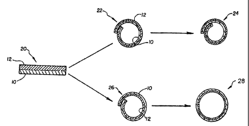
Referring now to Figures 2 and 3, two preferred alternate embodiments
of the invention will be described. If two elor,igated sheets 10 and 12 of
polymeric
material are superimposed together as shown at 20 in Figure 2, and as already
described with reference to Figure 1, to form a two layer laminate like
structure and
the sheets are then rolled into an elongate tube 21 as shown in Figure 3 in
perspective
view and in Figure 2 in end view, in two alternate forms 22 and 24 as shown in
Figure 2, respectively, two different types of clevice are produced depending
on which
layer 10 or 12 is rolled on the inside of the tubular structure. If layer 12
is to the
outside of the tube and layer 10 is to the inside: as seen at 22 in Figure 2,
the tube will
shrink in diameter as shown at 24 when layer 12 absorbs water or some other
absorbent. If, on the other hand, layer 12 is to the inside of the tube and
layer 10 is to
the outside as shown at 26 in Figure 2, the tube will expand in diameter as
shown at
28 when layer 12 absorbs water or some other absorbent.
The first type of device 22-24 of Figure 2 may find use as a sealing
device when placed around tubular conduits (not shown). In the presence of an
absorbate, the device 22-24 will shrink around such a tubular conduit in a
sealing
relationship. For example, in a medical application such a device may be
placed
around a blood vessel or other body conduit which has been opened or otherwise
requires patching and/or reinforcement on the exterior thereof. Such a device
including a water absorbent will shrink by absorbing body fluids to tightly
fit about
the vessel or other body conduit. Of course, appropriate sizing relationships
must be
taken into account but these may be readily determined depending upon the
particular
materials selected and combined for the device, etc.
CA 02275303 1999-06-17
WO 98/29148 PCTIUS97/23870
The presently most preferred embodiment of the invention involves the
application of the concept to stent devices. The device 26-28 shown in Figure
2 is
such an embodiment and will function as a seli:=expanding stent for use on the
inside
of vessels and other body conduits. Again, consideration must be given to
appropriate
sizing for any given application of such a stent. However, such stents may be
provided in any number of configurations and styles in addition to the
configuration
shown in Figures 2 and 3.
For example, a multi-layer structure according to the invention may be
formed in a helical configuration of a normal predetermined size as shown
generally at
30 in Figure 4. Since this is a stent, the absorbent layer 12 will be
positioned to the
inside. The stent may be loaded onto a suitable catheter (not shown) for
delivery as is
known in the art. To minimize its diameter during delivery, it may be tightly
wound
to a smaller delivery diameter as shown at 32 `vhen loaded onto the delivery
catheter
and covered with a removable sheath as is known in the art. Upon being
positioned in
the desired implantation location and exposed by removal of the sheath, stent
32 will
first expand to its normal size 30 and will then, upon absorbing water in
blood or
other body fluid, self-expand to a predetermine:d enlarged and expanded size
34, the
size depending on the inside diameter of the vessel in which it is to be used.
Other configurations, not limiteci to rolled tubular configurations, may
be used according to this invention for the two types of devices illustrated
in Figure 2.
For example, a perforated tubular configuration 36 is shown in Figure 5 and
another
configuration 38 is shown in Figure 6. Many other configurations and
structural types
will be readily apparent to those familiar with the graft and stent art.
For medical applications of the concept of the invention, it may be
desirable in certain instances such as stenting to make the device from all
biodegradable materials. Such materials are included in Tables 1, 2 and 3 for
example.
Although devices of more than two layers may make use of the concept
of the invention, the detailed description herein is limited to two layer
structures as
they are presently most preferred.
CA 02275303 1999-06-17
WO 98/29148 PCT/US97/23870
EXAMPLES
Bilayer membranes, one hydrophobic and one hydrophilic, were
prepared for demonstrating the application of the concept to stent usage.
Hydrophobic
layers of polycaprolactone (PCL) and layers oi' polydioxanone (PDS) were
prepared
by melting polymer on a hot plate and pressing it between two glass plates to
form a
membrane. Various thicknesses were prepareci in this manner.
Hydrophilic (water swellable) layers were prepared in composite form,
using gelatin particles in a polymer matrix of PCL or PDS. The gelatin was
powdered
in a mortar and separated by sieve to a 270 mesh size. The polymer was melted
on a
hot plate. The gelatin particles were mixed into it in 10% and 20% amounts.
Then
the melt was cast onto a warmed glass plate to form a membrane. Various
thicknesses
were prepared.
The membranes were superimposed together with tetrahydrofuran
(THF) as a solvent adhesive or with heat used for adherence.
Gelatin absorbs water at room temperature (RT) or lower and expands
in volume to become a gel without significant dissolution. At higher
temperatures
(about 70-100 C) it will dissolve into water. In a stent application, with
body
temperature being about 37 C, gelatin will absorb water and not dissolve
appreciably.
As can be seen from the above cliscussion, the two layers may make use
of the same polymer when a composite layer form is utilized. However,
different
polymers for the two layers may also be used. Also, the non-absorbing layer
may be
metallic in thin film form or in other forms. As already noted, biodegradable
or non-
biodegradable materials may be used. The layers may be superimposed together
with
adhesive or by means of heat melting. One layer can be placed on the other
layer as a
coating.
The expansion force of a bilayer formed as a coil or cylinder can be
controlled by the thickness of the layers, the loading amount of water
absorbent
material included in a composite type layer, the capability or capacity of the
particular
material for water absorbance and its expanding volume, and the type of
polymer and
its molecular weight.
CA 02275303 1999-06-17
WO 98/29148 -9- PCT/US97/23870
Specific examples are comprised of a PCL layer superimposed on a
PCL and gelatin composite layer. Several PCIL membranes were formed in various
thicknesses.
(1) 0.06 - 0.07 mm.
(2) 0.09 - O.:IO mm.
(3) 0.12 -0.13 mm.
(4) 0.13-0.14mm.
(5) 0.17 - 0.18 mm.
(6) 0.30 - 0.31 mm.
These membranes were formed by heating and melting the polymer in a glass vial
on a
hot plate at a temperature of about 70 - 80 C. The melt was placed on a flat
glass
plate. The thickness of the resultant membrane is dependent on the amount of
melt
placed on the plate and the pressure used in pressing it. A second glass plate
is placed
on top of the melt and the two plates are pressed together. The plates are
warmed
during this procedure. After the membrane has a smooth appearance and
thickness
the top plate is removed and the membrane is allowed to cool. The membrane is
then
peeled off of the remaining plate and cut to size, for example 8.0 mm. width
strips.
PCL and gelatin composite layers were also formed in various
thicknesses and gelatin loading.
10% by weight gelatin powder (particle size 270 mesh)
(1) 0.09-0.10mm.
(2) 0.13-0.14mm.
(3) 0.18 - 0.20 mm.
(4) 0.21 - 0.22 mm.
(5) 0.25 - 0.26 nun.
(6) 0.31 - 0.32 mm.
20% by weight gelatin powder (:270 mesh)
(1) 0.07-0.08mm.
(2) 0.09-0.12mm.
(3) 0.13 - 0.14 mm.
(4) 0.21 - 0.22 mm.
(5) 0.25 - 0.26 mm.
CA 02275303 1999-06-17
WO 98/29148 -10- PCTIUS97/23870
These membranes were formed by heating and melting the PCL
polymer in a vial on a hot plate at about 100 C. The gelatin was mixed into
the melt
and a quantity of the melt (the amount depending on desired thickness) was
transferred
to a warm glass plate. A second glass plate was placed on the melt and the
plates
were pressed together while heating them. After achieving a smooth appearance
the
top plate was removed and the membrane was allowed to cool on the bottom plate
after which it was peeled off and cut to size, for example 0.9 mm. width
strips.
Several rolled cylinder samples were made by superimposing various of
the membranes together. For example, a 10% gelatin and PCL membrane of 0.13
mm. thickness was combined with a PCL melr.ibrane of 0.10 mm. thickness. A 20%
gelatin and PCL membrane of 0.13 mm. thickiiess was combined with a PCL
membrane of 0.10 mm. thickness.
The two layers were combined by using a 1 % PCL solution in THF as
adhesive which was coated on one side of each membrane. They were then placed
together and held for one hour.
A heat gun was used to warm the bilayer which was then rolled to form
a closed cylinder having the configuration shown in Figure 3. Two interfitting
glass
tubes can be used to facilitate this procedure.
When these cylinders were dropped into water at room temperature,
expansion was observable within five minutes. Full expansion was observed
overnight with the 20% gelatin samples exhibiting greater expansion than the
10%
samples. This demonstrates that the amount o1' expansion is dependent on the
level of
gelatin loading.
Utilizing the following code, additional examples of stents were
prepared as above described.
PCL membrane, 9.0 mm. wide strips
D 1 0.06 - 0.07 nun. thick
D2 0.09-0.10mm. thick
Composite membrane 10% gelatin and PCL, 9.0 mm. wide strips
- X1 0.09 - 0.10 nun. thick
X2 0.13 - 0.14 mm. thick
Composite membrane 20% gelatin and PCL, 9.0mm. wide strips
CA 02275303 1999-06-17
Kl.\. 1 tJ\:L:1'.\ ~11 L':'.LIIL' uIj ::111 - 7 -:Jti = I .i . l ll . õ L_ "
,:, .)1l, 1 -= . ;I:,
JUL Illl/ VV' 1'~ IUI V lU1lU IIIU\L/ p rJIL1i1111111VU 1 llll :1V. VlL JVJ
JJVL 1. 1.
l.-
Y:? 0.13 - 0.14 mm. thick
Y5 0.25 - 0.26 mm. thick
The various combinations of these membranes are identified as follows:
D2 + X2 = DX22
D2 + Y2 = DY22
D1+Y2=DY12
When fornned into rolled cyliaders, the following expansion results
were observed in water at room temperature:
Sample Test Time (Opening in m.m.)
I hr. 2 hr. 4 hr. 1 day 2 days 5 days
DY12 9.77 11.16 11.65 11.25 11.93 11.54
7.03 8.81 8.96 9.01 9.41 9.10
DY22 4.99 7.68 8.40 8.61 9.19 9.44
3.59 5.09 6.68 6,81 7.68 7.43
DY15 3.55 6.97 7.84 8.45 9.05 9.09
4.72 5.88 8.94 9.80 9.77 9.58
DY25 3.12 4.94 6.35 6.95 7.63
4.81 6.48 8.17 9.67 9.81 9.99
DX22 3.28 4.20 4.29 4.35 4.40 4.54
3.13 3.84 4.39 4.44 4.47 4.57
Stents halving the configuration shown in Figure 4 were made utilizing
bilayer membranes 1.0 mm. wide and 44-55 mm. long of DY23 combination. The
strips were helically wound around a glass tube and oven heated at 52 - 57 C
for 15
mirnites. After cooling the stent held the glass tube size and a fixed
tubular/helical
shape.
Utilizing biodegradablc polydioxanne (PDS), melting temperature
204.7 C, as the poiymer- several bilayers were prepare&
AMENDED SHEET
CA 02275303 1999-06-17
WO 98/29148 -12- PCT/US97/23870
The polymer was melted and divided into two portions. One portion
was cast to provide membranes of 0.09-0.10 r.nm. thick. One portion was loaded
with
20% gelatin filler and cast to provide membranes 0.13-0.14 mm. thick. Bilayers
were
formed by bonding the membranes together bv heating them almost to melting or
by
pre-loading with PCL/THF and heating almost to melting.
The resultant bilayer was formed into a tubular/helical shape by cutting
the membrane into 1.8 mm. wide strips and wrapping them onto 3.0-4.0 mm.
diameter glass tubes which were inserted into larger tubes. These molds were
placed
in a 95 C oven for 15 minutes then cooled. T'hese stents when placed in water
expanded from 4.0 mm. to 5.6 mm. (OD).
All of the stents described above provided a strong holding force
believed to be appropriate for stent function.
The above Examples and disclosure are intended to be illustrative and
not exhaustive. These examples and description will suggest many variations
and
alternatives to one of ordinary skill in this art. All these alternatives and
variations
are intended to be included within the scope of the attached claims. Those
familiar
with the art may recognize other equivalents to the specific embodiments
described
herein which equivalents are also intended to be encompassed by the claims
attached
hereto.
CA 02275303 1999-06-17