Note: Descriptions are shown in the official language in which they were submitted.
CA 02277460 1999-07-13
' METHOD AND SYSTEM FOR HIGH RESOLUTION
ULTRASONIC IMAGING OF SMALL DEFECTS OR ANOMALIES
BACKGROUND OF THE INVENTION
Field of the invention
This invention relates to a method and a system for ultrasonic detection and
imaging of small defects inside or at the surface of an object by an improved
version of the Synthetic Aperture Focusing Technique, and particularly to
such method where ultrasound is generated by a laser and detected by either
a contact ultrasonic transducer or a laser interferometer.
Description of prior art
Ultrasound is a well-recognized technique for finding defects or
discontinuities in objects. Ultrasound provides not only information on the
presence of such discontinuities, but also an indication on their depth,
deduced from the arrival time of the echoes and the knowledge of the elastic
wave velocity. By scanning the surface with a piezoelectric transducer, the
object can be mapped out throughout its entire volume and the information
displayed as B-scans or C-scans. B-scans are planar cuts through the
material and indicate directly the depth of the discontinuities that are
found.
C-scans are more like views from the surface and provide depth information
by using a color or gray scale code. The coding may be associated either to
the arrival time of echoes or their amplitude. Ultrasound can also be used to
find flaws at the surface of objects by using waves propagating at their
surface (surface or Rayleigh waves) or when the object is a thin plate by
using Lamb waves.
High-resolution imaging and a better definition of the defects are obtained
by focusing ultrasound with acoustic lenses or curved transducers.
Alternatively, instead of physically focusing ultrasound inside the object (or
at
its surface), a numerical focusing technique, called Synthetic Aperture
Focusing technique (SAFT), can be advantageously used. SAFT allows a
1
CA 02277460 1999-07-13
lens with a very large effective aperture to bie realized numerically , which
leads in tum to improved resolution. SAFT has also the advantage of being
more easily applicable to objects with complex shapes, once the object
contour is known and does not require the realization of a special transducer
adapted to the shape of the object. SAFT is implemented by providing a
small ultrasonic source at the object surface with a focused transducer and
scanning this source over the surface. As shown in Figure 1 a, detection is
usually performed at the same location as generation (other schemes are
possible) resulting in a 2-D array of signals. SAFT performs a summation of
N signals shifted in time and taken from the measurement grid within a given
aperture (the synthetic aperture). The time shift of each signal is a function
of
the point where the signal is collected and the point at a depth z where the
presence of a defect is to be determined. The coherent summation increases
the SNR for defect detection by the factor . While maintaining the axial or
depth resolution ~z, the SAFT processing improves the lateral resolution ex.
It can be shown that the depth and lateral resolutions for defect sizing are
given by:
0x = a vot 0z = 2 vet ( 1 )
where v is the ultrasonic wave velocity, ~t is the ultrasonic pulse duration
and
a is the dimension of the synthetic aperture. Examples of implementation of
SAFT can be found in U.S. Patent Nos. 4,841,489 (Osaki et al.) and
5,465,722 (Fort et al.). See also the discussions in S. R. Doctor, T. E. Hall,
L.
D. Reid, "SAFT - the evolution of a signal processing technology for
ultrasonic
testing", NDT International, 19, 163 (1986) and J. A. Seydel, "Ultrasonic
synthetic aperture focusing techniques in NDT", in Research Techniques in
Nondestructive Testing Vol. 6, R. S. Sharpe, Ed. NY: Academic, 1983.
SAFT can also be advantageously applied when using lasers for the
generation and detection of ultrasound (a technique called laser-ultrasonics).
Laser-ultrasonics uses one laser with a short pulse for generation and
2
CA 02277460 1999-07-13
another one, long pulse or continuous, coupled to an optical interferometer
for
detection (see Fig. 1 b). Details about laser-t.lltrasonics can be found in C.
B.
Scruby, L. E. Drain, "Laser ultrasonics: techniques and applications", Adam
Hilger, Bristol, UK 1990 and J:-P. Monchalin, "Optical detection of
ultrasound," IEEE Trans. Ultrason. Ferroelectr. Freq. Control, 33, 485 (1986).
By relying on optics for providing the transduction of ultrasound, laser-
ultrasonics brings practical solutions for testing at a large standoff
distance,
for inspecting moving parts on production lines and inspecting in hostile
environments (for example, see J.-P. Monchalin et al., "Laser-Ultrasonics:
From the Laboratory to the Shop Floor", Advanced Performance Materials,
vol. 5, pp. 7-23, 1998). Generation of ultrasound can be pertormed either in
the ablation or thermoelastic regime. In the first case, a sufficiently strong
laser pulse provides vaporization or ablation of the surface. The recoil
effect
following material ejection off the surface produces strong longitudinal wave
emission. In the thermoelastic regime, the emission pattern depends on the
penetration of light below the surface, which could range typically from
microns to hundreds of microns in the case of polymers to practically no
penetration in the case of metals. Penetration produces a buried source and
a constraining effect that also favors longitudinal ultrasonic emission. In
all
cases, shear waves are also emitted. When tile source is small (smaller than
the acoustic wavelength) a complex pattern of emission is obtained, having in
some cases several emission lobes. It should be noted that with laser
generation, the ultrasonic source is located ~t the surface of the part and
follows automatically the contour. Regarding optical detection, the small
phase or frequency shift in the scattered light induced by the ultrasonic
surface motion is detected by an interferometric system. For applications
where the inspected part is scanned or is moving, a detection scheme that is
independent of the speckle or integrates over the whole speckle field is
needed. A passive approach based on time-delay interferometry may be
used or one can rely on an active one using nonlinear optics for wavefront
adaptation. Examples include those discussed in U.S. Patent Nos. 4,659,224
3
CA 02277460 2002-06-28
(Monchalin), 4,966,459 (Monchalin), 5,137,361 (Heon et al.), 5,131,748
(Monchalin et al.) and 5,680,212 (Blouin et al.).
For the detection of small defects, laser-ultrasonics has similar limitations
to conventional piezoelectric-based ultrasonics, caused by the wave nature of
the interrogation and diffraction effects. The spatial resolution of laser
ultrasonics depends upon the spot sizes of the generation and detection
lasers and may be inadequate for detecting small and deep flaws. The use of
a broad laser spot to produce an ultrasonic beam with little divergence gives
a
resolution essentially limited by the spot size. fn the opposite case,
focusing
the laser beam to a small laser spot yields a strongly diverging acoustic
wave,
leading also to poor resolution. Similarly to conventioC~al ultrasonics, SAFT
can be used in conjunction with laser-ultrasonics to improve resolution.
Examples of implementation can be found in US. Patent Nos. 5,615,675
(O'Donnell et al.) and 5,801,312 (Lorraine et al.). However the technique
described in these two patents presents several difficulties which limit their
applicability. A first difficulty originates from the fact that lasers have
usually
relatively low repetition rates, usually much lower than piezoelectric
transducers, which makes data acquisition time very tong, so a way to
minimize data acquisition duration while maintaining adequate resolution is
desirable. A second difficulty is the long time taken by SAFT processing with
the time domain approach used in these two patents. This approach is the
one that has been explained above. This times domain approach, while
straightforward in its principle and implementation, is not very efficient and
is
very computation intensive. A simple analysis reveals that the processing
time scales as n5 for a cubic data block, with n being the number of data
points along each axis. A third difficulty originates from the ultrasonic
pulse
produced by laser generation. This pulse has a unipolar shape so it does not
provide destructive interference at locations without defects, resulting in a
broad background around discontinuities. As indicated in the Lorraine's
patent, this problem was solved by filtering the low frequency components of
the waveform data to restore a bipolar pulse shape suitable for use with
SAFT. Considering that high spatial resolution relates to a short
4
CA 02277460 1999-07-13
pulse duration (see equation 1 ) or a large frequency bandwidth, filtering the
low frequency components does not appear to be optimal.
To solve the second difficulty just mentioned, i.e. to improve
computational efficiency, SAFT can be implerr~ented in the frequency domain
where advantage is taken of the fast Fourier transform (FFT) algorithms. Data
processing is performed in the 3-D Fourier space using the angular spectrum
approach of the scalar diffraction theory. They use of this method has been
reported by K. Mayer, R. Marklein, K. J. Langenberg and T. Kreutter, 'Three-
dimensional imaging system based on Fourie~r transform synthetic aperture
focusing technique", Ultrasonics 28, 241 (1990) and L. J. Busse, "Three-
dimensional imaging using a frequency-domain synthetic aperture focusing
technique", IEEE Transactions UFFC 39, 174 (1992). Even with improved
computation efficiency, the known frequency-domain method does not
provide a clue on how to get optimum resolution. A way to control the
aperture size is also missing, which is significant since the strength of the
ultrasonic wave and the detection sensitivity both decrease as the lateral
distance between the sampling point and the observation point increases and
adding contributions from highly offset points contributes more noise than
signal. This is straightforward in the time-domain SAFT, but not in the
frequency-domain SAFT. This control is particularly important for laser-
ultrasonics and in practice, the total opening angle of the synthetic aperture
is
expected to be limited to roughly 60°when longlitudinal waves produced
by an
ablation or constrained source mechanism ark used, which means a ~ z in
equation (1 ). When shear waves are used, the aperture should be annular.
Also, previous art related to SAFT processing does not teach how to minimize
the number of sampling points in order to minimize both data collection and
processing durations, while keeping adequate resolution.
It is an object of the present invention to provide a method that alleviates
the afore-mentioned limitations in the prior art.
SUMMARY OF THE INV~NTtON
5
CA 02277460 1999-07-13
According to the present invention there is provided a method for imaging
small defects or anomalies of a target object with a synthetic aperture
ultrasonic imaging system wherein ultrasound is generated at a plurality of
scanning positions constituting a measurement grid at the surface of the
target object, backscattered ultrasound from the measurement grid is
detected to provide an array of electrical signals which are digitally
sampled,
and a Fourier transform is performed on the array of signals in the time
domain to generate a new array of signals as a function of the temporal
frequency f. Each signal of the new array is deconvolved with a reference
signal to obtain an array of broadband deconwolved signals corresponding to
spike-like signals in the time domain, an image in real object space at depth
z
is derived from said deconvolved broadband signals, and the image is
displayed to show any defect or anomaly present at depth z.
Preferably, the image is derived by performing a Fourier transform on the
resulting new array of signals in the space domain to generate an array in 3 D
Fourier space with components as a function of the temporal frequency f and
spatial frequencies ax and ay; the 3-D Fourier space an-ay is backpropagated
from the surface of the target object to a plahe at depth z within the target
object to generate a new array in the 3-D Fourier space; the temporal
frequency components are summed over a given bandwidth to generate a
new array in 2-D Fourier space corresponding to the plane at depth z; and the
new array in 2-D Fourier space is Fourier transformed back to the real object
space corresponding to the plane at depth z.
In accordance with another aspect of the invention there is provided a system
for ultrasonic imaging small defects or anomalies of an object comprising
generating means for generating a small ultraspnic source at a given location
on the surface of the object, detecting means ftor detecting the backscattered
ultrasound and providing an electrical signal representative of the ultrasonic
motion at the detection location, means for digitizing said electric signal,
scanning means for creating a measurement grid at the surface of the object
and providing an array of said electrical signals, and processing means for
6
CA 02277460 1999-07-13
' performing a Fourier transform on the array of signals recorded at said
measurement grid, wherein the processing means deconvolves the
transformed signals with a reference signal to create a new array of signals
which are used to derive an image in real object space at depth z, and display
means are provided for displaying the subsurface image to show object
boundaries and any defect present at a depth x.
BRIEF DESCRIPTION OF THE DRAWINGS
The invention will be described in more detail, by way of example only,
with reference to the accompanying drawings, in which:-
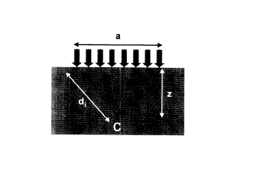
Fig. 1 a) is a schematic diagram showing, the collection of an array of
ultrasonic signals at the sample surface for its use with SAFT
known in the prior art,
Fig. 1 b) is a schematic illustration of a prior art laser-ultrasonic system.
Fig. 2a) is a schematic diagram of a laser-ultrasonic imaging system
according to one embodiment of the invention,
Fig. 2b) is a block diagram illustrating the various steps of the F-SAFT
method,
Fig. 2c) illustrates reconstruction speed of F-SAFT for a cubic data block
of size n.
Fig. 3a) illustrates a signal amplitude C-scan,
Fig. 3b) illustrates a signal amplitude profile,
Fig. 3c) illustrates a B-scan of F-SAFT processed data from a test
specimen.
Fig. 4a) illustrates profiles at the top of the 1.5 mm flat-bottom hole after
F-SAFT processing without deconvolution,
Fig. 4b) illustrates similar profiles as Fig. 4a but including deconvolution.
Fig.5illustrates influence of the aperture on the SNR after F-SAFT
reconstruction for the 0.34 mm diameter hole (solid circles) and the
0.5 mm diameter hole (open circles) pf the test specimen.
7
CA 02277460 2002-06-28
Figs. 6a) and 6b) illustrate C-scans after F-SAFT reconstruction for
two subsets taken from the original grid corresponding to a step
size of: 0.4 mm (Fig. 6a) and 0.2 mm (Fig. 6b),
Fig. 6c) repeats the C-scan in Fig. 3a obtained from the original grid with
step size of 0.1 mm.
Figs. 7a) and 7b) represent C-scans after F-SAFT reconstruction for the
two subsets of step size 0.4 mm (Fig. 7a) and 0.2 mm (Fig. 7b),
and including spatial interpolation to provide information at an
interval of 0.1 mm,
Fig. 7c) repeats the C-scan in Fig. 3a obtained from the original grid with
step size 0.1 mm.
DETAILED DESCRIPTION OF THE PREFERRED
EMBODIMENTS OF THE INVENTION
Referring now to Figure 2a the system according to one preferred
embodiment comprises a laser ultrasonic system collecting a 2-D array of
ultrasonic signals at the surface of the sample for imaging small defects at
its
inside. The generation laser is a pulsed laser source and the laser
interferometer for detecting backscattered ultrasound comprises a long pulse
laser or continuous laser coupled to an optical interferometer. The two laser
beams for generation and detection, are focused at the same location onto
the surface in a manner similar to the arrangement shown in Figure 1 b. A
scanning system is employed for generating and detecting ultrasound at a
plurality of scanning positions constituting the measurement grid at the
surface of the object. In the present embodiment, the array of signals is
obtained by scanning the beams on the sample surface with steered mirrors.
Alternatively, the sample could be moved using an X-Y translation table.
Preferably, the measurement grid has constant step sizes, 8x and 8y, in both
the x and y directions. The backscattered signal detected at each scanning
position on the measurement grid is digitally sampled by a digitizer, which
uses oversampling to better estimate each temporal frequency component,
and stored into a memory, thus providing an array of waveform data. A
8
CA 02277460 2002-06-28
processor unit comprising a single or several processors is used for SAFT
reconstruction and generation of the subsurface images using the method
described below. The images are displayed by a display unit.
Other embodiments where the two laser beams are offset from, each
other or are not simultaneously scanned can also be easily implemented.
With prior knowledge of the part shape, this laser-ultrasonic imaging system
can also be applied to samples with non-planar front surfaces.
The method of the present invention (called below F-SAFT) is based on
processing data in Fourier space and provides significant improvements over
previous frequency-domain SAFT methods. -fhe method can be used
advantageously with either a conventional piezoelectric-based ultrasonic
system or the preferred laser-ultrasonic imaging system described above.
Figure 2b shows an example of implementation of the proposed method and
the various steps it comprises.
Starting from the acoustic field S(x,y,z = o,t) at the sample surface of the
measurement grid, a 3-D Fourier transformation is first performed with
respect to variables (x, y, t) into a 3-D Fourier space represented by
variables
(ax, ay, f ) . Physically, these transformations can be seen as if the
acoustic
field of frequency f is represented by a superposition of plane waves at
different angles with spatial frequencies ax, 6Y . Then, the transformed field
S(aX,6Y,z=o,f) is backpropagated to any depth z using the expression:
S(6X~6y~z~f) = S(aX~6y~o~f) expC~2~iz (2f/v)2 -6X -aY~
with ~ corresponding to the sign of f and summed over the temporal
frequencies as follows:
~(6x'~y'z) - ~'S' (Crx,6y,z,f)
feS2
9
CA 02277460 1999-07-13
where s2 is the selected frequency bandwidth including negative components.
Finally, after to addition of zero values, an inverse 2-D Fourier
transformation
of E(ax,ay,z) with respect to variables (aX,ay) is performed, yielding the
space domain function ~(x,y,z). The back propagation and summing of
temporal frequency components is perfom~ed for a plurality of planes
corresponding to different depths within the object.
A flaw is present at position x, y and z if the function E at this point
exhibits a
peak. It is worth mentioning that windowing and smoothing techniques can
be applied either in the time or frequency domain prior to any of the above
Fourier transformations. For a cubic data block, the algorithm is found to
scale as n4 for computing equations (2) and ~3), and as n3log n for Fourier
transformations using a FFT (Fast Fourier Transform) algorithm. Therefore,
the frequency-domain SAFT method is inherently faster than conventional
time domain SAFT for moderately large values pf n.
Figure 2c shows the actual reconstruction speed obtained for different
values of n on a PC Pentium II 400 MHz. The speed is defined here as the
reciprocal of processing time (in min) divided by the size of the
corresponding
data block (in Mbytes, 16 bits per data point). The performance on this
machine appears optimal for the size n = 128, with a reconstruction speed of
7 MB/min. It is worth mentioning that for inspection over a large area, the
reconstruction can be made on many overlapping data blocks.
Although computationally efficient, the known frequency-domain method
can be significantly improved to get very high resolution images of small
defects and overcome the limitations mentioned when using laser-ultrasonics.
Figure 3 shows the results from the improved version of frequency-domain
SAFT (called F-SAFT) on laser-ultrasonic data obtained on a test specimen
made from an aluminum block 7 mm thick. All the key features of the
proposed F-SAFT method discussed next were used, except spatial
interpolation. To simulate buried flaws, four flit-bottom holes, 10 mm apart,
approximately 2 mm deep and of diameter 1.~, 1.0, 0.5 and 0.34 mm were
CA 02277460 1999-07-13
drilled on the back surface of a sample object. In the present embodiment,
the generation laser was a short pulse (~ 5 ns) Q-switched Nd:YAG laser
operating on its fourth harmonic. Generation of ultrasound was performed in
the ablation regime. A single mode, highly staible, long pulse (50 Ns) Nd:YAG
laser operated on its fundamental wavelengith of 1.064 ~,m was used for
detection of ultrasound. The light of the detection laser scattered off the
surface sample was sent to a confocal Fabry-Perot interferometer operated in
reflection mode (length of 1 m and mirror reflectivities of 89 %). The
frequency bandwidth of the system extended from 1 to 35 MHz. The two laser
beams were focused onto the surface of the specimen at about the same
location. The generation and detection spot sizes were 0.1 mm and 0.3 mm,
respectively. The step size of the scan was 0.1 mm and the inspected area
was 12.5 x 45 mm. For each node of the measurement grid, an ultrasonic
signal was collected, digitized and stored in the computer memory. The
whole block of data was then processed by the F-SAFT method.
Figures 3a, 3b and 3c show an amplitude C-scan and a B-scan of the
processed data after reconstruction at depths' from 3 to 7.5 mm with a step
size of 0.05 mm. To evaluate the SNR, a profile extracted from the C-scan
along a line crossing the holes is also shown irn figure 3. The laser-
ultrasonic
F-SAFT imaging provides very good detection of all the 5-mm deep defects,
with a SNR ranging from 24 dB for the 0.34-mm hole to 33 dB for the 1.5-mm
hole. The lateral resolution of the flat-bottom holes is also found to be
excellent. The apparent diameters (width at half maximum of the profile) of
the 0.34-mm and 1.5-mm diameter holes are 0.4 mm and 1.6 mm,
respectively.
A first feature indicated in Figure 2b of the, exemplary F-SAFT method is
the temporal deconvolution of the ultrasonic ~raveform data. The effect of
deconvolution is to replace the ultrasonic pulses by spike-like pulses.
Therefore, the ultrasonic pulse duration, fit, is reduced and consequently
both
depth and lateral resolutions are improved, as indicated by equation (1 ).
Preferably, Wiener deconvolution is used in the method and is well adapted to
11
CA 02277460 1999-07-13
the frequency domain calculations. Each signal S(x,y,f) at the sample surface
of the measurement grid is substituted by the function H(x,y,gy, given by:
_ R(~* e2R~~
H(x,y,f)=S(x,y,f) z a (4)
(R(f ~ + xZIR(f ~rnax
where R(f) is the reference pulse, ~ is the time delay required to shift the
reference pulse at time t=0 and x is a user-specified constant used to
quantify
noise in the signal. Notice that the symbol G*" denotes complex conjugate and
the subscript "max" denotes the maximum value. The value of user-
specified constant x, usually between 0.02 and 0.2, has to be carefully
chosen to avoid the deterioration of the SNI~t while trying to improve the
resolution. The reference pulse used in the data given as example is the
backwall reflection echo. Note that the brackef in equation (4) is a vector
that
has to be evaluated only once so this operal~ion has only minor effects on
processing time. Two other benefits of debonvolution are an improved
precision in the location of defects due to a more symmetric pulse shape of
the deconvolved signal and an accurate determination of the time origin when
the reference pulse is selected from the data block, due to the self
referencing of the deconvolution at t=0. The performance of this feature of
the
F-SAFT method is shown in Figures 4a and 4b. As shown in these figures,
the flat top shape of the 1.5 mm hole is revealed by the method (Figure 4b)
whereas standard frequency-domain SAFT does not provide similar
information (Figure 4a).
A second feature of the F-SAFT method its the control of the frequency
bandwidth of reconstruction. With respect t4 equation (3), the frequency
bandwidth control can be achieved by limiting tie sum to the actual frequency
bandwidth of the ultrasonic inspection system,'~2. Embedded in the F-SAFT
processing, this control is very effective and make any pre-filtering of the
ultrasonic signals unnecessary, as it would bye required using time-domain
SAFT. This frequency bandwidth control results in a reduced processing time
12
CA 02277460 1999-07-13
and an increase of the SNR, since noise contribution from high frequency
components without ultrasonic information are removed. The advantage of
oversampling during acquisition to better estimate each frequency
components within S2 is also preserved.
A third feature of the F-SAFT method consists in controlling the aperture
in the frequency domain. As already mentioned, the transformation to the 3-D
Fourier domain can be seen as representing tile acoustic field of frequency f
by a superposition of plane waves at different angles with spatial components
ax,ay . The direction cosines of the wave components are related to the
orientation of the wave vector k with respect to the coordinate axes.
Therefore, the angle of a given plane wave with respect to the z axis is given
by:
kz - (2f /v)2 -aX -av
k 2f ~v = cosec (5)
It will be observed that for simultaneously scanned generation and
detection, the acoustic propagation distances are doubled and this is
accounted for by the factor 2 appearing with the wave vector in equations (2)
and (5). Since it is expected that there is littlie signal at large values of
6Z,
because of a decrease of wave emission and detection sensitivity at these
values, one should limit 8Z to a maximum value 8 to avoid the addition of
noise, i.e. cos AZ > cos 8. It then follows a condition on the temporal
frequencies f to be included in the summation of equation (3):
2 2
~ ax + ar < f < f",~
2 sin8
where fm~ is the maximum frequency in the bandwidth s2. For increasing
values of the spatial components ax, ay , the mumber of temporal frequency
components f used in the summation is progressively reduced. Note that for
13
CA 02277460 1999-07-13
a total opening angle of 180°, eq. (6) automatically excludes the
contribution
from evanescent waves by restricting the argument in the exponential
function of eq. (2) to be imaginary. The influernce of the aperture on SNR for
the 0.34-mm and 0.5-holes is shown in Fig. 5 for the test specimen described
above. The SNR rapidly increases with aperture size, reaches a maximum at
around 60° and then progressively decreases, by at least 6 dB, as a
result of
including components that contribute more noise than coherent signal. In
addition, the processing time is reduced since Less data points are included
in
the SAFT processing by proper reduction of the aperture.
A benefit of the aperture control feature of the method is a reduction in
spatial sampling requirement. The choice of scanning step size 8 (either 8x or
8y) is a compromise between the smallest detectable defect and the time for
inspection and processing. A standard practice is to apply the criterion, 8 <
~min/2, where min is the smallest acoustic wavelength which is present or
required. With this criterion, propagating waved over an aperture of 28 =
180°
as well as evanescent waves are included in tMe direct and formal use of the
angular spectrum approach. However in the application to the F-SAFT
method, the control of the aperture limits the angular range over which the
plane waves in the expansion contribute. Therefore, the spatial frequencies
are limited to a value amp (either in the x or y direction) given by eq. (6):
sin a
amex . (7)
~ min
where ,min is the effective smallest wavelength equal to v I 2fm~. Following
Nyquist, adequate sampling requires a scanming step size 8 smaller than
1 /( 2a"~,~ ):
min
~2 CsinA)
14
CA 02277460 1999-07-13
Note that equation (8) reduces to the standard criterion, 8 < ~,m,~/2, for a
total
opening angle of 28 = 180°. For a nearl~r optimal aperture of
60°, the
sampling requirement is 8 < 7~min~ ~~esponding to a reduction factor of 2 (or
4
for the whole measurement grid) compared td the standard criterion, which
allows to decrease both inspection and proicessing times. If the effective
smallest wavelength is set equal to the size o~ the smallest detectable defect
w, then using an optimal aperture, the relation 8 ~ ~.mln ~ w is obtained. For
the test specimen described above, this corresponds to a sampling interval of
0.34 mm instead of the 0.1 mm used. To dembnstrate this point, two subsets
of signals were taken from the original grid of 126 x 451 signals and were
processed by F-SAFT. Figure 6a shows the C~scan obtained with a subset of
31 x 112 signals, corresponding to a step of 0.4 mm to be compared with
original C-scan obtained with a 0.1 mm step, shown in Fig. 3a and
reproduced in Fig. 6c. As expected from the previous argument, the 0.34-
mm hole is detected but with a poor SNR of 1'1 dB. Figure 6b shows the C
scan obtained with a subset of 63 x 225 signals corresponding to a step of
0.2 mm. In this case, the smallest 0.34-mm hole is well observed, with a SNR
of 21 dB close to the 24 dB obtained with the original 0.1 mm step. The
resolution and even SNR may be improved by the interpolation feature as
discussed next.
The proposed interpolation feature is well adapted to the frequency
domain and directly embedded in the F-SAFT imethod. For each depth z, the
algorithm simply consists in padding the function E(aX,ay,z) with zeros prior
to its inverse 2-D Fourier transformation with FFT, yielding the function
E(x,y,z) at intermediate points. This approach is demonstrated with the two
subsets discussed above and taken from the original grid.
Figs. 7a and 7b show the C-scans obtained respectively from the 31 x
112 grid (step size of 0.4 mm) and from the ~3 x 225 grid (step size of 0.2
mm), both including interpolation to provide information at intervals of 0.1
mm.
When compared to corresponding Figs. 6a and 6b, the small 0.34-mm hole
indeed appears more clearly using spatial interpolation. However, for the
coarser grid in fig. 7a, the size of the 0.34-mm hole appears overestimated
CA 02277460 1999-07-13
with reference to the 0.5-mm hole, indicative pf an imperfect reconstruction.
Another indication of critical sampling is the increase of SNR from 11 dB to
16
dB when including interpolation, a SNR which remains unchanged with 21 dB
using the finer grid having 0.2 mm step size. Then using a coarser grid, the
SNR can be improved by using an interpolation technique since a peak
associated with the presence of a defect in the function E(x,y,z) may be more
accurately resolved, with possibly a SNR gain by a factor of 2 corresponding
to 6 dB. While less accurate than using the original grid (reproduced in Fig.
7c), the C-scan in figure 7a appears quite acceptable, especially if one takes
into account the reduction by 16 in the inspection time and nearly by 5 in the
processing time.
The proposed method and system can ~Iso be readily applied to the
detection and imaging of surface defects using either ultrasonic plate (Lamb)
waves or surface (Rayleigh) waves. This approach has already been
described in the US. Patent No. 5,760,904 (L4rraine and Hewes), but using
the less efficient time-domain SAFT. In this case, only a single line scan is
required with the system, collecting ultrasonic signals at the sample surface
along direction x and imaging small surface defects at various distances from
the scanning line along direction z. If the ultrasionic waves are dispersive,
i.e.
the phase velocity is frequency dependent, this is easily accounted for in F-
SAFT by attributing for each temporal frequerncy during backpropagation to
distance z the velocity value at this frequency in eq. (2).
The proposed method and system can alsio be readily applied to the so-
called opto-acoustic imaging used in the medial field to detect anomalies in
tissue with enhanced contrast and resolution: The effect is the same as
mentioned above for the generation of ultrasound. The interest of the
technique and its enhanced contrast originaties from the variation of light
penetration between various tissues, partipularly between sound and
cancerous ones. Ultrasound is detected by a contact transducer which is
either piezoelectric or based on fiber optics. The approach combines the
advantages of optics (better contrast between tissues) and ultrasonics (less
scattering during propagation). Details about Qpto-acoustics can be found in
16
CA 02277460 1999-07-13
R.A. Kruger et al., "Photoacoustic ultrasound-reconstruction tomography",
Med. Phys. 22, 1605-1609 (1995), A.A. Oraev~ky et al., "Laser opto-acoustic
tomography for medical diagnostics: Principle" R.A Lieberman et al., eds.,
Proc. SPIE conf. on biomedical sensing, imaging and tracking technologies I,
2676, SPIE Press, Bellingham, WA, pp. 22-31 (1996). However, for a
clinically relevant and viable system, improvennents in 2-D detection have to
take place using both multiplexed array transducers and a suitable image
reconstruction method. For examples, see the recent works from R.O
Esenaliev et al., "Laser Optoacoustic imaging 'for breast cancer diagnostics:
Limit of detection and comparison with X-rays and ultrasound imaging", B.
Chance and R.R. Alfano, eds., Proc. SPIE cohf. on optical tomography and
spectroscopy of tissue: Theory, instrumentation, model and human studies II
2979, SPIE Press, Bellingham, WA, pp. 71-82 (1997) and C.G.A Hoelen et
al., 'Three-dimensional photoacoustic imaging of blood vessels in tissue",
Opt. Lett. 28 (1998). Therefore, this application can benefit from using the
above described F-SAFT method and system.
Of course, numerous other than described above embodiments of the
method and system may be envisaged without departing from the spirit and
scope of the invention.
17