Note: Descriptions are shown in the official language in which they were submitted.
CA 02277841 1999-07-09
WO 98131304 PCT/US97121948
-1-
Field Of The Invention
The present invention relates to the field of multilayer composite
tubes and, in particular, stems for use in surgical procedures. The present
invention provides a multilayer composite tubular structure for use as a
surgical
stent which is visible on a fluoroscope, yet does not mask anatomical
structures.
The present invention also provides a multilayer stmt which does not
delaminate
upon deployment and expansion.
Background Of The Invention
Stems are utilized in a wide range of surgical procedures. For
example, stents are used to repair and support injured tissue during the
subsequent healing process. The stmt is delivered to the target site and
expanded to several times its original diameter until the stem contacts the
surrounding tissue. This process is known as initial expansion of the stmt.
Next, the stmt is further expanded to imbed the stmt into the walls of the
surrounding anatomical structure, for example, an artery. This process is
known as imbedding. The process of initial expansion and imbedding is known
as deployment. Once the stent is expanded, it takes on a permanent set.
An example of a common surgical procedure involving the use
of a stmt is the placement of a stmt within.a coronary artery after removal of
plaque from within the artery. In that case, the stent is used to support the
vessel which has been blocked by atherosclerotic plaque.
Stems are also used in surgical procedures involving the ureters
or the urethra. For example, in prostate surgery, stems are used to hold open
tracts of the urinary system.
The placement and positioning of the stmt is crucial during all
surgical procedures such as those described above. Thus, various procedures
have been developed which allow a physician or surgeon to view a stmt in situ
CA 02277841 1999-07-09
WO 98/31304 PCT/CTS97/21948
-2-
for proper placement. The most common of these procedures is to view the
stmt using a fluoroscope.
Prior art stems are commonly made of a single material such as
stainless steel, tantalum, or NitinolTM, with the most common material used
being stainless steel. A major disadvantage of a stainless steel stmt is that
it is
transparent to a fluoroscope. Therefore, using a stainless steel stent
requires
that opaque dyes be injected in the bloodstream to make the stmt visible to
the
surgeon for positioning and deployment. These dyes dissipate very quickly,
making the stent visible for only a brief period of time. Thus, procedures
involving stainless steel stems and the use of dye to view the stmt require
rapid
placement and deployment of the stmt. Additionally, the lack of visibility of
the stmt makes it extremely difficult, if not impossible, to verify that the
stmt
has not changed location over time.
Tantalum is a radiopaque material widely used in stems. A solid
tantalum stmt must have a minimum thickness to be useful in deployment and
function. The required thickness of the solid tantalum stmt results in a high
luminosity on a fluoroscope, and in turn causes several problems. One is that
the fluoroscope image produced by tantalum stents is so luminous that it
obliterates the detail of the stmt pattern and the detail at the stent/vessel
interface. Because it is impossible to view the stent/vessel interface,
accurate
placement of the stem at vessel bifurcations is tedious. Moreover, the fact
that
the stent structure cannot be accurately observed makes more difficult the
determination of whether vascular conditions, such as restenosis, have
occurred
at the stmt site.
Because stents are used at various anatomical sites, it is necessary
to vary the thickness, and therefore the strength of the stmt, to compensate
for
anatomical variations. For example, stems may be used at anatomical sites
having varying degrees of muscle mass. A multilayer stmt would have to be
able to compensate for variations in muscle mass, for example, with different
thicknesses of the stmt layers. Additionally, it would also be desirable to be
able to vary the luminosity of a stent to compensate for different anatomical
variations and varying degrees of muscle mass.
Surgical stents undergo tremendous plastic deformation during
CA 02277841 1999-07-09
WO 98/31304 PCT/US97/21948
-3-
deployment. In a multilayer stmt, the layers of the stmt must not delaminate
or separate. Any delamination of a muitilayer stent could expose rough or
jagged edges, leading to thrombosis, and direct anatomical injury, including
the
tearing of vessels.
It is a usual surgical practice to deploy, expand, and imbed
surgical stems by means of a balloon unit. The use of these balloon units is
well known in the stmt art. The pressure employed to expand a surgical stem
using a ballon unit is critical. Stents that require a higher pressure to
expand
run an increased risk of balloon rupture, which could lead to an embolism.
There is, therefore, a need for a stent which is visible on a
fluoroscope, but does not mask anatomical structures.
There is also a need for a multilayer stmt which can expand
without delaminating.
There is also a need for a multilayer stmt whose strength can be
varied by varying its thickness to accommodate different anatomical
structures.
There is further a need for a multilayer stent where its luminosity
on a fluoroscope can be varied by changing the thickness of the layers of the
stem to accommodate different anatomical structures.
There is still further the need for a multilayer stent where the
stmt is expanded at a lower pressure and which provides a decreased risk of
balloon rupture upon deployment.
Summar~Of The Invention
The present invention is directed to a structure that satisfies the
need for a stent which is both visible on a fluoroscope, yet does not obstruct
the
details of the stmt itself or the anatomical structures surrounding the stmt.
The
present invention provides a truly innovative and effective solution to these
needs.
A structure having features of the present invention comprises a
multilayer composite stmt. One layer of the stmt is formed from a radiopaque
material. Another layer of the stent is formed from a biocompatible material.
The combination of these layers produces a structure which is both
biocompatible and visible on a fluoroscope, yet does not mask the stent
structure
or the surrounding anatomical structure.
CA 02277841 1999-07-09
WO 98/31304 PCTIUS97/21948
-4-
The present invention also provides for a method of making a
multilayer composite structure having properties of the stmt described above.
A multilayer composite structure which is the subject of the present invention
is created by reducing, such as by tube drawing, swaging, or deep drawing
multiple tubes or strip as a composite and applying heat treatment to cause
diffusion bonding of the layers. This results in a ductile structure that
permits
large deformation without delamination between the biocompatible and
radiopaque layers.
Description Of The Drawings
For the purpose of illustrating the invention, there is shown in the
drawings a form which is presently preferred; it being understood, however,
that this invention is not limited to the precise arrangements and
instrumentalities shown.
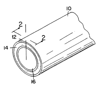
Fig. 1 shows a partial perspective view of a stent according to the
present invention, before etching or machining.
Fig. 2 shows a cross sectional view taken along line 2-2 in Fig.
1, showing the layers of the scent.
Fig. 3 is an electron micrograph of a portion of an actual stmt
in accordance with the present invention, after etching or machining.
Fig. 4 shows a fragmentary partial cutaway view of a stmt
according to another embodiment of the present invention, before etching or
machining.
Fig. 5 shows a cross sectional view taken along line 5-5 in Fig.
4, showing the layers of the stent.
Fig. 6 shows a cross sectional view of another embodiment of a
stmt according to the present invention, showing a metallic interleaf between
layers.
Figs. 7-9 show a partially cross sectional view of one process of
making a stmt according to the present invention.
Fig. 10 shows a cross sectional view of a stmt according to the
present invention the present invention in place in an artery.
Fig. 11 shows a side view of a stmt according to the present
invention prior to expansion.
CA 02277841 1999-07-09
WO 98131304 PCT/US97J21948
-5-
Detailed Description Of the Invention
Referring now to the drawings, wherein like numerals indicate
like elements, there is shown in Figs. 1 and 2 a stmt 10 in accordance with
the
present invention. In the preferred embodiment of the present invention shown
in Fig. 1, 2, and 3, a stent 10 comprises a multilayer composite tubular
structure having an outer layer 12 of biocompatible material, a middle layer
14
of radiopaque material, and an inner layer 16 also of biocompatible material.
In the preferred embodiment, the biocompatible material used for the outer
layer
12 and inner layer 16 is stainless steel, while the radiopaque middle layer 14
is
tantalum. These layers are bonded together using processes described below.
The stent 10 illustrated in Fig. 3 has been etched and machined to produce a
particular pattern.
It is recognized that other biocompatible materials can be
substituted for stainless steel. It is also recognized that the radiopaque
layer 14
is not limited to tantalum, and other materials, including but not limited to
gold,
platinum, and alloys of those materials, may be used without departing from
the
present invention.
In a stainless-tantalum-stainless stem such as stent 10, the
tantalum layer 14 must have sufficient thickness to provide a sharp and clear
image on a fluoroscope (not pictured). The thickness of the radiopaque
tantalum layer 14 can be varied to provide the optimum luminescence for
applications where the stmt is delivered to vessels close to the surface,
where
low luminescence is required, or procedures deeper within tissue such as
muscle, where a higher luminescence, and thus a thicker radiopaque tantalum
layer 14, is required. The thickness of the radiopaque tantalum layer 14 can
also be varied to accommodate for anatomical sites having tissue density
variations, as these locations require different stmt luminescence.
In another embodiment of the present invention, as shown in Fig.
4 and 5, thin radiopaque layers 24 can be deposited on the outer surface 26 of
a first tube 28 of biocompatible material through electroplating, or through
vapor, chemical, or other film deposition techniques. In Figs. 4 and 5, the
relative thickness of the radiopaque layer 24 is exaggerated for clarity. The
coated first tube 28 is then metallurgically bonded to a second tube 30,
forming
a diffusion layer 18, described below, between the radiopaque layer 24 and the
second tube 30. In another embodiment, the radiopaque layer 24 is deposited
CA 02277841 1999-07-09
WO 98/31304 PCT/US97/21948
-6-
on the inner surface of a first tube (not shown), which is then placed
coaxially
around and metallurgically bonded to a second tube.
The radiopaque layer 14 or 24 can therefore be from 1 % to 95 %
of the wall thickness of the stmt 10. Thus, the luminosity can be varied
widely
to accommodate for different tissue variations.
It is contemplated that additional layers can be added to the stmt
of the present invention to form stems of various compositions. For example,
a five layer stmt could be formed, having alternating layers of stainless
steel
and tantalum, with the stainless steel layers being the outer-most and inner-
most
layers.
Due to the large plastic deformation of the stmt 10 which must
occur during deployment and expansion, as previously described, the bond
formed between the stainless steel and tantalum layers is critical to the
proper
function of the structure. A mechanical bond is not adequate to meet the
1 ~ requirements of stems according to of the present invention. Instead, a
metallurgical bond, where diffusion of the material elements takes place, is
the
desired approach. This metallurgical bond is formed through the application of
pressure and heat to the materials, as described below.
As illustrated in Fig. 2, concurrent with the formation of a
?0 metallurgical bond between the layers of the structure, a diffusion layer
18 is
also created at the interface between adjacent layers 12 and 14, or 14 and 16.
'The characteristics of these diffusion layers 18 can be significantly
affected and
controlled by the proper heat treatment cycle, resulting in either a desired
ductile diffusion layer 18, or an undesirable brittle intermetallic layer.
25 Heat treatment, temperature, and time relationships control the
rates of transfer of the diffusing elements, resulting in diffusion layers 18
of
different elemental composition and thickness. Heat treatment cycles must be
optimized for different material combinations, so that the diffusion layer 18
maintains the ductility necessary for deployment. The diffusion layer 18 must
30 also be of minimal thickness necessary to ensure bond integrity and
ductility to
prevent delamination during stmt 10 expansion upon employment.
In another embodiment of the present invention, as shown in Fig.
6, the bonding together of materials which may not be readily compatible, and
which would result in an undesirable brittle intermetallic layer being formed,
35 can be accomplished through the use of a metallic interleaf 20. This
interleaf
CA 02277841 1999-07-09
WO 98/31304 PCT/US97I21948
20 acts to control both the diffusion rate and the elements which are
transported
across the diffusion region 22. For example, a gold interleaf can be used to
facilitate the formation of a proper diffusion layer 18.
A multilayer composite tubular structure having features of the
present invention can be formed using the following processes.
Examt~le #1
As previously noted, the diffusion layer 18 between the stainless
steel and tantalum is developed and controlled by the proper application of
pressure and thermal treatment. This is well known in the art of diffusion
bonding. In one example of a process that may be used in forming the present
invention, an outer tube made of a biocompatible material, a middle tube made
of radio-opaque material, and an inner tube made of a biocompatible, are
arranged coaxially, and reduced simultaneously, such as by swaging or tube
drawing, for example. The process of tube reduction in this fashion is well
known in the art. An example of a composite tubular structure arranged in this
manner is depicted in Figs. 1 and 3.
In the multilayer composite tubular structure according to the
invention, pressure at the interface between layers is developed as a result
of the
residual radial clamping stresses left in the tube after the composite drawing
operation. Those skilled in the art of tube drawing will recognize that
increasing the area reduction and varying the percentage of area reduction
versus wall reduction will either increase or decrease the magnitude of this
residual stress within certain limits.
In one example of this process, an outer tube of stainless steel,
a middle tube of tantalum, and an inner tube of stainless steel, are arranged
as
described above to form the composite structure. To facilitate proper bonding
between the layers, a residual clamping stress of at least 50 p.s.i. at the
interface should be developed. In addition, annealing of the composite tube
must be done within a limited range of time and temperatures. The lower limit
of this time and temperature range should be at least 1550°F for at
least six
minutes. The upper limit should be not greater than 1850°F for a period
no
greater than 15 minutes. Annealing of the composite tube within these
temperature ranges will provide a diffusion layer 18 of minimal thickness and
elemental composition to -maintain the required ductility to permit deployment
CA 02277841 1999-07-09
WO 98131304 PCT/US97/21948
_g_
and expansion at lower pressures, and still prevent delamination during
expansion.
Example #2
In another process of forming the present invention, a radiopaque
material layer is deposited on the outer surface of an inner tube of
biocompatible material. This arrangement is shown in Fig. 4. The radiopaque
material may deposited through a cladding process such as vapor deposition,
electroplating, spraying, or similar processes. An outer tube of biocompatible
material is then placed around the clad inner tube.
The composite tubes are then drawn together and progressively
reduced until the desired residual clamping stress is attained, as described
above. The tubes are then heat treated as described above, forming a diffusion
bond between the radiopaque layer and the inner surface of the outer tube.
It is recognized that this same process can be accomplished by
depositing the radiopaque layer on the inner surface of the outer tube, and
bonding that combination to the outer surface of the inner tube.
Example #3
Another process which can be used to form a multiple composite
tubular structure involves the use of a metallic interleaf 20. This is
illustrated
in Fig. 6. The interleaf 20 is placed between the biocompatible and radio-
opaque layers. and acts to control the diffusion rate and/or the diffusing
atoms
which are transported across the diffusion region. The multiple tubes are then
drawn together and progressively reduced until the desired residual clamping
stress is attained, as described above. The tubes are then heat treated as
described above, forming a diffusion bond between the radiopaque material and
the biocompatible materials, which is facilitated by the interleaf 20.
Example #4
Yet another process which can be used to form a multiple
composite tubular structure according to the present invention involves the
use
of deep drawing from a multilayer strip 42. The process of deep drawing is
well known in the art of tube formation.
In one embodiment, as shown in Figs. 7, 8, and 9, the multilayer
strip 42 has a top layer 54 of stainless steel, a middle layer 56 of
radiopaque
material, and a bottom layer 58 of stainless steel. This strip 42 is prepared
by
metallurgically bonding the layers prior to the deep drawing process. In the
CA 02277841 1999-07-09
WO 98/31304 PCT/US97121948
-9-
course of the deep drawing process, as shown in Fig 8, the strip 42 is placed
over a die 44, and the strip 42 is forced into the die 44, such as by a punch
46.
A tube 48 having a closed end 50 of a certain wall thickness is formed in the
die 44. This process is repeated using a series of dies of progressively
decreasing diameter until a multilayer tube 48 is formed having the desired
diameter and wall thickness. For certain material combinations, it may be
necessary to perform intermediate heat treatments, as described above, between
the progressive drawing operations. Once a tube of desired thickness and
dimensions has been formed, the closed end 50 and the curved edges 52 of the
tube 48 are cut off, as illustrated in Fig. 9. Then, the tube is heat treated
as
described above until the proper intermetallic bond is formed between the
layers.
An advantage of the composite structure described herein is that,
for a wide range of radiopaque layer thickness and materials, the stmt 10 can
be expanded at a lower applied force, which translates into lower deployment
pressures than those required for a solid stainless stent of the same wall
thickness. This is due to the lower modulus of the composite structure. The
lower modulus is caused by the lower yield strength of the radiopaque material
andlor a contribution from a lower strain hardening coefficient.
In a typical surgical procedure using a stmt, the stmt is initially
expanded with a low pressure balloon (not shown) until the stmt walls contact
a vessel to be held open, for example, the walls of an artery. A stmt 10 is
illustrated in Fig. 10 contacting the walls of an artery 11. The low pressure
balloon is then withdrawn, a high pressure balloon (not shown] is inserted,
and
the stmt 10 is further expanded by the high pressure balloon into the artery
wall
11. This second expansion is referred to as imbedding. The expansion and
imbedding pressures of a composite stmt having a 1:1:1 ratio of stainless
steel:tantalum:stainless steel, as compared to a common stainless steel stmt
of
the same thickness, are detailed in Table I below:
TABLE I
Stainless Multilayer Composite
steel
I:1:1
(stainlessaantalumatainless)
Deployment 4Atmospheres3.5 Atmospheres
Expansion (Imbedding)8 Atmospheres7 Atmospheres
CA 02277841 1999-07-09
WO 98/31304 PCT/US97/21948
-10-
The ability to use lower expansion and imbedding pressures for
the composite stmt, as compared to the solid stainless steel stmt, affects its
safety and reliability. Because less pressure is needed to expand and imbed
the
composite stmt of the present invention, there is less risk of tearing or
otherwise injuring surrounding anatomical features, tissues, or vessels.
Moreover, a lower stress is exerted on the balloon unit, thus decreasing the
risk
of balloon rupture and the concomitant risk of an embolism.
The present invention may be embodied in other specific forms
without departing from the spirit or essential attributes thereof and,
accordingly,
reference should be made to the appended claims, rather than to the foregoing
specification, as indicating the scope of the invention.