Note: Descriptions are shown in the official language in which they were submitted.
CA 02278035 1999-07-15
WO 98/31417 PCT/US98100966
LONGITUDINALLY ALIGNED DUAL RESERVOIR ACCESS PORT
BACKGROUND OF THE INVENTION
I . Figld of the Invention:
The present invention relates to implantable access ports and, more
specifically,
to dual reservoir vascular access ports.
2. Backeround Art:
Implantable vascular access systems are used extensively in the medical field
to
facilitate the performance of recurrent therapeutic tasks inside the body of a
patient. Such
a vascular access system generally comprises an implantable access port
attached to the
proximal end of a catheter. A typical access port comprises a needle-
impenetrable housing
having a fluid reservoir that is sealed by a needle penetrable septum. The
access port also
includes an outlet stem which projects from the housing and endures a fluid
passageway
that communicates with the fluid reservoir. The outlet stem is used to couple
the catheter
to the housing.
In use, the entirety of the catheter with the access port attached thereto is
implanted at an appropriate location in the tissue of the patient. The distal
end of the
catheter is disposed at a predetermined location where therapeutic activity is
to be
affected. Once the vascular access system is implanted, a needle attached to a
syringe can
selectively access the reservoir of the access port by penetrating the skin of
the
implantation site for the access port and then the septum. The needle and
syringe can then
be used to deliver to the reservoir medication or other fluids, which then
travel through
the outlet stem and catheter to be disposed in the body at the distal end of
the catheter.
Alternatively, the syringe can be used in aspiration to withdraw bodily fluids
located at the
distal end of the catheter.
Many access ports in use contain a single fluid reservoir through which
medication
can be delivered to a patient. Such structures can, however, be severely
limiting to
medical practitioners. For example, it is often desirable to deliver
medicaments to
separate locations within the body of a patient, or to deliver such
medicaments as are
incompatible, if mixed together in a single fluid reservoir before being
infused into the
body of the patient. Alternatively, it may be desirable to simultaneously
deliver the
SUBSTITUTE SHEET (RULE 26'~
CA 02278035 1999-07-15
WO 98/31417 PCT/US98/00966
-2-
medication to a patient and withdraw blood samples for testing. Such plural
functions
cannot be performed through the use of a single reservoir access port.
Instead, toward that end, dual reservoir access ports have been developed.
Dual
reservoir access ports typically comprise a housing having a pair of separate
reservoirs
formed therein. Each of the fluid reservoirs has a corresponding access
aperture that is
sealed by a discrete septum plug. The septum plugs are secured in place by a
jacket that
engages the housing.
An outlet stem housing a pair of fluid passageways projects from the exterior
of
the housing, usually at a between the pair of fluid reservoirs. This causes
the fluid
reservoirs to be spaced relatively far apart, increasing the overall size of
the access port.
Another problem with conventional dual reservoir access ports relates to the
method by which access ports are implanted. To do so, a subcutaneous pocket is
first
created to receive and house the access port. This is done by making an
incision in the
skin of the patient at the intended implantation site for the access port. The
access port
is then inserted beneath the skin through the incision.
The outlet stem of the access port must, however, always be received within
the
pocket last, after the rest of the access port. Only by so doing can a
catheter be coupled
to the outlet stem of the access port. The outlet stems of most dual reservoir
access ports
project from a longitudinal side of the housing, between the fluid reservoirs.
To implant
such access ports, an incision must be made at the implantation site that is
at least as long
as the access port. Only in this way can the access port be received through
the incision
followed by the outlet stem. The longer the incision, the longer the healing
process before
the access port can be freely utilized and the gt~eater the potential for
infection or other
complications.
An additional shortcoming of the conventional dual reservoir access ports is
their
inability to be coupled with a variety of different types of catheters. The
catheter is
typically attached to the access port by sliding the stem of the access port
within the lumen
of the catheter. A locking sleeve is then slid over the catheter having the
stem received
therein producing an effective seal between the outlet stem and the catheter.
Conventional dual reservoir access ports can only be coupled with a
corresponding
catheter made of a defined material having a defined lumen configuration. Such
a
r,
CA 02278035 1999-07-15
WO 98/31417 PCT/US98/00966
-3-
limitation precludes the medical practitioner from using a desired catheter
based on the
patient's needs rather than the access port used.
OBJECTS AND SUMMARY OF THE INVENTION
It is an object of the present invention to provide an improved dual reservoir
vascular access port.
Another object of the present invention is to provide such an access port
having
the same fluid capacity as a conventional dual reservoir access port, but
being smaller in
size than such an access port.
An additional object of the present invention is to provide a dual reservoir
access
port that can be implanted in small tissue areas in the body of a patient, and
can also be
used with small children and infants.
Yet another object of the present invention is to provide a dual reservoir
access
port that can be implanted subcutaneously through a small incision in the skin
of the
patient.
Finally, it is an object of the present invention to provide a dual reservoir
access
port that can be selectively attached to dual lumen catheters made of
different materials
or having different lumen configurations.
Additional objects and advantages of the invention will be set forth in the
description which follows, and in part will be obvious from the description,
or may be
learned by the practice of the invention. The objects and advantages of the
invention may
be realized and obtained by means of the instruments and combinations
particularly
pointed out in the appended claims.
To achieve the foregoing objects, and in accordance with the invention as
embodied and broadly described herein, an implantable delivery system is
provided. The
system includes an implantable dual reservoir access port. The access port
comprises a
housing having a floor with an encircling sidewall upstanding therefrom.
A proximal fluid reservoir is formed in a proximal end of the housing and
communicates with the exterior of the housing through a proximal access
aperture.
Likewise, a distal fluid reservoir is formed in a distal end of the housing
and
communicating with the exterior of the housing through a distal access
aperture.
CA 02278035 1999-07-15
WO 98/31417 PCT/US98/00966 ..
-4-
The housing fi,~rther includes a first fluid flow pathway formed in the
sidewall of
the housing. The first fluid flow pathway extends between the proximal fluid
reservoir and
a predetermined outlet location at the distal end of the housing. In addition,
a second fluid
flow pathway extends between the distal fluid reservoir and the predetermined
outlet
location.
Projecting from the housing at the outlet location is an outlet stem having a
free
distal end. The outlet stem includes a first and second outlet prong at the
distal end of the
outlet stem. Each of the first and second outlet prongs has an exterior
surface and an
inner face. The opposing inner faces define a slot in the distal end of the
outlet stem.
The outlet stem also includes a first fluid duct and a second fluid duct The
first
fluid duct extends longitudinally through the first outlet prong of the outlet
stem to the
first fluid flow pathway. The second fluid duct extends longitudinally through
the second
outlet prong of the outlet stem to the second fluid flow pathway.
A needle-penetrable compound septum overlies the proximal access aperture and
the distal access aperture. The compound septum comprises a septum web having
a top
surface and a bottom surface. Located on the bottom surface is a pair of plugs
that are
received within a corresponding one of the access apertures. Located on the
top surface
of the septum web is a pair of needle target domes that are individually
aligned with a
corresponding one of the plugs.
The access port also includes a clamp configured to compress and secure the
septum to the housing. The clamp includes a shoe having an interior surface
configured
to receive the floor of the housing. The clamp also includes a cap having a
pair of
apertures formed therethrough. The cap is configured to receive the compound
septum
and the housing, so that the needle target domes are received within the
apertures of the
cap. The cap then engages the shoe, compressing the septum against the housing
and
sealing the access apertures of the housing.
The fluid delivery system also includes a dual lumen catheter that is
selectively
attached to the outlet stem. The dual lumen catheter can be made of
polyurethane or
silicone and can have either D-shaped or trapezoidal shaped lumens.
Finally, a locking sleeve is used to secure the dual lumen catheter to the
outlet
stem. The locking sleeve comprises a proximal end, a distal end, and an
interior surface
r~
CA 02278035 1999-07-15
WO 98/31417 PCT/US98/00966
-5-
defining a passageway longitudinally extending therethrough. The interior
surface of the
locking sleeve radially, inwardly compresses a portion of the body wall of the
dual lumen
- catheter against a portion of the exterior surface of the stem. This is
accomplished when
the distal end of each of the first and second outlet prongs is individually
received in a
corresponding one of the lumens of the dual-lumen catheter and the dual lumen
catheter
with the stem received therein is positioned within the passageway of the
locking sleeve.
The inventive fluid deiivery system has a variety of unique benefits. For
example,
by positioning the outlet stem at the distal end of the housing, the
reservoirs are
longitudinally aligned with respect to the outlet stem. In such a
configuration, the access
port can be advanced longitudinally into a subcutaneous pocket at the
implantation site.
As a result, the incision at the implantation site need only be so long as to
receive the
width, rather than the length, of the access port.
Furthermore, the use of a unitary compound septum permits the fluid reservoirs
of the device to be positioned close together, decreasing the size of the
access port. By
minimizing the size of the access port, the access port can be implanted in
previously non-
conventional implantation sites in the arm of an adult patient, or even be
used with 'small
children and infants.
BRIEF DESCRIPTION OF THE DRAWINGS
In order that the manner in which the above-recited and other advantages and
objects of the invention are obtained, a more particular description of the
invention briefly
described above will be rendered by reference to a specific embodiment thereof
which is
in the appended drawings. Understanding that these drawings depict only a
typical
embodiment of the invention and are not therefore to be considered to be
limiting of its
scope, the invention will be described and explained with additional
specificity and detail
through the use of the accompanying drawings in which:
Figure 1 is a perspective view of a longitudinally aligned dual reservoir
access port
implanted within the upper arm of a patient and having a catheter attached
thereto with
an opposing end fed within the vascular system of the patient;
Figure 2 is an enlarged perspective view of the access port shown in Figure 1;
Figure 3 is a perspective view of the access port shown in Figure 2 in a
CA 02278035 1999-07-15
WO 98/31417 PCT/US98100966 ..
-6-
disassembled condition;
Figures 4A-4C are longitudinal cross-sectional plan views showing sequential
steps
in the manufacture of the outlet stem for the access port shown in Figure 3 ;
Figure 4D is an elevated distal end view of the outlet stem shown in Figure
4C;
Figure 4E is an elevated proximal end view of the outlet stem shown in Figure
4C;
Figure 4F is an elevated proximal end view of an alternative embodiment of the
outlet stem shown in Figure 4C;
Figure S is a cross-sectional side view of a subassembly of the access port
shown
in Figure 2 taken along section line S-5 shown in Figure 3;
Figure 6 is a cross-sectional top view of the subassembly of the access port
shown
in Figure 5 taken along section line 6-6 shown therein;
Figure 6A is a cross-sectional top view of an alternative embodiment of the
subassembly of the access port shown in Figure 6 wherein the fluid flow
pathway
extending from the proximal reservoir to the stem is formed in the sidewall of
the basket;
Figure 6B is a cross-sectional top view of an alternative embodiment of the
subassembly shown in Figure 6 wherein the basket has been replaced by a C-
shaped
sleeve;
Figure 6C is a cross-sectional side view of an alternative embodiment of the
subassembly of the access port shown in Figure 6 wherein the basket extends
through the
casing;
Figure 7 is a cross-sectional side view of the subassembly of the access port
shown
in Figure 2 taken along section line 7-7 shown therein;
Figure 8 is a perspective view of the compound septum in a compressed state as
shown in Figure 7;
Figure 9 is a cross-sectional top view of plugs projecting from the compound
septum shown in Figure 8 and taken along section line 9-9 shown therein;
Figure 10 is a cross-sectional top view of the connecting web of the compound
septum shown in Figure 8 and taken along section line 10- I 0 as shown
therein;
Figure I1 is a cross-sectional side view of the compound septum shown in
Figure 8 taken along section line 11-1 1 as shown therein;
Figure 12 is a schematic representation of the flow of the compound septum as
it
r~
CA 02278035 1999-07-15
WO 98/31417 PCT/US98/00966 - --
is compressed into the state shown in Figure 7;
Figure 13 is a cross-sectional side view of an alternative embodiment of the
access
port shown in Figure 3 wherein the basket has been replaced by an annular
sleeve not
having a floor;
Figure 14 is a perspective view of a multiple-catheter connection system where
one of three unique dual lumen catheters is selectively attached to the access
port shown
in Figure 3 by a locking sleeve;
Figure 15 is a cross-sectional top view of a selected catheter in preparation
for
attachment to the stem of the access port as shown in Figure 14; and
Figure 16 is a cross-sectional top view of the catheter attached to the access
port
shown in Figure 15.
DETAILED DESCRIPTION OF THE ILLUSTRATED EMBODIMENTS
Referring to Figure 1, a patient 10 is shown having a chest 12 with an arm 14
raised therefrom. A vein 16 extends through arm 14 into chest 12.
Subcutaneously
implanted at the inside of arm 14 is one embodiment of a longitudinally
aligned dual
reservoir access port 18 incorporating features of the present invention. Also
implanted
with access port 18 is an elongated, pliable dual lumen catheter 20 that is
coupled thereto.
Catheter 20 enters vein 16 in arm 14 and extends therein into chest 12 of
patient 10.
Catheter 20 is shown as having a proximal end 22 that is attached in fluid
communication with access port 18. Catheter 20 has a distal end 24 that has
been
advanced within vein 16 to a desired location within chest 12.
The needle of a syringe 23 can be used to transcutaneously deliver medication
to
either of the fluid reservoirs in access port 18. In turn, the medication
travels through
catheter 20 and is eventually discharged within the body of patient 10 at
distal end 24 of
catheter 20.
Alternatively, the needle syringe 23 can be received within access port 18 to
aspirate fluid samples. Bodily fluids located at distal end 24 of catheter 20
are drawn into
and though catheter 20 to access port 18, and then into syringe 23.
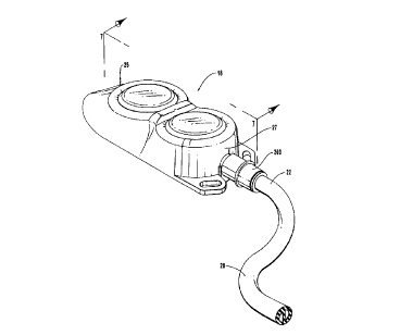
Depicted in Figure 2 is an enlarged perspective view of access port 18 shown
in
Figure 1. Access port 18 is shown as having a proximal end 25 and an opposing
distal
CA 02278035 1999-07-15
WO 98/31417 PCT/US98/00966
_g_
end 27. As will be discussed later in greater detail, proximal end 22 of
catheter 20 is
attached in fluid communication to distal end 27 of access port I 8 by a
locking sleeve 260.
To better appreciate the internal structure of access port 18, reference is
now made to
Figure 3, which shows an exploded view of the elements of access port I 8.
As depicted in Figure 3, access port 18 includes a rigid housing 26 which
comprises a casing 28 and an open-topped basket 32. Casing 28 has a proximal
fluid
reservoir 30 and an opposing distal cup 3 I . Distal cup 31 is configured to
receive open-
topped basket 32 which in turn defines a distal fluid reservoir 34. An outlet
stem 36 is
connected to housing 26 to enable fluid coupling to both proximal fluid
reservoir 30 and
distal fluid reservoir 34.
Access port 18 further includes a compound septum 38 that is secured against
housing 26 to cover proximal fluid reservoir 30 and distal fluid reservoir 34.
Compound
septum 38 is formed of a elastomeric, needle-penetrable material which enables
selective
needle access to either of reservoirs 30 or 34. A clamp 40, comprising a cap
42 and a
shoe 44, is used to compress and secure compound septum 38 against housing 26.
Outlet stem 36 has a proximal end 46 and an opposing distal end 48. Formed at
distal end 48 are a pair of adjacent prongs 50 and 52. As depicted in Figure
4A, outlet
stem 18 is manufactured from a substantially cylindrical stem body 54 having a
longitudinal axis L, a proximal end face 56, a distal end face 58, and an
encircling exterior
surface 60 extending therebetween Using a lathe or other comparable process,
an annular
groove 62 is formed around exterior surface 60 between proximal end 46 and
distal
end 48. Likewise, distal end 48 is tapered so as to have a substantially
frustoconical
shape.
Next, a pair of parallel large pilot holes 64 and 66 are drilled from proximal
end
face 56 a distance into stem body 54. Likewise, a pair of smaller parallel
pilot holes 68
and 70 are drilled from distal end face 58 so as to meet large pilot holes 64
and 66. The
intersection of large pilot holes 64 and 66 with smaller pilot holes 68 and 70
forms
passageways 69 and 71 that extend longitudinally through stem body 54.
A conventional wire electrostatic discharge machine (hereinafter "a wire EDM")
is then used to reconfigure the interior surface of passageways 69 and 71. To
do so, an
EDM wire 72 of a wire EDM is positioned extending through each of passageways
69
CA 02278035 1999-07-15
WO 98/31417 PCT/US98/00966
-9-
and 71. Each EDM wire 72 has a proximal end 74 positioned proximal of proximal
end
face 56. Likewise, each EDM wire 72 has a distal end 76 that is positioned
distal of distal
end face 58. Each EDM wire 72 is attached to a corresponding wire EDM that
supplies
each EDM wire 72 with a high frequency alternating current, but enables each
EDM
S wire 72 to have five dimensions of free motion. In this position, proximal
end 74 of each
EDM wire 72 is moved independently in a circular orientation in a plane
perpendicular to
longitudinal axis L of stem body 54. Distal end 76 of each EDM wire 72 is,
however,
independently moved in a D-shaped orientation in a plane perpendicular to
longitudinal
axis L of stem body 54.
The movement of each EDM wire 72 electrostatically removes the portion of stem
body 54 contacting each EDM wire 72. As depicted in Figure 4B, the movement of
each
EDM wire 72 converts passageway 69 into a first fluid duct 78 and converts
passageway 71 into a second fluid duct 80. Fluid ducts 78 and 80 are
adjacently disposed
and extend longitudinally between proximal end face 56 and distal end face 58
of stem
body 54. Each of fluid ducts 78 and 80 has a smooth interior surface 82 that
gradually
and continuously transitions in cross-section from a circular shape at
proximal end face 56
to a D-shape at distal end face 58. The smoothness of interior surface 82
helps prevent
damage to any living cells in the fluid flowing therethrough.
As best seen in Figure 4D, fluid ducts 78 and 80 at distal end 48 of outlet
stem 36
have a substantially D-shape transverse cross-section. In contrast, as
depicted in
Figure 4E, fluid ducts 78 and 80 at proximal end 46 have a substantially
circular transverse
crass-section.
Although the cross-sectional shape of fluid ducts 78 and 80 changes along the
length thereof, the transverse cross-sectional area of fluid ducts 78 and 80
are relatively
constant at all points between proximal end face 56 and distal end face 58.
This constant
cross-sectional area, optimizes the flow rate achievable through fluid ducts
78 and 80.
By having the D-shaped cross-section the same area as the circular cross-
section,
it necessarily follows that the D-shaped cross-section has a minimum inside
diameter that
is smaller than the inside diameter of the circular cross-section.
Accordingly, pilot
holes 68 and 70 which are subsequently formed into the D-shaped cross-section
are drilled
having a smaller inner diameter than the inner diameter of pilot holes 64 and
66. Although
CA 02278035 1999-07-15
WO 98/31417 PCT/US98/00966
- 10-
pilot holes 64 and 66 could be formed having the same small inner diameter as
pilot
holes 68 and 70, pilot holes 64 and 66 having a larger inner diameter minimize
the amount
of material that is subsequently removed by EDM wire 72 in forming the
circular cross-
sectional areas.
S As depicted in Figure 4C, once fluid ducts 78 and 80 are completed, prongs
50
and S2 are created by cutting a slot 81 between fluid ducts 78 and 80 at
distal end 48
Slot 81 may be formed using an EDM wire. Prongs SO and SZ are shown as having
a
proximal end 84, a distal end 86, and a curved, exterior surface 88 that
extends
therebetween.
Exterior surface 88 is shown as comprising a locking barb 92 positioned at
distal
end 86 of each of prongs SO and S2. Each locking barb 92 flares radially out
from distal
end face S8 to an outside ridge 94. Exterior surface 88 also includes a sloped
transition
shoulder 96 formed at proximal end 84. A cylindrical portion 98 extends
between
transition shoulder 96 and locking barb 92. Prongs SO and S2 have an inner
face 90 that
is substantially flat. Opposing inner faces 90 define slot 81. A portion 91 of
each inner
face 90 flares radially outwardly at distal end 86 to facilitate the
attachment to dual lumen
catheter 20.
Outlet stem 36 is further shown in Figure 4C as comprising a cylindrical
barrel 102
having a distal end face 103 and an opposing proximal end face 104. Proximal
ends 84
of prongs SO and S2 are formed on distal end face 103 of barrel 102 so that
prongs SO
and S2 project therefrom.
As shown in Figures 4C and 4E, proximal end face 104 of barrel 102 comprises
a semicircular projecting end face IOS and an adjacent recessed end face 112.
Recessed
end face 112 is also semicircular and is formed distal of projecting end face
IOS. Second
2S fluid duct 80 extends through recessed end face 112. Extending from
projecting end
face lOS is a cylindrical boss 106 having first fluid duct 78 extending
therethrough.
Boss 106 has a cylindrical sidewall 108 and an annular end face 110 that is
slightly curved.
In the illustrated embodiment proximal end 46 of fluid ducts 78 and 80 have a
circular cross-section, as shown in Figure 4E. In alternative embodiments,
however, fluid
ducts 78 and 80 can have any desired cross-sectional configuration. By way of
example
and not by limitation, as depicted in Figure 4F proximal end 114 of fluid duct
78 and
r. ,
CA 02278035 1999-07-15
WO 98/31417 PCT/US98/00966
-I1-
proximal end 1 15 of fluid duct 80 each have a D-shaped cross-section.
Although boss 106 in Figure 4E has a circular transverse cross-section, boss
106
can also be formed in a variety of alternative configurations. By way of
example and not
by limitation, depicted in Figure 4F is a boss 116 having a substantially D-
shaped
transverse cross-section.
Outlet stem 36 is made from a metal, such as stainless steel or titanium.
Alternatively, it is conceivable that outlet stem 36 can be formed from other
materials,
such as plastics, ceramics, or composites.
Returning to Figure 3, casing 28 comprises a top surface 118, a floor 120, and
an
annular sidewa.ll 122 extending therebetween. Casing 28 has a figure-eight
configuration
that extends longitudinally between a proximal end 124 and an opposing distal
end 126
Casing 28 could alternatively be circular or rectangular.
Formed in top surface 118 of casing 28 is a septum web recess 129. Septum web
recess 129 is in part defined by a horizontally disposed seat 128 and a
vertically oriented
interior sidewall 130. Interior sidewall 130 encircles seat 128 and extends
between
seat 128 and top surface 118 of casing 28.
Counter bored within seat 128 at proximal end 124 of casing 28 is proximal
fluid
reservoir 30. As best shown in Figure 5, seat 128 defines a space that
includes a proximal
access aperture 131 for proximal fluid reservoir 30 and a distal access
aperture 133 for
distal fluid reservoir 34. Proximal fluid reservoir 30 is further defined by a
cylindrical
sidewall 132 and a floor 134.
Referring to Figure 3, counter bored within seat I28 at distal end 126 of
casing 28
is distal cup 31 defined by a cylindrical sidewall 136 and a floor 138.
Extending between
proximal fluid reservoir 30 and distal cup 31 is a dividing wall 140.
A channel 142 is recessed within sidewall 136 of distal cup 3 I . Channel i 42
extends from dividing wall 140 to a predetermined outlet location 141 at
distal end 126
of casing 28. A transfer port 144 extends through dividing wall 140 to effect
fluid
communication between proximal fluid reservoir 30 and channel 142. An annular
outlet
port 146 extends through sidewall 122 of casing 28 at outlet location 141 to
effect fluid
communication between distal cup 31, channel 142, and the exterior of casing
28.
Open-topped basket 32 comprises an annular sleeve 148 having an interior
CA 02278035 1999-07-15
WO 98/31417 PCT/US98/00966 .--
-12-
surface I 50 and an exterior surface 152. Sleeve 148 extends between a top
edge 154 and
a floor 156. Top edge 154 defines distal access aperture 133 that is encircled
by
sleeve 148. Extending between interior surface 1 SO and exterior surface 152
is an annular
entry port 160. Alternatively, open-topped basket 32 can assume a smooth bowl-
shaped
interior rather than the cylindrical interior formed by sleeve 148 and floor
156.
Basket 32 and casing 28 are made from a metal such as stainless steel or
titanium,
but in the alternative could conceivably be formed from other materials, such
as plastic,
ceramics, or composites.
As depicted in Figure 5, distal cup 31 is configured to receive open-topped
basket 32 such that top edge 154 of basket 32 becomes flush with and forms
part of
seat 128. Top edge 154 is welded or otherwise secured to seat 128 in a fluid
tight
connection.
Basket 32 is disposed within distal cup 31 with entry port 160 of basket 32 in
alignment with outlet port 146 of casing 28. As best depicted in Figure 6,
barrel 102 of
outlet stem 36 is as a result received within outlet port 146, while boss 106
is
simultaneously received within entry port 160 of basket 32. Conventional
titanium
welding techniques or other securing processes are used to provide a fluid
seal between
barrel 102 of stem 36 and casing 28. Similar techniques are used to provide a
fluid seal
between boss 106 of outlet stem 36 and basket 32.
Boss 106 and barrel 102 each of outlet stem 36 can be formed in a variety of
alternative configurations as long as entry port 160 of basket 32 is
configured to
complementarily receive boss 106. Outlet port 146 of casing 28 must similarly
be
configured to complementarily receive barrel 102.- It has been found to be
easiest to align
and secure boss 106 into entry port 160 and barrel 102 into outlet port 146,
if entry
port 160 of basket 32 and boss 106 of outlet stem 36 have complementary
circular
configurations, and outlet port 146 of casing 28 and barrel 102 of outlet stem
36 have
complementary circular configurations.
With casing 28, basket 32, and outlet stem 36 interconnected as discussed
above,
discrete fluid communication is provided between proximal fluid reservoir 30
and outlet
stem 36 and also between distal fluid reservoir 34 and outlet stem 36. As
depicted by
arrows A in Figure 6, fluid in proximal fluid reservoir 30 flows through
transfer port 144
CA 02278035 1999-07-15
WO 98/31417 PCT/US98/00966 .-
-13-
in dividing wall 140 and enters a fluid flow pathway 162 to travel around the
perimeter
of basket 32. Fluid flow pathway 162 is completely formed only upon the
insertion of
basket 32 into distal cup 31. Fluid flow pathway 162 is thus bounded by
channel 142 of
casing 28 and exterior surface 152 of basket 32. From fluid flow pathway I 62,
the fluid
enters proximal end 46 of first fluid duet 78 and is subsequently discharged
from distal
end 48 of first fluid duct 78.
As depicted by arrow B in Figure 6, fluid in distal fluid reservoir 34 flows
directly
into proximal end 46 of second fluid duct 80 and therethrough for discharge at
distal
end 48 thereof.
Figure 6A illustrates an alternative embodiment of a housing 26A. As shown in
Figure 6A, although fluid flow pathway 162 is still bounded between casing 28
and
basket 32, channel 142 is shown as being recessed within exterior surface 152
of basket 32
rather than being recessed in annular sidewall 136 of distal cup 31.
In another alternative embodiment of a housing 26B shown in Figure 6B,
basket 32 has been replaced by a C-shaped sleeve 164 housing an interior
surface 163, an
exterior surface 165, and an outside edge 167. An entry port 160 extends
between
interior surface 163 and exterior surface 165. C-shaped sleeve 164 is large
enough to
cover transfer port 144, channel 142, and outlet port 146. As such, discrete
fluid
communication is still enabled from proximal fluid reservoir 30 to first fluid
duct 78 and
between distal fluid reservoir 34 and second fluid duct 80.
In housing 26B, distal fluid reservoir 34 is defined by the area bounded by
interior
surface 163 of C-shaped sleeve 164 and the portion of distal cup 31 not
covered by C-
shaped sleeve 164. To prevent fluid communication between proximal fluid
reservoir 30
and distal fluid reservoir 34, it may also be necessary to weld or otherwise
seal all points
of outside edge 167 of C-shaped sleeve 164 to distal cup 31.
Another alternative embodiment of a housing 26C is depicted in Figure 6C. As
shown therein, casing 28 is configured such that an open-topped basket 169
having a
floor 175 is received in a passageway 166 formed without interruption through
casing 28
from top surface I I 8 to and through floor 120. Floor 175 of basket 169 is
then flush with
floor 120 of casing 28 and visible from the exterior of casing 28. Basket 169
otherwise
has the same elements as basket 32 of the earlier embodiments and interacts
with
CA 02278035 1999-07-15
WO 98/31417 PCT/I1598/00966
- 14-
casing 28 in substantially the same way as previously discussed with regard to
basket 32.
Figure 13 also shows a final alternative embodiment of a housing 26D. As
disclosed therein, basket 32 has been replaced by an annular sleeve 246 having
a top
edge 247 and a bottom edge 249. Sleeve 246 also has an interior surface 248,
an exterior
surface 250, and an entry port 160 extending therebetween.
Sleeve 246 is used in the same way as discussed above with regard to basket
32.
The distinction between basket 32 and annular sleeve 246 is that annular
sleeve 246 does
not include floor 156. As a result, bottom edge 249 of annular sleeve 246
should be
welded or otherwise sealed to distal cup 31 to prevent fluid communication
between
proximal fluid reservoir 30 and distal fluid reservoir 34.
In one embodiment of the present invention, fluid coupling means are provided
for
efFecting a sealed fluid communication between second fluid duct 80 and distal
fluid
reservoir 34. By way of example and not by limitation, one embodiment of the
fluid
coupling means includes boss 106 of outlet stem 36, that is connected with
distal fluid
reservoir 34 through outlet port 146 of casing 28 in combination with entry
port 160 of
basket 32.
Alternative embodiments of such a fluid coupling means could include the
alternative configurations of boss 106 previously discussed with regard to
Figure 4F, and
the alternative structures through which entry port 160 is formed in the
embodiments of
housings previously discussed with regard to Figures 6A-6C and Figure 13. A
discrete
passageway could be formed through casing 28 and basket 32 in use for
communicatinu
with second fluid duct 80. Alternatively) boss 106 could project from basket 3
2 and pass
through casing 28 to connect with second fluid~duct 80 in outlet stem 36.
The present invention also provides fluid conduit means between casing 28 and
a
sleeve for placing proximal fluid reservoir 30 in fluid communication with
first fluid
duct 78 in outlet stem 36. By way of example and not by limitation, one
example of such
a fluid conduit means includes fluid flow pathway 162 formed by channel 142
recessed
within sidewall 136 of distal cup 31 and basket 32 received within distal cup
31, as
previously discussed with regard to Figures 3, S, and 6. Transfer port 144
allows fluid to
flow from proximal fluid reservoir 30 to fluid flow pathway 162. Outlet port
146 allows
fluid to flow between fluid flow pathway 162 and outlet stem 36.
CA 02278035 1999-07-15
WO 98/31417 PCT/LTS98100966
-15-
Alternative embodiments of such a fluid conduit means include a channel 142
recessed in an exterior surface of basket 32, as in Figure 6A or the various
structures
shown in Figures 6B, 6C, and 13 as being received in distal cup 31 so as to
close
channel 142.
The present invention also includes delivery means for effecting discrete
fluid
communication between each of fluid reservoirs 30 and 34 and the exterior of
housing 26.
By way of example and not by limitation, such a delivery means includes each
of the
disclosed structures and alternative embodiments of a fluid coupling means and
also each
of the disclosed structures and alternative embodiments of a fluid conduit
means.
Referring again to Figures 3 and 5, compound septum 3 8 is used for covering
and
sealing proximal access aperture 131 and distal access aperture 133. As shown
in
Figures 7 and 8, compound septum 38 includes a planar septum web 168 having a
top
surface 170, a bottom surface 172, and a side surface 174 extending
therebetween.
Compound septum 38 also extends longitudinally between a proximal end 171 and
a distal
end 173. As will be discussed later in greater detail, connecting web 168 is
configured to
be snugly received within septum web recess 129 of casing 28.
Projecting from bottom surface I72 of connecting web 168 is a cylindrical
proximal plug 176 and a cylindrical distal plug 178. As best seen in Figure 7,
each of
proximal plug 176 and distal plug 178 has a bottom face 180 with an annular
sidewall 182
extending between bottom surface 172 of connecting web 168 and bottom face
180.
Referring to Figure 8, compound septum 3 8 also comprises a proximal needle
target dome 184 and a distal needle target dome 186 each projecting from top
surface 170
of connecting web 168. Proximal needle target dome 184 is aligned with
proximal
plug 176. Likewise, distal needle target dome 186 is aligned with distal plug
178.
Compound septum 38 is preferably made of a compressible and resiliently
deformable material that, for example) enables a needle to pass through
proximal needle
target dome I84, connecting web 168, and proximal plug 176 into proximal fluid
reservoir 30. In one embodiment, compound septum 3 8 is made from a medical
grade
silicone. In alternative embodiments, compound septum 38 can also be made from
other
medical grade elastomers or rubbers.
Returning again to Figure 3, clamp 40 is used to secure compound septum 38 to
CA 02278035 1999-07-15
WO 98/31417 PCT/ITS98/00966
- 16-
housing 26. Clamp 40 includes a cap 42 and a shoe 44. Cap 42 is shown as
having a top
surface 188, a bottom surface 190, and a sidewall 192 that extends
therebetween. Cap 42
also has a proximal end 198 and an opposing distal end 200.
Extending through top surface 188 of cap 42 at proximal end 198 is a proximal
aperture 202. Likewise, extending through top surface 188 at distal end 200 is
a distal
aperture 204. Apertures 202 and 204 are configured to receive needle target
domes I 84
and 186 of compound septum 38, respectively.
Located on top surface 188 and extending between proximal aperture 202 and
distal aperture 204 is a bridge 206. Upstanding on bridge 206 is a tactile
locating
ridge 208 the position of which can be ascertained by a medical practitioner
through
palpating the skin of patient 10 at the implantation site for access port 18.
Once the
position of tactile locating ridge 208 is thusly ascertained, the position of
proximal fluid
reservoir 30 and of distal fluid reservoir 34 on either side thereof is also
automatically
determined. Tactile locating ridge 208 thus facilitates accurate targeting of
the needle of
syringe 23 into either proximal fluid reservoir 30 or distal fluid reservoir
34.
Projecting proximally outward at proximal end 198 of cap 42 is a sloped nose
210.
Sloped nose 210 enables easy insertion of access port 18 into a subcutaneous
implantation
pocket in the skin of patient 10. Extending through sidewall 192 of cap 42 at
distal
end 200 is a stem slot 212. Stem slot 212 is configured to receive outlet stem
36 when
housing 26 is disposed in clamp 40.
Radially projecting out from sidewall 192 of cap 42 at opposing sides of stem
slot 212 are tabs 214 that each have a suture slot 216 formed therethrough.
Suture
slots 216 are used to suturing access port 18~ in an implantation pocket in
the skin of
patient 10.
Cap 42 includes an interior surface 194 that defines a receiving chamber 196.
As
better seen in Figure 7, an annular attachment groove 217 is formed on
interior
surface 194 at bottom surface 190.
Interior surface 194 defines a cavity 218 at proximal end 198 of cap 42.
Cavity 218 is formed to minimize material costs and also to form cap 42 having
a
relatively constant thickness at all points about cap 42. Cap 42 is preferably
molded from
a medical grade plastic. In alternative embodiments, cap 42 can also be made
from metals,
CA 02278035 1999-07-15
WO 98/31417 PCT/US98/00966
-17-
ceramics, or composites. By forming cap 42 with a relatively constant material
thickness,
deformation resulting from difFerent rates of cooling of the molded plastic or
other
materials is minimized.
Shoe 44 is shown in Figure 3 as comprising an attachment ridge 219 having a
top
edge 220 and a bottom edge 222. Attachment ridge 219 also includes an exterior
surface 226 and an interior surface 228. Radially extending inward from
interior
surface 228 at bottom edge 222 is a lip 230. Shoe 44 also has a proximal end
232 and an
opposing distal end 234. Projecting from exterior surface 226 at proximal end
232 is a
tongue 236. Positioned at distal end 234 is a grooved stem carriage 238.
One of the novel features of the present invention is the configuration of
compound septum 38 and the use thereof to cover and seal proximal access
aperture 131
and distal access aperture 133.
A single compound septum 38 is used to cover and seal both of reservoirs 30
and 34. Compound septum 3 8 not only prevents the passage of fluid between
proximal
fluid reservoir 30 and distal fluid reservoir 34, but also prevents the
transfer of fluids
between the exterior of access port 18 and either of reservoirs 30 or 34. By
using a single
compound septum 38 rather than two individual septums, reservoirs 30 and 34
can be
positioned closer together in housing 26, thereby decreasing the overall size
of access
port 18.
Compound septum 38 is configured to have desired properties when compound
septum 38 is incorporated into access port 18. For example, when a needle is
passed
through compound septum 38 into one of reservoirs 30 or 34) septum 38
effectively seals
around the exterior of the needle to prevent the passage of fluids between
septum 3 8 and
the exterior of the needle. Septum 38 is also configured to exhibit a
predetermined
amount of needle retention force, once septum 38 is installed in access port
18. Needle
retention refers force to the tendency of septum 38 to resist removal of a
penetrating
needle.
The sealing effectiveness and needle retention of septum 38 is in part related
to the
amount of radial compressive force applied to septum 38 by housing 26 and
clamp 40.
In general, the greater the compressive force applied to septum 3 8, the
higher the sealing
effectiveness and needle retention.
CA 02278035 1999-07-15
WO 98/31417 PCT/US98/00966 .-
-18-
The compressive force on septum 38, however, must not be so great that
inserting
the needle through compound septum 3 8 results in the needle coring septum 3
8. Coring
occurs where the stress on the installed septum 3 8 is so high that when the
needle is
inserted into septum 38, a portion of septum 38 is forced inside the needle.
The portion
of septum 3 8 forced inside the needle is then severed from septum 3 8,
resulting in a small
passage extending through septum 3 8. Continued coring eventually results in
septum
failure.
Access port 18 is generally subcutaneously placed making it difl-icult to
direct
exactly where the needle will pass through septum 38. Compound septum 38 is
thus
configured to have substantially uniform properties across the exposed area
thereof when
installed in access port 18. That is, the interaction between septum 38 and a
needle should
be substantially similar independent of where the needle is passed through
septum 38.
To achieve the foregoing objectives with regard to compound septum 3 8,
proximal
plug 176 has an outside perimeter defined by sidewall 182. Furthermore,
proximal fluid
reservoir 30 has an inside perimeter defined by interior sidewall 130. The
outside
perimeter of proximal plug 176 is slightly larger than the inside perimeter of
proximal fluid
reservoir 30. The difference in perimeter sizes is sufficiently small to allow
proximal
plug 176 to be manually received within proximal fluid reservoir 30 without
causing
buckling of proximal plug 176. As a result of the size differential, radially
inwardly
uniform force is applied around the perimeter of proximal plug 176 when
proximal
plug 176 is received within proximal fluid reservoir 30. This radially inward
force applied
to proximal plug 176 is designated by arrows Rl shown in Figure 9.
Distal plug 178 has the same size relationship to distal fluid reservoir 34 as
proximal plug 176 has to proximal fluid reservoir 30. Accordingly, as also
depicted in
Figure 9, radially uniform force R, is applied to sidewall 183 around the
perimeter of distal
plug 178 when distal plug 178 is disposed within distal fluid reservoir 34.
Septum web 168 also has an outside perimeter defined by side surface 174
Furthermore, septum web recess 129 has an inside perimeter defined by interior
sidewall 130. The exterior perimeter of septum web 168 is larger than the
interior
perimeter of septum web recess 129. The difference in perimeter sizes is
sufficiently small
to allow septum web 168 to be manually received within septum web recess 129
without
CA 02278035 1999-07-15
WO 98/31417 PCT/US98100966
-19-
causing significant buckling in compound septum 38. As a result, interior
sidewall 130
of septum web recess 129 radially inwardly compresses side surface 174 of
septum
web 168, when connecting web 168 is received within septum web recess 129.
This
relatively uniform, radially inwardly directed force on septum web 168 is
designated by
arrows RZ shown in Figure 10.
As also shown in Figure 10, septum web 168 includes a proximal sealing
portion 240, which is defined as the area of septum web 168 above proximal
plug 176.
Septum web 168 also includes a distal sealing portion 242, which is defined as
the area
of septum web 168 positioned above distal plug 178. Furthermore, septum web
168 also
includes a central portion 244 between proximal sealing portion 240 and distal
sealing
portion 242.
As seen in Figure 10, the radial force RZ imposed by interior sidewall 130 of
housing 26 is not uniform around proximal sealing portion 240 and distal
sealing
portion 242. More specifically, no radial force is applied at central portion
244 of septum
web 168 that radially biases against sealing portions 240 and 242. As such,
the stresses
applied across sealing portions 240 and 242 by interior sidewall 130 of septum
web
recess 129 are not uniform.
To remedy this lack of uniformity in forces applied to septum web 168, the
height
of side surface 174 of septum web 168 is slightly greater than the height of
interior
sidewall 130 of septum web recess 129. As a result, when compound septum 38 is
received within septum web recess 129, side surface 174 projects above top
surface 118
of interior sidewall 130.
To assemble the components of access port 18, housing 26 and compound
septum 3 8 are received within receiving chamber I 96 of cap 42. Compound
septum 3 8
is positioned with proximal needle target dome 184 is within proximal aperture
202 of
cap 42 and distal needle target dome 186 within distal aperture 204 of cap 42.
Shoe 44
is then aligned with bottom surface 190 of cap 42. As shown in Figure 7, shoe
44 is
pressed against cap 42 to seat floor 120 of casing 28 on lip 230 of shoe 44
and to position
attachment ridge 219 of shoe 44 within attachment groove 217 of cap 42.
As a result, compound septum 38 is compressed between housing 26 and cap 42.
Septum web 168 is compressed between seat 128 of housing 26 and interior
surface I 94
CA 02278035 1999-07-15
WO 98/31417 PCT/US98/00966
-20-
of cap 42. This results in an axial compressive force V being applied to
septum web 168
as illustrated in Figure 11.
In Figure 12 dash lines 258 depict compound septum 38 prior to the application
of the vertical compressive force V, while the solid lines show the resulting
shape of
compound septum 38 after the application of compressive force V. As a result
of the
flexible nature of the material from which compound septum 3 8 is formed, the
application
of compressive force V results in a portion of the compressed material
radially flowing
inward towards proximal sealing portion 240 and distal sealing portion 242.
The flow of
material is represented by the arrows F. The portion of septum web 168 subject
to the
compressive force V decreases in thickness while the flow of material causes
the
remaining portion of septum 38 to increase in thickness.
Compressive force V is uniformly applied around the perimeter of each of
sealing
portions 240 and 242. As a result of the lateral flow F of material,
compressive force V
is converted into a horizontal compressive force that is uniformly distributed
around the
area of sealing portions 240 and 242 of septum web 168.
The combination of these forces being applied to compound septum 3 8 results
not
only in compound septum 38 sealing access apertures 131 and 158, but in
relatively
uniform compressive forces about the sealing portions 240 and 242 of septum
web 168
that produce desired needle sealing and retention properties in the installed
septum.
With compound septum 3 8 therebetween, cap 42 and shoe 44 are ultrasonically
welded or otherwise secured together at bottom surface 190 of cap 42. The
securing of
cap 42 to shoe 44 maintains compound septum 3 8 biased against housing 26 and
imparts
the above-described properties in compound septum 38. Cap 42 may only be spot
welded
to shoe 44 leaving an open seam therebetween that is large enough to admit a
sterilizing
gas, such as ethylene oxide, but small enough to preclude passage of blood
therethrough.
In one embodiment of the present invention, clamping means are provided for
securing septum 3 8 against housing 26 in sealing engagement with each of the
access
apertures therein. By way of example and not limitation, one embodiment of
such a
clamping means includes cap 42 and shoe 44 as previously discussed with regard
to
Figures 3 and 7. An alternative embodiment of the clamping means is depicted
in
Figure 13, where nose 210 is absent from cap 42 and shoe 44 includes no tongue
236.
r~
CA 02278035 1999-07-15
WO 98/31417 PCTIIJS98100966 - .-
-21 -
Sidewall 192 of cap 44 is substantially flat at proximal end 198. In yet other
embodiments, the ridge and groove configuration for connecting cap 42 and shoe
44 can
be reversed. Conventional connecting structures could be used to enable cap 42
and
shoe 44 to be snapped together.
As depicted in Figure 14, the present invention also includes a catheter
connection
system 252 for effecting a fluid tight coupling and a mechanical joiner
between a medical
device, such as access port 18, and a select dual lumen catheter 20 chosen
from among
a plurality of three dual lumen catheters. Catheter connection system 252
includes outlet
stem 36, as described above, and locking sleeve 260.
Locking sleeve 260 is shown as having a proximal end 290, an opposing distal
end 292, and an exterior surface 294 extending therebetween. Encircling and
extending
radially outwardly from exterior surface 294 is an annular bias ring 296. Bias
ring 296 is
used for gripping and advancing locking sleeve 260.
The present invention also includes gripping means for increasing frictional
engagement with exterior surface 294 of locking sleeve 260. By way of example
and not
by limitation, one embodiment of the gripping means includes bias ring 296. In
alternative
embodiments of the gripping means can include bias rings having a variety of
cross-
sectional configurations. The gripping means could also comprise an exterior
surface,
such a exterior surface 294, comprised of two different outside diameters with
a shoulder
extending for use to selectively bias locking sleeve 260.
Encircling exterior surface 294 at distal end 292 of locking sleeve 260 is an
annular
dye notch 259, which is co~gured to receive a dye, such as an ink, that
readily visually
distinguishes distal end 292 from proximal end 290. In this way, it is easy
for the user of
locking sleeve 260 to ascertain in which orientation locking sleeve 260 is to
be used. The
ink used for this purpose can advantageously be radiopaque by including, for
example, a
quantity of tungsten therein.
As shown in Figure 15, locking sleeve 260 also includes an interior surface
298
that defines a passageway 300 longitudinally extending between a proximal end
face 254
and a distal end face 256. Interior surface 298 is shown as comprising an
annular locking
ring 302, a first fi-ustoconical portion 304 that radially slopes outward from
locking
ring 302 toward proximal end 290, and a second frustoconical portion 306 that
radially
CA 02278035 1999-07-15
WO 98/31417 PCT/LTS98/00966
-22-
slopes outward from locking ring 302 toward distal end 292 of locking sleeve
260.
Recessed in proximal end face 254 is an enlarged cylindrical recess 308 that
extends to
first frustoconical portion 304. A cylindrical recess 310 is also formed at
distal end
face 256 and extends to second frustoconical portion 306.
Referring to Figure 14, selected catheter 20 is chosen from a first dual lumen
catheter 262, a second dual lumen catheter 264, and a third dual lumen
catheter 266.
Each of dual lumen catheters 262, 264, and 266 have a body wall 268 with an
exterior
surface 270 and an interior surface 272, as well as a septum 274 that extends
between
spaced locations 276 and 278 on interior surface 272 to define two distinct
longitudinally
extending fluid flow lumens 280 and 282 within body wall 268.
First catheter 262 is, however, made of silicone, while second catheter 264 is
made
of polyurethane. Third catheter 26b can be made from either silicone or
polyurethane.
Each of lumens 280 and 282 of first catheter 262 and second catheter 264 have
a
substantially D-shaped transverse cross-section. In contrast, each of lumens
280 and 282
of third catheter 266 have a trapezoidal shaped transverse cross-section.
Accordingly,
each of the catheters 262, 264, and 266 have a combination of material
composition and
lumen configuration that is distinct from the other.
During use, prongs 50 and 52 of outlet stem 36 are received in individual of
lumens 280 and 282 of select catheter 20 so that proximal end 254 of select
catheter 20
is biased against sidewall 100 of barrel 102. In this position, as shown in
Figure 16,
septum 274 is received in slot 81 between prongs 50 and 52.
Locking sleeve 260 is advanced over the portion of select catheter 20 on
prongs 50 and 52. Locking sleeve 260 is positioned so that sidewall 100 of
barrel 102 is
received within recess 308, and locking ring 302 is positioned proximal of
locking barb 92.
Locking sleeve 260 fi~nctions to form a sealed fluid coupling between prong 50
and lumen 282 and also between prong 52 and lumen 280. In part, this is
accomplished
by interior surface 298 of locking sleeve 260 compressing body wall 268 of
catheter 20
against exterior surface 88 of outlet stem 36. More specifically, locking ring
302
compresses body wall 268 against outlet stem 36 at a position just proximal of
locking
barb 92. This interaction between exterior surface 88 of outlet stem 36 and
body wall 208
of catheter ZO effects a sealed fluid communication therebetween.
CA 02278035 1999-07-15
WO 98/31417 PCT/US98/00966
-23-
Furthermore, the radial compressive force of locking ring 302 against prongs
50
and 52 wedges prongs 50 and 52 together at distal end 48. By compressing
partition
wall 274 between prongs 50 and 52, a fluid sealed is produced between
partition wall 274
and inner face 90 of each of prongs 50 and 52.
The D-shaped configuration of each of prongs 50 and 52 enables each of
catheters 262, 264, and 266 to be connected thereto in substantially the same
way as
shown above with regard to selected catheter 20, regardless of the cross
sectional
configuration of the lumens thereon.
The present invention may be embodied in other specific forms without
departing
from its spirit or essential characteristics. The described embodiments are to
be
considered in all respects only as illustrated and not restrictive. The scope
of the invention
is, therefore, indicated by the appended claims rather than by the foregoing
description.
All changes which come within the meaning and range of equivalency of the
claims are to
be embraced within their scope.