Note: Descriptions are shown in the official language in which they were submitted.
CA 02278193 1999-07-19
WO 98/33553 PCT/US98/00040
IMPEDANCE RESPIRATORY RATE MEASURING EDEMA MONITOR
BACKGROUND
This invention relates to irnplantable devices including but not limited to
tissue stimulators having measurement capability for determining impedance
measurements and is particularly well suited to measure long term edema
variations
within a living body.
Impedance monitoring has been used for determination of numerous
physiologic conditions within th.e body with implanted devices and has been
used in
Io external monitoring de~rices as well. It is commonly understood that
Transthoracic
Impedance measurements give a good indication of the level of edema in
patients.
Even as far back as 19',l l , in an article entitled "Transthoracic Electrical
Impedance
as a guide to Intravascc~lar Overload" by Berman et. al. (Archives surgery, V
102
P6i- 64 Jan. 1971), electrical impedance methods have been used to document
the
~5 accumulation of fluid in the living; tissue.
What's important about long term impedance measurement and noting
changes therein is that it is an valuable clinical indicator of the health of
the living
body which has heretofore been unavailable to physicians in a very useful
form.
While edema is a sign oi= many other conditions it is also a sign of the
failing
2o heart circulation which is our first concern. There are several mechanisms
or
diseases that can cause or affect edema. In general edema is a failure or over
response of homeostatic process within the body. The body normally prevents
the
build up of fluids by maintaining adequate pressures and concentrations of
salt and
proteins, and by actively removing excess fluid. If a disease affects any of
these
25 mechanisms the result c:an be edema. This includes heart failure, left
sided
MI(Myocardial Infarction), high blood pressure, altitude sickness, emphysema
(all
which affect pressures), and cancers that affect the lymphatic system,
diseases
. which disrupt the protein concentrations, ...etc. The list is large. The
point is that
by providing an adequate monitor of edema we can provide a physician and his
3o patient with a better tool to manage disease.
,i
CA 02278193 2002-11-25
66742-704
2
Unfortunately, ordinarily the first indication that a treating physician would
have of the occurrence of edema is very late in the disease process when it
becomes
a physical manifestation with swelling or breathing difficulties so
overwhelming as
to be noticed by the patient who then proceeds to be examined by a physician.
For a cardiac heart failure (CHF) patient, hospitalization at such time would
likely
be required. A device and system as proposed in this application can obviate
the
need for proactive hospitalization simply to monitor a patient's progression
of
to edema as hospital stays are discouraged whenever possible under the
emerging
health care delivery system in the world today. Therefore a strong incentive
or
need exists for devices that can allow a patient to be monitored for disease
symptoms over a long term without requiring hospitalization and allows for out-
of
hospital intervention when symptoms, in this case, due to edema suggest it.
t5 Additional need for this type of invention is found in the article "EFFECTS
OF PREHOSPITAL MEDICATIONS ON MORTALITY AND LENGTH OF
STAY IN CONGESTIVE HEART FAILURE," by Wuerz and Meador,
ANNALS OF EMERGENCY MEDICINE, 21:6, June, 1992, pp 669-74, in which
it is demonstrated that early pre-hospital treatment can save lives. A device
that
2o establishes new indications before the patient can be hospitalized by
allowing for a
readout of accumulated and trend data can be seen as an improvement in the
tools
available to save lives from CHF.
There are numerous devices and teachings which describe or are capable of
making impedance measurements including U.S. patents 5,534,018, 5,271,395,
25 5,370,6665, 5,233,985, and 5,282,840. Likewise, there are numerous mentions
of the use of impedance for determination of levels of edema in the literature
without the use of long term implantable devices which usually require
significant
and expensive monitoring, attention, and effort. Such monitoring and effort
may not
be available before hospitalization. However, there currently are no impedance
3o measurement devices suitable for providing an indication of when an edema
is about
CA 02278193 2002-11-25
66742-704
to become a serious problem for a patient, nor for closely monitoring it over
a
protracted period of time. ConseQueatty, as patients become out of titer on
their
edema management medication (diuretics, for example) or after they unknowingly
or unintentionally eat salty foods, hospitalization may be enhanced or
possibly
s avoided through the use of a device as described herein. Thus healthcare
resources
can be used more efficiently and effectively to help CIiF patients in
particular as
well as other patients which can benefit from similar information
availability.
It should be noted that adaptations of scone technologies for performing
some patient monitoring functions are developed as evidencxd by US Patems to
1o Yomotov, et al, Nos. 5,313,953 and 5,411,031.
However as yet there has not bees acceptance of implantable
devices for monitoring purposes alone, and certainly none for simply
monitoring
edema per se, nor for edema as a partial indicator of health, nor for use of
edema
monitoring as an adjunct indicator for therapy modification. Accordingly it is
is believed there is a large need for the teaching of this invention.
Additional measurements maybe particularly useful, including specifically
Respiration rate. Additional data can be found is the respiration rate which
can be
monitored as a separate and independent indicator of edema onset and
particularly
of pulmonary edema or increased lung water, since patients are known to breath
zo quickly when their Iungs fill with fluid. It is known, for instance that in
extreme
cases of pulmonary edema breathing rates of 60 per minute can occur. Because
it
would be impossible to clinically measure slight changes in long term
breathing
rates, for example a 5.5 to S.8 breath per minute change, but such monitoring
is
possible with an implanted device, by including a processor for sampling the
rate of
25 breathing and comparing it to next sampled rates over time, substantial
clinical
benefit can be obtained by making this data available.
BHI~ D~C..~,~'I IU1V UP 1 j~: DRAWINGb.
Fig. 1 is a cut away view of the body having a implanted impedance
measurement device for monitoring impedance within a body in accord with this
30 invention.
i1 : i
CA 02278193 2002-11-25
66742-704
4
Fig. 2 is an illustration of a common pacemaker
device for use with this invention.
Fig. 3 is a block circuit diagram of a stimulation
of an impedance measurement circuit for use with the
invention as described herein.
Fig. 4 is a block circuit diagram in accord with a
preferred embodiment with this invention.
Fig. 5 is a timing diagram for use with this
invention.
Fig. 6 is a heuristic block diagram of a body,
implanted devices and communication systems useful with this
invention.
Fig. 7 is a block diagram for device alternatives
to Fig. 1.
Fig. 8 is a circuit block diagram of a generalized
version of this invention.
Fig. 9 is a flow diagram for a control for use
with this invention.
Fig. 10 is a block diagram of a device in accord
with the invention.
Fig. 11 is a flow diagram illustrating steps for
evaluating edema using respiratory rate.
SUI~B~SARY OF THE INVENTION
In one aspect, the present invention provides an
implantable edema monitoring device having: an impedance
sensor for making impedance measurements in a living body
into which said device is implanted, and memory and
~r~
CA 02278193 2002-11-25
66742-704
4a
evaluation circuitry means for monitoring said impedance
measurements, and for generating a value representative of
respiratory rate based on evaluation of said impedance
measurement over time, edema value generator circuitry means
for monitoring changes in said respiratory rate value, so as
to generate an edema level value indicative of a measured
value of edema in said living body based solely on said
changes in said respiratory rate value.
In a second aspect, there is provided a method of
determining patient pulmonary edema comprising: storing a
history of respiratory rate in an implanted device,
monitoring changes in the respiratory rate of the patient,
determining a level of edema based on said changes in said
respiratory rate that have occurred during said monitored
history.
In a further aspect, there is provided a method of
determining patient pulmonary edema comprising: storing a
history of respiratory rate in an implanted device,
monitoring changes in the respiratory rate of the patient,
determining a level of patient health based on said changes
in said respiratory rate that have occurred during said
monitored history.
In another aspect, the invention provides an
implantable edema monitoring device having: an impedance
sensor for making impedance measurements in a living body
into which said device is implanted, and memory and
evaluation circuitry means for monitoring said impedance
measurements, and for generating a value representative of
respiratory rate based on evaluation of said impedance
measurements over time, patient health value generator
circuitry means for monitoring changes in said respiratory
rate value, so as to generate a patient health level value
i
CA 02278193 2002-11-25
66742-704
4b
indicative of general physical health in said living body
based solely on said changes in said respiratory rate value.
A system for determining, generating, monitoring,
and using signal representative of edema in a living body is
described herein.
It includes an implantable apparatus for
production of impedance measurement in a subcutaneous region
of the living body having at least two electrically isolated
electrodes, preferably but not necessarily on the outer
surface of its housing and having within the housing an
energy pulse delivery mechanism to deliver electrical pulses
to living body and means for receiving electrical impulses
on the surface of the housing so as to determine the
impedance of the body between the two preferred or less
preferred pair of electrodes.
The energy pulse delivery mechanism may
advantageously be provided with an adjustment control that
can be used to customize the output for a patient, assist in
optimization of the Signal to Noise Ratio (SNR), and avoid
local muscle stimulation. Automatic feedback control loops
may be used for this purpose, but in the presently preferred
embodiment, both the determination of the preferred pulse
CA 02278193 1999-07-19
WO 98/33553 PCT/I1S98/00040
delivery electrodes and the values used for the impedance energy pulse to
initiate
measurement are either factory set or controlled by a telemetry link to the
implant
during the implant or adjustment procedure.
This invention can be used in conjunction with traditional pacemaker
systems and implantable defibrillators, and other implantable devices, or may
be
incorporated into them. For example, the electrode configuration for impedance
measurement may include a cardiac electrode tip in the heart and an electrode
on the
surface of a pacemaker blousing i:o~r one measure of impedance and an
additional
pair of electrodes both l~xated on t:he housing would enable the use of two
different
to measures of impedance ;end facilitate the use of comparisons between the
resultant
signals to refine the signal and provide additional information.
Additional means for discriminating impedance noise signals which are not
representative of edema may be iincluded within the housing. Apparatus and
method for determining long term and short term average values (LTA and STA
t5 values, respectively) and also for determining when to sample impedance
measurements are included. In preferred embodiments trigger means for
determining diagnostically significant events based on long term and short
term
average values are also iincluded..
Memory for storage andprocessors and signaling for managing the storage
2o so that optimal diagnostic data c<ln be stored based on edema measurement
signals
are also provided.
Enhancements include various ways to produce power within the device,
programmable output v~ilues of impedance pulses, optimization of electrodes
synchronization of stimulus pulses to physiologically recurring signals. Alarm
25 means, base station means, telephonic links, and diagnostic assistance may
also be
additionally provided.
For instantiation~~ where the invention is installed into pacemakers or drug
pumps or other implantable devices, it can be used to alter the delivery of
drugs and
stimulation pulses to respond to the onset of edema automatically.
CA 02278193 1999-07-19
WO 98/33553 PCT/US98100040
6
In Cardiac Heart Failure (CHF) patients the infusion of diuretics to manage
edema automatically is a prime example of how this invention would be most
useful.
Also, for providing additional useful data or for reference by automatic
triggering apparatus to store data(in looping or non-looping memories) or
generate
alarms or take other actions based on significant events, ECG signal reading,
pedal
impact or other activity sensors, and sensors for measuring temperature,
pressure,
oxygen saturation, and so forth may advantageously be included. Where such
triggers are used the device can be constructed to perform an appropriate
device
behavior from a range of preconditioned device behaviors.
Preferred embodiments of an implantable edema monitoring device have
an impedance sensor for making impedance measurements in a living body into
which said device is implanted, and memory and evaluation circuitry means for
monitoring said impedance measurements, and for generating a value
representative
of respiratory rate based on evaluation of said impedance measurements over
time,
along with edema value generator circuitry for monitoring changes in said
respiratory rate value, so as to generate an edema level value indicative of
the
amount and presence of edema in said living body based solely on said changes
in
said respiratory rate value. It can further store at least two edema values
taken
over time and comparing these at least two edema values to so as to determine
whether and in what direction a change in this body edema may be occurring, as
well as recording changes in said edema value for later transmission to a
second
device adapted to receive such information outside the body from said
implanted
monitoring device. It could have an alarm for generating an alarm based on
predetermined changes in said edema level value. Further, the evaluation
circuitry
for monitoring said impedance measurements may sense DC changes in impedance
indicative of interstitial fluid build-up from these measurements and generate
a
second edema level value signal indicative of the presence of edema, based
solely
on this DC change, with said respiratory rate filtered out, and may generate
an
CA 02278193 1999-07-19
WO 98/33553 PCT/ITS98/00040
7
alarm based on predetermined c:hinges in both said edema level value and said
second edema level value.
Other determination circuitry means may determine if said respiration rate
value exceeds a Cheyne-Stokes(CS) minimum value and if so, then determines if
after a predetermined period of exceeding said minimum and if said respiration
rate
then falls substantially, to generate a CS episode indicator signal to
indicate that an
episode of Chyene- Stokes syndrome has occurred. Additionally, sensor and
evaluation means for evaluating a level of patient activity independent from
impedance based measurement .of respiratory rate and generating a value for
said
t o other activity level evaluation, and memory storage circuitry for storing
respiratory
activity and said activity level value or a ratio thereof over time may be
included.
The device may also store a parameter that is a ratio or other relation
representing
respiratory rate and activity levels and a timing circuit may cause said
parameter to
be stored on a daily basis.
is Also, using this parameter and having a second physiologic indicator sensor
circuit for generating a second physiologic sensor level signal for
communication to
a storage trigger circuit: and this trigger circuit can generate a trigger
signal to
cause said respiratory hate to activity level parameter to be stored in said
storage
circuit when said second sensor trigger circuit determines a second
physiologic
2o indicator signal matches a predeae:rmined trigger condition.
This can also bf~ stored historically, longitudinally over time and the device
may also have an edema level indicator circuit for monitoring said parameter
level
longitudinal history to ,generate a signal value representative of an amount
of
worsening or improving edema based on evaluation of said parameter history.
Zs Any of these various configurations can additionally have a circuit process
for discriminating impedance noise signals generatable by said living body's
electromyographic and motion signals so as to remove the noise such body
activities
would otherwise introduce into impedance measurement signals. This could
comprise a rest cycle determinv:lg timing circuit means for generating a
timing
3o signal to limit impedance measurements to times that the body is determined
by said
CA 02278193 1999-07-19
WO 98/33553 PCT/US98/00040
8
rest cycle timing and determining circuit means to be at rest. In general also
the
device memory means stores data related to significant events and history of
impedance measurements. It can have power generator means for continuous long
term operation of said device, programmable output values of impedance pulses,
and even have the impedance measurement switchable between more than two
electrodes to obtain best position for signal to noise.
The implantable device housing can additionally house or communicate with
other implanted devices like Pacemaker, Implantable Defibrillator, Drug pump,
Loop ECG recorder, and or heamodynamic parameter recorder/monitors.
to The device can use said activity sensor to remove from consideration those
respiratory rate signals not taken at rest. It may further comprise an
activation
circuit for activating drug therapy responsive to impedance value changes.
In one form the implantable edema monitoring device has an impedance
sensor for making impedance measurements in a living body into which said
device
is is implanted, memory and evaluation circuitry means for monitoring said
impedance measurements, and for generating a value representative of
respiratory
rate based on evaluation of said impedance measurements over time,and a
patient
health value generator circuitry means for monitoring changes in said
respiratory
rate value, so as to generates a patient health level value indicative of
general
2o physical health in said living body based solely on said changes in said
respiratory
rate value.
DETAILED DESCRIPTION OF THE PREFERRED EMBODIM NTS
In the heuristic drawing of Fig. l, a section of a Body 11 is shown with a
cut-away area 12 to allow for illustration of the inventive device 10. The
device 10
25 has two electrodes 15a and 15b on the surface of its shell 14. Power is
provided to
the circuitry internal to the shell 14 by a power supply 18 which drives a
stimulation circuit 16 which sends electrons through various pathways in the
body
(such pathways are heuristically illustrated as being primarily in the area
surrounded
by dotted line) 13 between electrodes 15a and 15b. An impedance measurement
3o device 17 determines the impedance of the circuit pathway 13.
I~ 3
CA 02278193 2002-11-25
66742-704
9
A description of the application of impedance sensing for determining
minute ventilation would be helpful to determine the scope of relevant
variability
available for using this invention with different electrode configurations,
leads,
Locations and test pulse characteristics. We recommend review of the book:
Clinical Cardiac Pacing, Kenneth Ellenbogen, et al, published by WB Saunders
Company, copyright 1995, pages 219-233.
Because of the possible poor signal characteristics that may be found using
to the same electrodes for generating the impedance test pulse signal and
taking the
measurement from the same electrodes, we prefer to measure in a uniform pan
(or
relatively noiseless area) of the field. Both the configuration of electrodes
and the
values of the test pulses should be programmable. One way to do this is usiag
one
electrode, electrically isolated from the large surface indifferent
electrode(like the
can or housing of a pacemaker, device 10, or other implant) to deliver the
test
pulse, and a second electrically isolated electrode to measure the voltage
difference
in the tissue between the indifferent electrode and this second electrode.
Another
preferred arrangement would use two completely independent electrodes in the
field
to measure the impedance, thus having a quadripolar system. In various
2o configurations of this invention additional electrodes can be imagined for
flexibility
where needed or to use electrodes on leads locatable in specific places within
the
field created by the test (or as we sometimes call it the "excite") pulse.
This
acceptable variety of configuration to achieve different edema measurement
signal
values is~ illustrated in Fig. 7 wherein an implantable device ID has
electrodes e1,
e2, eg and em and either electrodes e1 or e2 can be used for developing the
test
pulses. The value being measured (voltage or impedance of the tissue between
these electrode pairs) is taken between another electrically isolated
measuring
electrode em and the indifferent or ground electrode eg; between em and e1; or
between em and e2 in the preferred forms. Or, of course, the measurement could
CA 02278193 1999-07-19
WO 98/33553 PCT/US98/00040
be taken between the two test pulse delivery electrodes a 1, and eg; or
between e2
and eg in the least preferred forms.
As will be described with reference to various figures below, substantial
variation can be used for each of the elements described with reference to
Figs. 1
5 and 2, and still be within the scope of this invention.
In Fig. 2 an alternative apparatus for housing the invention is shown in a
body (Body) having a heart. A pacemaker (IPG) is implanted on the right side
and
has a lead L extending through the Right Atrium (RA} and into the Right
Ventricle
{RV) of the heart. By using the circuits and teachings of this invention an
apparatus
1o such as a pacemaker and lead combination implanted into a living body like
that
illustrated here can be used to implement this invention. Alternative types of
implantable devices may also be used to house this invention including for
example,
defibrillators, drug infusion devices, spinal cord stimulators or any other
implantable device having the minimum external number of electrodes and being
provided with an impedance stimulation and measurement circuit.
So as to describe a workable device, a preferred form of the invention is
described with reference to Fig. 3, in which a block diagram 30 is included
which
illustrates the addition of edema monitoring circuit to a dual chamber two
lead
pacemaker system. In Fig. 4. , a block diagram 40 is provided to describe
elements
2o useful in finding diagnostically valuable edema signals. For additional
beneficial
data generation purposes other sensors may be included in the implanted device
and
data therefrom temporally matched with edema data to provide additionally
beneficial diagnostic data. Each sensor can be thought of as a system for
providing
an indication of patient condition, either when it's output is taken alone or
combined
in manners known to those in the art to determine patient condition. Such
included
sensor systems or subsystems could include, for example, diurnal cycle
indicators,
position or posture indicators, resting indicators, heart beat cycle
indicators,
breathing indicators, movement indicators, and so forth, each providing a
signal
value that could be stored or used to trigger an activity of the implanted
device.
CA 02278193 1999-07-19
WO 98/33553 PCTlUS98/00040
I1
Referring now t:o Fig. 3, it will be understood of those by ordinary skill in
the art that a ventricular lead VL, will have a V tip electrode and a V ring
electrode
and an atria lead AL will have a A ring and an A tip electrode and that these
electrodes are adapted to be inserted into the ventricle and atrium of a
patient. A
case electrode (or neutral electrode as it may be called) is also provided to
the
circuit so that measurement may be made between any one of the four electrodes
and the case, (or between any t:wo electrodes if it is desired not to measure
the
impedance between an extended lead electrode and the case). In any device
having
an electrode in the hea~~t and an electrode located substantially away from
the heart
to such as here with the case electrode in the pacemaker pocket, the kind of
transthoracic impedance measurement that will be obtained provides a direct
measurement of pulmonary ede:m~a.
(We also provide localized edema or impedance measurement ( see Fig. 7)
in our most preferred e:mbodim~ents and compare these two values as described
with
t 5 reference to Fig. 4. )
Protection circuits are often provided in implanted devices such as circuits
31A and B in order to :protect the more sensitive electronics of the device
from
electrosurgical cautery in, or defibrillation of, the patient. A lead
interface 32
(usually within the pacemaker shell itself and not in the connector block)
provides
2o connection between the electrodes and sources of electrical stimulation as
well as
circuits for measurement. An excitation circuit 34 (usually associated with a
current reference circuit 35) and ;~ control logic circuit 36 also supply
input to the
lead interface 32. As various switching circuits are well know to those of
ordinary
skill in the art the use of a large: scale line 33 {a control bus) to provide
electrical
25 connection to the measurement block 37 is shown here to obviate the need to
show
all possible connections. This block 37 captures the resulting voltage from
the
excitation provided by circuit 34 and functions as a sample and hold circuit
between
measurements. The input impedance of this block is preferably very large
compared to the excitation and measurement path so as not to affect the
result.
3o Preferable values for capacitors C:l-C4 are substantially within the ranges
of 2pF -
CA 02278193 1999-07-19
WO 98/33553 PCT/LTS98/00040
12
SOpF based on the current excitation to allow complete charging in an
excitation
cycle and realization in a integrated circuit design. Measurement circuit 37
is run
of course by a clock which in this embodiment has three signals, illustrated
in this
Fig. and in Fig. 5 as CLK 1, CLK 2, and CLK 3 to time the switches. During
CLK 1 the top plate of capacitor C 1 is connected to the ring and the bottom
plate is
connected to the case ( the reference). The capacitor C3 top plate is
connected to
the tip electrode and its bottom plate is connected to the case. This
arrangement
and timing stores the positive peak voltage on capacitors C 1 and C3.
During CLK 2 the top plate of capacitor C2 is connected to the case
to electrode and the bottom plate is connected to the ring. The capacitor C4
top plate
is connected to the case electrode and the bottom plate is connected to the
tip
electrode. This results in the peak voltage during the negative phase of the
excitation being stored on capacitors C2 and C4.
The clock signal phase CLK 3 connects the top plate of capacitors Cl to the
top plate of capacitor of C2 with the reference connected to the ground. The
top
plate of capacitor C3 is also connected to the top plate of capacitor C4. This
results in the peak-to-peak excitation voltage on capacitors C 1 plus C2 and
peak-
to-peak measurement voltage on capacitors C3 and C4.
Numerous alternative circuit arrangements are within the skill of the
ordinary artisan and could be employed rather than the circuit described here,
but it
is believed that it will be advantageous to design the circuit with certain
constraints.
Particularly relevant is having the test pulse delivery occur synchronously to
the
timing of the impedance measurement. Also depending on the location of the
electrodes used for measurement, it is wise to consider synchronization to the
heart
beat cycle and the respiratory cycles or the variation in measurement
resulting from
measuring at inconsistent times within these cycles may cause insurmountable
difficulties in extracting useful signal from the impedance changes created by
these
cycles.
For example, using the pacemaker as the vehicle for this invention the
3o transthoracic impedance measurement made between a ring or tip and can
electrode
CA 02278193 1999-07-19
WO 98/33553 PCT/US98/00040
13
pair will vary more dw°ing the lileart beat cycle than the expected
range for
significant edema indicative variation in the DC signal. The same will be true
to a
lesser extent for the breath cycle. If the impedance measurement is taken in
some
- other transthoracic vector, the breath cycle may be a greater influence than
the
heartbeat cycle on the noise. V~Jhere the implant is designed to measure only
local
edema these heartbeat ~~rld breath cycle noise sources will be less of a
problem,
especially where the device is to be located in an extremity, but one can
certainly
design in protections to filter out or otherwise obviate these sources.
(Exertion and
movement noise source's are addressed later in this disclosure).
to In one preferred embodi.m.ent in which we want to filter out heart beat
variation and/or breathing variation from the DC signal, we would employ a
buffer
filter circuit 38 so as to find both a excitation path baseline impedance, DCZ
and a
measurement path trar~s-impedance DCZX . The impedance of DCZ ( Z) is equal
to (Vring-Vcase)/ I ring to case). DCZX impedance is equal to (V tip -V case
)/ I
(ring to case).
Filtering can be implemented in this block as desired. Preferably band
pass poles of 0.05 Hz to 0.5 Hz can be realized and we prefer a switched
capacitor
bandpass filter.
Depending on the device configuration, it may be preferable to convert the
2o voltage values to digital values or to sum the values directly over time to
eliminate
the cardiac component;. in the impedance signal as well as other short
duration
impedance changes. Baseline impedance can be determined by the following
equation.
Z Transthoracic = ~;DCZ+I)C:ZX/2n
where n represents an ;adequate sampling over a extended period. The sampling
period in preferred emlbodiments should be greater than the longest
respiration cycle
or at five breaths a minute, and the summation period should be at least
twelve
3o seconds. A longer period of thirty two to sixty four seconds would further
reduce
CA 02278193 1999-07-19
WO 98/33553 PCT/US98/00040
14
the respiration component in the measurement signal. Impedance measurements
can of course be made at sampling intervals appropriate to the device.
In taking a Long Term Average (LTA) signal especially but also for all
signals, the potential for super-long term changes should be recognized and
s compensated for as well. A specific example would be the effect of weight
gain on
the long term edema signal level, and it's differing effects on trans thoracic
measurements and those of local measurements. The differences can be used to
algorithmically process and remove or highlight the variance over time in the
output
data, and can then be used to more accurately asses the amount of tissue edema
over
i o the long term changing condition. This additional processing can be done
manually
or in a computer system outside the patient preferably, but as processing
power
needs decrease or clear trends in the data become available, it may become
feasible
to include this kind of processing to the implant itself, allowing for
automatic
recallibration over the long term and for additional indications of patient
health to
15 be monitored.
In Fig. 5, the timing diagram for switching the CLK switches (CLK 1-3) and
their timing in relation to the stimulation signal STIM, are shown. It should
be
recognized that the current (I) ranges from about 1mA peak-to-peak lOuA peak-
to-
peak and can be selected depending on the device used for the impedance
measurement and other factors which would be apparent to one of ordinary skill
in
the art. The convenient current reference block 35 of Fig. 3 could be used for
this
adjustment.
Another preferred way to remove heart and breath cycle noise is to
synchronize to the measurement so as to measure only at the same point in the
25 cycles. This is made possible in pacemakers that already use impedance
measurements to detect Minute Ventilation (such as can be found in the Kappa
400)
pacemakers by Medtronic, or the META DDDR by Telectronics/St. Jude Medical,
since they keep track of the breathing cycle and the heart cycle already. By
simply
adding a trigger signal generator to signal the measurement circuits at a
particular
3o value, and then storing the impedance at the time such signal is generated
thereby
CA 02278193 1999-07-19
WO 98/33553 PCT/US98/00040
designating it the DC impedance value, the required result is accomplished for
that
cycle. The trigger signal should preferably coincide with the nearest upcoming
preprogrammed measurement tirr~e, preferably at 5 - 15 minute intervals, but
anything up to each cy~~le or every hour would be reasonable. The value could
be
5 stored and held so long as it doesn't change. (Many alternatives for
filtering are
available such as using a sampling rate that corresponds to a factor of one of
the
cycles and averaging the measurements, for example could be used for one or
the
other of these cyclic major noise sources.) If the filtered impedance value is
seen to
change sufficiently(most simply determined by comparing it to the last value
1 o accepted for an impedance measurement and determining whether the absolute
value
of the difference meets a predetermined value minimum), this change criterion
can
be used to generate a warning or just held for later data retrieval as an
additional
data point.
If other mean; or circuits are used to determine a propitious and consistent
1 s cycle moment for measurement (other than MV impedance testing pulses and
internal cardiac cycle timing values as described generally in the preceding
paragraph), such other circuits can be used to trigger a signal in any
implanted
device. For example, if the device is implanted in a surgically created chest
pocket
and measures breathing; cycles with a motion sensor, strain gauge on a
diaphragm,
or other activity sensor and uses electrodes to measure a subcutaneous ECG,
the
output of these sensors can be monitored to determine periodicity of these
cycles
and exclude measurements made or prevent measurements from being made during
inconsistent parts of these cycles. These variations on the inventive concepts
taught
here are within the ordinary skill of the artisan in this field to implement
without
undue experimentation now that t:he invention has been revealed. In the memory
and under program control, the device can then respond to the accumulated and
processed data to determine appropriate device behavior from a range of
preconditioned device behaviors. For example, if the edema level has changed
greatly within a short period of time and the pressure values measured confirm
that
3o there is a physiologic problem, a drug pump can add diuretic as well as
blood
CA 02278193 1999-07-19
WO 98/33553 PCT/US98/00040
16
pressure medicaments to the patient's body automatically, whereas if the blood
pressure has not increased, only the diuretic can be added. These responses
could
be preprogrammed and modified as an overseeing physician directs.
Referring now to Fig. 4, a circuit block diagram 40 is shown having
electrodes SOa-n either situated in leads or located directly on the housing
depending on the configuration of the device connected to switching system 49.
The circuit 40 has a stimulation circuit 41 and a measurement circuit 42
connected
to the electrodes as appropriate by the control circuit 51. (Of course, if
only two
or three electrodes are used the multiplexor design can be quite simple or
reduced to
1 o a switch mechanism. )
The measurement circuit 42 is essentially the same circuit illustrated in Fig.
3 (circuit 39) wherein the value of the measurement impedance M 1 and the
excitation impedance M2 are provided as outputs to a circuit 43 which in Fig.
4's
circuit 40 provides a measure of the difference between the two (M1 and M2) to
a
is long term average circuit 44 and short term average 45. Manipulation of the
stored
values of long term and short term average circuits 44 and 45 which in the
preferred embodiments are multiplace register circuits for holding digital
values, is
accomplished in a comparison circuit or combination circuit 46, which in the
preferred embodiment subtracts the short term average value from the long term
2o average value providing as output a sign and absolute value to a physical
event
detection criteria circuit 47 which in turn provides an output signal to a
output
circuit 48 which enables the useful application of this value to the patients
health.
The measurement circuit 42 is adapted to have input to the preferred
embodiment from a synchronization circuit 52 and a trigger and /or sampling
25 frequency circuit 53.
The synchronization circuit can only exist in those preferred embodiments in
which the device itself makes a determination of the timing of either
respiration,
Rwaves, or Pwaves so as to make the sample measurements at the same points in
those cycles. A trigger device could be used if desired so that a patient may
3o activate the storage of a event (i.e.), these measurements were made around
the
CA 02278193 1999-07-19
WO 98133553 PCT/US98/00040
17
time of the activation) a.t a given time or an attending physician or
automatic device
may be provided to do so. A sampling frequency circuit can be provided also
based
on a clocking device within the inventive device such that it determines the
time
appropriate to take a sample reading.
In all preferred embodiments we would prefer to make measurements during
the rest periods of the patient, since this time obviates the need to remove
muscle
movement and body activity noise sources. Where this is not possible, the
addition
of activity sensors and common mode rejection of EGM or other signals from the
measurements should be: employed.
to The control circuit 53 is preferably adapted to fire the stimulus circuit
so as
to deliver a programmable output and pulse width designed to obtain the
maximum
useful signal. If a circuit like circuit 40 is found in a pacemaker type
device and
Bipolar leads are used within thc: Atria and Ventricle, M1 is the value of the
Unipolar impedance anal M2 is the value of Bipolar impedance. However if a
unipoIar lead is used, NI2's value is zero.
Circuit 47 in the preferred embodiment operates by determining if the sign is
positive and the value i;~ greater than a programmable percentage of the long
term
average (or some other programmable value determined to be appropriate) If
this is
true, circuit 47 will cause a detection signal to be generated. A memory
circuit
2o may be provided to store short term and long term average values as well as
data
from any of the circuits, other sensor outputs, or output paths . These data
may be
useful for diagnostic purposes in the patients future.
It will now be apparent tlhat not only a single data point (LTA-STA) can be
used to determine the extent and progression of the edema. A chart or history
of
the impedance (LTA-S7'A) versos time could be used to determine the
effectiveness
of drug therapy by including an index circuit or memory circuit with
comparative
values in the device. A determination of how severe the disease is by how
quickly
the edema progresses (i.e. if the change was seen over the course of two
weeks,
versus one day) becomes a measure that has value to the patient and physician
and
CA 02278193 1999-07-19
WO 98/33553 PCT/US98/00040
18
can be a stored value kept in a memory circuit by a device made in keeping
with
this invention.
With respect to configuration of the electrodes 50, preferred embodiments
include four electrodes with one common electrode, three electrode's, and two
electrodes. (Reference to other potential electrode configurations was
described
above at page 6) The determination of pulmonary edema or local edema will be
based upon comparison of long term average impedance value compared to the
short term average value. A discrimination of pulmonary edema from local
edema (or the location of the edema) could be made by comparing the change in
the
1o LTA-STA in the local (pocket located) electrodes compared to the change in
the
LTA-STA in transthoracic electrodes.
The number of samples for long and short term averages are dependent upon
the sampling frequency. As we mentioned above it is up to the reader to chose
a
preferred sampling rate, but examples of sampling rates having some usefulness
would include 5 minutes, 15 minutes, 1 hour, 2 hours, 1 day, and so forth. It
depends on the capacity of the device battery to continue to make measurements
and
the period of time preferred to monitor that the selection of periodicity is
based on.
If it is sufficient to sample only when the patient is asleep, once a day may
be
enough. Long term average preferably represents the number of days (in the
most
2o preferred embodiment three to thirty) while the short term average
represents the
number of hours (preferably one to forty-eight). However, if the device is
expected
to detect very short term rises in impedance, a short term value should be
measured
in minutes and long term in less than a week. This kind of measurement timing
set-
up would accommodate the rapid rise that sometimes hospitalizes patients after
2s eating a salty meal, for example, allowing a warning to be generated or
direct
intervention started without the need for hospitalization. When the difference
between the average signal value of the long term average storage circuit
verses the
value in the short term average storage circuit reaches a critical level
(which should
be programmable for the particular patient and adaptable during implant), a
3o determination is made that the edema is problematic. Other circuits or
processes
n :~ r
CA 02278193 2002-11-25
66742-704
19
could be used to make this kind of discrimination to avoid noise, whether
cyclic or
transient. In general these can be referred to as noise discrimination circuit
processes since they may require use of data processing techniques or analog
circuitry or both to implement the noisc reduction plan. Both types of design
are
known to those of skill in this art and suggestions for preferred forms have
been
suggested already.
It is~worth mentioning for completeness that any of the data stored in the
device memory is read out by telemetry preferably. Many types of communication
with implanted devices is lmown including telemetry from the pacemaker art,
1o including US Pat. No. 5,354,319, issued to Wyborny or 5,324 315 and
5,292,343
and x,117,825 issued to Grevious et al, amongst many others. For example, the
US
Pat. No. 4,987,897 issued to Funke describes a communications system that uses
the body as a transmissions medium. Since many are known and available, it is
therefore sufficient to state that any means or apparatus system could be used
to
read out data collected by a device such as the ones described herein without
deviating from the intent of this inventive teaching. We prefer the standard
pacemaker telemetry approaches for our preferred embodiments but only because
they are the most familiar.
2o It should also be noted that a patient trigger or activator can be used by
a
communications apparatus in conformity with the last paragraph to which a
button
or other patient manipulable device is attached to signal the implant to begin
data
readout, store a more complete form of data representing perhaps more
measurements per unit time after the trigger is activated, and so forth. This
kind of
device could also be employed to enhance transtelephonic communication with a
medical care provider if it only additionally included apparatus to connect
the
activator to the telephonic network.
Such a device could advantageously be like the device WD in Fig. 6. It
would merely require a handy user interface like a button, switch or knob and
3o perhaps lights to acknowledge the trigger and so forth. Alternatively by
tapping on
CA 02278193 1999-07-19
WO 98/33553 PCT/I1S98/00040
the body in the vicinity of the device or holding a magnet over it or some
other
activation scheme known in the art may be used.
Additional triggers for storing data on measurements made at the time of a
trigger signal could be automatic, based on regular measurements made by the
5 implanted device. For example, if a doctor believes a 10% change in LTA over
a
two week period should invoke hospitalization, he could set such a trigger in
the
implanted device memory. The device then would incorporate an automatic
process
for checking the data against the automatic trigger values, and when the
comparison
indicated a trigger condition, cause activation of either a readout, or an
alarm or a
to change in the type of measured data stored, depending on how the device
were
programmed. Alternatively, of course, this automatic trigger could be located
in
the external device that reads out the data from the implant. This would be
preferable in most situations since the external device need not be as power
constrained as the implant. Thus it could process the data more cheaply for a
15 determination of whether the automatic trigger values have been reached.
There
are, of course many kinds of automatic rigger mechanisms that can be used,
including the reaching of an absolute measurement value, pattern matching with
an
overall change in values plus reference to an absolute value in a limited
time,
absolute change between the LTA and STA values and so forth. Any of these can
2o be factory set or set by physicians/attendants in the field, and if
transtelephonic
communications are used, from remote locations.
Trend data retrieved from the implant's memory can be used to chart the
variation over time between the two values (LTA and STA) or any other stored
values, which could be used to enhance the diagnostic value of the data for
the
physician.
Refer now to Figs. 8 and 9 in which the more generalized forms of the
invention are illustrated, a block diagram 80 having a similar input/output
setup to
circuit 40 of Fig. 4, such that the impedance stimulus pulses and measurements
may
be gathered through the same electrode system, that is, in Fig 8, electrodes
8Ia-n
3o and multipiexor or switching circuit 82. In this more generalized device, a
bus or
CA 02278193 1999-07-19
WO 98/33553 PCT/US98/00040
21
set of connections transports digital signals between functional elements of
the
device including the m.icrocontroller or processor 87/86, respectively, a
memory
88, the impedance stimulus circuit 83, the impedance measurement circuit 85
and
the other sensor circuits 89. With these parts a device can have the
controller 87/86
schedule the delivery of any stimulation pulses needed and their intensity to
each of
the sensor systems 89a. or 89 when such stimulus is required and allow for the
transport of measured data from such systems, as needed also. A control and
storage mechanism ma.y be a unitary memory or a plurality of memories here
illustrated as memory .88. The ~;.ontroller would allow sensor data to be
routed to
to memory for storage and later c:o nmunication to the world outside the
implantable
device through a telemetry subsystem 99. One of ordinary skill in the art is
familiar
with how to employ such components to build a system as described. In fact,
some
variation can be easily introduced, such as having all sensor subsystems
communicate analog data to a single analog to digital converter(not shown).
The
way it is illustrated in Fig. 8 A/L> conversion and it's converse are made
part of the
sensor subsystems as illustrated in the edema measurement subsystem 89a by
having
AID and D/A circuits in the measurement and stimulus circuits.
For convenient reference the kind of data available from sensor subsystems
(89) would be ECG data, marker channel data, other device state data,
pressure,
2o pedal impacts, activity, body position, temperature, heart rate, (or when
compiled
over time) heart rate variability, .heart events {fibrillation or
tachyarrhythmia
episodes and their frequency and duration), respiration, blood oxygen levels,
patient
symptoms monitor triggered measurements due to patient activation by a patient
monitor activator (which could be a part of device WD in Fig 6, for example)
as
well as the described in detail pulmonary or transthoracic edema and local
edema
related impedance values or an,y other sensor subsystems and combinations
thereof
that are now available. It is ever. possible to use the inventive concepts
described
herein for these other sensor subsystems without edema if desired.
Fig. 9 illustrates the general flow 90 of progress in developing diagnostic
3o data from each of the sensor subsystems and can be incorporated into highly
CA 02278193 2002-11-25
66742-704
a
complex algori~s for rrmltiple subsystems and for coordinating the storage and
m~asuremen~t of all sensor data. Beginning with the data gang stage, if a
stimulus pulse is roquirod it is given aid the measurement is made 91. No
atimnlns
pulse would be required for exa~le is an aarvity sensor that was self
reporting.
s These values are scored in the dcvicx m~orlr 92, and then acted upon
according to
a program or circuit 93 that locates the u~a~sure~~t vahies in relation to or
with
r~pax to time and oth~i sensor measnran~ This program can have bras to
alarms or initiation roaring to change the behavior of as implanted device
basal on
the values scared at this stage if desired. Iin this stage also,. calibration
agaimt
to ~ ' stored referee valves of stag upon near refer~oe vahies is handled.
If changes are raduired, say for example in the level of stimulus requira~ to
elicit useful sensor response, they are detaminod at the neat step 94. Such
changes
in the value of the stimulus test pulses can be due to customizaation needs
for a
particular pati~t, to optimize the Signal-to Noise Ratio(SNR), to prevent
accidental
1s or unwanted local muscle stimulation, etc. Alternatively, the excitation
and
~asur~~at paths (which bodes are selocted for stimulus and measurement)
should be switchable to determine the location of the edema and as part of the
algorithm for detxting changes in edema, or simply to improve the.SNR. The
program will then execute these changes, wait for the next appropriate time to
take
2o a measurement from this sensor subsystem and return to step 91.
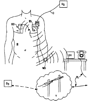
Fig. 6 Illustrate a patiatt body B having an implanted device in accord with
the invention ID for determining a value of importance indicative of edema.
For
example purposes also ilhtstrated implanrable device OD, as illustrated
here, a pacanaker having a lead L.
25 In the clinical se~g the doctor will use a programmer device which is
either a hand held unit like the PH unit which is held in proximity to the
body B or
a hand held device associated with a larger "programmer" which in common use
is
similar in power to a personal computer running an Intel 80486 or ~Pentium*
processor. 3'his unit is illustrated heuristically here with the numeral D2,
connected
3o to the telem~ry containing head PH by a conaaxor cord C. S~bstanbal
diagnostic
*Trade-mark
CA 02278193 1999-07-19
WO 98/33553 PCT/US98/00040
23
testing and processing .can be done, giving the patient stress testing, drug
treatments, and so forth with the doctor in attendance since the immediate
data from
the implant can be acceased andl used by the physician to test the body's
physiologic
responses to various test conditions. Various configurations of D2 and PH with
s respect to telemetric co~mmunic,ations to implants are well known and any
such
systems could be exploited for ~hi.s invention. The new data now available
from the
implanted inventive de~~ice ID enhances this physician function by providing
an
edema and edema history signal. containing data about the changes in the edema
parameter over time to the physician. This data can be processed with data
from
other implanted sensor s which may be distributed between the implanted
devices
OD and ID, etc., or all from one device, such as a pacemaker like device OD.
It is
already known in the ant to have and use implantable sensors to monitor and
record
data including activity sensed, heart rate, heart rate variability,
respiration, minute
ventilation and variability of, arrhythmia frequency and duration, averages of
these
1s values over long term, pressure at various sensor locations, 02 saturation
at various
sensor locations, time located patient activated data records for holding
various
pieces of these data sets around a temporal marker set by a patient activated
signaling device(which could be incorporated into a device such as WD, or some
other convenient unit) for diagnosing patient symptoms, and so on. However,
the
2o use of an impedance sensor dedicated to generate data specific to edema
conditions
and history has not heretofore been seen. Combining these edema measurements
with any of these other signal data provides an enhanced diagnostic and
patient
management efficacy to all the devices that can be used in the Fig. 6
environments.
For example, the ID device could contain a looping memory activated by trigger
25 signals sent either through a patient activator (which advantageously can
be located
in device WD) or by physiolagic signals generated by one of the sensor systems
(systems 89 or 89a of Fig. 8).
Continuing to refer to Fi:g. 6, the inventive implantable device ID can also
be used in settings remote from direct physician contact to further its
usefulness. A
3o wearable device WD can be used to extend the communications range without
i~ l d ~ I ...._,_...
CA 02278193 2002-11-25
66742-704
24
substantially it~sing ba~ay usage by the implant 1D for sending communication
signals. A patent illust<atmg this concept is U.S. patent No. 5,113,869
showing
that a wearable device ~uld be used in such a manner. As power considerations
change and oommttnicatioa w implants b~oo~,s less costly, it is feasible to
eliminate an intermediate device such as WD, btrt for the foracrable future
such
devices are eto be used for yoaic communications with implarus.
A borne monitor device such as D 1 may be kept in a location within range of
the
oo>mnuiueatiom c~abilities of the device WD anti oo~max to a tell lip TL
system to a telephony or cortnmuaications network T (which may be worldwide in
~ scope) or fbuough otlur communications c~am~els not shown to reach a devicx
D3
that is -located at a physician station, hospital or clinic and which can
perform the
same fumxions descn'bed for the pmdevice (D2) above.
Preferrably, devicx D3 can also comtaunic~te through the telephone or radio
frequency chapel to the wearable device WD. In this way too, this communicated
is information could be used to modify the functioning of the implanted
devices, OD
and/or 1D. These changes could be modification to data collection methods,
type of
data collected or trigger release values for drug or swmrlation therapies.
In some preferred embodiment devices we would ttse the respiratory rate as
an indicator of edema or lung water. This can be monitored long term like the
DC
2o impedance signal, but instead of filtering out the respiration signals as
noise, we
would for these devices use the respiration varied impedance measures to
determine
breath rate. It is well known how to do this. Medtronic produced two
pacemakers,
the Legend bus*series and the Kappa*400 more rxe~tly that m~asvre mite -
ventilation, which requires a ddermiaation of a breathing rate to provide a
rate
2s response funcxioa. Another called the RDP3 or MB-1 was reported in "Rate
responsive Pacing with a Pacemaker that detects respiratory rate (Biorate);
Clinical
advantages and complications," by Lau et al, CLINICAL CARDIOLOGY, May,
1988, 11(5) p318 24, ISSN 0160=9289, Therefore it can readily be seen that
determining respiration rate is not outside the skill of those of ordinary
skill in this
30 -art. Ia our device we use the signals described above for counting
breaths. An
*Trade-mark
CA 02278193 1999-07-19
WO 98/33553 PCT/US98/00040
equation representative. of how this function works is Respiratory Rate =
Minute
Ventilation divided by TV, where TV is the maximum value of DCZ.
It is up to the designer of the device, of course, but alternative methods are
available to generate a reliable respiration rate signal; for another example
see
5 Plicchi and Canducci, US Pat. No. 4,576,183. All that is required is that a
reasonably reliable count of respiration per unit time be maintained and
reviewed on
an ongoing basis to determine the change vector. A single memory element could
be used to record whether each sample is moving up or down in rate if desired,
but
we prefer to keep some: value taken at several measurement so that the data
can be
to relayed to a doctor for careful analysis.
It is expected that measurement over sleep periods, with reduced muscle
movement artifacts will yield a cleaner signal, so we prefer using an
additional
trigger to time the measurements to occur during the patient's sleep cycle. A
choice of a suitable sleep trigger is up to the designer, but it could be done
in a
t5 number of ways such a.s waitinl;(perhaps based on a timer and a timeout
period) for
a period of, say, 15 minutes in which no noise artifacts show up in the
signal,
timing the sampling periods tocorrespond with the patient sleep times using a
24
hour clock in the device, and so forth. Many other devices and methods for
determining sleep periods are known and may be applied here if desired.
2o A uniquely valuable enhancement would be the addition of an algorithm to
detect Cheyne-Stokes respiration which often is present during sleep as a well
known indicator of fluid build-up. By simply monitoring for rapid respiration
rates
and then closely observing the respiration signal after short episodes of
rapid rates,
one can easily generate: a marker in memory to indicate that such an episode
has
25 occurred when shallow breathing or absence of breathing is detected after
bursts of
high respiration rate.
To exclude from consideration for edema monitoring those rises in
respiration rate that occur because of patient exercise or other activity, we
use an
activity sensor like the activity crystal sensors or accelerometers commonly
used for
3o activity measurement in modern pacemakers to generate a signal indicative
of
CA 02278193 1999-07-19
WO 98/33553 PCT/US98/00040
26
patient activity Level. If the level respiration rises only during or after
periods of
indicated increased patient activity, the value of a signal based on
respiration rate
should be decreased. The respiratory rate taken during periods of no activity
is
preferred to measure long-term breathing rate trends . In the preferred
embodiment
we use a Respiratory Rate/Activity or Accelerometer Parameter coefficient that
is
calculated on a daily basis . Thus two independent values are generated. One
value
is generated for activity and a value is generated for respiration rate and
the ratio of
these two is monitored. A change in this ratio causes an increase in the value
used
to indicate a troubling rise in edema. In other words, the change may indicate
to more or less breathing during exertion, thus indicating an improvement or
deterioration in lung fluid levels.
A device can be constructed using the respiration rate as described alone or
in combination with the DC impedance measurement of edema described above.
If both are used a mixed value should ultimately be relied upon to indicate
the level
of edema the patient is subjected to. Many ways can be devised to combine
these
signals, including simply adding them throughout the potential range and using
the
total value for the indicator; relying on one measure only, in situations
where it is
superior, or simply recording both separately and reporting them out through
telemetry for analysis together by a doctor or other attendant (human or
automatic).
2o Alarms can be initiated thorough whatever patient or doctor communication
system
is used with the device at whatever level is programmed in for the variable or
variables used to evaluate these measures of edema.
In our most preferred embodiment, we would use a combination of
breathing rate rise over 10 % per 24 hour period with or without any
indication of
DC impedance measurement as sufficient to generate an alarm. Likewise a 20 %
rise in edema measured by DC filtered signal value should suffice to generate
an
edema alert alarm. Finally the presence of a newly discovered Chyene-Stokes
episode or, if expected, perhaps the appearance of two Cheyne-Stokes episodes
in
one 24 hour period or one with either of the other just mentioned edema signal
3o value percentage changes should be sufficient to generate the alarm.
CA 02278193 1999-07-19
WO 98/33553 PCT/US98/00040
27
In situations where the device is communicating with or a part of a therapy
delivery device, response to edema levels, however measured may be used to
trigger or adapt therapies, in any situation described above with reference to
the
alarm.
In Fig. 10, an idealized implantable device 110 is illustrated heuristically
with a body compatible shell 110b (which could of course be composed of
Titanium
or ceramic, or any other body compatible material suitable for housing an
implant
in a living body) surrounding its component circuits 111-124 and any others
desired
(but not shown here) by the designer of the device 110 to supplement the
inventive
1o circuitry arrangement described herein. For evaluation of impedance
measurements
to derive a respiratory ,rate signal one requires an impedance measurement
circuit
111 connected to an electrode means on the shell or a lead or similar
appendage
attached to the shell (he;re illustrated as two bumps 110a) and a circuit to
evaluate
changes in the value of the impE:dance measurements over time. As is well
known
is in the art (and mentioned above implemented in present pacemakers), The
impedance measurement can find a value for a respiratory rate signal from
counting
breaths represented by an approximation of a sinusoidal variation in the
impedance
occurring within the pa~tential range of breathing rates. In Fig 10, this
determination of respiration ratf: occurs in the circuit 114. It should be
understood
2o that this ability to determine breathing rate is known in the art through
at least the
methods described in L:~S Pat. I\fo. 4,576,183 (Plicchi and Canducci) and
5,271,395
(Wahlstrand et al.). Generally in block 114 a circuit should provide
conversion of
an AC impedance signal from the change in impedance tot a respiration rate
through
bandpass filtering, zero-crossing detection, and if desired, template matching
of the
25 digitized waveform. Any design that gives a fairly accurate assessment of
respiration rate is acceptable, however.
For completeness(in some embodiments), a DC value of Z may be
maintained to take advantage of the potential for determining edema directly
from
the changes in the impf;dance signal as described above, and this function
occurs in
3o Fig 10 in circuit 112.
CA 02278193 1999-07-19
WO 98/33553 PCT/US98/00040
28
A memory circuit is provided in device 110 which herein is illustrated as
being in 4 potential parts, memory circuits 113, 115, 119 and 123. Of course
the
designer may subdivide or consolidate a memory circuit in any way desired and
this
is indicated in Fig 10 by the dotted line 116, suggesting by way of
illustration that
the configuration of the memory circuitry is best left to the final designer
of a
particular implementation of this invention.
A timing or clock circuit 125 should be provided to coordinate the storage of
data related to each evaluation so that histories of such data can be
maintained. As
illustrated here, the DC impedance is stored as a secondary or second value
set of
edema values in memory I13, the respiratory rate values in memory 115 and the
respiratory rate change that indicates an edema value (here a primary or first
edema
value)is stored in memory 119.
An alternate and independent activity sensor and associated signal value
generation circuit I22(any known kind could be used) may be provided for a
preferred embodiment. This activity sensor and value generator can have its
own
historical values stored in a piece of memory too, here memory 123.
At any time desired in accord with the needs of the preferred design, the
current value of any one of the stored values can be compared to a current or
past
value of either the same the same or any other one of the stored values. This
means
2o that the change in rate, as found in circuit 117 can be used to evaluate
the
respiration rate, and that the respiration rate can be compared. If desired a
memory
controller 127 can coordinate these evaluations according to a program that
may
also be in software stored in the memory, but currently in preferred designs
hardwired circuitry (not shown) cycles through comparisons required by the
particular design. In this way the illustrated flexibility is compromised for
cost
reasons. The comparitor circuit 117 output should be qualified by an
evaluation
circuit 120, preferably each time a comparison is made to determine if the
results of
that comparison indicate a need to generate other signals that trigger an
alarm or a
readout or a therapy, depending on the configuration of the device. For
example, if
3o the respiratory rate has risen very quickly (thus meeting a criteria stored
in or
CA 02278193 1999-07-19
WO 98/33553 PCT/US98/00040
29
accessible to circuit 120, a signal should be sent to the Cheyne Stokes
indicator
circuit 121 to see if the change should generate an alarm. If circuit 121
determines
that it is appropriate it :should send a signal to the transceiver circuit 12b
to perform
its function suitable to receiving a signal from said CS indicator circuit
121. Circuit
126 may be a circuit to transmit data through telemetry to a device adapted to
receive such telemetered data outside the body as illustrated in earlier
Figs., or it
could be a simple alarm. that vibrates or generates a sound from within the
patient.
It can be triggered by the evaluation circuit any time appropriate criteria
are met by
the output of the comparitor circuit 117, all of which are described above.
to Additionally circuit 12E~ may readout the memory contents or parts thereof
on
receiving an inquiry signal from an external device, or automatically
telemeter out
data based on an the ex~?iry of a timer or other signal generated by clock
circuit
125.
As with any imylanted device there should be a source of power, here
illustrated as power supply 124, which of course could be a battery or power
generator of any kind suitable for powering implanted devices, so as to supply
power to the circuits used in the device and to generate pulses for impedance
measurement.
In Fig. 11 a flow diagram 130 illustrates a method for using the measure of
2o respiratory rate over time to generate an evaluation of edema or lung
water. It is
assumed that he impedance measures are made in the thoracic region of the body
by
an implanted device as ~describeci above.
Step l la measures the impedance and stores the history, repeating itself
continuously as indicated by the curved arrow. In Step l 1b, each breath count
is
determined by review of the history of impedance measurements. A rate is then
determined and stored as a current in Step i lc. On their own appropriate
cycle
times, as would be app~irent to one of skill in this art, both steps llb and l
lc iterate
and store their determined value, as history. As can easily be appreciated,
the past
history of a prior step rnay be il;nored once a new datapoint is generated by
a later
CA 02278193 1999-07-19
WO 98/33553 PCT/US98/00040
step if the only data desired is that determined in the later step. Thus
history
memory circuitry can be conserved.
In Step l 1d, the history of respiratory rate from l lc is evaluated. Here a
determination is made as to whether it is tending faster or slower and whether
it
5 meets preestablished criteria to proceed to step 11 a and to generate an
alarm. If the
device in which this method is used does more than generate an alarm, it
should
readout the storage for analysis by a health professional, Step l 1f. The
preestablished criteria can be stored in the device during manufacture or may
be set
and reset by an attending physician depending on patient conditions. They may,
of
1o course, be comparative criteria indicating simply a vector of edema
measurements
or may be combined with other physiologic indicators as described above to
establish more complex or more reliable criteria.
An easy way to turn the data into diagnostically useful data is to store a
respiratory rate to activity level parameter history longitudinally over time.
Then
t5 an edema level indicator circuit can generate a signal value representative
of
whether edema or lung water is worsening or improving, based on evaluation of
said parameter history. The doctor can simply look at the change in this value
over
time to assist in his care of the patient. This could be called simply a
measure of
patient health, rather than edema since respiratory rate may correspond to
other
2o simultaneously occurring changes in patient physiologic condition.
The invention as described and illustrated herein is only limited by the
following appended claims.