Note: Descriptions are shown in the official language in which they were submitted.
CA 02278537 1999-07-22
CVO 9917348 - PCT/CB98I03510
lviETiiob aND APPAR4TUS >FOR nETECTiNG At~r OBJECT
' The present invention relates to a method and apparatus for detecting and/or
imaging an object.
Imaging through turbid media is often carried out using Time of Flight
measurements. Various methods of time of flight measurement are known. The
majority of these methods measure the time taken by pulses of light to travel
a return
path. Since the velocity of light is both constant and known for most
materials,
measurements of time of flight can be readily converted into images of the
medium
through which the light has passed.
Time of slight imaging i~ particularly usaful when imaging over large
distances or in semi-turbid media. I~owever, when dealing with short distances
image
resolution decreases rapidly, since the speed of light is so great that, for
example, a
spatial resolution of lm will require a detector with a temporal resolution of
ins. The
shortest measurable distance and the image resolution obtainable using Time of
Flight
imaging is thus limited by the response time of the detectors used. A further
disadvantage of Time of Flight imaging is that the reset time of the detectors
used is
considerably longer than the j fitter time) so that the detectors are only
capable of
detection for a very short period of their total operating time. An
alternative method
of imaging through turbid media is to use acoustic waves (eg, ultrasound
imaging).
However, acoustic imaging suffers from lack of spatial resolution due to the
large
divergence of acoustic waves.
A further known method of imaging comprises forming an image of an object
from interference of two beams of coherent light, one of which bas been
scattered
from a target (ie. holographic imaging). Holograms have been used to analyse
non-
visible parameters of a target, for example, vibration of an engine block.
Holographic
imaging suficrs from several disadvantages. Firstly, holograms require two
investigative beams that must interfere and be coherent ever the distance to
the target.
Secondly, holograms are not suited to imaging through opaque media. An image
cannot therefore be produced ak a distance greater than a single photon
transport path
of the light used to obtain the h logtant. Thirdly, holograms are unsuited for
. SUB SHEET (RULE 25)
CA 02278537 1999-07-22
WO 99127348 - .- PCT~GB98103510
1
measurement through media that exhibit dynamic scattering) and the scattering
will
reduce the quality of images obtained.
Several mown imaging techniques exist where an investigative wave is
perturbed as it passes, scatters) reflects or is absorbed by a target. These
techniques
require that the form of energy usCd for the investigative wave, and its
frequency,
must be chosen to interact with the target and cannot thus be fully optimised
for
detection (i.e. low absorption and~or high spatial .resolution and/or high
signs! to
noise).
It is an object of the present invention to overcome or substantially mitigate
the above disadvantages.
According to a first aspect of the present invention there is provided a
method
of detecting an object located within a dynamic scattering media) the method
comprising:
i) directing a continuous coherent light wave of predetermined
wavelength into the media;
ii) detecting dynamically scattered tight emerging from the media;
iii) correlating the detected light photons in the time or frequency domain;
iv) determining the presence of an object from analysis of differences
between said correlation and the correlation which would arise from photons
scattered
by the media only; and
v) determining the approximate position of the object within the media
from said an$lysis of the correlation and knowledge of the mean transport path
of the
light wave of predetermined wavelength within the media.
The term "light wave" is not limited to visible light but is to ba interpreted
as
encompassing electromagnetic radiation of any suitable wavelength.
The media may be a turbid n4edia of relatively high density and the object may
be more or less dense and more or less viscous than the media. 'rhe object may
be an
object which absorbs and/or reflects the incident light waves.
1'hc invention (from hereon referred to as Diffuse Wave Imaging) incorporates
aspects of the known technique of Uiflusing ~,Vavc Spectroscopy (DWS). DWS is
used for sub-micron particle: siri g and bulk rheology measurements in dense
suspensions or emulsions. nWS i not applicable to the imaging of singlt
objects
SUBSirITUTE SHEET (RULE 26j
CA 02278537 1999-07-22
_ i
WO99f17348 _ .. ~ 1'CTlCB98N35t0
3
within turbid media. Furthermore) DWS is not used to identify individual
particles.
The suppression of photons close to the axis of the auto-correlation tracts is
treated as
a limitation of DWS_ The inventors have realised that this suppression of
photons
which have undergone multiple scattering events can be used to determine the
presence of an object, and by taking many measurements, to form an image.
The invention allows images to be obtained using photons which have
uravelled optical distances greater than 2 photon transport paths. This is
opposite to
conventional imaging of dense media, which generally removes or ftlters out
light
which has been scattered more than once_ Since the invention does not require
photons which have undergone ballistic and low order scattering, it is
suitable for
imaging very dense suspensions where ballistic scariering is limited.
The steps (i) to (v) may be iterated either sequentially or simultaneously
using
coherent tight waves of different predetermined frequencies having different
mean
transport paths within the dynamic scattering media, to thereby obtain further
information as to the approximate position of the detected object from the
analysis of
the respective cornlations and knowledge of the respective mean transport
paths. For
instance, light of three different wavelengths may be used.
The pr each iteration of steps (i) to (v) may be repeated) either sequentially
or
simultaneously, for additional locations within the media, the results then
being
combined to construct an image of the object within the media. Where only one
wavelength of light is used the image will be two dimensional. However) using
two
or more different wavelengths as mentioned above (which effectively probe into
different depths of the media and/or object) enables the construction of a
three
dimensional image.
The light emerging from the media may be detected at one or more
prcdcternlined scattering angles, preferably a scattering angle of 180°
andlor 0°.
The media tnay be modulated to induce, or enhance) dynamic motion within
the media to provide or enhance the required dynamic scattering, Similarly)
when the
object is an obj~eet which is at least partially reflective of the or each
light wave, the
object may be modulated to enhaneo phase changes in light reflected therefrom.
The method muy included tl a step of selecting for detection light .which has
a
predetermined component of polari anon. The selection may be accomplished
usins
SUB9T1TUTE SHEET (RULE 26)
CA 02278537 1999-07-22
WO 99IZ73a8 . . PCT/GB9$/o3510
4
polarising fillters or fibre optic cables which preserve only one particular
component
of polarisation.
The or each light wave may be passed through a window prior to entering the
media. the window being arranged tQ reflect light which is detected together
with light
emerging from the media) thereby producing a heterodyne signal. The window may
be adjustably displaced relative to tl~c origin of said Iight wave to allow
control of the
intensity of the reflected light which is detected. 'fhe window may be
arranged to
cause the reflected light to undergo multiple reflections before being
detected, thereby
enabling the path length travelled by the reflected light to be controlled.
The method may be performed on a human or animal body to detect the
presence and approximate position) or construct an image, of a pathological
entity
within the body.
According to a second aspect of the present invention there is provided a
method of detecting the presence of~a pathological entity within the human or
animal
body) the method comprising:
i) directing a continuous coherent light wave of a first predetermined
wavelength into the body;
ii) detecting dynamically scattered light emerging from the body;
iii) correlating the detected light photons in the time or frequency domain;
and
iv) determining the presence of a pathological entity from analysis of
differences between correlation and the correlation arising from photons
scattered by
the media surrounding the entity only.
According to a third aspect of the present invention there is provided
apparatus
for detecting an object located within a dynamic scattering media, the
apparatus
comprising means for directing a continuous coherent light wave of a
predetermined
wavelength into the media, means for detecting dynamically scattered light
emerging
front the media, means for correlating the detected light photons in the time
or
frequency domain, whereby the preseance of an object cart be determined from
analysis
of differences between said correlation and the correlation which would arise
from
photons scattered by the media oniy,~the approximate position of the object
within the
SU851ITTUT)' SH
~r ~u~ zs~
CA 02278537 1999-07-22
. I VVO 99!17348 - PGT/GB98I03510
3
media can be dctetzrlined from said analysis of the correlation and Imowledge
of the
mean transport path of the light wave of predetermined wavelength within the
media.
A plurality of detectors maiy be arranged in an array to provide a series of
measurements simultaneously. The array of detectors may be coupled to a CCD
camera.
Preferably the or each detector is either located adjacent the emitter or is
displaced from the emitter and is located an the axis of emission of the
emitter
The coherent light producing means produces both visible light and infrared
li ght.
Polarising filters may be located in front of the detection means to select
either
light with a polarisation perpcndioular to the light emitted from the emitter,
or to
select light with the same polarisation as the light emitted froth the
emitter.
An object located within a medium may be caused to modulate to increase the
contrast of the phase of scattered light, and thereby improve the resolution
of the
image,
The invention may be used in combination with Time of Flight apparatus to
provide imaging over both short and long distances.
Where heterodyne detection is to be used, the coupling means may be
provided with a window which will reflect a fraction of the light towards the
detection
means, thereby providing a heterodyne signal.
The window may be adjustably displaced from the coupling means to allow
eotttrol of the intensity of reflected light incident at the detection means.
The window may be arranged to cause the reflected light to undergo multiple
reflections before being incident at the detection means, thereby allowin5 the
path
length travelled by the reflected light to be controlled.
'The coupling means and detectiory means may comprised polarisation
preserving optical fibres. The Fbr~ may be mounted so as to be rotatable
through 90
degrees) thereby allowing modifict~tion of the effective nurrierieal aperture
of the
fibres.
Preferably the means for producing coherent light comprises a laser.
1'refcrubly) lour lasers o erablc at a different wavelength are used
concurrcntlv.
5UB5 SNE>='T (RULE 26)
, W0991Z'13d8 . ~ CA 02278537 1999-07-22
PCT/GB98I03S10
6
Preferably) two of the lasers arc arranged to produce orthogonally polarised
light; and a polarising beamsplitter cube is provided to couple the light into
a
polarisation preserving optical fibre.
The coupling means and deflection means may comprise optical fibres with
terminations located in a probe comprising a cylindrical head.
According to a fourth aspect of the present invention there is provided
apparatus for detecting the presence of a pathological entity within the human
or
animal body, the apparatus comprising means for directing a continuous
coherent light
wave of a first predetermined wavelength into the body, means for detecting
dynamically scattered light emerging from the body, and means for correlating
the
detected light photons in the time or frequency domain, whereby determining
the
presence of a pathological entity may be determined from analysis of
differences
between said correlation and the cprrelation arising from photons scattered by
the
media surrounding the entity only.
According to a fifth aspect of the present invention there is provided a
method
of detecting the presence of an object within a media, the method comprising:
i) inducing vibration in the object at a predetermined frequency which
does not propagate efficiently within. the media;
ii) generating a continuous coherent light wave;
iii) modulating the generated light wave at a second predetermined
frequency;
iv) directing the modulated coherent light wave into the media;
v) detecting scattered lig~tt emergins from the media;
vi) analysing the detected scattered light for the existrnce of a beat signal
corresponding to the beat frequency between the first and second predetermined
frequencies thereby indicating the presence of the object.
The frequency of the induced vibration is selected to correspond to the
resonant frequency of the object, or~ regions within the object to be
detected. Such
regions may for instance be regions of stress) such as craek.,s in the object.
The vibrating frequency may be varied until the object, or pans of the object
to
be detected, resonates, artd wherein he size of the resonating object, or
region of tttc
object) is determined as a function of~tho resonating frequency.
SU85'~IN>!E SHEET (RULE 2b~
CA 02278537 1999-07-22
WO 991s73~8 _ PCIYGB9aID3510
7
A,n image of the object, or parts of regions of the object to be detected, may
be
constructed by detecting light scattered from different parts of the object.
The method may be performed on a human or animal body to detect the
presence, or construct an image, of a pathological entity within the body.
The invention also provides apparatus for detecting the presence of an object
within a media, the apparatus comprising:
i) mEans for inducing vibration in the object at a predetermined
frequency which does not propagate efficiently within the media;
ii) means for generating a continuous coherent light wave;
iii) modulating the generated light wage at a second predetermined
frequency;
iv) means for directing the modulated coherent light wave into the media;
v) means for detecting scattered light emerging from the media;
vi) means far analysing the detected scattered light for the existence of a
beat signal corresponding to the beat frequency between the first and second
predetermined frequencies thereby indicating the presence of the object.
The method and apparatus according to the fifth aspect of the invention may
be used to investigate properties that cannot be imaged directly such as
scaling in an
oil pipe car cracks in a metal structure,
The modulation frequency may be used to perform a secondary function on the
object. For example, resonance may be used to cause descaling of a pipe whilst
a~
imagt (by means of measurement of the change in the magnitude of resonance at
a
single frequency or preferably the chattgc in the resonant frequency) is used
to
monitor the descaling operation in real time. Similarly, where the methods is
used on
a human or animal body, the same method may be used to remove or destroy
pathological entities such as tumourt; (wherein the resonance may heat and
kill the
ta~et), 'all stone (wherein the res~nanee will physically break down the
target),
kidney stones, and other growths) foreign bodies and abnormalities. A similar
method
may be used to remove blockages eto. io non-medical applications. such as to
remove
blocl:a5cs from target objects such as underwater pipe lines or cables.
Resonance rnay also be used to modify the target object means of chemical
release or activation.
SUBSTiITUTE SHEET (RULE 26)
CA 02278537 1999-07-22
WO 99n~f'348 - .. PC"T/GB98/03510
8
'The invention may utilise a naturally occurring source as the source of the
i nvestigative wave.
The present invention may be combined with existing detection or imaging
systems, such as time of flight systems) CAT, electron resonance etc.
Other possible features of the invention will become apparent from the
description below.
Specific embodiments of the invention will now be described,. by way of
example only, with reference to the accompanying drawings) in which:
Figure 1 is a schematic diagram of an imagng apparatus according to a first
aspect of the present invention;
Figure 2 is shows three auto-aorrclation traces which illustrate the operation
of
the apparatus of Fig 1;
Figure 3 is a schematic diagram of a first heterodyne window suitable for use
with the present invention;
Figure 4 is a schematic diagram of a second heterodyne window suitable for
use with the invention;
Figure S is a schematic diagram~of a third heterodyne window suitable for use
with to the invention;
Figure 6 is a schematic diagram of a light generating apparatus;
Figure 7 is a schematic diagram of a detection apparatus;
Figure 8 is a schematic diagram of a probe for use in the present invention;
Figure 9 is a schematic diagram of a fibre block for use in the present
invention;
Figure 10 is a schematic iilustratiori of an imaging apparat(is~ according to
a
second aspect of the invention; . -- '.
Figure 11 is a schematic illus9ration of the operation of the apparatus of
Figure
10.
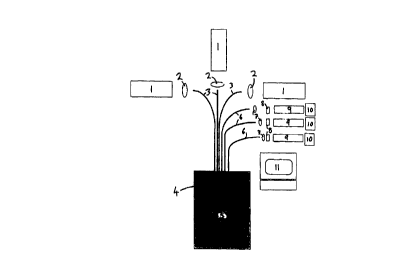
Figure 1 shows an imaging apparaws tomprising three lasers I operating at
different wavelengths. The tlvee lasers produce light ;rt wavelengths 4S8nm
(argon
ion), 633nm (helium neon) and 106 nm (Nd:YAG) respectively, the light from
each
laser l having :t Single transverse a d longitudinal mode. Optics ? (an
example of
SUBS~ITUT>' SHEET (HUt:E 26)
WO99~.t7348 ( ~ CA 02278537 1999-07-22 ~~~ggg~p3510
9
which is described in more detail below with refere»ce to Figure 6) couple
light from
each laser 1 into a single mode polarisation maintaining optical fibre 3.
Light from
each fibre 3 is launched into a turbid medium 4 within which is located art
absorbing
object 5.
Light scattered by the turbid medium 4 is collected by three optical fibres 6
which may have the same construotion as the launch fibres 3. The collected
light is
collimated usi»g further optics 7 (an example of which is described in more
detail
below with reference to Figure 7) and directed at polarising filters 8 and
laser-line
optical filters 9. Light transmitted by the laser-line filters 9 is monitored
by detectors
10) a respective detector 10 being arranged to detect scattered light of each
of the three
laser 1 wavelengths. The detectors 10 are photon counting photon multiplier
tubes or
photon counting avalanche photodiodes) whichever is appropriate for the
wavelength
of lisht to be detected. An output signal from each detector 10 passes to a
digital
correlator which produces a cotrdlation trace (described below with reference
to
Figure 2). The correlation trace may be displayed on a monitor 11.
The illustrated apparatus is configured to detect light scattered through 180
degrees (back-scatter), The apparatus could be configured to detect Light
scattered
thmugh other angles including light which, although scattered, exits the
turbid
medium 4 in the same direction as the incident light (i.e, scattering through
0
degrees). Whilst operation of the apparatus at any angle is possible, the
mathematical
modelling required to obtain imaging becomes more complicated for scattering
angles
other than 180 degrees or 0 degrees and thus these two configurations are
preferred.
Figures 2a to ?c show three correlation tracts obtained from the scattered
light
detected at each of the detectors 10. Each correlation is obtained by
correlating
detected light with itself (i.e. the known technique of auto-correlation).
Each
correlation trace shows the logarithm of the number of photons detected
(vertical axis)
versus the square root of the corralator delay time (i.c. the delay time of
the auto-
eorrelation of the detected signal). The region of the correlation trace near
to the
vertical axis represents photons which have undergone a ?rcat deal of
scattering,
lesser amounts of scattering are inddcated further from the vertical axis (for
light with
purely random phase the correlatio ' would be a flat line). It is important to
note that
some form of motion of the mediut~t 4 is required so that scattering of the
li~tht by the
StIBSmVTE SHEET (MULE 26)
CA 02278537 1999-07-22
WO 99IZ'1348 - j ' PCT/G898103510
medium will modify the phase of the light (Hrownian Motion is often sufftcient
for
this purpose).
The correlation trace of Figure 2a is obtained from detection of the shortest
wavelength light (i.e- 488ntri). The turbid medium 4 will scatter the
relatively short
wavelength light efficiently, and consequently in the illustrated example the
avera?e
pcnetxation of photons into the turbid medium 4 is less than the distance to
the
absorbing object 5. Thus) the short wavelength light is largely unaffected by
the
absorbing object ~, and the correlation trace obtained will have a shape
characteristic
of random scattering by the mediutxs 4 (i.c. a straight line sloping
downwatdly away
from a maximum value of 1 ). Thus the correlation trace indicates that the
depth of the
object S within the medium 4 is greater than the average scattering depth of
the
488nm light (the average scattering depth of 48Bnm light in a given medium may
be
known from a reference text or from expe~mentxtion).
The correlation trace 2b is olptained from detection of the medium wavelength
light (i.c. 633nrtt), which has a greater average penetration depth than the
relatively
shorter wavelength Light. The trace shows some attenuation close to the
origin, which
indicates that those photons which have travelled the longest path throw the
medium
4 (approximating to diffuse photons) have been preferentially depleted by the
absorbing object S. Further away from the origin of the trace those photons
which
have travelled a shorter path are unaffected by the object S, and the downward
sloping
straight line of the trace is unchanged. The shape of the correlation trace
thus
provides information pertaining to the depth and shape of the object S.
Correlation trace Zc represents long wavelength light (i.e. 1464nm) which has
a greater average scattering depth than the 633nm light. The effect of the
object ~ on
the trace is more accentuated than the effect on correlation trace B, since
more of the
1~64nm photons penetrate sufficiently far into the medium 4 that many of them
arc
absorbed by the object S. ). Thus thd shape of the correlation trace ?c
provides further
information pertaining to the depth and shape of the object ~.
The invention thus uses light of three different wavelengths to determine the
position of the absorbing object S. The depth of the object is determined by
comparison of the shapes of the thre correlation traces 2a-c.
sues sHl,»t ~tu~ ~
CA 02278537 1999-07-22
WO 951I~7348 - _. PCT/GB98103510
A single measurement as described above will describe the position of a small
area of the object ~_ To obtain an image of the object the position of the
optics (not
shown) which launch light >iom the fibres 3 into the medium, and/or the
position of
. the optics (not shown) which coupe light from the medium into the fibres 6,
are
translated betweea measurements to provide a grid arrangement of measured
areas.
The wavelengths used may be pre-selected to provide a desired range of
penetration depths into a given tnedium. It is noted that a coherent light
source
capable of being tuned over a wide range of wavelengths (for example an
Optical
Parametric Oscillator) would provide the capability to pre-select
wavelcngtlts) and
also to obtain many measurements at different wavelengths (although the three
wavelengths used abo~o should be stt~cient to obtain accurate imaging).
The apparatus may comprise single emitters and multiple detectors, thereby
allowing many point measurements. The fibres collecting scattered light could
be
coupled directly (or via an intensifier) to arrays of detectors such as charge-
couple
device (CCD) cameras.
Inclusion of the polarising filters $ enables further resolution of the depth
of
the object 5. When polarising filters 8 arc set so as to transmit light which
is polarised
transverse to the light from the lasers l, only photons which have undergone
many
scattering events will be detected. This orientation of polarising filters
thus
suppresses detection of photons which have penetrated a relatively small
distance into
the medium 4. When the polarising filters 8 arc rotated to transmit light
which has the
same polarisation as the light from the lasers l, photons which have undergone
many
scattcrin, events are suppressed, and photons which have penetrated a
relatively small
distance into the medium 4 arc detected.
When optical Fbres 3,6 which support only a single eigcn-mode of light at an
appropriate wavelength are used) the polarising, ftltcts 8 are not needed.
This is
because the transverse polarisation of the fight produced by the lasers 1 will
be
preserved by the fibres 3 which launch the light into the medium ~. The
oriemazion o~
the fibres 6 which couple scattered light from the medium can be sec so as to
couple
light with the same polarisation ;s.~s lhat emitted from the lasers I, or
light with
polarisation orthogonal to that emttc d from the lasers, This will allow
discrimination
of penetration depth ns described ab~vc,
SUBS~tTI;ITE SHEET (RUI.F 26)
CA 02278537 1999-07-22
WO 9917348 - ~ PC'1'IGB98103510
12
Optical fibres 3,5 capable of supporting multiple wavelengths would reduoe
the number of fibres 3,6 required by the apparatus (it is noted that the use
of
polarisation maintaining bcamsplitters would be advantageous in this
arrangement to
allow the combination of beams from the different lasers 1 ).
A fibre having multiple cores would allow different emitter/detxtor spacing
within that fibre.
It is noted that the invention may be used to image reflecting objects, in
addition to imaging absorbing objects as described above. Light reflected from
the
object will cause a perturbation at or close to the vertical axis of a trace.
The invention may be applied to imaging of the human body) for example to
detect tumours. For body imaging, utilisation of both 180 and 0 deg~ee
measurements
in a number of positions will produce the best results. The position of launch
of light
into the body) or of collection of scattered light, may be varied by scanning
the
apparatus appropriately or by using multiple emitters and/or detectors. After
operating the apparatus at a series df positions correlations obtained arc
normalised
and compared. Diffexenccs in coSrelation filnctions close to the vertical axis
will
suggest a change in the effective 'viscosity' or 'refractive index' of the
body at that
point) which may indicate the presence of, for example, a tumour.
Heterodyne sigtal processing may be used prior to auto-correlation. An
apparatus which will provide a heterodyne signal is shown in Figure 3. bight
is
transmitted through emission optics 12 into a window 13. At a lowermost
surface of
the window 13 the light passes into;the turbid medium 4, and a action of the
light is
reflected duo to the difference in re&active index of the window 13 and the
medium 4.
Detection optics 14 collect scattered light and retleeted light which
interferes to
produce a beat signal, and the beat signal is processed to provide imaging in
the same
way as described in relation to Figure 2. The triangular area 13 located
beneath the
detection optics 1~4 represents the dttcction area of the optics 1~4 (i.e.
corresponds to
the numerical aperture of the optics 14). The emission optics 12 and detection
optics
14 are index-matched to the window 13 to prevent waveguading of light within
the
window l;.
The apparatus shown in Fig re 3 has two (imitations. The BrSt limitation is
that the ratio of heterodyne (i.e. re erred) light relative to scattered (i.e.
homodyne)
suB rTUTE sH~r (RV~ zsll
CA 02278537 1999-07-22
WO 991~73d5 - .. PCT/GB98103510
13
light cannot be controlled. The second limitation is that the heterodyne light
and
homodyne light travel different path lengths, as a consequence of which
stringent
coherence of the beam is required. These two limitations arc linked, since
when the
homodyne light has a short path length in the medium 4 the intensity of
scattered
homodyne light detected will be high and a strong heterodyne signal will be
required.
An apparatus which overcomes the first of the above limitations is shown in
Figure 4. Emission optics 12 and detection optics 14 arc located above an
optical
window 13. The optics 12,14 are not index-matched to the window 13, and a
proportion of light emitted from the optics 12 will be reflected by the window
13 and
collected by the detection optics 14. The window is index-matched to the
turbid
medium 4 so that there are no reflections from the lowermost surface of the
window
13. Light scattered by the medium 4 interferes with light reflected from the
window
13 to provide a beat signal for processing as before. However) the separation
of the
window 13 from the optics 12,14 allows the optics to be moved relative to the
window 13, thereby controlling the proportion of reflected light which is
collected by
the detection optics t4 (i.e. moving the window 13 further kom the optics
1?,l4 will
increase the proportion of reflected light collected).
The apparatus illustrated in Figure 4 does not allow control of the heterodyne
path length (i.e. path length of the reflected signal). The mean path length
of the
heterodyne signal may be controlled using the window 13 as a guide for
multiply
reflections ss shown in Figure 5. The emission optics ( 3 and detection optics
14 arc
not index-matched to the window 13, and the window is not index-matched to the
turbid medium 4) so that a portion of the emitted light is reneged from the
uppermost
and the lowermost surfaces of the window 13. Heterodyne light may enter the
detection area (4) by multiple reflections tTOm the surfaces of the window 13.
Moving the window 13 toward the emission optics 13 and the detection optics 14
will
reduce the heterodyne signal) and increase the mean optical path length of the
heterodyne signal. Reduction oC the path length of the heterodyne component
(by
movin5 the window I3 further from the probe) will increase the heterodyne
signal
strength. Thus, using. a window l; Qf selected material, optical coatings and
thickness
it is possible to pro~idc a hetcrodym~ signal ot'the desired path lensth and
intensity.
SUB SHEET (RULE 26)
CA 02278537 1999-07-22
WO 9911734a . P~T/GB98/03510
14
It is noted that since light will be emitted by the emission optics with a
finite
divergence, there will be a spread in the path lengths travelled by the
heterodyne light.
Figure 6 illustrates a configuration for coupling light from several lasers
into
two optical fibres. A configuration based on that illustrated may be used to
couple
light from Three lasers into three separate optical fibres. as is required by
the apparatus
shown schematically in Figure 1 arid described above.
The primary lasers are: a single longitudinal mode frequency doubled
horizontally polarised Nd:YAG laser 16 {Smw of output power at 532nm), and a
stabilised horizontally polarised 675nm laser-diode unit 17. Two further
lasers may
be used to provide light at wavelengths of 580-833nm (eg. 780nm stabilised
diode
laser 18) and 430 - 670nm (cg. 488nm single transverse mode argon ion laser
19).
Light from the Nd:YAG laser 16 and diode laser 17 is directed to a polarising
beamsplitting cube 20 via baffles 21 and mirrors 22. The dimensions of the
baffles 21
and mirrors 22 are minimised to reduce the possibility of multiple reflections
of light
re-entering the lasers 16,17. The minrors 22 are capable of rotational and
translational
movement. Light from the lasers 16,17 may pass through f aser line filters 23
(eg.
birefiingent filters) if required.
Since the light fmm the le~sers 16,17 is horizontally polarised it will pass,
without reflection, through the polarising beamsplitter cube 20. Any component
of
the light which is not horizontally polarised will be directed down to a prism
24. Onc
short side of the prism 2~4 is painted black, to minimise reflection of the
light back to
the polarising beamsplitter cube 20.
The polarising beamsplitter cube ?0 is used to combine (vertically polarised)
light from the 488nm laser 19 with the light from the Nd:YAG laser 16, and to
combine (vertically polarised) ligft from the 780nm stabilised diode laser 18
with
light from the 675tun laser-diode unit 17. Light froth the 488nm single
transverse
mode argon ion laser 19 is coupled to the beamsplitter cube 20 using a
suitable lens
25. The lens 2~ may for example be piano-convex) anti-reflection coated on a
curved
side and courted to the polarising beamsplitter cube 20 to reduce reflections
from the
interface between the lens ?~ and the polarising beamsplittcr ZU. 'Che 780 nm
Iaser 1 S
muy be collimated viu a s~radi~nt index lens which is index-matched to the
beamsplitter cube 30.
SUBSTITUTE SHEET (RULE 26)
CA 02278537 1999-07-22
WO 99rZ'1348 _ PCT/G898/03510
The beams pass from the polarising beamsplitter cube 20 to two achromatic
lenses 2G which focus the light into aligned fibres 27 held in two blocks 28.
Light at 532nm from the Nd:YAG laser 16 is launched onto the fast axis of a
first of the optical fibres 27a, and light from the as8nm laser 19 is launched
onto the
slow axis of the first optical fibre 27a. Similarly, light from the 675nm
laser-diode
unit 17 is launched onto the fast axis of a second of the optical fibres 27b)
and light
from the 780nm stabilised diode laser 18 is launched onto the slow axis of the
second
optical fibre 27b. Using the slow axis of the fibre 27 (which has a high
numerical
aperture) For the shorter wavelength 488nm Light accentuates the increased
scattering
suffered by the lower wavelength) since the 488nm light will have a wider
angle close
field of view compared with the ~32nm light, The 675nm light is directly
comparable
with the ~32nm light since both pass down a fast axis of a fibre 27. However,
the
G7~nm Light will suffer less scattering within a turbid medium and will thus
analyse
material weighted deeper in the sample.
The blocks 28 arc capable of rotating the optical fibres 27 through 90 degrees
to allow the eigenmodcs of the fibres 27 to be chanbcd. The part of the fibres
27 held
in the blocks 28 is mode-stripped to prevent light being launched into the
cladding of
the fibres 27.
An avalanche photodiode may be used to detect the 780nm light, the other
wavelengths being detected using photo-multiplier tubes.
Coupling light into a medium using low numerical aperture optics) and
detecting light polarised at right angles to the coupled light will allow
objects as deep
as possible to be detected) and ensures that only high order multiple
scattering is
detected, which scattering is mast suited to the known multiple light
scattering
models. By altering the wavelength of the coupled light, the depth monitored
by the
technique will be significantly affected, since scattering of light is
wave)ength
dependant. Where both transmission (0 dE~ec scattering) and back-scatter ( 1$0
degree scattering) measurements are possible, information pertaining to the
depth of
an object in the medium could be obtained from the difference bcoYeen ThcSV
two
measurements.
SU8 ~ ITUTE SHEET (RULE 26)
__ CA 02278537 1999-07-22
. . . . , W0 99/Z7368 . PCT/GB98fi3510
16
Figure 7 illustrates apparatus for detecting light scattered by a turbid
medium,
and may comprise part of the apparatus shown schematically in Figure 1 and
described above,
In the apparatus illustrated, light is coupled by detection optics (not shown)
into an optical fibre 29. An emission end of the fibre 29 is held in a block
30 that is
fixed in one of two positions to alloNV selection of polarisation modes. An
achromatic
Lens 31, of higher numerical aperture than the fibre to allow for
misalignment,
collimates light from the fibre 29. The Light may be attenuated by a simple
moving
beam block 32; this is more efficient than attenuating or reducing the laser
intensity
since both the signal and the background are attenuated. The light passes
through a
laser line (or other) filter 33 which removes unwanted laser frequencies and
background noise. A polarising filter 34 selects the required polarisation
state, and
the light is then focused by an achr4matic lens 35 onto a photo-multiplier
tube ;6 (or
avalanche photo~.diode). The signal detected by the photo-multiplier tube 36
is
amplified and filtered 37 prior to signal analysis.
Figure 8 shows a fibre pmbe which combines ctnission fibres 38,39 and
detection fibres 39,40. The fibre probe may be used as part of the apparatus
shown
schematically in Figure 1 to couple light into and out of a turbid medium:
Light of 488nm and 532tim is emitted down a first fibre of the probe 38 and
collected by a second fibre 40, Both fibres 38,40 are bow tie (polarisation
preserving)
fibres, their cigen modes being set ~t 90 degrees so that only light which has
suffered
a polarisation rotation through 90 degrees during scattering in the turbid
medium will
be detected. The 675 and 780nm light is similarly emitted and detected by
fibres
39,41. The fibrc3 38-41 arc glued at their tips 42 into a short Capillary 43,
typically a
few mm long. The tips 42 of the detection fibres 39,41, and an adjacent
portion
thereof, are stripped of Gbre cladding, and black paint is applied around the
outside of
the fibre to molt-strip the colh:eted light (i.c) prevent light being coupled
into the
cladding). The short capillary 43 i$ filled with black paint. The emission
38,40 and
detection 39.41 fibres are held in separate jackets (not shown), beyond the
capillary
43. to prevent cross talk of light between then.
.~ s~eond capillary ~4 Burro ds the first capillary 43 and fibres 3$-4l
(typical
diameter 3mml, and further eapill~ries may be added. ThE second capillary 44
is
SU8S1TITUT1' SHEET (RULE 1b1
CA 02278537 1999-07-22
WO 99IZ7348 ~ ._ PCT/GI398/03510
17
encased to within O.Smm from the ends of the fibres 38-41 with a protective
steel
jacket 4~. The jacket 4~ is a loose sliding Ct on the capillary 44 and is
affixed with
silicone, to stop thermal sct'essing. 'Ihe probe is Ftted with a window holder
46 that
forms a close sliding fit with the steel jacket 45. A window 47 may be glued
or fused
to the holder 46. A temperature sensor (not shown) may be attached to the
window
holder 46.
A fibre block of the type which has been shown in Figures 6 and 7 is
illusnatcd in detail in Figure 9. The fibre block 48 is cylindrical and is
provided with
two chamfered holes 49 spaced apart by 90 degrees about the block. A single
ball
bearing 50 provided in a block-mounting (not shown) is resiliently biased to
locate
within either of the holts 49, thereby allowing the block to be rotated
accurately
through 90 degrees about its axis. The block 48 has a recessed face 51 to
allow
polishing of a fibre held therein 52 and an inner capillary 53 without
contamination.
The inner capillary 53 is located in an outer capillary ~4 allowing that part
of the
fibre ~Z which is mode-stripped to be protected. The fibre 52 is glued to the
inner
capillary ~3 and supported by silicone ~5 as it exits the outer capillary 55.
The urea of a medium which is.imagcd by the apparatus described above will
be influenced by the distance and angle between the detectors and emitter
optics,
Auto-correlation of the detected signal, or heterodyne detection) may be
substituted by any other processing which is sensitive to the dynamics of a
system
being imaged, for example pulse arrival distribution or a frequency scan from
a ctalon.
Where the object is reflective the objtct itself may be used to obtain a
heterodyne signal.
The methods according to the present invention have been in terms of light of
visible frequency, however any wave form that may be produced with a coherence
equivalent to the maximum path length diffettncc of the quanta may be used.
The invention is applicable to any system where infomtation is to be
transmitted ttuough turbid media) although it is particularly suited to body
imaging
and undersea surveillance.
ror imaging of a reDeetive object, where insufficient dynamic information is
present to obtain a useful eotrclati n (clue tv an insufficient amount of
Brownian
Motion in the target), a modulator~ntay he used to induce vibration of the
object.
SUB UTE SHEET (RULE 26)
CA 02278537 1999-07-22
WO 99II7348
PCT/GB98J03510
18
Inducing resonance of an object will thus improve the contrast obtained via
diffuse
wave imaging (the resonance of the object will result in a hetexodyzte
signal).
For imaging of an absorbi~lg object, the vibration may be induced in the
medium surrounding the object may be in the event that there is insufficient
Bmwnian
Motion to pmvide dynamic scattering of light in the medium.
A specific application of dif~usc wave imaging in which resonant modulation
of a target may be useful is imaging of the human body) for example where the
body
has a tumour growth, foreign body or other abnormality surrounded by normal
tissue,
A sonic wave may be used to set-up resonance of the target tissue without
inducing
significant modulation in the tissue surrounding the target. Tissue will give
a very
low frequency dynamic scattering signal (Brownian type motion) as panicles in
the
tissue arc constrained. and scattering centres are located in a soft solid.
The resonance of the target tissue will depend upon its viscoclastic
properties
and size of the primary particles constituting the target. DifFtrse wave
imaging
apparatus may be used to view the target directly. Resonance of the target may
also
be used to treat the foreign body as it is being viewed, i.e. to break a gall
stone, or to
heat a tumour. Resonance may also be used to trigger a secondary (or morn)
chemical
reaction such as a drug release whilst the target is imaged.
An alternative technique for obtaining an image of a modulating object which
dots not use Diffuse Wave Imaging will rtow be described with reference to
Figures
and 11 (the technique will be re~etrcd to as Antenna lmaging). Antenna Imaging
uses a modulation of an investigative wave (usually optical) arid from hereon
referred
to as investigative beam) and modulation of a target by a modulation wave
(usually
acoustic). The frequency of the modulation wave is chosen to be between 10 and
90%
of the frequency applied to the investiSative beam. Therefore) if the
investigative
beam has a frequency F, the modulation wave may have a frequency of 0.75F. A
fraction of the investigative beam is diverted towards a detector, without
Frst entering
the medium, thereby providing a hettrodyne beam.
The investigative beam will be scattered from the tar?et, and a proportion of
the investigative beam ~~-ill also be scattered by the medium surrounding the
target.
Light from the investigative beam th t has not been seatter~ed From the target
will have
a modulation frequency F. although there will be a spread of modularion
firequencies
SUBSTITUTE SHEt~' (RULE 26)
CA 02278537 1999-07-22
W099It73d8 . _. ~ PCT/GB98I0351b
l9
due to dynamic light scattering in the media. This li'ht is combined with the
heterodyne beam prior to detection to produce a beat signal, which in this
case will
have a frequency of OHz, Light that hits the target will have the modulation
frequency
of the target applied to it. When this light is combined with the heterodyne
beam it
will product beats of frequency 0.2~F (ie. F-0.75F)) thus indicating the
presence of
the target.
The investigative beam andlor the modulation wave may be scanned (both
spatially, or by frequency), thereby allowing an image to be built up. The
wavelength
and other physical properties (eg. polarisation) of the investigative wave and
the
detection means may be used to alter the depth of penetration of the
investigative
wave into the media surrounding the target.
~4"here the media is absorbing of the investigative wave, and the absorption
is
a function of wavelength then a series of wavelengths may be used to analyse
the
depth of the target. Whore the media is scattering and the scattering is a
function of
wavelength, this property may also be used to analyst depth of the target.
All returning light will have a slight frequency spread due to dynamic light
scattering by the.media. However) the detected signal is integrated between
O.1F and
0.4F before forming an image, thus lessening the effect of the frequency
spread.
Multiple scattering of the light (or other investigative wave) may lead to a
slight loss
of resolution, but since the invention uses the magnitude and position of the
centre
frequency of the detected light to form the image, dynamic scattering of the
light will
have only a limited effect.
Two forms of resonance modulation may be used to induce modulation of the
target. The first form of modulation is modulation of the entire target) where
the
wavelength is a harmonic of the target. In this form the resonant frequency
may be
used to size the object very accurately. '1 he second form of modulation is
modulation
of constituents of the target, wherein primary particles within the target are
made to
modulate. The modulation could have a secondary fuaction of heating or
breaking
down the taretet or objects attached to it
The medium surrounding the object could be made to modulate whilst the
object to be imaged is not modular d. For example, in imaging of the human or
animal body it may be practical to choose an acoustic wavelength that passes
through
SUBSTTnJTE SHEET (RULE 26)
CA 02278537 1999-07-22
WO 99167348 - _. P~"t'~B98I0351o
l0
normal tissue but is highly attenuated by a tumour (in order to induce motion
in the
tissue, and thereby obtain dynamic light scattering). Con:elation traces thus
obtained
will be of the same form as thoso shpwn Figwre 2, but with increased contrast.
In one specific example of an application of the invention) an undersea pipe
may be modulated directly at an oil platform or a shorn base, at a resonant
frequency
that does not propagate efficiently through water. An antenna camera may then
be
used to track the pipe and provide an image of the pipe. Alternatively) the
pipe rnay
be modulated by a transducer pcrrnanently located on the pipe.
The pipe may be imaged directly, for example to look for damage (ie, a source
of light and detector may be located immediately adjacent the pipe). In this
case a
transducer may be included as part pf a single piece of equipment which also
contains
the light source and detector.
Resonant modulation of the pipe may allow imaging Qf cracks, blockades and
build-up within the pipe which wpuld not otherwise be visible. This is done by
choosing a modulation frequency which corresponds to a frequency of, for
example,
blockages located within the pipe. Sensitivity to a blocka?e may be improved
by
varying and measuring the resonant frequency at the blockage.
An apparatus which may be used to provide an image of a pipe located on the
sea bed is shown in Figure 10. A light source 56 emits coherent light which is
coupled through a polarising filter 57 and an optical filter 58. The light is
then
rnodulatcd at a frequency F by an optical modulator 39, and passes via a
variable
mixer 60 into a medium in which the target 61 is located (in this example the
medium
is water, and the target 61 is a pipe).
A proportion of the coherent light is diverted towards a detector 62 without
entering the water, and this light forms a reference. The remaining light is
coupled
into the water, and is scattered by the water and from the pipe 61. Scattered
light is
collected and passes through the variable miter b0, via a polarising filter 63
and an
optical filter 64 to the detector 63.
A modulator 63 is located on the pips 61 and is used to cause resonant
modulation of the pipe G 1. This resonant modulation will be applied to the
coherent
light when it is scattered from the ipe (i l . When scattered light is
detected it will
interfere with the reference light to roduce a bast signal. witla a frequency
deterniined
SU8 SHEET (RULE 24)
CA 02278537 1999-07-22
W099/~9348 ~ PCT/G898/03510
?1
by the difference between the frequency of modulation of the pipe 61 and the
ftequency of modulation of the coherent light by the modulator 59. The
generation of
the beat signal is described above. A band-pass filter 66 is used to select
the beat
frequency from the signal produced by the detector 62.
The polarising filters 57,63 are set at orthogonal polarisations when the
apparatus is used to detect light that has travelled more than one photon
transport
path) or when the target is not a spacular reflector, since this minimises
detection of
photons which have been scattered by the water rather than the pipe 61. When
the
apparatus is used in this way 50% of the light that has been scattered from
the pipe 61
will be filtered out by the polarising filter 63. However, the suppression of
photons
which have undergone low order scattering in the water offsets this loss of
signal.
The optical filter 58 may be used in conjunction with a spatial filter (not
shown) to
improve the coherence of the light prior to modulation_
Figure 11 illustrates the operation of the apparatus of Figure 10. Figure 2a
shows the frequency F of the modulation applied to the coherent light by the
modulator 4. Fi;ure 2b shows the frequency applied to the pipe 61 by the
acoustic
modulator 10. Figure 2e shows the frequency spread of li?ht incident at the
detector
63. Light which has been scattered from the pipe 61 will have a frequency
component
labelled 'S'. There is significant broadening of the detected frequency about
'S' due
to scattering of the light by the water. There will also be a sigrtiftcant
noise band at
OHz due tv homodyne scattering of the light) and the line-width of this light
will be
broadened due to scattering. Figure 2d shows a trace obtained by auto-
correlation of
the detected signal. The auto-correlation is used to provide imaging of the
pipe.
Heterodyne imaging (i.e. where the investigative light is made to mix with
light that has travelled a different path) does not necessarily roquire direct
modulation
of the investieative beam as described about. Heterodyning of the
investigative beam
could also occur between light modulated by the target and light scattered by
the
media, although this will limit control of the relative magnitudz of the
reference
signal. . Heterodyning of the investigative beam could also occur between
light
modulatzd by the target and a portion of light taken from the incident beam
(conventional heterodyne). This gi ~cs a sisnal around Ofiz and is ideal for
digital
correlation of quanta but is noisy for~ignal analysers.
SUB5IriTUTE SHEET (RUIN 26)
CA 02278537 1999-07-22
WO 99IZ73~8 - . _ , PCT1GB98103510
zz
Apparatus for providing a heterodyne signal obtained directly from a source is
illustrated in Figures 3 to 6 as described above.
SHEEF (RUt,.E 2t)