Note: Descriptions are shown in the official language in which they were submitted.
CA 02278540 1999-07-16
WO 98/31285 PCT/1898/00143
_1_
CALIBRATED HOLLOW PROBE FOR ULTRASOUND IMAGING
Descr' tion
Technical Field
This invention relates in general to a medical apparatus and in particular
to probes such as hollow probes used for medical procedures and the ultrasonic
imaging of such probes.
Backaround of the Invention
The accurate localization of hollow probes, such as needles and catheters,
by ultrasound, particularly those needles and catheters used in diagnostic and
surgical procedures, is described in several articles (for example,
Ultrasonics, 1988,
vol. 26, pp 27-30; Journal of Ultrasound in Medicine, 1990, vol.9, pp 243-245;
and
Medical Electronics, April, 1995, pp 64-65) and in several patents (for
example, EP
A 0083973; EP A 0453251 / US 5095910; / GB A 2157828). This technology
arose from the need for fast, accurate location of the tips of needles and
catheters.
For example, in aspiration biopsy, it is important that the tip of the biopsy
needle be
accurately placed into the area of interest before a tissue sample is
collected.
A disclosure relating to a method for enhancing the visibility of a hollow
probe to a Doppler ultrasound imager is described in US Patent 5,549,1 12.
This
disclosure provides an apparatus comprising a hollow tubular probe, such as a
needle, which is adapted for insertion into body tissue, the hollow probe
being
provided with a transducer which is substantially mechanically isolated from
the
probe and coupled to a fluid column within the probe, the transducer being
arranged
to generate a longitudinal oscillation of said fluid column at a sub-
ultrasonic
frequency which enhances the visibility of the probe tip to Doppler ultrasound
imaging. One means by which the transducer might be mechanically isolated from
the hollow probe is to couple the transducer to a column of air or gas in a
flexible
connecting tube, the internal lumen of which communicates with the bore of the
probe.
One problem with the disclosure described in US Patent 5,549,112 is that
the optimal visualization of the tip of hollow probes of different lengths and
CA 02278540 1999-07-16
WO 98/31285 PCT/IB98/00143
-2-
diameters may require the generation of longitudinal oscillation of different
frequencies and/or amplitudes. The colour of a region of a Doppler ultrasound
image
is dependant on the velocity of the corresponding region of tissue and this
velocity
will vary with the frequency and amplitude of the oscillation of the end of
the fluid
column at the probe tip. In turn, the frequency and amplitude are dependent on
both
the mechanical properties of the tissue being vibrated by the fluid column and
on the
mechanical properties of the connecting tube and/or the probe itself.
Accordingly, it may be necessary for the user of such a device to adjust,
recalibrate or reprogram the frequency delivered by the transducer each time
that a
7 0 probe system, consisting of a connecting tube and probe, with a different
length
and/or diameter and/or material is utilized.
eject of the Invention
One object of the current invention is to provide for efficient transfer of
oscillation signals to the hollow probe.
Another object of the current invention is to provide for an efficient and
safe mechanism to automatically select the most appropriate frequency,
generated
by devices such as those described above, for optimal visualization the tip of
a
particular hollow probe, such as the tip of a biopsy needle, to Doppler
ultrasound
imaging.
The invention may not only replace a tedious manual adjustment but also
reduces the risks of errors associated with such manual processes, such as
incorrect
adjustment, fatigue, and lack of concentration.
~umma~ of the Invention
In one form the invention may be said to reside in a medical apparatus
including a probe, the probe having a characteristic which is useful to assist
with the
positioning of the probe in a human or animal body, and an information
encoding
component on the probe, a selected value of the information encoding component
corresponding to the characteristic of the probe.
Preferably the probe is a hollow probe.
The characteristic may be a resonant frequency of the probe which
enhances the visibility of the tip of the probe to Doppler ultrasound imaging.
CA 02278540 1999-07-16
WO 98/31285 PCT/IB98/00143
-3-
The information encoding component may be selected from the group
which comprises an electrical resistor, a capacitor and a programmable device.
The
programmable device, may be for example a non-volatile memory device, such as
a
RAM or ROM. Alternatively, the programmable device might be a PROM, EPROM or
E2PROM.
The probe may comprise a hollow tube and a connector at one end thereof
and the information encoding component may be associated with the connector.
The medical apparatus may further comprise a signal generating device for
the probe and an interrogating means associated with the frequency generating
device to interrogate the information encoding component.
The probe may be a dual lumen needle comprising an inner needle and an
outer needle and the distal and of the inner needle may have a closed end and
a side
hole.
Preferably, the generating device provides an acoustic signal.
In an alternative form the invention may be said to reside in a medical
apparatus comprising a hollow probe, a signal generating device and a
connecting
tube between the hollow probe and the signal generating device, a first
connector
between the signal generating device and the connecting tube, the first
connector
including a first portion associated with the signal generating device and a
second
portion associated with the connecting tube, an information encoding component
associated with the second portion and an interrogating means associated with
the
first portion wherein when the first and second portions of the first
connector are
connected the interrogating means engages and is able to interrogate the
information
encoding component.
Preferably the first connector provides a smooth air conduit between the
signal generating device and the bore of the connecting tube.
The apparatus may further comprise a second connector between the
connecting tube and the hollow probe.
The hollow probe may be a dual lumen needle comprising an inner needle
and an outer needle.
CA 02278540 1999-07-16
WO 98/31285 PCT/IB98/00143
-4-
Preferably the second connector provides a smooth air conduit between
the bore of the connecting tube and the bore of the inner needle.
The second connector may include means to enable disconnection of the
coupling tube from the outer needle and removal of the coupling tube and inner
needle while leaving the outer tube insitu.
The distal end of the inner needle may have a closed end and a side hole.
in an alternative form the invention may be said to reside in a medical
apparatus comprising a hollow probe and a signal generating device and a
connector
between the signal generating device and the hollow probe, the connector
comprising
a first portion associated with the signal generating device and a second
portion
associated with the hollow probe, an information encoding component associated
with the second portion and interrogating means associated with the first
portion,
wherein when the first and second portions of the connector are connected the
interrogating means engages the information encoding component.
Preferably the connector provides a smooth air conduit between the signal
generating device and the bore of the hollow probe.
In an alternative form the invention may be said to reside in a medical
apparatus comprising a signal generating device in a housing and a hollow
probe, the
hollow probe having a hub at one end thereof and an aperture through the hub
providing a fluid passage into the hollow probe, a connection assembly between
the
housing and the hub, a signal port in the housing extending from the signal
generating device to the aperture in the hub, characterized by a smooth fluid
conduit
extending from the signal port through the aperture to the hollow probe.
Preferably the aperture is tapered to connect with a Luer fitting.
The hub may have an information encoding component and the housing
have an interrogating component wherein the connection of the hub to the
housing
by means of the connection assembly enables the interrogating component to
engage
the information encoding component.
In an alternative form the invention may be said to reside in a medical
apparatus comprising a hollow probe and a signal generating device and a
connection
assembly between a signal generating device and the hollow probe, the hollow
probe
CA 02278540 1999-07-16
WO 98/31285 PCT/IB98/00143
-5-
having a first bore and the signal generating device having a signal port
having a
second bore, the connection assembly including a first portion associated with
the
signal generating device and a second portion associated with the hollow probe
and
a hub associated either the first portion or the second portion, the hub
including an
aperture providing a smooth transition between the first bore and the second
bore.
Preferably the aperture is of frustoconical shape having a diameter at one
end equivalent to the diameter of the first bore and the diameter at its other
end
equivalent to the diameter of the second bore.
The medical apparatus may further include the hollow probe having an
information encoding component and the signal generating device having an
interrogating component wherein connection of the hollow probe to the signal
generating device by means of the connection assembly enables the
interrogating
means to engage the information encoding component.
In an alternative form the invention may be said to reside in a connection
between a signal generating device and a hollow probe, the connection
providing a
smooth air conduit from the signal generating device to the hollow probe for
the
transmission of acoustic signals therethrough.
Preferably the connection includes a tapered portion associated with the
hollow probe to enable connection thereto of a Luer fitting.
Further the connection may include an information encoding component
associated with the probe and an interrogating component associated with the
signal
generating device and making the connection enables the interrogating means to
engage the information encoding component.
The medical apparatus according to the various embodiments of this
invention may further including an electronic control circuit adapted to
interrogate the
information encoding component and to adjust the frequency of the signal
provided
by the signal generating means in response to the interrogation.
Hence it will be seen that according to one aspect of the invention there
is provided a hollow probe which includes an information encoding component, a
known characteristic of which is selected to correspond to those
characteristics of
CA 02278540 1999-07-16
WO 98/31285 PCT/1898/00143
-6-
the probe that are important for optimal localization of the probe tip by
Doppler
ultrasound imaging.
According to another aspect, the invention provides a smooth transition
from the signal generating device to the hollow probe for efficient transfer
of the
signal to the tip of the probe.
Brief Description of the Drawings
This then generally describes the invention but to assist with
understanding the invention will now be described with reference to particular
preferred embodiments of the invention by way of example with reference to
accompanying draws wherein:
FIG. 1 shows a schematic representative diagram of one embodiment of
the medical apparatus comprising a hollow probe and the signal generating
device,
FiG. 2 shows a schematic representation of one embodiment of a coupling
between the flexible connecting tube and the needle that is part of the probe,
FIG. 3 shows a schematic representation of one embodiment of a coupling
of the distal end of a flexible connecting tube, being that part of the probe
containing
the encoding component, to the signal generating device,
FIG. 4 show a schematic representation of one embodiment of the contact
between the information encoding component and the interrogating means of the
signal generating device,
FIG. 5 shows a schematic representation of the use of one embodiment
of the medical apparatus comprising a probe and the signal generating device,
FIG. 6 shows an alternative embodiment of a coupling between the flexible
connecting tube and the needle that is part of the probe,
FIG. 7 shows an alternative embodiment of the medical apparatus of the
present invention comprising a hollow probe and the signal generating device,
FIG. 8 shows a section view of the embodiment shown in FIG. 7, and
FIG. 9 shows a disassembled view of the medical apparatus shown in FIG.
7.
CA 02278540 1999-07-16
WO 98/31285 PCT/IB98/00143
_7_
Detailed Description
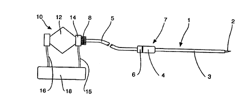
FIG. 9 shows one embodiment of medical apparatus having a hollow probe
1, a flexible connecting tube 5 and a signal generating device 10. The hollow
probe
1 consists of an inner needle 2 and an outer needle 3 and a connection portion
4.
The attached connecting tube 5 has a connection portion 6 which together with
the
connection portion 4 forms a connection 7. The bore of the inner needle 2 is
continuous with the inner lumen of the attached connecting tube 5. The other
end
of the connecting tube 5 has a connection assembly 8 that allows it to be
couple to
the signal generating device 10.
The signal generation device 10 has a housing 12 which contains an
acoustic signal generator, a connection portion 14 that allows it to be fitted
to the
connection tube 5 by means of the connection assembly 8 and electrical wires
15
and 16 which connect the signal generating device 10 to an electronic control
circuit
18. The electronic control circuit 18 is connected to the interrogation
assembly
within the connection portion 14 by the wires 15 and this reads the
information
encoding component within the connection assembly 8 and hence provides
frequency
control for the signal generating device 10 to give an optimum frequency
acoustic
signal transmitted down the hollow probe.
FIG. 2 shows the preferred embodiment of the coupling 7 of the hollow
probe 1 to the connecting tube 5. This coupling is constructed in such a way
that
the inner needle 2 and the connecting tube 5 can be uncoupled and withdrawn
from
the outer needle 3. This leaves the outer needle 3 insitu for various medical
procedures such as aspiration or sample collection or accurate placement of
pharmaceuticals. The inner needle 2 and the connecting tube 5 are joined by a
fitting
20. The inner lumen 21 of the joining fitting 20 is formed in such a way that
there
is a smooth transition, free from perpendicular surfaces, from the inner walls
of the
connecting tube 5 to the inner needle 2. By this means there is minimal loss
or
distortion of the acoustic signal. The fitting 20 is coupled to the hub 22 on
the outer
needle 3 by means of a rotating locking ring 23 which locks to the hub 21 by
means
of a mating threads 24 and 25 on the two components respectively.
CA 02278540 1999-07-16
WO 98/31285 PCT/IB98/00143
_g_
FIG. 3 shows a preferred embodiment of the coupling of connecting
portion 8 on the connecting tube 5 and connection portion 14 on the signal
generating device 12. This coupling is constructed in such a way an
information
encoding device 30, situated on a hub 31 at the end of the connecting tube 5
of the
hollow probe, makes contact with an interrogating means 34 situated on fitting
14
on the signal generating device. The interrogating means 34 transmits
appropriate
information from the encoding component 30 to an electronic control circuit 18
(refer FIG. 1 ) via .an electrical wire 35. The inner lumen 36 of fitting 14
is formed
in such a way that there is a smooth transition, free of perpendicular
surfaces, of the
walls of the inner lumen 36 from the signal generating device 10 to the
connecting
tube 5 through the hub 31. By this means there is minimal loss or distortion
of the
acoustic signal. The hub 31 is coupled to fitting 14 by means of a rotating
locking
ring 32 which locks to fitting 14 by means of mating threads means 37 and 38
on
the two components respectively.
FIG. 4 shows detail of one embodiment of the information encoding
component 30 on hub 31 which is part of the fitting 8 (refer FIG. 1 ) at the
proximal
end of the hollow probe 1, and the interrogating means 34 on fitting 14 of the
frequency generating device 10 (refer FIG. 1 ). The information encoding
component
30, for example a surface-mount resistor of known value, is situated on the
end of
a post 40 on hub 31. The interrogating means 34 consists of two metal contacts
42 each supported by a spring 44. When the hub 31 is coupled to fitting 14 the
metal contacts 43 on the information encoding component 30 make contact with
the
metal contacts 42 of the interrogating means 34 which transmits appropriate
information from the encoding component 34 to an electronic control circuit 18
(refer
FlG. 1 ) via electrical wires 35.
One example of the use of this embodiment of the invention will now be
described with reference to FIG. 5.
The selected hollow probe 1 has at the distal end of the inner needle 2 a
closed end 46 and a side hole 47. Fitting 8, at the end of the probe system,
is
attached to fitting 14 on the signal generating device 10. The interrogating
means
34 in fitting 14 transmits appropriate information from the information
encoding
CA 02278540 1999-07-16
WO 98/31285 PCT/IB98/00143
_g_
component 30 in fitting 8 to an electronic control circuit 18 via the
electrical wire 15.
The electronic control circuit 18 subsequently controls the signal generating
device
10, via an electrical wire 16, such that the signal generating device 10
generates the
appropriate frequency and amplitude for the selected probe, as coded by the
information encoding component 30.
The needle assembly of inner needle 2 and outer needle 3 on the hollow
probe 1 is inserted into the tissue 50 from which a biopsy is to be taken. As
it is
inserted, the signal generating device 10 generates a longitudinal oscillation
of the
air column at a sub-ultrasonic frequency, within the connecting tube 5 and the
inner
needle 2, which induces an oscillation 52 of the tissue near the tip of the
needle
around the side hole 47, enhancing the visibility of the needle tip to Doppler
ultrasound imaging from an imaging device 54.
When the tip of the needle is situated in the appropriate position within the
tissue, the inner needle 2 is withdrawn from the outer needle 3, without
disturbing
the position of the outer needle 3, and suction is applied to the hub 22 at
the end of
the outer needle 3, in order to collect tissue or liquids for biopsy.
It should be appreciated that the scope of the present invention is not
limited to the example described above where it is used to locate the tip of a
biopsy
needle but also relates to the localization of the tip of any hollow probe for
any
reason.
FIG. 6 shows an alternative embodiment of the connection assembly 7
between the needle assembly of the inner needle 2 and outer needle 3 and the
connecting tube 5. It should be noted, however, that a similar connection
system
could be used where the needle assembly of the inner needle 2 and outer needle
3
is connected directly to the signal generating means without the use of the
connection tube 5.
In this embodiment the connecting portion 4 has an inner lumen 60 which
is of a frustoconical shape which is the same taper as standard Luer
connection used
in medical technology. This allows the direct connection of the outer needle
to a
syringe or vacuum extraction device after positioning and removal of the inner
needle
2.
CA 02278540 1999-07-16
WO 98/31285 PCT/IB98/00143
- 10-
It may be noted, too, that an inner needle need not be used in all cases
with either the connecting tube 5 or the signal generating means connecting
directly
to the outer needle 3.
FIGs. 7, 8 and 9 show an alternative embodiment of the medical apparatus
of the present invention comprising a hollow probe and a signal generating
device.
The medical apparatus comprises a needle portion and a signal generator
portion.
The needle portion comprises a needle hub 70 with a needle 71 extending
therefrom. The needle hub 70 has a threaded annular flange 72 at the end
opposite
from the needle 71. The thread is a male thread 74. The needle hub has an
inner
lumen 75 which is smoothly tapered to the needle connection point 76. Within
the
needle hub 70 there is also an information encoding component 78 such as an
electrical resistor, capacitor and/or programmable device.
The signal generator portion comprises a body 80 with a rear housing 84
enclosing a speaker 82 and a locking ring 86. The locking ring 86 has a female
thread 88. The body 80 has an inner lumen 90 which is smoothly tapered from
the
speaker output cone 92 to the beginning of the inner lumen 75 of the needle
hub 70.
Also in the body is an interrogating means 94 which when the needle hub is
connected to the body engages the information encoding component 78. Wires 91
extend to the speaker 82 and wires 95 extend to the interrogating means 94.
A suitable alignment groove 79 in the needle hub and projection 93 on the
body ensures correct mating of the signal generator portion to the needle hub.
The
locking ring 86 is freely movable on the body 80 so that once the alignment
groove
79 in the needle hub and projection 93 on the body have been engaged the male
thread 74 can engage the female thread 88 to securely connect the two portions
and
ensure correct alignment of the respective inner lumens 75 and 90 and correct
connection of the interrogation means 94 with the information encoding device
78.