Note: Descriptions are shown in the official language in which they were submitted.
CA 02280087 1999-08-04
WO 98/35215 PCT/US98/01285
SEE-CjEI-CATIQN
LASER CAPTURE MICRODISSECTION ANALYSIS VESSEL
BACKGROUND OF THE I]'~VENTION
1. Field of the Invention
The present invention relates to laser capture microdissection. More
particularly, the present invention relates to consumables for laser capture
microdissection and to liquid reagent vessels for holding transfer films used
in laser
capture microdissection.
2. The Prior Art
Diseases such as cancer have long been identified by examining tissue
biopsies to identify unusual cells. The problem has been that there has been
no
satisfactory prior-art method to extract the cells of interest from the
surrounding
tissue. Currently, investigators must attempt to manually extract, or
microdissect,
cells of interest either by attempting to mechanically isolate them with a
manual tool
or through a convoluted process of isolating and culturing the cells. Most
investigators consider both approaches to be tedious, time-consuming, and
inefficient.
A new technique has been developed which can extract a small cluster of
cells from a tissue sample in a matter of seconds. The technique is called
laser capture
microdissection (LCM). Laser capture microdissection is a one-step technique
which
integrates a standard laboratory microscope with a low-energy laser and a
transparent ethylene vinyl acetate polymer thermoplastic film such as is used
for the
plastic seal in food product packaging.
-1-
CA 02280087 1999-08-04
WO 98/35215 PCT/US98/01285
In laser capture microdissection, the operator looks through a microscope at a
tissue biopsy section mounted on a standard glass histopathology slide, which
typically contains groups of different types of cells. A thermoplastic film is
placed
over and in contact with the tissue biopsy section. Upon identifying a group
of cells
of interest within the tissue section, the operator centers them in a target
area of the
microscope field and then generates a pulse from a laser such as a carbon
dioxide
laser having an intensity of about 50 mW and a pulse duration of between about
50
to about 500 mS. The laser pulse causes localized heating of the plastic film
as it
passes through it, imparting to it an adhesive property. The cells then stick
to the
localized adhesive area of the plastic tape directly above them, whereupon the
cells
are immediately extracted and ready for analysis. Because of the small
diameter of
the laser beam, extremely small cell clusters may be microdissected from a
tissue
section.
By taking only these target cells directly from the tissue sample, scientists
can
immediately analyze the gene and enzyme activity of the target cells using
other
research tools. Such procedures as polymerase chain reaction amplification of
DNA
and RNA, and enzyme recovery from the tissue sample have been demonstrated. No
limitations have been reported in the ability to amplify DNA or RNA from tumor
cells
extracted with laser capture microdissection.
Laser capture microdissection has successfully extracted cells in all tissues
in
which it has been tested. These include kidney glomeruli, in situ breast
carcinoma,
atypical ductal hyperplasia of the breast, prostatic interepithielial
neoplasia, and
lymphoid follicles. The direct access to cells provided by laser capture
microdissection will likely lead to a revolution in the understanding of the
molecular
-2-
CA 02280087 1999-08-04
WO 98/35215 PCT/US98/01285
basis of cancer and other diseases, helping to lay the groundwork for earlier
and
more precise disease detecuon.
Another likely role for the technique is in recording the patterns of gene
expression in various cell types, an emerging issue in medical research. For
instance,
the National Cancer Institute's Cancer Genome Anatomy Project (CGAP) is
attempting to define the patterns of gene expression in normal, pre-cancerous,
and
malignant cells. In projects such as CGAP, laser capture microdissection is a
valuable
tool for procuring pure cell samples from tissue samples.
Laser capture microdissection, like all emerging techniques, still has room
for
improvement. With further refinement of the plastic film and activation by a
more
tightly focussed laser bearn, the technique extraction of single cells can
easily be
foreseen.
It is therefore an object of the present invention to provide a liquid reagent
biological analysis vessel which will facilitate laser capture microdissection
analysis.
It is another object of the present invention to provide a liquid reagent
biological analysis vessel f'or use with laser capture microdissection
analysis which
allows greater automation of the analysis process.
Yet another object of the present invention is to provide a liquid reagent
biological analysis vessel for use with laser capture microdissection analysis
which is
less susceptible to DNA contamination than is presently encountered.
-3-
CA 02280087 2005-11-30
50378-1
BRIEF DESCRIPTTON OF THE INVENTION
Designs for laser capture microdissection consumables according to the
present invention involve incorporating a laser capture microdissection lift-
off
substrate comprising a transparent thermoplastic film into substrate. The
substrate
may be, for example a cap or other structure which may be associated with an
analysis vessel such as an Eppendorf TubeTM.
According to its broadest aspect, the present invention comprises any method
to hold a thermoplastic lift-off substrate film in a fixed position relative
to a
biological analysis vessel. For example, the film may be affixed to a
structure which
is inserted into the vessel and is held therein such that the sample is held
in a fixed
position relative to an observation port on the vessel.
Because analysis of cells dissected from samples using the laser capture
microdissection process employs small volumes of liquid reagents such as
proteinase
solutions, the sample is held in a fixed position relative to the observation
port of the
vessel so that is may be verified that the tissue sample has been immersed in
the
liquid reagent.
In the simplest embodiments of the present invention, the present invention
comprises a cap for the analysis vessel. The cap is formed from a machined or
injection molded material such as plexiglas, and includes a window for holding
the
lift-off substrate film. If the cap is formed from a transparent material, the
film-may be
disposed upon a window portion of the cap. The film may also be disposed
across
an aperture formed in the cap with the laser propagating through the through
hole
in the cap.
-4-
CA 02280087 2005-11-30
50378-1
According to a first embodiment of the present invention, an aperture is
disposed through the cap of an analysis vessel, such as an epindorf tube. A
thermoplastic lift-off substrate film is fastened across the aperture by
gluing, by
welding the thermoplastic around the periphery of the aperture or by some
other
known fastening means which holds the film in place. The film itself acts as a
transparent window that allows for inspection of the lifted tissue sample.
To perform the laser capture microdissection, the cap and film assembly is
placed in a suitable holder in the laser capture microdissection apparatus and
is
aligned in contact with the slide containing the tissue sample. The operator
then
centers the cells of interest on the laser beam target and then activates the
laser pulse
to weld the selected cell group to the film.
After welding the tissue sample of interest to the film affixed to the cap, a
small volume of proteinase solution is placed in the ependorf tube, the cap is
placed
on theEpPendorf TubeTM and the tube is inverted. The tissue is dissolved and
the DNA is
free to enter the solution. The solution is then pipetted out of the tube and
into the
PCR mixture for analysis. The positioning of the film over the through hole
permits
inspection to insure that the sample has been dissolved.
According to a second embodiment of the present invention, the cap
comprises a solid piece having transparent optical quality windows on its top
and
bottom surfaces. The film is deposited or otherwise affixed to the surface of
the
bottom window of the cap using a process such as spin coating, dipping or
spraying.
The tissue is then welded to the top of the cap as described for the first
embodiment
with the laser propagating through the optical windows on the cap.
-5-
CA 02280087 2005-11-30
50378-1
The vessel may be the well known Eppendorf TubeTM,
but persons of ordinary skill in the art will appreciate
that the techniques described herein may be used for caps
that are configured to fit other standard biological fluid
analysis sample holders such as 96 well microtiter plates.
According to a third embodiment of the present
invention, the LCM transfer film is affixed to a disk or
plug which may be inserted into the analysis vessel.
According to the present invention, if it is
desired to combine more than one tissue sample in a single
analysis vessel, a first tissue sample is dissolved in the
vessel. The first cap, disk, or plug is then removed from
the analysis vessel and a second cap, disk, or plug is then
inserted into the vessel. In this manner, more than one
tissue sample may be easily combined in a single analysis
vessel.
According to one aspect of the present invention,
there is provided a carrier for laser capture
microdissection analysis comprising: a substrate, said
substrate structured and configured to matingly engage an
analysis vessel; and a laser capture microdissection
transfer film mounted on said substrate.
According to another aspect of the present
invention, there is provided an assembly comprising: a
holder having a plurality of indexed positions thereon; a
plurality of carriers for laser capture microdissection
analysis, each carrier comprising a substrate and a laser
capture microdissection film mounted on said substrate; and
one of said carriers disposed at each indexed position in
said holder, said indexed positions configured to precisely
locate the position of each said substrate relative to an
- 6 -
CA 02280087 2005-11-30
50378-1
element of a laser capture microdissection tissue transfer
apparatus.
BRIEF DESCRIPTION OF THE DRAWING FIGURES
FIGS. la and lb are side and top views,
respectively of a cap for an analysis vessel including a LCM
transfer film according to a first embodiment of the present
invention.
FIG. 2 is a side view of an analysis vessel
positioned to mate with the cap of FIGS. la and lb.
FIGS. 3a and 3b are side and top views,
respectively, of a cap for an analysis vessel including a
LCM transfer film according to a second embodiment of the
present invention.
FIG. 4 is a side view of an analysis vessel
positioned to mate with the cap of FIGS. 3a and 3b.
- 6a -
CA 02280087 1999-08-04
WO 98/35215 PCTIUS98/01285
FIG. 5 is a side view of an analysis vessel cap according to the present
invention shown positioneci over a glass slide and tissue sample and placed
under a
microscope objective, illustrating a laser capture microdissection procedure.
FIG. 6 is a perspective view of a cap for an analysis vessel including an
identifying serial number according to another aspect of the present
invention.
FIG. 7 is a perspective view of a cap for an analysis vessel showing a
procedure for affixing the LCM film thereto.
FIG. 8 is a perspective view of a cap for an analysis vessel showing an
alternate procedure for affixing the LCM film thereto.
FIG. 9 is an exploded perspective view of an analysis vessel including a cap
and a disk carrying an LCN[ filrn which may be disposed inside the vessel
according
to the present invention.
FIG. l0a is an exploded perspective view of an analysis vessel including a
cap and a plug carrying an LCM film which may be disposed inside the vessel
according to the present invention.
FIG. 10b is a side view of the analysis vessel of FIG. l0a showing the plug
disposed inside the vessel according to the present invention.
-7-
CA 02280087 1999-08-04
WO 98/35215 PCT/US98/01285
FIG. 11 is a diagram of a substrate carrier that slides into a mating mount
which is indexed to precisely locate the position of the substrate relative to
an
element of an LCM tissue transfer apparatus.
FIG. 12 is a diagram of an alternative method for mounting the substrate
carrier.
DETAII.ED DESCRIPTION OF A PREFERRED EMBODIMENT
Those of ordinary skill in the art will realize that the following description
of
the present invention is illustrative only and not in any way limiting. Other
embodiments of the invention will readily suggest themselves to such skilled
persons.
Referring first to FIGS. la and lb, side and top views, respectively, are
presented of a cap 10 for an analysis vessel including a LCM transfer film
according
to a first embodiment of the present invention. FIG. 2 shows Cap 10 positioned
to
mate with analysis vessel 12, which may be, for example, an eppindorf tube.
Cap 10 may be formed from a material such as machined or die cast plexiglas,
and includes an aperture 14 formed therethrough and a LCM transfer film layer
16,
which may be formed from a thermoplastic film material such as ethylene vinyl
acetate (EVA) polymer, is attached to cap 10 covering aperture 14. LCM
transfer
film 16 may be fastened to the surface of the underside of cap 10 by various
known
fastening means. Such films for LCM application have thicknesses typically in
the
range of from about 10 to about 500 microns.
-8-
CA 02280087 1999-08-04
WO 98/35215 PCT/US98/01285
The thermoplastic iFilm is preferably a 100 micron thick ethyl vinyl acetate
(EVA) film available froni Electroseal Corporation of Pompton Lakes, New
Jersey
(type E540). The film is chosen to have a low melting point of about 90 C.
This
particular EVA film has been doped with an infrared napthalocyanine dye,
available
from Aldrich Chemical Company (dye number 43296-2 or 39317-7). These dyes
were chosen because they have a strong absorption in the 800 nm region, a
wavelength region that overlaps with an high power AlGaAs laser diode (model
OPCA001-808FC available from Opto Power Corporation, Tucson Arizona) which is
used to selectively melt the film. The dye is mixed with the melted bulk
plastic at a
temperature of about 120 C. The dyed plastic is then manufactured into a film
using
standard film manufacturing techniques. The dye concentration in the plastic
is
about 0.001 M.
As may be seen most easily from an examination of FIG. l b, aperture 14
provides an unobstructed view of LCM transfer film 16 through the body of cap
10.
According to the present invention, a tissue sample may be attached to LCM
transfer film 16 by placing the cap 10 over the slide containing the sample
and
generating a laser pulse as is known to perform laser capture microdissection.
Cap
10, with the captured tissue sample, may then be mated with analysis vessel
12.
After the cap 10 has been mated with analysis vessel 12, aperture 14 provides
a convenient way to observe the sample in analysis vessel 12. This is
particularly
useful for procedures in which the sample is to be dissolved by a small volume
of
proteinase solution placed in the vessel 12. Aperture 14 permits observation
of
whether the sample has contacted the solution and has been dissolved.
Referring now to FIGS. 3a and 3b are side and top views, respectively, are
shown of a cap 20 for an analysis vessel including a LCM transfer film
according to
-9-
CA 02280087 1999-08-04
WO 98/35215 PCT/US98/01285
a second embodiment of the present invention. FIG. 4 is a side view of an
analysis
vessel 22 positioned to mate with the cap of FIGS. 3a and 3b, which may be,
for
example, an eppindorf tube.
Cap 20 includes an optical window 24 through its body and a layer of LCM
transfer film 26 disposed over the surface of optical window 24 at the
underside of
the cap 20. If cap 20 is made from a material such as plexiglas, window 24 is
easily
formed by controlling the surface characteristics of the cap as is known in
the art.
LCM transfer film 24 may be affixed to the window surface of the underside of
cap
20 using various known fastening techniques.
As with the aperture 14 of the embodiment of FIGS. la and lb, window 24
provides an unobstructed view of LCM transfer film 26 through the body of cap
20.
As with the embodiment of FIGS. la and lb, a tissue sample may be attached to
LCM transfer film 24 by placing the cap 20 over the slide containing the
sample and
generating a laser pulse to perform laser capture microdissection. Cap 20,
with its
captured tissue sample, may be mated with analysis vessel 22.
After the cap 20 has been mated with analysis vessel 22, window 24 provides
a convenient way to observe the sample in analysis vessel 22. This is
particularly
useful for procedures in which the sample is to be dissolved by a small volume
of
proteinase solution placed in the vessel 22 since window 24 permits
observation of
whether the sample has contacted the solution and has been dissolved.
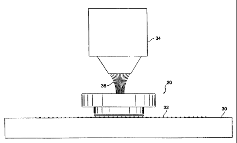
FIG. 5 is a side view of an analysis vessel cap 20 according to the present
invention shown positioned over a glass slide 30 and tissue sample 32. The
slide 30
and cap 20 are placed under a microscope objective 34, and a laser pulse,
shown
-10-
CA 02280087 1999-08-04
WO 98/35215 PCTIUS98/01285
diagrammatically at reference numeral 36 is directed at a selected region 36
of the
tissue sample 32 to perfoirm laser capture microdissection of the tissue
sample 32.
FIG. 5 illustrates the ease of use of the present invention, since the
integral cap and
LCM transfer film assemibly is easily handled, either manually or by automated
means, and thus greatly i'acilitates obtaining the LCM sample and decreases
the
possibility of DNA contzunination of the sample during handling and transport.
Those of ordinary skill in the art will appreciate that an alternate
configuration
which may be employed is an inverted microscope wherein the tissue sample may
be
viewed from underneath the sample slide. Such skilled persons will appreciate
that
the present invention may easily be used in such a configuration.
According to another aspect of the present invention, the cap of FIGS. la and
lb and 3a and 3b may be provided with an identifying serial number. FIG. 6 is
a
perspective view of a cap 20 for an analysis vessel including an identifying
serial
number 38 according to this aspect of the present invention.
As an example of this aspect of the present invention, cap 20 is equipped
with a marking means 38 such as a UPC label or laser etched label. Serializing
all of
the caps provides for easy iidentification and tracking of cell sarnples. The
label may
be read by a sensor, such as a UPC label sensor or OCR sensor which is mounted
in
or on the laser capture microdissection apparatus.
According to this aspect of the present invention, the serial number 38 is
placed on the top of the cap. Placirig the serial number on the top of cap 20,
and the
thickness of cap 20 is selected to be larger than the depth of field of the
microscope
objective. Thus, the microscope can be focussed on the tissue sample below the
-11-
CA 02280087 1999-08-04
WO 98/35215 PCT/US98/01285
bottom surface of the cap and not have the label or serial number interfere
optically,
since the label is far from the focal plane of the imaging lens and is thus
out of focus.
There are several ways in which the LCM transfer film may be affixed to the
surface of the caps according to the present invention. FIG. 7 is a
perspective view
of a cap 40 for an analysis vessel illustrating a step in a first procedure
for affixing
the LCM film thereto.
First, a small, e.g., about 1 cm square piece of EVA film 42 is cut. The fiim
42 is
gently pressed onto the bottom surface of the cap 40 making it stick thereto.
A
glass microscope slide 44 is heated to about 100 C on a hot plate. A
0.002 inch thick piece of mylar plastic release liner 46 is placed on the
slide. As is
known in the art, a release liner is a plastic sheet that is coated with a
silicone
coating so it does not stick to the EVA film material or the glass slide.
The cap 40 with its attached film 42 is pressed onto the release liner/slide
assembly for about 5 seconds or until the film melts. The cap 40 with attached
film
42 and release liner 46 is then removed from the hot glass slide 44, cooled
down to
room temperature, and the release liner 46 is peeled off. Finally, the excess
EVA film
is trimmed off.
FIG. 8 is a perspective view of a cap for an analysis vessel showing an
alternate procedure for affixing the LCM film thereto. According to this
method, a
piece of transparent double sided adhesive tape 48 (such as standard double
stick
tape available from 3M Corporation) may be used to tape the EVA film 42 to the
bottom of the cap 40. The excess EVA film may then be trimmed off.
-12-
CA 02280087 1999-08-04
WO 98/35215 PCT/US98/01285
As will be appreciated by those of ordinary skill in the art from an
examination of the disclosure, the present invention involves holding the LCM
transfer film in a fixed position relative to a biological analysis vessel. In
addition to
the vessel cap embodiments of FIGS. la and lb and 3a and 3b, other embodiments
of
the present invention are contemplated herein. In general, the LCM transfer
film
may be affixed to something that may be placed into the vessel such that the
film
and its adhered tissue sample is held in a fixed position relative to an
observation
port on the vessel.
FIGS. 9 and l0a and lOb illustrate embodiments of the present invention
wherein the observation port may be located on the cap or may be located
elsewhere on the vessel. Referring now to FIG. 9,an exploded perspective view
is
presented of an analysis vessel 50 including a cap 52 and a clear plastic disk
54
carrying an LCM film 56 which may be disposed inside the vessel 50 according
to
the present invention. The methods disclosed herein and other known methods
may
be used to affix LCM transfer film 56 to disk 54. A viewing port 58 may be
provide
at either the cap 52, the bottom of analysis vessel 50, or at both locations.
As will be appreciated by those of ordinary skill in the art, disk 54 has a
shape
which mates with the cross. section of the interior of analysis vessel 50 and
is sized
for a slip fit such that it may be easily inserted therein but constrained
from
significant lateral motion orice inside the analysis vessel 50.
Referring now to FIGS. l0a and lOb, yet another embodiment of the present
invention is depicted. FIG. l0a is an exploded perspective view of an analysis
vessel 60 including a cap 62 into which a plug 64 carrying an LCM transfer
film 66
may be inserted according to the present invention. FIG. 10b is a side view of
the
-13-
CA 02280087 2005-11-30
50378-1
analysis vessel of FIG. 10a showing the plug disposed inside
the vessel according to the present invention. The LCM
transfer film may be affixed to the surface of plug 64 as
taught herein. A viewing port 58 may be provided at either
the cap 62, the bottom of analysis vessel 60, or at both
locations.
Those of ordinary skill in the art will appreciate
that the embodiments of FIGS. 9, 10a and lOb may be used
without the need for a solid cap for the analysis vessel.
Techniques such as use of low vapor pressure oils are known
for reducing evaporation of the liquid reagents used in LCM
analysis and the present invention may be employed with such
techniques.
Those of ordinary skill in the art will recognize
that other orientations are contemplated according to the
present invention, such as ones wherein the film is held in
the analysis vessel in a fixed position with respect to a
viewing port located at other positions on the analysis
vessel. In general, the present invention provides in
combination a LCM film mounted on a substrate which may be
transparent if necessary for the particular application.
While embodiments described herein comprise caps, disks and
plugs, the substrate of the present invention is not limited
to such particular forms. Because the LCM film is mounted
in a fixed position on the substrate, it may be easily
oriented with respect to other elements of an LCM tissue
sample transfer device or analysis vessel.
As will be appreciated by those of ordinary skill
in the art, the carrier comprising the substrate and mounted
LCM transfer film of the present invention is easily used
with apparatus for performing LCM tissue sample transfers.
A plurality of substrates may be arranged according to the
- 14 -
CA 02280087 2005-11-30
50378-1
present invention to provide a degree of automation to the
LCM process.
- 14a -
CA 02280087 1999-08-04
WO 98/35215 PCT/US98/01285
Referring now to FIG. 11, a diagram is presented of a substrate carrier 70
that
slides into a mating mount 72 which is indexed to precisely locate the
position of the
substrate relative to an element of an LCM tissue transfer apparatus. Many
substrates can be evaluated in sequence by using a dovetail slide 70 which may
be
mounted to the side of the translation stages located on the microscope used
in the
LCM apparatus. This slide 70 can contain strip 72 holding a number of
substrates
74 and vials in a parallel row of vial holder apertures 76 as indicated in
FIG. 11. The
dove tail slide can be indexed to provide precise locating of the substrates
and vials
to simplify loading and unloading. The substrates are contained in a plastic
carrier
that can be bar coded to provide a unique identification number for this set
of
substrates. The individual substrates can each be individually labeled on the
top
surface thereof as seen in F'IG 6 to provide a unique identifier. Since the
top surface
of the substrate 74, shown in FIG. 11 in the form of a cap, is several
millimeters from
the location of the transfen=ed tissue the labels will not effect the
illumination or the
viewing of the tissue sample since the label will be far from the focal plane
of the
imaging lenses and out of focus. FIG. 12 is a diagram of an alternative method
for
mounting the substrate carrier.
While embodiments and applications of this invention have been shown and
described, it would be apparent, to those skilled in the art that many more
modifications than mentioned above are possible without departing from the
inventive concepts herein. The invention, therefore, is not to be restricted
except in
the spirit of the appended cl.aims.
-15-