Note: Descriptions are shown in the official language in which they were submitted.
CA 02280757 1999-08-12
WO 98/35606 PCT/US98/03069
METHOD AND APPARATUS FOR MINIMALLY INVASIVE PELVIC SURGERY
Field of the Invention
The present invention relates to methods and devices for improving urinary
incontinence. More particularly,
the present invention relates to methods and devices for creating a cavity
near the urethral floor, methods and
devices for placement of a urethral sling or other device in such a cavity,
and methods and devices for driving bone
piercing guides into and through the pubic bone for use in stabilizing the
urethral or pelvic floor.
Background of the Invention
The present invention relates to the treatment of stress urinary incontinence
"SUI," and to improved
methods and surgical devices for the surgical treatment of SUI. The devices
disclosed herein are additionally useful
in a wide variety of other surgical procedures.
Genuine stress incontinence is the involuntary loss of urine due to a sudden
rise in intra~abdominal pressure.
It has been estimated that between 40~° and 50% of young, healthy
nulliparous women admit to occasional mild
stress incontinence; however, at least 80% of stress incontinence patients are
in the perimenopausal age group and
are multiparous. Raz has suggested that the female urethral continence
mechanism is dependent on the interaction
of four urethralfactors: urethralclosing pressure, urethrallength,
urethrotrigonafanatomy, and urethralreception of
intra~abdominal pressure. Raz, S., Modified bladder neck suspension for female
stress incontinence, Uroioay, 17:82,
1981.
The urethral closing pressure is predominantly a result of the interaction of
smooth and striated muscle
sphincter activity, but there is also some contribution by nonmuscular
urethral factors such as the submucosal
vascular plexus, the elastin and collagen content of the urethral tissues, and
a sphincter like effect of the mucosa.
There has been considerable diversity of opinion regarding the anatomic
structure and the innervation of the urethral
sphincters, and a variety of views have been expressed in the literature.
Urethral length is important in the maintenance of continence. However,
although it certainly interacts with
other factors to contribute to continence, a short urethra alone will not
produce incontinence. Urethral length varies
considerably in normal women, and women with proven genuine stress urinary
incontinence do not invariably have
urethral shortening.
Urethrotrigonal anatomy, which can be demonstrated by lateral
cystourethrography, should fulfill certain
criteria. The bladder base should lie above the level of the inferior ramus of
the symphysis, and with straining should
not descend more than 1.5 cm. There should be a normal urethrotrigonal
alignment with an angle normally less than
100 degrees, and the urethras axis should be approximately 35 degrees from the
vertical. In the hypermobile
situation loss of all of the normal anatomic features may occur, a radiologic
finding that correlates with the clinical
finding of cystourethrocele. However, clinical experience has shown that the
coexistence of cystourethrocele and
incontinence does not predict that the incontinence is of a genuine stress
variety.
CA 02280757 1999-08-12
WO 98/35606 PCT/US98/03069
2.
The transmission of intro-abdominal pressure to the intro-abdominal portion of
the proximal urethra is also
reported to be important in the maintenance of continence. This is a passive
phenomenon, and is the result of the
normal anatomic configuration just described. Whenever there is a rise in
infra-abdominal pressure during such
stresses as coughing or straining, the pressure is transmitted not only to the
bladder hut also to the proximal urethra,
with resultant increase in the closing pressure, and prevention of leakage. If
the urethra) axis is altered, rotational
descent will drop the proximal urethra and bladder base from its intro-
abdominal location, and will obviously impair
such pressure transmission.
A wide variety of operations have been used to correct this condition,
generally involving the principles of
elevating the bladder neck anteriorfy andlor elongating and narrowing the
proximal urethra. Two of the most popular
operations today for stress incontinence are the Marshall-Marchetti-Krantz and
Birch vesicourethropexies. The
Marshall-Marchetti-Krantz technique has at least an eighty-five percent
success rate, against which other operative
success rates must be measured. Recently, the Pereyra operation and its
modifications have enjoyed some popularity,
but less than basic techniques.
Notwithstanding the foregoing, however, there remains a need for an improved
treatment for SUI.
Preferably, the treatment is as noninvasive as possible under the
circumstances, and will eliminate or minimize
hospitalization and the use of general anesthetics. In addition, there remains
a need for improved medical
instrumentation such as tissue cavity dilators, incision guides, bone-piercing
guide drivers, and Quick-connect slings
and suture-securing devices for use in connection with SUl treatment and other
medical procedures. U.S. Patent No.
5,611,515, issued March 18, 1997 to eenderev et al., introduces pioneering
minimally invasive percutaneous and
transvaginal bladder neck stabilization approaches. The percutaneous approach
of Benderev et al. involves stabilizing
the bladder neck using a bone anchor which is percutaneously introduced from
the abdominal side of the patient.
The transvaginal approach of Benderev et al. involves stabilizing the bladder
neck using a staple or bone anchor
which is transvaginally placed into the pubic bone. The methods and devices of
the present invention may be used
in several urethra) or bladder neck stabilization procedures that are less
invasive than many of those currently
available.
Summary of the Invention
It is an objective of this invention to provide a means and method for
relatively sterile placement of urethras
slings. It is a further objective of this invention to provide an apparatus
for straight line positioning for bone
piercing, so as to achieve proper placement of urethra) slings and to minimize
difficulty in aligning a bone-piercing
apparatus with the ultimate target in a tissue cavity. Another objective is to
provide apparatus and a method for
reconstructing and stabilizing the urethra) or pelvic floor by affixing
devises placed to support the urethra) or pelvic
floor to a fixed reference tissue such as a bone. A further objective is to
provide improvements over current
techniques that require drilling holes in a bone, and the placement of bone
anchors therein.
This invention has the additional objective of providing rapid and simple
surgical connections for connecting
a suture to a medical device inside a tissue cavity or other structure in the
body that may be in need of stabilization.
CA 02280757 1999-08-12
WO 98/35606 PCT/f<JS98/03069
-3
This invention also seeks to provide alternatives to transvaginal methods of
urethral and pelvic floor reconstruction
and stabilization, to minimize the risk of infection, and to enable surgeons
to approach the urethral or pelvic floor
from different locations. Finally, it is a further objective to provide
minimally invasive means and methods of securing
a target tissue to an immoveable reference tissue, such as the pubic bone. One
of more of these objectives is
satisfied by various embodiments of the invention.
The invention provides a dilator for creating a cavity in tissue. The dilator
has two functional portions:
an insertion spreader and handles. The insertion spreader includes of two
facing guides that may be semi-cylindrical.
The spreader has open and closed positions. In the closed position the guides
are close together and the dilator may
have the appearance of a split tube or cylinder, while in the open position
the guides are separated. In bath
positions the guides remain essentially parallel to each other.
The insertion spreader may be attached to the handles for manipulation of the
guides. The handles can
be joined together with a pivot, so that pivoting the handles translates to a
movement of the guides either toward
or away from one another. The dilator may also have a ratcheting lock for
maintaining the insertion spreader in a
fixed position. The penetrating ends of the guides also may be sharpened to
facilitate penetrating the target tissue.
The dilator aspect of the invention also provides a method of creating a
tissue cavity by using the dilator.
With the insertion spreader in the closed position the spreader is advanced
into the target tissue. When the spreader
reaches the desired depth the handles are moved to separate the guides. The
separation of the guides causes a
tearing of the tissue, creating a cavity therein. This method also may be
employed by first advancing a needle
partially into the tissue to create an insertion path. The guides of the
spreader are positioned about the protruding
part of the needle and inserted into the tissue along the same path created by
the needle.
This method for spreading tissue with the dilator of the invention may be used
to create a cavity in the
vaginal hiatus. The term "vaginal hiatus" refers to the tissue between the
urethra and the vagina. This term may
apply to the exterior surface between the distal urethra and the vaginal
orifice as well as to the deeper tissue
between the urethra and the upper vaginal wall. In some cases spreading may be
facilitated by performing an
episiotomy of the skin of the vaginal hiatus. The method of this aspect of the
invention also may be performed
transvaginally to create a cavity, for example in the vaginal wall. Whatever
the tissue, the method may be preceded
with a fluid-dissection of the target tissue, wherein a solution is injected
into the tissue to create a fluid bolus. The
fluid bolus forms a pocket in the tissue, and the dilator is used to create an
opening connecting the outer surface
to the pocket.
The invention also provides an insert card for advancing a medical device, for
example a urethral sling, into
a tissue cavity. One end of the card holds the sling to be used for
stabilizing tissue or internal structures of the
urethral or pelvic floor. The other end of the card is fashioned to permit a
physician to grasp and manipulate the
card, or to align or connect the card with other external devices, such as
those disclosed herein. The sides of the
card may be adapted for use with the dilator mentioned above. Thus the card
can be used to enhance both the
sterility and the positional precision in a sling-placement procedure.
CA 02280757 1999-08-12
WO 98/35606 PCT/US98/03069
-4-
The card and the dilator may thus be used in a method of advancing a sling
into a tissue cavity. The
tissue cavity is created by the dilator as described above, and the spreader
is locked in the open position. A sling
is placed in the proper position on the card, and the card is positioned so
that its lateral edges align with and slide
into the spreader guides. The spreader guides provide a track for the
insertion of the card to the desired depth
within the cavity. This method of sling placement may be used in procedures
employing a variety of techniques for
securing the sling, including techniques adapted for slings that are to be
secured with sutures, quick connect devices,
bone anchors, staples, and the like.
Also provided in this invention is an incision guide for creating a cavity
between the urethra and the vagina.
The incision guide has a catheter that is inserted into the urethra. This
catheter expands and straightens the
urethra, essentially immobilizing the urethra in an easily identified
position. Also part of the incision guide is a cutter
that slides along the catheter and makes an incision into the vaginal hiatus
that is a fixed distance from, and
therefore parallel to, the urethra. The catheter may display graduation marks
or other indicia to enable a surgeon
to determine the position of the catheter or the cutter relative to the
bladder neck.
The incision guide of the invention may also have a stop, such as a block or a
ring, that locks in place on
the catheter. The stop abuts the cutter and prevents insertion of the cutter
beyond the desired depth of incision.
The cutter portion of the incision guide may be a needle, a blade, a bipolar
knife, or other incision device
adapted for slidably mounting to the rigid catheter. One example of such an
adapted incision device is the dilator
of the invention as described above.
The incision guide aspect of the present invention provides a method of
creating a cavity in the vaginal
hiatus. The method includes the steps of inserting the catheter into the
urethra, determining the position of the
bladder neck by using the catheter, and inserting the attached cutter into the
vaginal hiatus. The catheter allows
straight-line tracking for the cutter and indicates the depth of incision,
thus avoiding injury to the bladder.
This method of creating a cavity in the vaginal hiatus may be used in concert
with the method of placing
a sling in a cavity by use of the card, as discussed above. The card
supporting the sling may advance into the
cavity having its edges in contact with the hiatal tissue along the sides of
the cavity. Alternatively the dilator of
the invention also may be used to serve as a guide for the card, after the
cavity is made using the incision guide
of the invention. When the card reaches the intended depth in the cavity, the
sling is in proper position for fastening
in place.
An additional aspect of the present invention provides a driver for driving a
guide into or through the pubic
bone. The driver has two jaws and a slide bar. The first jaw has a distal end
that inserts into a tissue cavity and
a proximal end that attaches to the slide bar. The second jaw slides along the
slide bar toward the first jaw. The
second jaw has a bone-piercing guide attached to it such that the guide moves
toward the first jaw when the second
jaw is advanced along the slide bar. The guide connected to the driver may be
a cannula, a needle, or a like device
adapted for driving through bone.
The driver provides a method of driving a guide through the pubic bone. The
steps include: inserting the
first jaw of the driver into a tissue cavity, locating the pubic bone,
positioning the driver to align the pubic bone
_~........_
CA 02280757 1999-08-12
WO 98/35606 PCT/US98/03069
-5
between the first jaw and the second jaw, and advancing the second jaw toward
the first jaw to drive the guide
through the pubic bone.
The invention further provides a method for passing a device through the pubic
bone. The guide is driven
through the pubic bone as outlined above. The guide is next retracted, leaving
a path through the bone, and the
device is passed through the pubic hone along the path made by the guide. The
device passed by this method may
be a suture, a suture passer, a quick-connect fastening device, and the like.
In an additional method of this aspect of the invention, the driver of the
invention is used to advance a
cannula through the pubic bone. The lumen of the cannula constitutes a channel
through the pubic bone. A device
may then be passed through the bone within the lumen of the cannula. Devices
that may be passed by this method
include a suture, a suture passer, a quick-connect fastening device, and the
like.
A further method of pelvic surgery provided by the invention includes the
following steps. A cannula is
driven through the pubic bone with the driver of the invention. The cannula is
further driven into the tissue cavity
in which the first jaw of the driver is positioned. The first end of a suture
is passed through the cannula and
secured to a structure within the cavity. The second end of the suture is
secured to the pubic bone, thereby
stabilizing tissue adjacent to the cavity. According to this method, the
cannula may be removed from the bone
before either end of the suture is secured, or the suture within the tissue
cavity may be secured before withdrawal
of the cannula. The suture within the tissue cavity may be secured by
stitching the suture through a tissue mass
of the cavity, or by attaching the suture to a structure introduced into the
cavity for stabilizing the tissue of the
cavity, such as a suture button.
The invention also provides a method of pelvic surgery wherein a cannula is
driven through the pubic hone
and into the tissue cavity as described above, and a suture is passed through
the cannula and into the cavity. The
suture is passed through a structure therein to stabilize the tissue adjacent
to the cavity, then the suture is passed
back out along the same path through the bone, and both ends of the suture are
secured to the pubic bone.
Yet another method of the invention involves driving a cannula through the
pubic bone and into a tissue
cavity in a first location to make a first path. The suture is then advanced
into the cavity along the first path.
The suture is passed through a structure of the cavity to stabilize the tissue
adjacent to the cavity. The cannula
is then driven through the bone and into the cavity along a second path, and
the suture is withdrawn from the cavity
along the second path. Both ends of the suture are then secured to the pubic
bone.
The foregoing methods focus on the path of the suture: the suture may be
advanced one-way into the
cavity and affixed there, or the suture may be advanced and withdrawn from the
cavity along the same path through
the bone, or the suture may be advanced and withdrawn along two separate paths
through the bone. Regardless
which method is used, the tissue cavity of the method may be the vagina.
Alternatively, the cavity may be a hiatal
cavity made according to a method of the dilator or incision guide aspects of
the present invention. Further, the
tissue cavity of the method may be a transvaginally created pocket into the
plane of the vaginal hiatus. Also
regardless which method is used, the method may advantageously be performed on
the left side of the cavity and
CA 02280757 1999-08-12
WO 98/35606 PCT/US98/03069
-6-
on the right side of the same cavity by piercing the pubic bone on both sides
lateral to the pubic symphysis. The
method may also include a step of tensioning the suture to elevate or
otherwise stabilize the tissue mass.
Further provided is a method of stabilizing a urethral sling relative to the
pubic bone. This is done by
creating a tissue cavity and creating a path through the pubic bone by driving
a guide through the bone. Then a
urethral sling is placed into the cavity. A suture is passed through the pubic
bone along the path, and is attached
to the tissue mass. The suture is then secured to the pubic bone to stabilize
the tissue.
Another aspect of the invention provides a driver frame assembly for
positioning and stabilizing a bone-
piercing guide driver relative to the patient. The driver frame assembly
includes an upper clamp and a lower clamp,
as well as a catheter, a cavity tongue, and the driver. The upper clamp has a
head portion, a descending arm, and
a base portion. The head portion has a compression foot for compressing the
patient's abdominal surface against
the pubic bone. Stabilizing pins extend downward from the compression foot and
penetrate the abdominal surface
adjacent to the superior surface of the pubic bone. The base portion of the
upper frame attaches to the catheter
and the tongue. The catheteris used to expand and straighten the urethra; the
tongue inserts into the cavity,
providing counterpressure to oppose the pressure of the compression foot. The
lower clamp has a buttock plate for
insertion beneath the patient, so that the patient's weight rests on the plate
to secure the frame assembly relative
to the patient. The lower clamp also has an ascending arm that connects with
the base portion of the upper clamp.
Finally, at least one driver is attached to the descending arm of the upper
clamp. There may be mare than one
driver mounted to the frame assembly, or there may be one driver that drives
two bone-piercing guides, which may
be displaced to the left and right of center relative to the patient.
This aspect of the invention provides a method for stabilizing pelvic tissue
by relatively non-invasive pelvic
surgery. The foregoing frame assembly is installed on the patient. The bone-
piercing guide is positioned and is driven
through the hone and into the cavity. A stabilizing device is passed along the
path through the hone created by the
guide and secured in the cavity, thus stabilizing the targeted tissue of or
adjacent to the cavity. This method may
employ two or more guides, or one guide in various positions, to create more
than one path through the pubic bone.
The path created may be directly through the bone, after removal of the guide,
or may be through the guide itself,
if the guide is a cannula.
The stabilizing device thus passed through the bone may be a suture, a suture
passer, a quick connect
device, and the like. The cavity may be the vagina, a cavity of the vaginal
hiatus, or a cavity made by entry through
the vaginal wall. The method of stabilization may be a suture stitching of the
cavity tissue or the placement of a
quick connect device to a sling or suture button. The tissue stabilization is
achieved by securing the suture to the
bone with a quick-connect bone suture fastener. A sling, suture button, or
like device that attaches to the suture
or quick connect may be positioned in the tissue cavity by using the card
discussed above in cooperation with the
frame. The tongue of the frame may be adapted to cooperate with the card much
like the dilator of the invention,
such that the proper placement of the tongue as part of frame installation
assures proper positioning of the device
to be carried on the card for binding the device to a suture or a quick
connect device. This card may be advanced
into position in a cavity of the vaginal hiatus, the vagina, or a cavity made
in the vaginal wall.
~......._ ,
CA 02280757 1999-08-12
WO 98/35606 PCT/US98/03069
7.
Another aspect of the invention provides a system for attaching a urethral
sling to a suture. The system
includes a urethral sling and a connector. Part of the sling is a ring member.
The ring member has a central opening
that cooperates with the sling to allow unidirectional passage of the
connector through the opening, and to prevent
retrograde passage of the connector through the opening. The connector and
ring member may have a variety of
configurations. One such configuration provides a ring member having several
flanges and a substantially conical
connector with a shoulder that contacts the flanges, preventing withdrawal of
the connector from the ring member.
Another configuration provides a connector having an elongate axial segment
and a leading segment that is flexibly
perpendicular to the axial segment. This "T" connector may cooperate with a
ring member that is simply an opening
in the urethral sling. The connectors of any configuration may be a attached
to a suture.
This aspect of the invention provides a method for securing a sling for
urethral and pelvic floor
reconstruction. A sling having a suitable ring member is placed in position in
a tissue cavity. A suture with a
suitable connector is passed through the pubic bone, and the connector is
advanced through the ring member of the
sling. The suture is then fastened to the pubic bone, thus securing the sling
in the cavity. The cavity may be the
vagina, a cavity of the vaginal hiatus, or a cavity in the vaginal wall.
A closely related aspect of the invention provides a system for attaching a
securing device to a suture.
The system includes a securing device with a ring member, and a connector that
attaches to a suture. The ring
member and the connector cooperate as described above. The securing device may
be a suture button, a staple,
or a quick connect.
The method provided in this aspect of the invention is a method for securing a
target tissue to the pubic
bone. The securing device with a ring member is placed within or adjacent to
the target tissue. A suture with a
suitable connector is passed through the pubic bone, and the connector is
advanced through the ring member of the
securing device. The suture is fastened to the pubic bone, thus securing the
target tissue to the bone.
Also part of the present invention is a bone eyelet having a sleeve and at
least one crosspiece. The sleeve
has an outer surface and an inner surface. The outer surface is adapted for
inserting into a bone, and the crosspiece
is attached to the inner surface to transect the sleeve, providing a plurality
of channels in the sleeve. The crosspiece
may be a plane or a rod. Alternatively, the crosspiece may be created by a
piercing or crimping of the sleeve. The
sleeve may have an external friction surface for contacting with the bone. h
may have a flange rim for suspending
the sleeve at the surface of the bone. The sleeve may also have a conical
shape to facilitate advancing the sleeve
into and contacting it with the bone.
The invention provides a method for securing a suture to a bone. The bone is
pierced and a suture is
passed through the bone. Suture ends are passed through at least two channels
in the bone eyelet and the bone
eyelet is placed in the opening in the bone. The suture ends are then tied,
thus securing the suture to the bone.
The bone may be pierced with a drill or with a driver as described above. The
suture may be connected directly
to a tissue or to a medical device, such as a sling. a quick connect device, a
suture button. a staple, an implant,
or to itself. Appropriate tension on the suture may be provided, for example
with use of a suture tensioner.
CA 02280757 1999-08-12
WO 98/35606 PCT/US98/03069
-8-
The invention also provides a quick-connect bone suture fastener for fastening
suture to a bone. The suture
fastener consists of a sleeve and a sleeve plug. The sleeve has at least two
openings through which suture may
pass. The sleeve is adapted for inserting into a bone, and has a surface for
frictionally contacting the sleeve plug,
which functions to occlude at least one of the openings. The friction surface
of the sleeve may be threaded for
contacting with a threaded sleeve plug; the friction surface also may be a
plurality of flanges that overlie the top
of the sleeve plug after the plug is inserted into the sleeve. There also may
be a friction surface on the outside of
the sleeve for contacting with the bone. The sleeve may have a flange rim for
suspending the sleeve at the surface
of the bone. The sleeve may also have a conical shape to facilitate advancing
it into and contacting it with the
bone.
This aspect of the invention provides a method for quick connection of a
suture to a bone. A bone is
pierced and a suture is passed through the bone and through the sleeve. The
sleeve is then pressed into the opening
in the bone. The sleeve plug is then inserted into the sleeve, and the suture
is secured. The bone may be pierced
by drilling or by driving a guide through the bone. The suture may be attached
to tissue or to a device, as
described above. The suture may be tensioned with a suture tensioner prior to
placement of the sleeve plug.
Brief Description of the Drawinns
Figure 1 shows the urethra, the vaginal wall and the vagina in transverse
cross section.
Figure 2a represents the insertion of a needle into the vaginal hiatus.
Figure 2b illustrates the insertion of a dilator into the vaginal hiatus over
the needle of Figure 2a.
Figure 2c depicts the withdrawal of the needle of Figure 2a and further
insertion of the dilator of Figure
2b.
Figure 2d represents the dilator of Figure 2c in the open position.
Figure 2e illustrates the alignment of the insert card with the guides of the
dilator of Figure 2d.
Figure 2f shows the insertion of the insert card between the guides of the
dilator of Figure 2d.
Figure 2g shows the insert card in position in the hiatus after withdrawal of
the dilator.
Figure 3 is a partial longitudinal cross section of the vagina, urethra, and
bladder showing a rigid catheter
in place in the urethra.
Figure 4 is a cross section as in Figure 3, and shows a rigid catheter with
the stop attached.
Figure 5 is a cross section as in Figure 3, and shows an incision guide
assembly with a catheter, stop, and
cutter in place.
Figure 6a is a transverse cross section taken along the line 6-6 in Figure 5
and illustrates the hiatal region
of the patient with the catheter in place and the cutter as a needle.
Figure 6b is a transverse cross section as in Figure 6a and shows the hiatal
region with the catheter in
place and the cutter as a blade.
Figure 7 is a cross section view of the pelvis as in Figure 3 with the driver
positioned above the pubic bone.
~~.....~ r
CA 02280757 1999-08-12
WO 98/35606 PCT/US98/03069
.g.
Figure 8 is a cross section including a driver as in Figure 7, depicting the
passage of the bone-piercing guide
through the pubic bone lateral to the urethra and into the vagina.
Figure 9a is a transverse cross section taken along the line 9-9 in Figure 8,
showing the distal end of the
first jaw of the driver in position in the vagina, with cannulas forming a
passage through the pubic bone and into
the vagina.
Figure 9b is a cross section as in Figure 9a that illustrates passage through
the cannulas of a suture and
connecting device.
Figure 10a is a cross section as in Figure 9a, showing the pubic bone, the
urethra, the hiatal region and
the vagina, with sutures attached on the right and left sides of the upper
vaginal wall.
Figure 106 is a cross section as in Figure 10a, showing elevation of the
urethra resulting from tensioning
of the sutures.
Figure 11 is a cross section view of the pelvis as in Figure 3 with the upper
clamp of the driver frame
assembly in place, articulating with the rigid catheter and the tongue.
Figure 12a is a cross section taken along the line 12-12 in Figure 11, and
illustrates the hiatal region
depicting a flat insertion tongue.
Figure 12b is a cross section taken along the line 12-12 in Figure 11, and
depicts a concave insertion
tongue in a hiatal cavity.
Figure 13 is a cross section view of the pelvis as in Figure 3 showing the
complete driver frame assembly
in place.
Figure 14 is a cross section view taken along the line 14-14 in Figure 13,
showing left and right
displacement of the bone-piercing guides mounted on the driver.
Figure 15 is a cross section as in Figure 3, and depicts the driver frame
assembly with the bone-piercing
guides penetrating to the hiatal cavity.
Figure 16 illustrates the pubic hone with the guides passing through the bone
left and right of the pubic
symphysis.
Figure 17 is a cross section view of the pelvis as in Figure 3 and the driver
frame, with the tongue
supporting an insert card and a sling in position.
Figure 18 is a cross section taken along the line 19-19 in figure 17, and
shows the position of the rigid
catheterinside the urethra, the tongue, the insert card, and the sling.
Figure 19 is a cross section as in Figure 17 with the driver frame in place,
and shows the driver frame with
the driver removed and a cannula in position.
Figure 20 corresponds to Figure 19, but shows the driver frame with the
cannula in place and a suture with
quick-connect device passing through the cannula.
Figure 21 is a detail view of the area described by the curved arrows in
Figure 20, and shows detail of
the quick-connect device passing through the cannula toward the sling.
CA 02280757 1999-08-12
WO 98/35606 PCT/US98/03069
10-
Figure 22 corresponds to Figure 21, and provides detail of the quick-connect
device articulating with the
ring member of the sling.
Figure 23 is a cross section taken along the line 23-23 in Figure 22, and
shows the position of the rigid
catheter, left and right side cannulas, and a quick-connect device in the left
cannuia articulated with the ring member
of the sling.
Figure 24a illustrates in cross section the position of the concave insertion
tongue with contact pins relative
to the urethra, the pubic bone, and the compression foot of the driver frame
assembly, prior to application of
counterpressure on the pubic bone by the concave tongue.
Figure 24b corresponds to Figure 24a and shows the concave tongue and contact
pins providing
counterpressure against the inferior posterior face of the pubic bone.
Figure 25 is a view of the insertion tongue from the direction 25-25 of Figure
24a, and provides detail of
the elevated edge of the concave insertion tongue, showing the position of the
contact pins and the passage gap.
Figure 26a corresponds to Figure 24b, and shows the compression foot with bone
driver guides passing
through the pubic bone and passage gap of the tongue.
Figure 26b corresponds to Figure 26a, and shows the withdrawal of the guides
and the position of the
sutures.
Figure Z7a corresponds to Figure 26b, and shows the orientation of the
sutures, the tongue, and the hiatal
cavity before tensioning of the sutures.
Figure 27b corresponds to Figure 27a, and shows the elevation of the hiatal
cavity and the urethra after
tensioning of the sutures.
Figure 28 is a cross section view as in Figure 3 showing a sling with the ring
member of a quick-connect
device in place.
Figure 29 is a plan view of a sling with the ring member of a quick-connect
device in place.
Figure 30 is a cross section view taken along the line 30-30 in Figure 29,
showing the sling with the quick-
connect ring member in place.
Figure 31 is a plan view showing the bone eyelet in position in a bone with
suture on either side of the
crosspiece.
Figure 32a is a cross section taken along the line 32-32 in Figure 31, and
depicts the bone eyelet with a
planar crosspiece in position in the pubic bone and connected by suture to an
arrowhead configuration of the quick-
connect device articulated with the ring member of the sting.
Figure 32b corresponds to Figure 32a, but shows a bone eyelet wherein the
crosspiece is formed by
crimping or piercing the eyelet sleeve.
Figure 33 corresponds to Figure 32a, but shows a bone eyelet with a rod
crosspiece in place in the pubic
bone connected via suture to a T-configuration of the quick-connect device.
Figure 34 is a perspective view of the T-configuration of the quick-connect
device.
CA 02280757 1999-08-12
WO 98/35606 PCT/US98/03069
.11.
Figure 35 is a side elevation that depicts the passage of the T.configuration
of the quick.connect device
through a cannula toward the ring member of a sling.
Figure 36 is a perspective view of a bone suture fastener and a sleeve plug
oriented above the pubic bone.
Figure 37a is a cross-section taken along the line 37.37 in Figure 36 and
illustrates suture passing through
the sleeve with the sleeve plug in place.
Figure 37b is a cross section view similar to Figure 37a showing the
zipper.lock configuration of the bone
suture fastener with the sleeve plug in place.
Figure 3B is a cross section view similar to Figure 37a showing the threaded
configuration of the bone
suture fastener with the sleeve plug in place.
Detailed Description of the Preferred Embodiments
The treatment of incontinence for intrinsic sphincter deficiency (ISD) can
often be corrected surgically with
the placement of a sling. This sling may consist of a wide variety of well
known biocompatible materials: bovine
pericardium, autograft, synthetics, cadaveric tissue, collagenlsynthetic
blends and the like. The sling also may be
placed through a variety of surgical procedures. Slings suitable for use in
urethral or bladder neck stabilization or
suspension procedures and methods for implanting them are disclosed in the
copending U.S. Patent Application
entitled "Stabilization Sling for Use in Minimally Invasive Pelvic Surgery"
(VESITEC.023A?, filed February 13, 1998,
and the identically titled U.S. Provisional Patent Application Serial No.
601038,379, filed February 13, 1997. The
extent of surgical intervention is a surgeon's preference, but all present
surgical interventions require a vaginal
incision. The presence of microorganisms is high in the vagina; in procedures
utilizing slings of non.autologous
material, a high rate of infection has been reported. The procedure described
herein approaches sling placement in
a different manner from that requiring a vaginal incision. The vaginal hiatus
is approached just under the distal
urethra and a cavity is dilated within the tissue parallel to the urethra and
upper vaginal wall. This device and
resultant pocket provide access for placement of the sling in the treatment of
ISD and urethral hypermobility. The
dilator also may be used in an approach from within the vagina to create a
pocket in the desired location
approaching the bladder neck.
Turning now to the drawings, Figure 1 shows the urethra and the vagina 4 with
the vaginal wall 8 in
between. The dotted line in Figure 1 represents an incision site in the
vaginal hiatus 2. The vaginal hiatus 2 is the
external tissue between the urethra and the vagina 4, as well as the tissue
deep to that external tissue. The vaginal
wall 8 is intended to refer to all interior surfaces of the vagina 4.
The series of figures from 2a to 2g demonstrates a sequence having to do with
one aspect of the present
invention, referred to herein as the dilator 10. The dilator 10 consists of
two distinct functional units, the insertion
spreader 12 and the handle 18. The insertion spreader 12 can have the
appearance of a split tube, and each half
of the insertion spreader 12, or each half of the split tube, can he an
elongated semi.cyfindrical spreader guide 14.
The invention contemplates spreader guides 14 shaped other than semi-
cylindrically, such as spreader guides 14
CA 02280757 1999-08-12
WO 98/35606 PCT/US98/03069
12-
whose cross section when joined would describe a square, a hexagon, and the
like, depending on the application for
which it is used, and depending on the configuration of the card, to be
discussed below.
The spreader 12 has an open position in which the spreader guides 14 are
separated from each other, as
in Figure 2d, as well as a closed position in which the spreader guides 14 are
closely aligned, substantially forming
a cylinder, as in Figure 2c. The preferred separation of the spreader guides
14 in the open position is approximately
2.5to4cm.
The spreader guides 14 are substantially parallel to each other whether the
spreader 12 is in the open or
closed position, or is moving from one position to another. The spreader
guides 14 have a distal end 15 and a
proximal end 16, the distal end 15 being for insertion into the tissue 9, and
the proximal end 16 being for
attachment to the handles 18. In a preferred embodiment of this invention, the
spreader guides 14 are sharpened
at the distal end 15, to facilitate entry into a tissue 9 and passage
therethrough. The distal ends 15 of the spreader
guides 14 also may be shaped to cooperate with a needle 24, which may be
inserted first into the tissue 9 (see
Figure 2a) before insertion of the spreader guides 14, to provide a path for
the spreader guides 14 to follow into
the tissue 9 as they are inserted over the needle 24 (Figure 2b).
The handles 18 of the dilator 10 have first 19 and second 21 ends. The first
end 19 of each handle 1B
is connected to the proximal end 16 of each guide. The second end 21 of each
handle 18 is adapted for a physician
to grasp and manipulate the handles 18. The handles 18 are joined at a pivot
20 and may be moved about the pivot
relative to one another, in such a way that movement of the handles 18
translates to displacement of the
spreader guides 14 relative to one another. In a preferred embodiment of the
invention, a ratcheting lock 22 is also
20 part of the handles 18, and provides a mechanism for the handles 18 to be
locked into a particular position, thus
also locking the spreader 12 in a particular position.
This aspect of the invention provides a method for creating a cavity in a
tissue 9. The needle 24 is
optionally first inserted into the tissue 9 as in Figure 2a. The spreader
guides 14 of the spreader 12 are placed in
a closed position and are inserted into the tissue 9 over the needle 24
(Figure 2b) or directly into the tissue 9. The
needle 24, is used, is then withdrawn (Figure 2c). When the spreader guides 14
are inserted to the desired depth,
the handles 18 of the dilator 10 are moved together, as shown in Figure 2d.
This causes a separation of the
spreader guides 14 until the dilator 10 is in the open position. The movement
of the spreader guides 14 away from
each other toward the open position creates a cavity in the tissue 9.
In a preferred embodiment of the method of this invention, the tissue 9 is the
vaginal hiatus 2. In some
cases, the practice of this method may be facilitated with the additional step
of preforming an episiotomy on the
skin of the vaginal hiatus 2. In an alternative embodiment, this method may be
practiced on a tissue 9 of the
vaginal wall 8, for example the upper vaginal wall, to create a cavity between
the vagina 4 and the urethra. Also
contemplated in this invention is the practice of this method in any tissue 9
wherein it may be advantageous to
simultaneously create a cavity and provide guide tracks for placement of a
medical device within the cavity.
An additional preferred embodiment of the method of the invention has as a
first step the insertion of a
needle 24 into the target tissue 9, such as the vaginal hiatus 2 or vaginal
wall 8, as in figure 2a. The needle 24
r
CA 02280757 1999-08-12
WO 98/35606
13-
PCT/US98/03069
may be calibrated or otherwise marked to indicate the depth of its insertion,
to allow a physician to accurately
determine the proximity of the tip of the needle 24 to an internal structure,
such as the bladder neck 47. In addition
to a determination of the depth of penetration, the needle 24 may also provide
a path for simplified insertion of the
spreader 12 of the invention.
Using an embodiment of the dilator 10 wherein the distal ends 15 of the guides
14 are adapted for
cooperating with a needle 24, the spreader 12 is moved to the open position,
and the spreader 12 spreader guides
14 are placed near the needle 24, then the spreader 12 is moved to the closed
position. In the closed position the
spreader guides 14, at least at their distal ends 15, substantially conform to
the shape of the needle 24 and may
follow its path into the tissue 9, as in Figure 2b. The spreader guide 14 is
then inserted to the desired depth, at
which point the needle 24 may be withdrawn, as in Figure 2c. Then, as before,
the handles 18 are moved closer
together, thus moving the spreader guides 14 away from each other to the open
position (Figure 2d). The ratchetittg
lock 22 portion of the handles 18 holds the handles 18 together and the
spreader guides 14 apart. Movement of
the spreader guides 14 to the open position creates the cavity desired for
insertion of a medical device, or for
performing a desired surgical procedure.
i5 As an alternative embodiment of this method, an additional step may be
preformed to facilitate creation
of the cavity. In this embodiment, the target tissue 9 is fluid~dissected by
injecting a solution into the tissue 9 prior
to advancing the insertion spreader 12 into the tissue 9. This additional step
of hydro-dissection may be preformed
using a variety of physiologically suitable buffers or solutions. This
additional step provides an advantage in some
cases, because hydro~dissection may be tissue~sefective with respect to the
vaginal hiatus 2 and the urethra. That
is, hydro-dissection may tend to preferentially dissect hiatal tissue without
impinging upon urethras or bladder tissue.
Accordingly, a first step of hydro-dissection that creates a saline bolus, may
predissect the tissue without affecting
the integrity of the urethra. The subsequent step of passing the insertion
spreader 12 into the tissue is therefore
simplified, and the movement of the spreader guides 14 into the open position
is also simplified, because a
substantial portion of the cavity is already created by the process of
hydro~dissection.
In one method of hydro-dissection and subsequent cavity opening with use of
the dilator 10, a needle is
inserted into the upper vaginal wall 8 and the saline solution is delivered
into the deep tissue of the vaginal hiatus
2. The deep tissue of the vaginal hiatus 2 is thereby dissected by the
injected solution. Subsequent insertion of
the spreader 12 through the external skin of the vaginal hiatus 2 provides a
route of entry that is less susceptible
of infection than may be the case where a tissue cavity is created entirely
transvaginally. Because the interior of
the vagina 4 harbors more microorganisms than the surface of the vaginal
hiatus 2, and is also much more difficult
to surface sterilize, the exterior vaginal hiatus 2 may often be the preferred
route of entry for creating a tissue
cavity for urethral and pelvic floor reconstruction. However, certain
circumstances may dictate creation of a tissue
cavity transvaginally; the dilator 10 of the invention and the methods of its
use are fully adaptable to creation of
a cavity transvaginalfy. Thus, the present invention provides a surgeon with a
convenient means of opening a tissue
cavity and with alternative avenues of entry to the tissue cavity. Additional
devices and methods for transvaginal
urethral or pelvic floor reconstruction and urethral or bladder neck
stabilization or suspension, suitable for use in
CA 02280757 1999-08-12
WO 98/35606 PCT/US98/03069
14-
connection with the present invention, are disclosed in the copending U.S.
Patent Application Serial No. 081744,439
entitled "Transvaginal Anchor Implantation Device," filed on November 8, 1996.
Another aspect of the present invention is a card 30 far advancing a medical
device into a tissue cavity
as shown in Figure 2e. The card 30 has lateral edges 32, a distal portion 34,
and a proximal portion 38. A part
of the proximal portion 38, the articulation opening 40, may be adapted for
articulation with additional devices that
may be useful in positioning or stabilizing the card 30 in certain methods of
use. The distal portion 34 of the card
30 is adapted for carrying a medical device into a tissue cavity. In one
embodiment of the invention the medical
device is a urethra) sling 42. The card 30 enables sling 42 manipulation
without touching the sling 42. This reduces
contamination, and establishes the sling 42 position within the body and
relative to other devices that may he used
in positioning and securing the sling 42. The lateral edges 32 of the insert
card 30 may be specially adapted to
articulate with spreader guides 14 that provide a path into the tissue cavity,
such as the spreader guides 14 of
dilator 10. The distal portion 34 of the card 30 is inserted into the cavity
by aligning it with the proximal ends 16
of the spreader guides 14. Once the card 30 is thus aligned, the edges 32 of
the card 30 easily slide along the
semi-cylindrical spreader guides 14 into the cavity until reaching the proper
depth in the cavity.
The card 30 and dilator 10 of the invention thus may be used in a method for
inserting a medical 'device
into a tissue cavity. In a preferred embodiment of this invention, the medical
device is a sling 42. Other medical
devices that may be positioned with use of the card 30 include pharmaceutical
implants, therapeutic devices,
closures, staples and clips. In the preferred method, a urethra) sling 42 is
placed at the distal region of the insert
card 30. A cavity is formed in a target tissue 9 as described above. Briefly,
the spreader 12 is placed in a closed
position and the spreader guides 14 are positioned against the surface of the
target tissue 9. The spreader 12 is
inserted into the target tissue 9 and is then moved to the open position by
moving the handles 1 B of the dilator 10
together. The spreader 12 is held in the open position by the ratcheting lock
22. With the sling 42 on the card
and the cavity opened, the card edges 32 are aligned with the semi-cylindrical
spreader guides 14 of the dilator
i 0 and the card 30 is inserted into the cavity until it reaches the desired
depth. The card 30 is manipulated by
25 its proximal portion 38, and may be manipulated by means of an accessory
tool contacting the card 30 at the
articulation opening 40.
There are several advantages to this method of the invention. One advantage is
that the medical device
can be placed without excessive contact between the device and the patient.
Excessive contact between the surgeon
and the device also may be avoided, which allows a reduction in handling and a
reduced likelihood of contamination.
30 This fact minimizes the risk of infection in the placement of the device.
Another advantage is that the card 30
provides support for the device in subsequent steps of attaching the device in
place inside the tissue cavity. A
further advantage is that the spreader guides 14 provide tracks along which
the card 30 may enter, minimizing
difficulties and variability in the location of the sling 42 in the desired
position.
This method is applicable to cavities made in the vaginal hiatus 2 as well as
in the vaginal wall 8,
specifically in the upper vaginal wall. Other uses for this method, involving
the placement of a medical device
_.~_ ....~
CA 02280757 1999-08-12
WO 98/35606 PCT/US98/03069
-15
supported on a card 30 with the assistance of the dilator 10 of the invention,
will be evident to those of skill in
the art.
Figures 2e, 2f and 2g show the steps of the method after the cavity is
created. In Figure 2e the card 30
holding the sling 42 is aligned with the spreader guides 14 of the dilator 10.
In Figure 2f the card 30 is inserted
into the cavity by sliding the sides of the card 30 along the semicircular
tracks provided by the spreader guides 14
of the dilator 10. Figure 2g shows the card 30 in position in the tissue
cavity after removal of the dilator 10. As
can been seen in Figure 2g the proximal portion 38 of the card 30 and the
articulation opening 40 remain outside
the cavity for continuing or subsequent interaction with accessory tools, such
as those which are disclosed below
in a discussion of other aspects of this invention.
The placement of the sling 42 or some other medical device by the method of
this aspect of the invention
preferably precedes the securing of such a medical device inside the tissue
cavity. The invention contemplates
securing the sling 42 or other medical device in several different ways. In
one embodiment the sling 42 may he
placed in the cavity to be sutured therein by a suture 88 entering the cavity
from the upper vaginal wall 8. !n
another embodiment the sling 42 may be stapled or anchored into place
subsequent to its positioning with the use
of the card 30. Suturing of the sling 42 into position also may be
accomplished percutaneously, or with the suture
being advanced from above or through the bone. Additional devices and methods
for percutaneous and hiatal
approaches for urethra! or pelvic floor reconstruction and urethra! or bladder
neck stabilization or stabilization, suitable
for use in connection with the present invention, are disclosed in the
copending U.S. Patent Application entitled
"Percutaneous and Hiatal Devices and Methods for use in Minimally Invasive
Pelvic Surgery" (UESITEC.029A), filed
February 13, 1998, and the identically titled U.S. Provisional Patent
Application Serial No. 60!038,171, filed February
13, 1997.
Preferred methods of securing the sling 42 in place may involve anchoring the
sling 42 to a bane via a
suture 88 and a bone anchor, or may involve attaching the sling 42 to a suture
88 which passes through a bone,
such as the pubic bone. This preferred embodiment of the method of attaching
the sting 42 into place after it has
been delivered into a tissue cavity by the card 30 of the invention will be
discussed below in connection with other
aspects of the present invention. It will be evident to those of ordinary
skill in that art that the method of this
aspect of the invention will be applicable to the positioning of several kinds
of medical devices. Such medical devices
may be secured into place after their positioning by one of several known
techniques.
Another aspect of the invention provides an incision guide 50 (see Figure 5)
for cutting a cavity between
the urethra and the vagina 4, in the hiatal tissue. The incision guide 50
consists of a rigid catheter 52 and a cutter
54, and may also consist of several other accessories to enhance or vary the
performance of the incision guide 50.
The rigid catheter 52 is a modified Foley-type catheter, preferably having a
shaft of metal or other rigid material over
the surface of the catheter.
The catheter 52 is inserted into the urethra and an integral bladder neck
balloon 53 is inflated. (Figure
3.) The rigid catheter 52 straightens the urethra and extends externally to
provide a guide for attachment of devices
which advance parallel to the urethra along the central hiatus plane. The
balloon 53 holds the catheter 52 in place.
CA 02280757 1999-08-12
WO 98/35606 PCT/US98/03069
16-
Such devices which track along the rigid catheter 52 are used for dissecting
the hiatus 2 laterally between the
urethra and the upper vaginal wall 8 from the proximal urethra. A number of
different methods are contemplated.
The catheter 52 is therefore insertable into the urethra and is adapted for
indicating the position of the
bladder neck 47. The rigid catheter 52 functions to expand and straighten the
urethra, providing a fixed reference
point in the soft tissue of the urethral floor and hiatal plane. This fixed
reference function also facilitates a
surgeon's determination of the lateral position of the urethra by palpation,
or with any of several forms of
instrumentation.
The catheter 52 is designed to be of sufficient length to reach to the bladder
46 and also to extend outside
the body of the patient. The catheter 52 will preferably have graduation marks
or other indicia 58 thereon to
indicate the distance from the surface of the distal urethra to the bladder
neck 47 and the bladder 46. (Figure 4.)
The catheter 52, therefore, as it runs from the bladder neck 47 to the distal
urethra and beyond, provides access
for mounting and guidance of other devices, such as the cutter 54. (Figure 5.)
The cutter 54 is used far forming the desired cavity at a position that is a
fixed distance from, and
therefore parallel to, the urethra. The cutter 54 has a longitudinal axis of
similar dimensions to the catheter 52,
and has a cutting end 56 and a connecting end 57. The cutter 54 is adjustable
at its connecting end 57 with the
exterior portion of the rigid catheter 52 and can slide along the catheter 52,
thus providing tracking guidance for
the cutter 54. (Figure 5.)
The manner of attachment between the cutter 54 and catheter 52 will determine
the amount of offset
between the cavity and the urethra. The preferred distance of offset between
the cutter 54 and the catheter 52
is approximately 0.5 cm. This distance would in most patients position the
cutter 54 to roughly bisect the distance
between the upper vaginal wall 8 and the urethra. Because of the variability
in the anatomy of patients, and the
other ways in which this approach can be applied, a preferred range of offset
may be from .25 cm to .75 cm. In
other embodiments a useful range may be from .1 cm to .9 cm. Again, because of
the variability in patient anatomy,
in some cases it may be advantageous to further offset the cutter 54 from the
catheter 52 by a distance of 1 cm
or more.
The cutter 54, being adapted to articulate with and slide along the axis of
the catheter 52, provides a
means for creating a cavity in the vaginal hiatus 2. This cavity is in a
predictable and optimally safe plane between
the urethra and the upper vaginal wall 8. The desired dimensions of the cavity
may vary widely depending on the
anatomy of the patient and the purpose for which the cavity is made. In some
cases, a preferred cutter 54 for
attachment to the catheter 52 is a needle 62. (Figure 6a.) In other cases a
preferred cutter 54 is a blade 64.
(Figure 6b.)
A third preferred cutter 54 is a bipolar knife, providing lateral dissection
of the vaginal hiatus 2 that is
bloodless, by cutting and coagulating the tissue simultaneously. With use of
the bipolar knife, it is preferable to
equip the metal portion of the catheter 52 with thermistors along its length,
to measure the heat generated by the
bipolar knife and provide temperature information to the surgeon. One
embodiment of the bipolar knife, also known
~.......
CA 02280757 1999-08-12
WO 98/35606 PCT/US98/03069
17-
as the bipolar cutting loop, consists of a pair of wires, one flexible and one
rigid, through which a current is passed
to heat the loop.
Preferred dimensions of the cavity that is created may be from 1 to 3.5 cm
deep and may have a width
ranging from the width of a needle 62 to approximately 3.5 cm. The most
preferred width for applications in which
a sling 42 is to be installed is approximately 2 to 4 cm. The preferred depth
of penetration of the cutter 54 is, of
course, a function of the particular anatomy of a given patient, and is to be
determined by the surgeon after
insertion of the rigid catheter 52 and reference to the indicia 58 thereon.
Modified embodiments of the incision guide 50 include the attachment of
various other devices to further
optimize the control that a surgeon may exercise over the depth and direction
of penetration of the cutter 54 device.
One such modification is to add one or more stops 60 to the device, as shown
in Figure 5. In one embodiment of
this modification, one stop 60 is movably and lockably positioned on the
catheter 52. This stop 60 may have the
form of a ring or a block, and may be locked at a particular position of the
catheter 52 by means of a thumb screw
or a spring snap that articulates with the indicia 58 on the catheter 52 at
various positions along its length. The
stop 60 slides onto the end of the end of the catheter 52 and may be locked in
a certain position on the catheter
52 to prevent advancement of the cutter 54 past the position of the stop 60.
The cutter 54 may also have a block
61 intended to abut the stop 60 that is placed on the catheter 52. Therefore
the cutter 54 which slides along the
catheter 52 may attach by means of a stop block 61, or by other means of
attachment that may similarly function
as a stop block 61.
This aspect of the invention provides a method for creating a cavity in the
vaginal hiatus 2. The method
begins with insertion into the urethra of the rigid catheter 52. (Figure 3.1
The preferred catheter 52 is, as discussed
above, provided with indicia 58 to indicate the position of the bladder neck
47. The catheter 52 extends from the
distal urethra of the patient, providing a linear guide for the cutter 54.
According to the method, the position of
the bladder neck 47 is determined. Next the cutter 54 is positioned on the
catheter 52 and is advanced toward the
patient along the catheter 52 until the cutter 54 contacts and penetrates the
vaginal hiatus 2. (Figure 5.) The
cutter 54 is then inserted into the vaginal hiatus 2 to a predetermined depth,
thus creating a cavity in the vaginal
hiatus 2 that does not extend to the bladder neck 47.
This method allows a surgeon to make an incision into the vaginal hiatus 2 in
a way that optimizes the
safety, reproducibility, and reliability of the procedure. With a preferred
embodiment of the incision guide 50 as
discussed above, the depth of insertion may be very precisely controlled,
preventing damage to the bladder neck 47.
Likewise, the displacement between the urethra and cutter 54 is maintained
constant, thus assuring that creating
the cavity will not compromise the urethra or the vaginal wall 8.
The various embodiments of the incision guide 50 also provide precise control
of the width of the incision.
For example the incision width may be that of a needle 62 (Figure 6a) of a
selected gauge, or it may be the width
of a selected blade 64 (Figure 6b), or it may be the width determined by the
dimensions and orientation of the wires
in a bipolar knife. An additional benefit of the method of the invention is
that, because the cutter 54 tracks along
the rigid catheter 52, and therefore tracks along the urethra itself, there is
a constant lateral relationship between
CA 02280757 1999-08-12
WO 98/35606 PCT/US98/03069
18-
the dimensions of the pocket and the position of the urethra. This assures
that the cavity will have the dimensions,
orientation, and position to optimize placement of a device within the cavity.
The depth of incision and the distance of offset between the urethra and the
cavity thus created is
determined by the dimensions of the attachment block 61 between the cutter 54
and the rigid catheter 52. (Figure
5.) In a preferred embodiment of the invention, wherein the rigid catheter 52
has attached thereto a stop 60, the
stop 60 may be precisely positioned to prevent the advancing of the cutter 54
to a depth that would create a risk
of damaging structures of the bladder 46.
Where the rigid catheter 52 is also equipped with thermistors, providing
temperature feedback for safe use
of the bipolar knife, an incision may be made rapidly and bloodlessly. Because
of the potential damage caused by
a bipolar knife in tissue close to critical structures such as the bladder 46
and urethra, many physicians would
ordinarily hesitate to make incisions with such an instrument. However, this
concern is addressed through the use
of the present method because the orientation of the cutter 54 and the
catheter 52 provides very precise control
over the offset between the cutter 54 and the catheter 52 as well as over the
depth of penetration of the cutter
54.
The incision guide 50 may be combined with the insert card 30 to provide a
method for inserting a medical
device into a cavity. In this method a tissue cavity is created according to
the steps of the method provided
immediately above, and a card 30 supporting a medical device is inserted into
the cavity. The medical device may
be a pharmacologically active implant, a prosthetic balloon, or a therapeutic
device. In a preferred embodiment of
this method, the medical device is a urethraf sling 42, as in Figure 2e. After
the cavity is created with use of the
incision guide 50, the card 30 may be inserted directly into the cavity,
depending on the dimensions of the card 30
and the cavity. The medical device may be secured within the cavity by a
variety of means, after which the card
may he withdrawn. Alternatively the card 30 may be withdrawn before the device
is secured.
An additional embodiment of the method of the invention combines use of the
dilator 10 of the invention
together with the incision guide 50 and the card 30. A cavity is created with
use of the incision guide 50, as
25 explained above, and then the cavity is stabilized and further defined by
insertion of the spreader 12, which is then
moved to the open position, as in Figure 2d. With the spreader 12 in the open
position, the cavity may be stretched,
if necessary. The cavity is also provided with tracks along which the card 30
may easily slide to enter the cavity,
as in Figures 2e and 2f. Thus, the invention contemplates the use of the
devices of the invention alone or in
combination, in order to achieve the desired result.
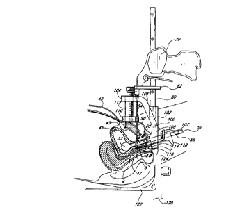
30 With reference to Figures 7, 8, 9 and 10 in another aspect, the present
invention provides a driver 70 for
driving a bone-piercing guide 84 through the pubic bone. Originally, passing a
suture 88 through the pubic bone was
done by drilling a hole through the generally anterior portion of the pubic
bone using a drill guide attached to a
stabilizer and vaginal retractor device. A suture 88 was then passed through
the drilled hole with a suture passer.
The present invention does not require the use of a drill and is capable of
creating small passages through the pubic
hone, sufficient to allow passage of a suture 88 through the bone. The pubic
bone is particularly well suited for
this adaptation because it is relatively easily pierced, due to its low
density.
CA 02280757 1999-08-12
WO 98/35606 PCT/US98/03069
-19
The driver 70 of the invention may be described as having four basic parts: a
first jaw 72, a slide bar 80,
a second jaw 82, and a bone-piercing guide 84. (Figure 7.) The first jaw 72
has a distal end 74 and a proximal
end 78. The distal end 74 is adapted for inserting into a tissue cavity and
the proximal end 78 of the first jaw 72
is attached to the slide bar 80. The slide bar 80 connects the first or fixed
jaw 72 with the second or moveable
jaw 82. The second jaw 82 slides along the slide bar 80, with a releasable
ratcheting action, toward the first jaw
72. The bone~piercing guide 84 attaches to the second jaw 82, and advances
toward the first jaw 72 as the second
jaw 82 is ratcheted along the slide bar 80.
A stop 86 on the slide bar 80 prevents further closing of the jaws once the
sharp end 96 of the cannula
90 exits the bone and is even with the first jaw 72. The first jaw 72 has a
slot 76 so that the sharp end 96 of
the cannula 90 does not actually contact it when exiting the bone. (Figure 8.)
The driver 70 may be equipped with
a double cannula jaw 82 (not shown) so that parallel passages may be created
through the bone simultaneously.
It is the function of the first or fixed jaw 72 inside a tissue cavity to
provide a counterpressure on the bone
opposite the pressure applied by the bone-piercing guide 84. Accordingly, the
distal end 74 of the first jaw 72 may
have a shape adapted to provide positions that can oppress the inferior region
of the pubic bone 45 lateral to the
pubic symphysis without crushing the urethra 6. Such a configuration of the
distal end 74 of the first jaw 72 is
shown in cross section in Figures 24a and b, 26a and b, and 27a; a detail view
of a portion of the edge of the
distal end 74 of the first jaw 72 is shown in Figure 25. In these figures, the
distal end 74 comprises a tongue 114
with a central depression 136 and elevated edges 138. The edges 138 may have
contact pins 140 adapted for
piercing the tissue lying between the pubic bone 45 and the elevated edge 138
of the tongue 114. At the elevated
edge 138 there also may be a gap 144 through which a guide 84 may pass without
contacting the tongue 114.
Any device with opposing jaws having one jaw adapted for insertion into a
tissue cavity may preferably
have a tongue configuration as described above. The choice of a desirable
configuration of the distal end 74 of the
first jaw 72 may be determined by one of ordinary skill in the art, taking
into account anatomical considerations,
the particular procedure involved, and the like.
The pubic bone is an especially important structure for piercing in surgical
applications. This is true far
at least two reasons: the first is that there are soft tissue structures in
the proximity of the pubic bone whose
dimension or displacement can result in several medical problems. The second
reason is that the pubic bone is a
relatively low density bone and therefore may he pierced without the
application of undue force, if the force is
properly oriented. The fact that the pubic bone may be pierced creates the
possibility of stabilizing a soft tissue
structure near the pubic bone by attaching a device or a suture to the soft
tissue structure and stabilizing it by
attachment to the relatively immovable pubic bone. In addition, by piercing
through the bone, the suture locking and
tissue securing method may be accomplished from the superiorlanterior bone
surface, which is much more accessible
than the posteriorlinferior surface. The prior need to work near or at the
posterior(inferior surface of the pubic bone
arose from the proximity of this surface to the structures most often sought
to be stabilized. With the methods and
devices of the present invention, however, passage of suture through the pubic
bone combines the desired proximity
to structures beneath the bone, with the convenience and simplicity of
introducing and securing suture through the
CA 02280757 1999-08-12
WO 98/35606 PCT/US98/03069
-20-
upper surface of the hone. Therefore the bone driver 70 of the present
invention provides a device of potentially
wide applicability for stabilizing structures of the pelvis, particularly in
reconstruction and stabilization of the urethral
and pelvic floor.
Alternative approaches to stabilizing structures of the urethral and pelvic
floor or other soft structures of
the pelvis by attachment to a fixed reference tissue have relied on drilling a
hole into the surface of a bone and
placing into the hole a hone anchor to which a suture is attached. The
difference between such approaches and
the present approach is that the present invention allows a much smaller
opening to be made. This opening traverses
the bone rather than being simply on the surface of the bone. Through this
much smaller passage may be passed
a suture, without the need of a bone anchor. As used herein, a bone anchor is
a device that attaches a suture to
the surface of a bone, wherein the suture thus attached does not pass through
the bone. The present invention
provides devices for connecting sutures to the bone, wherein the sutures have
passed through the bone. This is the
basis for the distinction, made in this specification, between "bone suture
fasteners" and "bone anchors."
A preferred embodiment of the driver 70 of the present invention provides a
first jaw 72, whose distal end
74 is adapted for insertion into the vagina 4. (Figure 8.) An alternative
embodiment provides a jaw 72 whose distal
end 74 is adapted for insertion into a cavity created in the vaginal hiatus 2
as discussed above. A further
embodiment may provide a first jaw 72 whose distal end 74 is adapted for
insertion into a transvaginally created
cavity in the hiatal plane.
As alternative embodiments to the preferred ratcheting motion of the second
jaw 82 toward the first jaw
72, the jaws also may be brought together by various other mechanical
advancing means, such as a threaded bar,
in combination with a thumb screw. The bone-piercing guide 84 may be hollow or
solid; examples of bone-piercing
guides 84 may be a needle, a cannula, or a solid rod. The guide 84 also may be
a cannula with a removable
obturator, so that the guide 84 behaves essentially as a solid rod while
piercing the bone, but then can be converted
to a hollow configuration for passing various devices along the lumen thereof.
A preferred cannula size is believed
to be approximately 14 gage. In a preferred embodiment the guide 84 is
sharpened and relatively stiff, thus
minimizing the possibility that the guide 84 will bend or skim along the
surface of the bone, and increasing the
tendency of the guide 84 to pierce directly into the bone along a straight
line between the first jaw 72 and the
second jaw 82.
An advantage of the bone-piercing guide driver 70 is that the device does not
require that a hole be drilled
through the bone. The passage remains open and completely accessible until the
cannula 90 is removed, whereas
the drilled hole is often lost once the drill bit is removed. Also, the drill
requires additional incisions on both sides
of the pubic bone to expose the bone, otherwise tissue is twisted around the
drill as it turns.
The driver 70 of this aspect of the invention may be used by itself or in
combination with devices of other
aspects of the invention. Accordingly, the driver 70 may be used in connection
with the dilator 10, for example,
by creating a tissue cavity with the dilator 10 and then placing the first jaw
72 of the driver 70 in the tissue cavity
created by the dilator 10. likewise a tissue cavity may be created by the
incision guide 50 and the first jaw 72
of the driver 70 may be placed inside the cavity thus created. Furthermore,
the insert card 30, capable of
...~"......_. ,
CA 02280757 1999-08-12
WO 98/35606 PCT/US98/03069
21-
introducing into a cavity a medical device, also may be used in connection
with the driver 70 of the invention. In
this particular combination the first jaw 72 of the driver 70 may be
configured to support the card 30 or connect
with the articulation opening 40 of the card 30 such that introduction of the
first jaw 72 into the tissue cavity
places the device in the appropriate position within the cavity. Subsequent
operation of the driver 70 directly
positions the bone-piercing guide 84 in the proper orientation with respect to
the device supported on the card 30.
Therefore, use of the driver 70 in combination with devices of other aspects
of the present invention may result in
several beneficial methods for surgery on the urethralfloor and other
structures of the pelvis.
This method creates a path through the pubic bone, which path is useful for
passing sutures 88 or medical
devices through the pubic bone. There are several alternative embodiments of
this method. In ane embodiment the
guide 84 is passed through the bone to create a path through the bone and then
the guide 84 is removed, leaving
the path in the bone and in the tissue. After removal of the guide 84, medical
devices such as a suture 88, a suture
passer, or a suture securing device 126 may be passed along the path through
the bone that was created by the
bone-piercing guide 84. Any device capable of passing a suture through tissue
may be used in accordance with the
present invention, including the suture passers and methods of their use,
disclosed in U.S. Patent No. 5,611,515,
issued March 18, 1997 to Benderev et al.
The driver 70 of the invention also may be used in a different orientation
such that the movable jaw is
adapted for insertion into a tissue cavity and for driving a device into the
pubic bone from the posterior-inferior
surface. As an example of a preferred embodiment, a sling 42 with pre-attached
push-in hone anchors (not shown)
is positioned on an insert card 30 and is placed into a tissue cavity with the
assistance of the dilator 10 or the
incision guide 50 of the invention, or by using both in combination. Next, the
push-in anchors are oriented to face
and contact the pubic bone. Finally, the movable jaw of the driver 70 is
placed below the push-in anchors of the
sling 42 and the fixed jaw of the driver 70 is placed against the patient's
abdominal surface such that the pubic
bone lies between the fixed jaw and the mavabie jaw. The movable jaw is then
advanced toward the fixed jaw such
that the push-in bone anchors are driven into the posterior-inferior surface
of the pubic bone and the sling 42 is
secured in place.
Other means of securing a sling 42 in place by using push-in type bone anchors
are also contemplated in
the invention. For example, a pivoting ar otherwise manipulable tongue or
insert card 30 that supports and positions
the sling 42 may be forcefully angled against the pubic bone sufficient to
drive into the pubic bone the push-in type
anchors from the posterior-inferior surface of the bone. In another
embodiment, cannulas 90 are driven through the
bone, both left and right of the midline, such that a path into the pubic bone
is provided for initial guidance of push-
in anchors. As the cannulas 90 on either side of the midline are withdrawn
from the pubic bone, the push-in anchors
loot shown) are pressed upward against the bone and initially follow the path
of the cannulas 90. Thus, a guide
hole is created by the cannulas 90 for the anchors. The application of
additional upward pressure seats the anchors
and the sling 42 is secured in place.
A preferred embodiment of the invention uses a cannula 90 as the bone-piercing
guide 84. (Figure 9a.)
The lumen of the cannula 90 constitutes a path through the tissue and through
the pubic bone. Accordingly, the
CA 02280757 1999-08-12
WO 98/35606 PCT/US98/03069
22~
path through the lumen of the cannula 90 allows passage of sutures 88 or other
devices through the bane without
the difficulty of locating the path through the bone. (Figure 9b.) In this
method a cannula 90 (Figure 9a) is attached
to the driver 70, as shown in Figure B, and the first jaw 72 of the driver 70
is placed inside the tissue cavity, the
pubic bone is located and the driver 70 is positioned to align the pubic bone
between the first jaw 72 and the
second jaw 82. The second jaw 82 is then advanced toward the first jaw 72,
pushing the cannula 90 through the
pubic bone and through the soft tissue on either side of the pubic bone.
Subsequently, a suture 88 is passed
through the cannula 90 and into the cavity.
Depending on the internal diameter of the cannula 90, other devices also may
be passed inside the lumen.
For example, devices such as a suture passer, a quick connect device, and the
like, may be passed through a cannula
90 of sufficient internal diameter. In a preferred embodiment, the cannula 90
has a sharpened tip 96, and has a
relatively high degree of stiffness.
This aspect of the invention provides a method of pelvic surgery that uses the
driver 70 of the invention
wherein the guide 84 of the driver 7D is a cannula 90. The first jaw 72 of the
driver 70 is inserted into the tissue
cavity, and the pubic bone is located. The driver 70 is positioned to align
the pubic bone between the first jaw 72
of the driver 70 and the second jaw 82 of the driver 70. The second jaw 82 is
advanced toward the first jaw 72,
forcing the cannula 90 into the tissue and through the pubic bone. After the
cannula 90 passes through the pubic
bone it is further advanced along the same line into the cavity, approaching
the first jaw 72 of the driver 70.
Alternatively, a first cannula 90 can be used to pierce the pubic bone, and a
second cannula 90 may be passed
within the lumen of the first cannula 90 to its final position on either side
of the urethra. A suture 8B is then
passed into the lumen of the cannula 90 and is advanced through the cannula 90
until it enters the tissue cavity.
The end of the suture 88 that is in the tissue cavity is secured there and the
second end of the suture 88 is secured
to the pubic bone. The attachment thus created between the pubic bone and the
tissue cavity stabilizes the tissue
cavity and structures nearby. (Figure 10b.)
There exist several alternative embodiments of the method of this aspect of
the invention. In one
embodiment, the cannula 90 is removed after the first end of the suture 88
passes through the cannufa 90 and into
the cavity. The cannula 90 is therefore withdrawn, leaving both ends of the
suture 88 on opposite sides of the
pubic bone prior to the attachment of either end of the suture 88 in its
place. In another embodiment of this
method, the suture 88 is attached within the cavity and then the cannula 90 is
withdrawn, followed by the
attachment of the second end of the suture 88 to the bone.
The made of and purpose for attachment of the suture 88 within the cavity is
variable, and the factors
affecting the selection of the mode will be appreciated by one of ordinary
skill in the art. For example, the suture
88 may be affixed in the cavity with one or more stitches 92 (Figure 10a) to
tissue therein. Alternatively, the suture
88 may be attached to a device far distribution of pressure across relatively
widened area of tissue, such as suture
button 94 or a sling 42. (Figure 9b.) The suture 88 also may be attached to a
staple (not shownl within the cavity,
such that the staple anchors the suture 88, so that appropriate tension on the
suture 88 elevates the tissue to which
the staple is attached.
_r_ .._
CA 02280757 1999-08-12
WO 98/35606 PCT/US98/03069
~23
Without the use of an additional device, attaching the suture 88 to tissue
within the cavity may make
possible amelioration of certain incontinence conditions that arise from
hypermobility of the urethra or from intrinsic
sphincter deficiency. This is done simply by attaching the suture 88 to tissue
and then providing an appropriate
tension. This tends to elevate the tissue, which therefore elevates the
urethra at that position a distance A (compare
Figure 10a to 10b1, eliminating or easing the condition causing urinary
incontinence.
An alternative embodiment of this aspect of the invention uses the driver 70
to insert a cannula 90 into
the tissue cavity by passing it through the pubic bone as described above. In
this embodiment, however, the end
of the suture 88 which is advanced through the cannula 90 and into the tissue
cavity is passed through a structure
93 within the tissue cavity. It is subsequently passed back out of the cavity
and secured to the pubic bone, along
with the second end of the suture 88. In this embodiment of the invention, the
structure 93 through which the
suture 88 may be passed include a suture button 94 or grid, a sling 42, and a
tissue mass adjacent the cavity
(creating stitches 921. The cannula 90 advantageously has a sharpened end 96
and a reinforced stiffness, to
facilitate its passage in a straight line through the bone and to minimize
skimming along the surface of the bone.
Further, for embodiments of this method, the cavity is preferably a cavity in
the vaginal hiatus 2 created according
to one of the other methods of this invention. The cavity also may be the
vagina 4, or a cavity in the hiatal tissue
created transvaginally.
This method is advantageous particularly for the stitching of a tissue mass
for stabilization of the urethra
floor or other pelvic structures. (Figure 10.) Because the suture 88 enters
and then again exits the tissue cavity,
it may be passed through several stitching 92 points in the tissue before it
is withdrawn through the cannula 90
and out to the pubic bone. Both ends of the suture B8 may be secured to the
pubic bone in a variety of ways,
many of which will be explained in detail below.
This method may be advantageously practiced with the additional assistance of
a suture passer device.
The suture passer is advanced through the cannula 90 and into the tissue
cavity upon completion of the desired
number of sutured stitches 92 within the cavity, the suture 88 is grasped by
the suture passer, and withdrawn
through the cannula 90 to properly position it at the surface of the pubic
bone for attachment. This method may
be preferably used for tensioning, stabilization, or elevation of a tissue
mass adjacent to the urethra or adjacent to
another soft tissue structure in the pelvis that may be in need of
stabilization or reorientation. The fact that the
suture 88 is secured to the bone creates a stability for the target tissue
mass that is desirable in many cases. It
also may be desirable to use this method in securing in a tissue cavity a
pharmacological implant, a prosthetic device,
or a therapeutic device.
This aspect of the invention provides an additional method for pelvic surgery
wherein the driver 70 is used
to pass a guide 84 through the pubic bone along a first path, proceeding into
the tissue cavity at a first position.
A suture 88 is then passed through the cannula 90 into the cavity. The driver
70 is then used to create a second
path through the pubic bone, arriving at a second location within the tissue
cavity. The suture 88 which was passed
along the first path through the pubic bone and into the cavity may then be
passed along the second path out of
the cavity and through the pubic bone at the second position. Bath ends of the
suture 88 may then be secured to
CA 02280757 1999-08-12
WO 98/35606 PCT/US98/03069
-24-
the pubic bone. In this method, the suture 88 that passes through the cavity
may preferably also be passed through
a tissue mass of the cavity, a suture button 94 or a grid, or a sling 42,
prior to exiting the cavity along the second
path through the pubic bone. Alternatively, by simply passing the suture 88
through the cavity and then tensioning
it properly, the suture 88 may serve to elevate the tissue mass and stabilize
the structures adjacent thereto.
The invention thus provides several alternatives for stabilizing structures of
the urethral floor or other
structures of the pelvis by securing the soft tissues to the pubic bone. The
methods differ primarily in the paths
along which the suture 88 is advanced. However, whether one chooses to advance
the suture 88 into the cavity
and anchor one end of the suture 88 therein, or to advance the suture 88 into
the cavity and return it out of the
cavity along one path, or to advance the suture 88 into a cavity along a first
path and retract it ftom the cavity
along a second path, all three methods may be adapted to several variations.
In all of these methods, the tissue
cavity may be the vagina 4. It also may be a cavity in the hiatal tissue
created, for example, either by the dilator
10 or by the incision guide 50 of the present invention as previously
described. Alternatively, the cavity may be a
cavity created transvaginally by opening a pocket in the hiatal tissue through
the upper vaginal wall 8.
Furthermore, in any of the methods of this aspect of the invention, a
preferred embodiment would perform
the method on both the right and left side of the midfine of the patient, to
equally distribute points of attachment
on either side of the urethra. Likewise, another preferred embodiment of any
of these three methods would involve
the additional step of tensioning the suture 88 prior to its attachment to the
pubic bone. Suture tensioning may
be accomplished in a variety of ways, one of which is with the use of suture
tensioning device. This type of device
has a handle with which the surgeon can place the device next to the location
where the suture 88 will be tied.
Attached to the handle is another structure of the suture tensioner around
which the suture 88 will be wrapped,
and upon which the suture 88 may be tied. The external dimensions of this
structure control the degree of slack
that the suture 88 retains after the suture 88 is tied. Therefore, based on
the linear distance between the bone
and the soft tissue sought to be stabilized, as welt as the amount of slack
needed to achieve a particular objective
for a given patient, a suture tensioner with an appropriate diameter will be
selected so as to provide the proper
amount of tension in the connection between the bone and the soft tissue.
The several methods of passing a suture 88 through one or more paths in the
pubic bone may be
particularly applied to a method for stabilizing a urethral sling 42 relative
to the pubic bone. In this method a cavity
is created in the vaginal hiatus 2. The cavity may preferably be made using
the dilator 10 or the incision guide 50
of the present invention, or both in combination. The dimensions of the cavity
and the amount of offset from the
urethra will be determined according to the size of the sling 42 that is
needed, the dimensions of the patient's
urethra, and the surgeon's preference. After the cavity is formed, guide 84 is
driven through the pubic bone to
create a path. This is done by placing the distal end 74 of the first jaw 72
of the driver 70 into the tissue cavity
followed by positioning the second jaw 82 of the driver 70, such that the
pubic bone Pies in a straight line between
the first jaw 72 and second jaw 82. A urethral sting 42 is placed into the
cavity either before or after the cannula
90 is driven through the pubic bone and into the cavity. The sling 42 may be
placed into the cavity with the aid
CA 02280757 1999-08-12
WO 98/35606 PCT/US98/03069
~25~
of an insert card 30 as discussed above, or by other surgical procedures known
in the art. With the cannula 90
in the cavity adjacent the sling 42, the suture 88 is attached to the sling 42
and to the pubic bone.
This method is embodied in a technique wherein the cannula 90 is withdrawn
before the suture 88 is
secured on either end. The method also contemplates withdrawal of the cannula
90 after the sling 42 is secured
but before the suture 88 is secured to the bone. In addition to attaching the
sling 42 with sutures 88, this method
contemplates an indirect attachment of the sling 42 to sutures 88 by directly
attaching the sling 42 to devices which
are themselves attached to sutures 88. An example of such an indirect
attachment to a suture 88 is a securing
device 126 as disclosed herein. This method may advantageously involve use of
the devices of other aspects of the
present invention. The tissue cavity may be made either by the dilator 10 or
the incision guide 50, or by using both
together, and the sling 42 may be delivered to its proper position in the
cavity with aid of the insert card 30 as
discussed above. Depending on the particular manner in which the insert card
30 articulates with a driver 70, the
cooperative use of the insert card 30 may precisely position the sling 42.
This will cause the bone~piercing guide
84 to meet the sling 42 at the appropriate straight line position as is
desirable in this method.
Another aspect of this invention provides a driver frame assembly 100. The
driver frame assembly 100
serves to align, support and stabilize both the patient's pelvis and the
devices of the invention in procedures for
reconstructing the urethral and pelvic floor or for performing other methods
of pelvic surgery. The driver frame
assembly 100 has an upper clamp 102, a rigid catheter 52, a cavity tongue 114,
a lower clamp 120, and at least
one driver 70. The upper clamp 102 of the driver frame assembly 100 has a head
portion 104, a descending arm
106 and a base portion 108. The head portion 104 has a compression foot 110
that is used to compress a
patient's abdominal surface against the pubic bone. The compression foot 110
has stabilizing pins 112 that extend
downward therefrom and that pierce the patient's skin 48, penetrating the
abdomen at a position adjacent to the
superior surface of the pubic bone. The compression foot 110 may be pressed
against the abdomen of the patient
with, for example, a threaded screwing mechanism, a ratcheting or piston
mechanism, or a linkage mechanism. The
stabilizing pins 112 are designed to be of a length that makes it impossible
for the pins 112 to cause damage to
any abdominal or pelvic organs. At the same time the stabilizing pins 112 have
dimensions making them sufficiently
strong to resist the lateral forces exerted with operation of the driver 70.
The rigid catheter 52 portion of the driver frame assembly 100 functions much
as the rigid catheter 52
of the incision guide 50, as discussed above. The catheter 52 is intended to
straighten and elongate the urethra,
as well as to assist in identifying the position of the urethra, the bladder
46, and bladder neck 47. The rigid
catheter 52 is of sufficient length to extend outward beyond the distal
urethra of the patient. This external
extension of the rigid catheter 52 provides a structure with which the frame
assembly may articulate and attach.
(Figure 11.)
The tongue 114 portion of the driver frame assembly 100 is adapted for
insertion into a tissue cavity, such
as the vagina 4 or a hiatal cavity prepared prior to insertion of the tongue
114. In a preferred embodiment, the
tongue 114 has a central depression 136 and elevated edges 138 (Figure 12b1,
allowing compression of the tongue
114 sides against the inferior surface of the pubic bone to counter the
pressure of the compression foot 110.
CA 02280757 1999-08-12
WO 98/35606 PCT/US98l03069
26-
Because of the shape of the tongue 114, this type of pressure may be applied
without crushing the urethra. The
tongue may also have a relatively broad elevated edge 138 contact pins 140 for
contacting the pubic bone. (Figures
24 and 25.) A flat tongue 114 does not allow the application of a strong
counterpressure against the bone, because
of the potential damage to the urethra. (Figure 12a.) The tongue 114 has a
first end 116 for inserting into the
cavity, and a second end 118 which is adapted for articulating with the base
portion 108 of the upper clamp 102.
This articulation may be, for example, by means of a threaded connector 107
that joins the tongue 114 to the base
portion 108 of the upper clamp 102.
The lower clamp 120 of the driver frame assembly 100 has a buttock plate 122
for insertion beneath the
patient such that the patient's weight rests on the plate to further secure
the frame assembly. Attached to the
buttock plate 122 is an ascending arm 124 that is adapted for articulating
with the base portion 108 of the upper
clamp 102. (Figure 13.) Thus, the upper clamp 102 attaches to the rigid
catheter 52 and the cavity tongue 114,
and then the upper clamp 102 also attaches to the lower clamp 120, which is
anchored under the patient's body.
The effect of these multiple attachments is to compress a patient's pelvic
region to provide stability for operation
of the bone-piercing guide driver 70 as well as to enhance the straight line
precision of driving a guide 84 through
the pubic bone.
In a preferred embodiment, an insert card 30 capable of supporting a medical
device is an additional
component of the driver frame assembly 100. (Figure 18.) In such an
embodiment, the card 30 adapted to slide
along the tongue 114 to reach the proper position within the cavity. The
articulation opening 40 of the card 30
also may be used for attachment or positional control of the card 30 with
respect to the driver frame assembly 100.
The driver 70 portion of the driver frame assembly 100 is similar to the
driver 70 of a previous aspect of
the invention as discussed above. The driver 70 of this aspect of the
invention, however, does differ in the respect
that it attaches to the driver frame assembly 100 via the slide bar 80 of the
driver 70. Accordingly, the driver
70 of this aspect of the invention has the slide bar 80 and the moveable jaw,
but does not have its own immovable
or first jaw 72. However, upon attachment of the driver 70 to the driver frame
assembly 100, both the tongue 114
portion and the buttock plate 122 portion of the driver frame assembly 100 can
simultaneously function as fixed
jaws toward which the immovable jaw advances as it moves along the slide bar
80. (Figure 15.)
As mentioned above, the function of the first or fixed jaw is to provide
counterpressure to the pressure
applied to the pubic bone by the advancement of the guide 84 through the pubic
bone. The preferred driver 70
portion of the driver frame assembly 100 has two positions for attachment of
bone-piercing guides 84 to the second,
or movable, jaw 72. (Figure 14.) These positions are left and right of center,
being laterally spaced to provide
sufficient offset from the patient's midline so as to prevent any contact of
the bone-piercing guides 84 with the
urethra. Thus, the driver 70 of the assembly is intended to simultaneously
advance two separate bone-piercing
guides 84 through the pubic bone, one left and one right of the patient's
midline, as shown in Figure 16.
This aspect of the invention provides a method for reconstruction of the
urethral and pelvic floor or for
stabilizing a tissue of the pelvic region. In this method a rigid catheter 52
is placed in the urethra so that the
_~.. . .._
CA 02280757 1999-08-12
WO 98/35606 PCT/US98/03069
27-
urethra is elongated and straightened. (Figure 11.) The catheter 52 also aids
in determining the position of the
bladder 46 and the bladder neck 47. The tongue 114 of the driver frame
assembly 100 is placed into the tissue
cavity, and also may be attached to the rigid catheter 52. (See Figures 11 and
12.) Preferably an insert card 30
holding a medical device, for example, a urethral sling 42, is inserted into
the cavity using the tongue 114 as a
guide. (Figure 18.) The upper clamp 102 is attached to the tongue 114 and to
the catheter 52, and the
compression foot 110 is compressed against the patient's abdomen, after
palpation to determine the proper position
of the stabilizing pins 112 relative to the superior surface of the pubic
bone. The compression foot 110 is tightened
against the patient's abdomen to prevent slipping of the bone-piercing guide
84, and to further immobilize the soft
tissues of the pelvis. (Figure 11 ). Further stabilization is achieved with
attachment of the lower clamp 120 which
is done by sliding the buttock plate 122 beneath the patient and firmly
attaching the ascending arm 124 of the lower
clamp 120 to the base portion 108 of the upper clamp 102. (Figure 13.)
With the driver frame assembly 100 properly installed along the patient's
midline, the driver 70 portion of
the driver frame assembly 100 may be mounted on the descending arm 106 of the
upper clamp 102. (Figure 13.)
Care is taken to assure that the hone-piercing guides 84 of the driver 70 are
positioned such that they will enter
the pubic bone. Then the second removable jaw of the driver 70 is advanced
downward along the slide bar 80 and
the guides 84 are advanced into the abdominal surface and through the pubic
bone, emerging within the tissue
cavity near the medical device supported on the card 30. (Figure 15.1
In a preferred example, the bone-piercing guides 84 are cannulas 90 and the
medical device supported on
the card 30 is a sting 42. (Figures 17 and 18.) In such an embodiment, the
proximity of the cannulas 90 with the
sling 42 permits suturing or other attachment through the lumen on the cannula
90 to the desired location on the
urethral sling 42. (Figure 19.) After attachment of suture 88 directly or
indirectly (see Figures 21-23) to the sling
42, the cannulas 90 are removed, leaving behind the suture 88, which is then
secured to the pubic bone. (Figure
28.)
In another embodiment of this aspect of the invention, a concave insertion
tongue 114 is used to provide
counterpressure for driving cannulas 90 through the pubic bone 45. The concave
tongue 114 is inserted into a
cavity, such as, for example, a hiatal cavity or the vagina 4, and the
compression foot 110 of the driver frame
assembly 100 is placed against the patent's pubic bone 45. (Figure 24a.) The
concave tongue has a central
depression 136 and elevated edges 138, allowing the edges 138 of the tongue
114 to provide counterpressure to
the posteriorfinferior surface of the pubic hone 45, while the compression
foot 110 and the cannulas 90 apply
pressure to the opposite fanteriarlsuperior) surface of the bone 45. (Figure
24b.)
The central depression 136 of the tongue 114 prevents the urethra 6 from being
crushed upon application
of counter pressure on the bone 45 by elevated edge 138 of the tongue 114.
(Figure 24b.) The edge of the tongue
may also have a gap 144 to allow the cannula 90 to pass through the plane of
the elevated edge 138 without the
cannula 90 impacting against the tongue 114. The edge 138 may also have
contact pins 140 of a configuration
and position to pierce through soft tissue and fascia to contact the surface
of the pubic bone 45 and prevent
slippage of the tongue 114. (Figure 25.)
CA 02280757 1999-08-12
WO 98/35606 PCT/US98/03069
28-
When pressure is applied to the opposite sides of the bane 45, as shown in
Figure 24b, the cannulas 90
are driven through the pubic bone 45 and into the cavity. (Figure 26a.) Suture
88 or other devices may be passed
through the cannulas 9D and into the cavity. (Figure 26b.) The suture 88 may
be attached within the cavity either
by stitches 92 or by attaching the suture 88 to a suture button 94. (Figure
26b.)
After proper attachment of the suture 88 within the cavity, the pressure on
both sides of the bone 45 is
released, and the compression foot 110 is raised. (Figure 27a.) The tongue 114
is removed from the cavity, and
the sutures attached therein are fastened to the bane 45 with, for example,
either a bone eyelet 150 or a bone
suture fastener 170. The urethra 6 is elevated by the tension that is applied
to the sutures 88 in the step of
fastening the sutures 88 to the bone 45. The resulting elevation of the
urethra 6 is shown as distance B in Figures
27a and 27b.
The advantage of the method of this aspect of the invention is that the driver
frame assembly 100, if
properly installed, provides rigidity to the driver 7D to allow for highly
accurate straight line passage of a bone-
piercing guide 84 through the pubic bone and to a target sight within the soft
tissue. Because the driver 70 is able
to advance two different guides 84 through the bone at the same time, and
because the contact between the guide
84 and the sling 42 is relatively precise, the entire procedure can be done
very rapidly and with minimal invasion
compared with other procedures used to achieve a similar objective. In
addition, the method of this aspect of the
invention, being minimally invasive, greatly reduces the risk of infection
arising from the procedure. Also reduced
is the number of incisions, resulting in a shorter recovery time for the
patient and less scarring, since the external
abdominal wounds are all puncture wounds, and the only incision is a
relatively small incision or a knife stick in the
vaginal hiatus 2 or in the upper vaginal wall 8. In a preferred embodiment of
this aspect of the invention, the
sutures 88 which pass through the bone are secured to the bone after being
tensioned with a suture tensioning
device as discussed above.
Another aspect of the invention provides a system for attaching urethral sling
42 to a suture 88 as shown
in Figure 21-23. The system includes a urethral sling 42 and a connector 130;
the urethras sling 42 has a ring
member 44 attached thereto (Figures 29-30) and the connector 130 is adapted to
cooperate with the ring member
44 to permit unidirectional passage of the connector 130 through the ring
member 44 (Figures 21-23, 32-351. The
ring member 44 and the connector 130 further cooperate to prevent retrograde
movement of the connector 130
through the ring member 44.
The invention contemplates several embodiments both of the ring member 44 and
of the connector 130.
The ring member 44 may be as simple as a hole in an appropriate part of the
sling 42, suited for passage
therethrough by a connector 130. (Figure 33-35.1 The ring member 44 also may
be a more elaborate structure, such
as a reinforced ring with flanges 142 protruding into the central opening 128
of the ring member 44. (Figures 29,
30, 32.1
In a preferred embodiment, the connector 130 has a conical tip 131 attached to
a cylindrical portion 133
behind the tip 131 to which a suture 88 may attach. This embodiment is
referred to as the arrowhead connector
132. (Figure 32a.) The cylindrical portion 133 behind the tip 131 has a
smaller circumference than the widest part
r
CA 02280757 1999-08-12
WO 98/35606 PCT/US98/03069
-29
of the conical tip 131, such that there is a substantially flat shoulder 129
behind the tip 131. Accordingly, the
arrowhead connector 132 is adapted for cooperating with the reinforced ring
member 44 having flanges 142 that
protrude into the central opening 128. As the tip 131 penetrates the central
opening 128, the flanges 142 move
aside, allowing passage of the tip 131. However, once the entire conical
portion of the tip 13i has passed through
the central opening 128, the flanges 142 return to their initial orientation
around the central opening 128 and rest
against the shoulder 129, resisting retrograde movement of the connector 130
through the ring member 44. (Figures
22, 23, 30, 32.) Since the cylindrical portion 133 behind the shoulder 129 of
the arrowhead connector 132 has
a means for connecting to a suture 88, the passage of the connector 130
through the ring member 44 creates a
connection between the suture 88 and the sling 42. (Figure 32a.1 By this
method of connecting suture 88 to sting
42, there is no need for stitching or tying knots to the sting 42, which
greatly simplifies and accelerates the securing
procedure.
Another preferred embodiment of the securing device 126 has a connector 130
with two perpendicular
cylindrical members 135, 137 wherein the members 135, 137 are substantially
flexibly attached one to the other
at a flexible joint 139. (Figure 34.) The rear cylinder 135 has a means of
attaching to a suture 88, while the
leading cylinder 137 is adapted for passage through the ring member 44. As
this embodiment of the connector 130,
referred to herein as the T connector 134, is inserted into a cannula 90 for
advancement toward the sling 42, the
perpendicularity between the two cylindrical members 135, 137 is distorted,
and the leading cylinder 137 assumes
a position that is more closely parallel, rather than perpendicular, to the
rearward cylinder 135. (Figure 35.)
However, after the T connector 134 has passed through the cannula 90 and the
leading cylinder 137 of the T
connector 134 has also passed through the ring member 44 of the sling 42, the
angular relationship between the
two cylindrical members 135, 137 reverts to perpendicular. /Figure 33.) In
this conformation, the connector 130
may not pass back through the ring member 44, and a secure connection between
the suture 88 and the sling 42
is therefore established.
This aspect of the invention provides a method for simple, minimally invasive
placement and securing of a
sling 42 in a tissue cavity. A tissue cavity, such as a cavity in the hiatal
tissue, is created by use of, for example,
the dilator 10 or the incision guide 50 of the invention. Alternatively an
existing tissue cavity, such as the vagina
4, may be selected for placement of the sling 42. In addition, a tissue cavity
in the hiatal tissue may be created
by inserting a dilator 10 or an incision guide 50 through the upper vaginal
wall 8. A sling 42 is then placed into
the cavity in its desired location. This step is preferably performed with the
use of the insert card 30 of the
invention, wherein the insert card 30 carries and supports the sling 42 in its
appropriate position within the cavity
until it secured there. (Figure 18.)
A cannula 90 is driven through the pubic bone using, for example, either the
driver 70 of the invention or
the driver frame assembly 100 of the invention. (Figure 18.) The cannula 90 is
then further driven through tissue
until it approaches and aligns with the sling 42, (Figure 19.) which is
equipped with ring members 44. A connector
i 30 attached to the suture 88 is then inserted into the lumen of the cannula
90 and advanced therethrough until
it contacts with and passes through the ring member 44 of the sling 42.
(Figures 20, 21, 22.) At this point, the
CA 02280757 1999-08-12
WO 98/35606 PCT/US98/03069
30-
suture 88 is tested for the integrity of the connection between the connector
t 30 and the ring member 44, and the
cannula 90 is withdrawn from the pubic bone. The suture B8 may then be
appropriately tensioned and secured to
the pubic bone (Figure 28) as will be discussed below. Also contemplated as
embodiments of this aspect of the
invention are similar connections between other kinds of medical devices and
suture 88 via, for example, arrowhead
connector 132 or T connector 134 passing through a cannula 90 which has been
driven through the pubic bone.
As a further embodiment of the present invention, the securing devices 126 of
the invention may be used
to secure a urethral sling 42 or other medical device after such a device has
been positioned in a tissue cavity that
was created transvaginally. For example, a cavity may be opened in the upper
vaginal wall 8 by hydrodissection,
or by means of the spreader 12 of the invention, or by blunt dissection. A
medical device such as a urethral sling
42 may then be placed into the cavity, wherein the medical device has one or
more ring members 44 capable of
permitting unidirectional passage of a connector 130 of the securing device
126. The appropriate connector 130
is then passed through the corresponding ring member 44, and the device is
secured in place in the transvaginally
created tissue pocket. (See Figure 23.)
This invention is further characterized by a bone eyelet 150 for securing
suture 88 to a bone. The bone
eyelet 150 consists of a sleeve 152 and at least one crosspiece 160. (Figure
31 J The sleeve 152 has an outer
surface 154 that is adapted for inserting into and contacting with a bone, and
also has an inner surface 156 such
that the sleeve 152 is a substantially hollow structure with openings at
either end. (Figure 32a.) The crosspiece
160 is attached to the inner surface 156 of the sleeve 152, and creates a
plurality of channels within the sleeve
152.
Several alternative embodiments of the bone eyelet 150 are contemplated in the
present invention. In one
preferred embodiment, the crosspiece 160 is a single rod 164. (Figure 33.) fn
another preferred embodiment, the
crosspiece 160 is a plane 162. (Figure 32a.) In both of these embodiments, the
presence of the single crosspiece
160 produces two channels 168 through which a suture 88 may pass. In another
embodiment, the crosspiece 160
is created by crimping or piercing the sleeve 152. In this embodiment, the
thus-distorted portion of the sleeve 152
becomes the crosspiece 160, as shown in Figure 32b. In other embodiments,
multiple crosspieces 160 may be
present in the sleeve 152, to produce more than two channels 168 through which
a suture 88 may pass. In another
embodiment, the sleeve 152 includes a perpendicular flange rim 158. This
flange rim 158 suspends the sleeve 152
at the surface of the bone and prevents it from sliding into the hole in the
bone. (Figures 31-33.)
This aspect of the invention provides a method for securing a suture 88 to a
bone. In this method, a
suture 88 is passed through a bone in which a path for suture 88 has been
created, for example, by driving a bone-
piercing guide 84 according to this invention. Where two ends of a suture 88
both extend past the surface of the
bone, each end may he advanced through one of the channels 168 among the
plurality of channels 168 provided in
the bone eyelet 150. The suture 88 ends may then be tied together and will be
prevented from sliding into the bone
by the action of the crosspiece 160 of the bone eyelet 150. (See Figures 32,
33.)
Another embodiment of this method includes a step of tensioning the suture 88
during the tying step to
achieve a desired elevation of the structures to which the suture 88 is
attached. For example, this method may be
_.r......
CA 02280757 1999-08-12
WO 98/35606 PCT/US98/03069
-31
employed in connection with a securing device 126 discussed above to greatly
accelerate and simplify the steps in
securing a tissue mass or a urethral sling 42 in a tissue cavity. (Figures 32,
33.) Accordingly, the bone eyelet 150
may be used in connection with the driver 70 aspect of the this invention, as
well as the driver frame assembly 100
aspect of the invention. It may further be adapted for use with other means of
advancing a suture 88 through a
pubic bane.
A further aspect of the present invention provides a bone suture fastener 170
for quick and simplified
connection of a suture 88 to a bone. (Figures 36-38.) The suture fastener 170
consists of a sleeve 172 having
an opening 174 at each end, and a sleeve plug 178. The sleeve 172 is provided
with a friction surface 180 for
contacting the sleeve plug 178 and for preventing disengagement of the plug
178 from the sleeve 172. Different
embodiments of the bone suture fastener 170 have a sleeve 172 that is
substantially conical or cylindrical and a
sleeve plug 178 that is likewise substantially conical or cylindrical. In one
embodiment the friction surface 180 is
a plurality of friction flanges 184 partially occluding one opening 174 of the
suture fastener sleeve 172. (Figures
36, 37a.) These flanges i 84 flex is one direction to allow insertion of the
sleeve plug 178 into the sleeve 172 and
then prevent release of the sleeve plug 178 from the sleeve 172. In another
embodiment, the friction surface 180
is threaded 182 and the sleeve 172 is substantially cylindrical. (Figure 38.)
The sleeve plug 178 is likewise
threaded 162 and is adapted for ~frictionalfy contacting the threads 182 of
the suture fastener sleeve 172. An
additional embodiment, the sleeve plug 178 may have a series of distortable
angled rings 179 along its length,
wherein the circumference of the top of each ring 179 is smaller than the
circumference of the bottom of the same
ring 179. The suture 88 may be tied directly to the sleeve plug 178. In this
embodiment, the friction surface 180
of the sleeve 172 has a rim 183 adapted to allow unidirectional passage of the
sleeve plug 178 and to prevent
retrograde passage thereof, resulting in a one-way zipper-lock action. This
embodiment allows a surgeon to adjust
the tension art the suture 88 simply by pulling the sleeve plug 178 through
the sleeve 172 to the desired position.
(Figure 376.) In any of these embodiments, a flange rim 186 may extend around
the circumference of one end of
the sleeve 172 to prevent sinking of the bone suture fastener 170 past the
surface of the bone. (Figure 38.)
This aspect of the invention provides a method for Quick and simple securing
of a suture 88 that has
passed through a hone. According to the method, one or multiple ends of a
suture 88 may be passed through the
sleeve 172 of the suture fastener 170 and the suture fastener 170 may be
advanced along the suture 88 until it
contacts the bone through which the suture 88 passes. (Figure 36.) The sleeve
172 is secured in the bone near
the bone surface either by a friction surface 180 on the outside of the sleeve
172, or by a flange rim 186 extending
around the circumference of one end of the sleeve 172. With the sleeve 172 in
place, and the suture BB passing
therethrough, the suture 88 may be tensioned to approximately the desired
tension and the sleeve plug 178 partially
inserted into the opening 174 of the sleeve 172 at the surface of the bone.
Any desired tensioning or release of
tension in the suture 88 is done prior to final seating of the sleeve plug 178
in the sleeve 172 against the friction
surface 180. (Figures 37a and 38.) When the sleeve plug 178 is appropriately
seated against the friction surface
160, the suture 88 is secured in place at the surface of the bone, and excess
suture 88 is cut off by the surgeon.
CA 02280757 1999-08-12
WO 98/35606 PCT/US98/03069
~32-
Another embodiment of this aspect of the invention contemplates the use of the
bone suture fastener 170
to secure a suture 88 that is connected to a device or a stitch 92 in a
transvaginally created tissue cavity. For
example, a tissue cavity is dissected between the urethra and the upper
vaginal wall 8 by hydrodissection, blunt
instrument dissection, or by the spreader 12 of the invention. Subsequently, a
sling 42 or other medical device may
be placed in the cavity, or a suture 88 may be passed through a tissue mass to
elevate or stabilize the tissue mass
(compare Figure 27a to Figure 27b and note distance of elevation B, see also
Figures 9, 10, 261. In cases where
the suture 88 is passed through the tissue and follows a path through the
pubic bone, the end of the suture 88 that
protrudes from the pubic bone may be anchored thereto by means of the bone
suture fastener 170 of this aspect
of the invention. (Figure 28.)
This method of the invention may be practiced in cooperation with several
other methods and devices of
the present invention. In any aspect of the invention in which a suture 88 is
passed through a bone, the suture 88
may be advantageously secured to the bone in this manner. Thus, the quick
connect bone suture fastening may be
used in coordination with the quick connect securing devices 126 used to
connect a sting 42 or other internally
placed medical device with a suture 88. They also may be used to secure the
ends of a suture B8 that has been
passed through a tissue mass by stitching 92. They also may be used in
connection with the bone-piercing guide
driver 70 or with the driver frame assembly 100 of the invention.
Accordingly, the several aspects of the present invention cooperate to achieve
the desired effect of providing
a variety of surgical options for and solutions to problems associated with
stress urinary incontinence and related
dysfunctions or deformations of the urethral or pelvic floor. Also
contemplated within the overall scope of the
present invention are other applications for securing a soft target tissue to
a relatively fixed reference tissue, such
as the pubic bone. It is understood that the examples of embodiments and
methods provided herein are merely
representative of the invention, and are not taken to limit the invention
beyond the express limitations of the
following claims.
~~,.. .