Note: Descriptions are shown in the official language in which they were submitted.
CA 02280794 1999-08-12
WO 98/37417 PCT/US98102995
Plasmon Resonant Particles, Methods and Apparatus
Field of the Invention
The present invention relates plasmon resonant entities (PREs), or particles,
to methods of
interrogating a field containing PREs, and to apparatus for carrying out the
method, and to various
applications of PREs.
Background of the Invention
There are a number of important commercial and scientific applications of
interrogating a target
for information about the target. For example, the aim of analyte diagnostic
tests and methods is to
detect the presence and/or amount of an analyte (the target). The target
analyte may be detected by
reacting the analyte with a detectable reporter that (i) can bind specifically
to the analyze and (ii) is
detectable with suitable detecting tools. The reporter may, for example, be a
colored or fluorescence
molecule, or a colloidal metal, or a reporter such as a radiolabel that
requires special film or scintillation
equipment for its detection.
In some diagnostic applications, it is desirable to detect proximity
relationships in a target
analyte, as evidenced by the interaction between two proximately located
probes on the target analyte.
This forms the basis of so-called homogeneous assays, where the presence of an
analyze is determined
by a detectable probe proximity effect observed when two distinct probes are
brought together on closely
spaced sites on the analyte. As an example, two fluorescent molecules, when
brought together, may
exhibit a detectable fluorescence quenching or a non-radiative energy transfer
effect that acts to shift the
Stokes radius between the excitation and emission peaks.
A chemical, biochemical, or biological target may be interrogated by a variety
of chemical and
spectrographic methods to determine chemical structure, the presence of
certain chemical groups, or the
environment of the chemical groups. Notable among these methods are magnetic
resonance methods
for determining chemical structure and chemical group environment,
spectroscopic methods, such as
UV, IR, Raman, ORD, and CD spectroscopy, for detecting specific chemical
groups, and mass
spectroscopy for determining structure by fragment molecular weight analysis.
Surface chemical analysis of a target sample may be carried out by bombarding
the surface with
high-energy particles, e.g., electrons, and detecting the energy of atoms that
are ejected from the
surface. Electron Spectroscopy for Chemical Analysis (ESCA) is an example of
such an approach.
Often it is desirable to establish spatial and/or distance relationships in a
target, generally
requiring interrogation by microscopy. Light microscopy has the advantage of
simplicity, ease of
sample preparation, and the feature that the sample can be examined in a "wet"
condition. Its
disadvantage is the relatively low resolving power, directly related to the
wavelength of the illumination
CA 02280794 1999-08-12
WO 98/37417 PCT/US98/02995
source (in the 400-650 nm range) and inversely proportional to the numerical
aperture of the lens
systems (at best, about 1.4), limiting resolution to several hundred nm).
High-energy beam microscopes, such as the transmission electron microscope
(TEM) and the
scanning electron microscope (SEM) can achieve resolution down to the low nm
range, but require a
high-vacuum environment of the target sample, limiting applications with
biological samples. Atomic
force microscopy (AFM) is useful for interrogating surface features of a
target sample, also with a
resolution in the low nm range. The method is limited to surface effects.
Radiographic and scintigraphic methods for detecting andlor localizing sites
of high-energy
emission are also widely used. These methods tend to be quite sensitive, being
able to detect very low
numbers of high-energy emission events, but suffer from relatively high-cost
and poor resolution when
target spatial information is desired.
Despite the variety of methods currently available, there are a number of
target-interrogation
tasks of commercial and scientific interest that are difficult or impossible
with current methods. Among
these are:
1. Detecting single (or only a few) molecular events, such as the presence of
one or a few
binding sites, or one or a few enzymic sites on a target. This capability
would open up new diagnostic
applications, e.g., related to the presence or absence of specific
intracellular events, and reduce the
amount of sample material needed for a reliable assay and allow
miniaturization of the assay.
2. Resolving sub-wavelength distance relationships in a biological target in a
natural hydrated
state. As noted above, subwavelength resolution by high-energy beam microscopy
requires the sample
target to be in a desiccated state, precluding the observation of natural
cellular processes, including
subwavelength movement of cellular components, and allows the user to perturb
the sample during
observation.
3. Direct spatial mapping of selected target sites on a biological target,
such as direct mapping
of selected sequences in a chromosome for purposes of chromosome mapping.
Currently, this type of
information is either not practical, or in the case of chromosome mapping, is
not possible at high
resolution and precise localization of gene sequences.
4. Optical reading of microencoded information. The ability to detect unique
patterns of
individual reporter groups would have important applications in forensics,
information storage,
metrology, and security identification microcodes.
It would therefore be desirable to provide a method and apparatus for
interrogating a field for
the type of information outlined above that is impractical or impossible to
obtain by prior art methods.
2
CA 02280794 1999-08-12
WO 98/37417 PCTIUS98/02995
It would also be desirable to apply the method to various diagnostics
applications, to achieve
improved sensitivity, spatial and distance information, ease of sample
preparation, and flexibility in the
type of target sample that can be interrogated.
Summar5r of the Invention
In one aspect, the invention includes a method of interrogating a field having
a plurality of PREs
distributed therein. The method includes the steps of illuminating the field
with an optical light source,
and detecting a spectral emission characteristic for individual PREs and other
light scattering entities in
the field. From this information is constructed a computer image of the
positions and values of the
emission spectral characteristic of individual PREs and other light-scattering
entities present in the field,
as a basis for discriminating PREs with a selected spectral signature from
other light-scattering entities
in the field, to provide information about the field.
The illuminating step may be carried out at different frequency bands, where
the spectral
emission characteristic of individual PREs and other light scattering entities
in the field are detected at
each such band.
Alternatively, the illuminating step may include exposing the field to a
plurality of narrowband
pulses of light which vary in duration, to detect variations in emitted light
intensity produced by
variations in duration.
In another embodiment, where the field preferably includes at least some non-
spherical PREs,
the illuminating step may involve exposing the field to polarized light at
different orientations andlor
different angles of incidence. The detecting step includes detecting a change
in value of a spectral
emission characteristic as a function of incident light polarization
orientation or angle of incidence.
The detecting step may include simultaneously detecting the values of a
spectral emission
characteristic of individual PREs and other light scattering entities in the
field at a plurality of defined
spectral bands. Alternatively, the spectral emission characteristic values of
individual PREs and other
light scattering entities in the field may be detected sequentially at a
plurality of defined spectral bands.
The PREs may be formed in or added to the field by metal enhancing nucleation
centers in the
field, by adding pre-formed PREs to a target in the field, or by making PREs
at target sites in the field,
e.g., by photolithographic methods.
The method may be practiced to discriminate PREs with a selected spectral
signature from all
other light-scattering entities in the field. The spectral emission
characteristic that is detected, as a basis
for the discrimination, is typically peak position, peak intensity, or peak
width at half intensity of the
spectral emission curve, peak halfwidth in the image plane, andlor
polarization or angle of incidence
response. Other emission spectral characteristics, such as response to pulsed
beam illumination, are also
contemplated.
3
CA 02280794 1999-08-12
WO 98/37417 PCT/US98/02995
The same spectral characteristics, either alone or in combination, are useful
for discriminating
(i) PREs from non-PRE light-scattering entities, (ii) one selected type of PRE
from another, and (iii)
PREs that are interacting through proximity effects from non-interacting PREs
(typically PRPs).
In another embodiment, the PREs have a surface localized fluorescent or Raman-
active
molecular entities, e.g. , Raman-active molecules, and the detecting includes
detecting plasmon-resonance
induced fluorescence emission or Raman spectroscopy emission from one or more
of said entities.
The method may be carried out to yield information about (i) the total number
of PREs of a
selected type in a field, (ii) the spatial pattern of PREs having a selected
range of values of a selected
spectral characteristic in the field, (iii) a distance measurement between two
adjacent PREs, particularly
PREs separated by a distance less than the Rayieigh distance, (iv) a change in
the environment of the
field, e.g., dielectric constant, that affects the value of a plasmon
resonance characteristics, or (v)
motion of PREs in the field.
In another aspect, the invention includes apparatus for interrogating a field
having a plurality
of PREs distributed therein, for example, in practicing the above method for
interrogating a field. The
apparatus includes an optical light source for illuminating the field, and an
optical detector for detecting
values of a spectral emission characteristic of individual PREs and other
light scattering entities in the
field, when the field is illuminated by the light source.
Also included in the apparatus is an image processor operatively connected to
the detector for
constructing, from signals received from the detector, a computer image of the
positions and detected
values of the emission spectral characteristic of individual PREs and such
other light-scattering entities
present in the field, and a discriminator for discriminating PREs with a
selected spectral signature from
other light-scattering entities in the computer image, i. e. , a selected
range of values of a selected spectral
emission characteristic. The apparatus is constructed to display (or store)
information about the field
based on the information about the selected PREs.
One preferred light source is a bright field/dark field lens for directing
light onto the field. The
illumination source may alternatively be a bright field lens, a dark field
lens, a polarizer for producing
polarized-light illumination source, such as a plane-polarized light source, a
TIR, a pulsed beam, an epi
illumination system in which light is reflected by a half silvered mirror
through a dark field/bright field
lens, and a dark field condenser lens. The light source may include means for
separately with field with
light having different excitation wavelengths.
The optical detector may include structure for spectrally separating light
emitted from the PREs.
The detector in this embodiment operates to form a computer image of the
positions and emission
spectral characteristic values of individual PREs and such other light-
scattering entities present in the
field at each of a plurality of different emission wavelengths.
4
.T... ~...,. , ..
CA 02280794 1999-08-12
WO 98/37417 PCT/US98/02995
The optical detector may include a two dimensional array of optical fibers, a
grating or prism
for responding to the output of the optical fibers when aligned to act as a
line source of light from the
array, and a two-dimensional detector array for responding to the spread-out
spectral light from each
fiber in the line source of light.
The image processor may operate to construct an image of field positions and
associated values
of peak position, peak intensity, or peak width at half intensity of the
spectral emission curve, peak
halfwidth in the image plane, and/or polarization or angle of incidence
response.
In other embodiments, where the PREs have surface associated fluorescent or
Raman-active
molecular entities, the image processor operates to construct an image of
field positions and fluorescence
peak of plasmon-resonance induced fluorescence, or a Raman spectral feature in
plasmon-resonance
induced Raman spectral emission.
The discriminator may operate to discriminate a selected type of PRE from all
other light-
scattering entities in the field, PREs from non-PRE subwavelength light-
scattering particles, including:
(i) PREs from non-PRE light-scattering entities, (ii) one selected type of PRE
from another, and (iii)
PREs that are interacting through proximity effects from non-interacting PREs
(typically PRPs).
The information displayed by the apparatus may be related to information about
(i) the total
number of PREs of a selected type in a field, (ii) the spatial pattern of PREs
having a selected spectral
characteristic in the field, (iii) a distance measurement between two adjacent
PREs, particularly PREs
separated by a distance less than the Rayleigh distance, (iv) a change in the
environment of the field,
e.g., dielectric constant, that affects a plasmon resonance characteristics,
or (v) motion of PREs in the
field.
In another aspect, the invention includes a composition of plasmon resonant
particles (PRPs)
having one or more populations of PRPs. The composition is characterized by:
(a) the PRPs have a
width at halflleight of less than 100 nm; (b) the PRPs in a single population
are all within 40 nm of a
defined wavelength; (c) at least 80% of the PRPs in the composition satisfying
criterion (a) are in one
or more of the populations and have a spectral emission wavelength in a single
range > 700 nm, 400-
700 nm, or <400 nm; and (d) each population has at most a 30% overlap in
number of PRPs with any
other population in the composition. The composition may be used in practicing
the above target-
interrogation method, and/or in conjunction with the above target-
interrogation apparatus.
In one embodiment at least 80% of the PRPs in the composition are in one or
more of the
populations and have a spectral emission wavelength in the 400-700 nm
wavelength range. Also in this
embodiment, the particles have a composition formed of a solid silver
particle, a silver particle with a
gold core, or a particle with a dielectric core and an outer silver shell of
at least about Snm.
5
CA 02280794 1999-08-12
WO 98137417 PCT/L1S98/02995
In one general embodiment, for use particularly in a variety of diagnostic
applications, the
particles have localized at their surfaces, (i) surface-attached ligands
adapted to bind to ligand-binding
sites on a target, where the ligand/ligand-binding sites are conjugate binding
pairs, (ii) fluorescent
molecules, (iii) Raman-active molecular entities, and (iv) a blocking reagent
to prevent non-specific
binding, (v) a coating with functional groups for covalent coupling to the
PRPs, or (vi) combinations
of (i)-(v).
The localized ligand may be one of a conjugate pair, such as antigen/antibody,
hormone/receptor, drug/receptor, effector/receptor, enzyme/substrate,
lipid/lipid binding agent and
complementary nucleic acids strands.
The composition may have first and second populations of PRPs having first and
second
different surface localized molecules or entities. For use in identifying a
target having first and second
ligand-binding sites, the first and second surface bound molecules are first
and second ligands effective
to bind to the first and second ligand-binding sites, respectively. As an
example, the first and second
surface-localized molecules are oligonucleotides having sequences that are
complementary to first and
second proximate sequence regions of a target polynucleotide. As another
example, the first and second
surface-localized entities may be Raman-active molecular entities with
different Raman spectral
characteristics.
The composition may contain first and second populations of PRPs, each with a
different shape,
at least one of which is spherical or tetrahedral.
In still another aspect, the invention includes a diagnostic method for use in
detecting the
presence of, or information about, a target having a molecular feature of
interest. The method includes
contacting the target with one or more PREs (preferably PRPs) having surface
localized molecules, to
produce an interaction between the molecular feature and the localized
molecules, illuminating the target
with an optical light source, and determining the presence of or information
about the target by
observing a plasmon resonance spectral emission characteristic of one or more
PRPs after such
interaction with the target. The diagnostic methods may be carried out, for
example, by the above
target-interrogation method above, using the above target-interrogation
apparatus.
In a general embodiment, the target contains a ligand-binding site, and the
surface-localized
molecule is a ligand capable of forming a ligand/ligand-binding complex with
the target. The binding
interaction is detected by detecting a plasmon resonance spectral emission
characteristic of the complex.
The surface localized ligand may be, for example, a polynucleotide,
oligonucleotide, antigen, antibody,
receptor, hormone, enzyme, or drug compound.
In a solid-phase format of the method, the target is washed to remove PRPs not
bound to the
target through a ligand/ligand-binding interaction, before detecting complex.
6
CA 02280794 1999-08-12
WO 98137417 PCT/US98/02995
In a homogeneous phase of the method, the interaction of the PRE(s) with the
target is effective
to produce either a plasmon-resonance spectral emission characteristic which
is distinguishable from that
of the non-interacting PREs, or separate diffraction centers, where the two
PREs have different peak
wavelengths. By detecting one of these features, the presence of the
diagnostic interaction can be
determined.
In one homogeneous-phase embodiment, the PRE(s) contain surface-localized
fluorescent
reporter molecules, and the interaction of a PRE with the target or with
another PRE at the target is
effective to delectably alter a plasmon-resonance induced spectral emission
characteristic of the
fluorescent molecules on the PRE.
In another embodiment, the PRE(s) contain surface-localized Raman-active
molecular entities,
and the interaction of a PRE with the target or with another PRE at the target
is effective to delectably
alter a plasmon-resonance induced spectral emission characteristic of the
Raman-active molecular entities
on the PRE.
In still another embodiment, the target has two or more proximately spaced
ligand-binding sites,
and the complex that forms includes at least two proximately spaced PREs that
have a spectral emission
signature different from that of PREs in the absence of binding to the target,
e.g., a change in the
spectral emission curve of the complex, where the two PREs have substantially
the same peak
wavelength. Alternatively, where the two PREs have different peak wavelengths,
the individual PREs
may be interrogated at the two different wavelengths, and the distance between
PREs determined by the
distance between centers of the two diffraction patterns in the image plane.
The embodiment may be
practiced, for example, by reacting the target with first and second
populations of PREs having surface-
localized first and second ligands, respectively, for binding to the first and
second ligand binding sites,
respectively.
For use in forming a spatial image of the target, where the target has
multiple ligand-binding
sites, contacting the PREs with the target produces binding at multiple sites.
The detecting step includes
constructing a spatial image of the sites of PRE attachment to the target,
which is indicative of the
relative spacings of the ligand-binding sites in the target.
One application involves the mapping of closely spaced regions in a
polynucleotide, where the
detecting includes observing the spacing between centers of the diffraction
patterns of the PREs in the
image plane of the PREs.
Another application involves gene mapping, e.g., by binding PREs with
different
complementary surface-localized oligonucleotides to a target polynucleotide,
with such in an extended
condition.
7
CA 02280794 1999-08-12
WO 98/37417 PCT/US98/02995
In another embodiment, for use in detecting target sequence mutations or for
sequencing by
hybridization, the target is an array of different-sequence oligo- or
polynucleotides. The array is
contacted with one or more PREs having one or more surface-localized test
polynucleotides, under
conditions which allow the PRE's surface-localized poIynucleotides to
hybridize with the target array
oligo- or polynucleotides. After washing the target to remove unbound PREs, a
spectral emission
characteristic of PREs at each region of the array is detected, to determine
the pattern of polynucleotide
binding to the array.
In another embodiment, the target is a polynucleotide present as a separated
band in an
electrophoresis gel, and the contacting is carried out by exposing the surface
of the gel to PREs under
hybridization conditions. This method simplifies the Southern hybridization
method by eliminating a
DNA band transfer step.
In another general embodiment of the method, the molecular feature of interest
is a molecule
which functions catalytically to break a bond between two atoms in a molecular
chain. The PRE reagent
in the method is a pair of PREs linked by said chain, where the linked PREs
may have a spectral
emission spectrum different from thaE of the individual, i. e. , separated,
PREs. The contacting is carried
out under conditions effective to cleave the molecular chain. The presence of
the cleaving agent is
detected by the disappearance of the linked-PRE spectral emission signature,
or the appearance of the
individual-PRE spectral emission characteristic, or a change in the detected
distance between the two
PREs.
In another aspect, the invention includes a composition of plasmon resonant
particles (PRPs)
characterized by: (a) the PRPs have a width at halflleight of less than 100
nm; (b) at least 80% of the
PRPs in the composition satisfying criterion (a) are in one or more of the
populations and have a
spectral emission wavelength in a single range > 700 nm, 400-700 nm, or < 400
nm; and (c) surface
localized ligands adapted to bind to ligand-binding sites on a target, where
the ligand/ligand-binding sites
are conjugate binding pairs, (ii) fluorescent molecules, or (iii) Raman-active
molecular entities.
The invention further includes a variety of PRE compositions and methods
discussed in Section
VI of the Detailed Description of the Invention.
These and other objects and features of the invention will become more fully
apparent when the
following detailed description is read in conjunction with the accompanying
drawings.
Brief Description of the Drawings
Fig. 1 is a graph of the relative scattering intensity of two optically
observable plasmon resonant
entities with disparate peak scattering wavelengths.
Fig. 2 is a graph of the relative scattering intensity of four optically
observable plasmon
resonant entities with similar peak scattering wavelengths.
8
CA 02280794 1999-08-12
WO 98/37417 PCT/US98/02995
Fig. 3 is a schematic illustration of one embodiment of a darkfield microscope
detection system
suitable for the observation of plasmon resonant entities.
Fig. 4 is an illustration of a liquid analog to a solid-immersion-lens which
may be used to
observe plasmon resonant entities.
Fig. 5 is an illustration of a total internal reflection type sample stage
suitable for use in the
observation of plasmon resonant entities.
Fig. 6 illustrates a reflecting brightfieldldarkfield lens suitable for PRE
imaging.
Fig. 7 is a reproduction of a transmission electron microscope image of two
plasmon resonant
particles.
Fig. 8 is a graph of light intensity as a function of position in the image
plane at two different
bandwidths emitted by the plasmon resonant panicles shown in Fig. 7.
Fig. 9 is a graph showing the results of an assay performed with plasmon
resonant labels.
Fig. l0A illustrates a focused light beam having intensity profile
characteristics measurable with
plasmon resonant entities
Fig. lOB illustrated the placement of a plasmon resonant entity within the
focused light beam
of Fig. 10A.
Fig. 11 is a Raman signature from a Raman-active PRE.
Fig. 12 is a chicken skeletal muscle section whose ryanodine receptors have
been labeled with
anti-ryanodine PRPs.
Fig. 13 is a DroSOphila polytene chromosomes where a specific gene has been
labeled by PRPs.
Detailed Description of the Preferred Embodiments
I. Definitions
The following terms have the definitions given below, unless indicated
otherwise:
"Plasmon resonant particle" or "PRP" denotes a single piece or fragment of
material, e.g.,
spherical particle, which elicits plasmon resonance when excited with
electromagnetic energy. A
plasmon resonant particle can be "optically observable" when it exhibits
significant scattering intensity
in the optical region, which includes wavelengths from approximately 180
nanometers (nm) to several
microns. A plasmon resonant particle can be "visually observable" when it
exhibits significant
scattering intensity in the wavelength band from approximately 400 nm to 700
nm which is detectable
by the human eye. Plasmon resonance is created via the interaction of incident
light with basically free
conduction electrons. The particles or entities have dimensions, e. g. ,
diameters preferably about 25 to
150 nm, more preferably, about 40 to 100 nm.
9
CA 02280794 1999-08-12
WO 98/37417 PCT/US98/02995
The term "plasmon resonant entity" or "PRE" is used herein to refer to any
independent
structure exhibiting plasmon resonance characteristic of the structure,
including (but not limited to) both
plasmon resonant particles (PRPs) and combinations or associations of plasmon
resonant particles as
defined and described above. A PRE may include either a single PRP or an
aggregate of two or more
PRPs which manifest a plasmon resonance characteristic when excited with
electromagnetic energy.
A "field having a plurality of PREs distributed therein" is a one-, two-, or
three-dimensional
region, for example, a target or portion or region of a target having PREs
attached or otherwise
distributed therein, such that the PREs in the field, when illuminated with an
optical light source, exhibit
plasmon resonance.
A "spectral emission characteristic" refers to a spectral scattering
characteristic of a PRE related
to the plasmon resonance of the PRE, as discussed in Section III. As used
herein, "emission", as
applied to PREs, means scattered light produced or excited by plasmon
resonance.
The "value" of a spectral emission characteristic is the qualitative or
quantitative value of the
emission feature, e.g., the value of the detected peak intensity, peak
wavelength, or peak width at half
maximum.
A "selected spectral signature" refers to a selected range of values of a
selected spectral
emission characteristic, e.g., a range of spectral peak intensity values.
A "computer image of the positions and values of the emission spectral
characteristic" refers
to a matrix which associates each region in a field being interrogated with
one or more spectral emission
characteristic values or signature measured for a light-scattering entity in
that region. The image may
be a matrix of stored values, or may be an actual image showing the locations
of light-scattering entities
in one dimension or plane, e.g., the x-y plane, and the associated spectral
emission value in another
dimension, e.g., the z-axis.
A "ligand" is a chemical species, typically a biochemical species, that is
capable of forming a
specific, typically high-affinity bond with a "ligand-binding" site or
molecule. The ligandlanti-ligand
form a conjugate pair that can include, for example, antigenlantibody,
hormonelreceptor, drug/receptor,
effector/receptor, enzyme/substrate, lipid/lipid binding agent and
complementary nucleic acids strands.
A "Raman-active molecular entity" is a molecule, molecular complex, or
particle, e.g., silicon
particle, that displays a Raman spectroscopic signature, preferably through
resonance Raman excitation,
when excited by electric fields of a plasmon-resonating particle to which the
molecular entity is attached.
"Surface-localized" ligands and other species refer to molecular species that
are attached to a
PRE by covalent or other molecular forces, e.g., electrostatic or dispersion
forces, or which are
embedded in a shell or other surface coating on a PRE.
II. Plasmon Resonance
CA 02280794 1999-08-12
WO 98/37417 PCT/US98/02995
The present invention utilizes one or more of a number of spectral emission
characteristics of
conductive plasmon-resonance particles (PRPs or PREs) to interrogate a field
for a variety of types of
information, including the presence or absence of a target, spatial features
of a target, the environment
of a target, number and/or spatial distribution of a selected type of target
binding sites, and distance
relationships in the target, as will be detailed in Sections III-VI below.
Plasmon resonant entities (PREs) or plasmon resonant particles (PRPs) scatter
incident light,
and the resulting scattered light has a frequency spectrum characteristic of
the particle. A general theory
describing the interaction of an incident electromagnetic wave with a
spherical particle which
successfully predicts this resonant scattering was developed early in the 20th
century (H.C. Van Ve
Hulst, Light Scattering by Small Particles, Wyley, N.Y., 1957). In a metallic
sphere, the incident
electromagnetic field induces oscillations, referred to as "plasmons", in the
nearly free conduction
electrons of the metal, and these plasmons produce an emitted electromagnetic
field. For some
materials, and for the optimum choice of particle size, shape, and morphology,
there will be a maximum
scattering efficiency at a wavelength characteristic of the scattering
particle and its surrounding medium.
For some materials, the intensity of the emitted light is sufficient for
observation under an optical
microscope. Silver particles are the most notable exhibitors of this effect,
as the wavelength of the
resonantly -scattered light can be in the visible region of the spectrum.
Theoretical calculations correctly predict that the resonantly scattered
wavelength of a spherical
particle will increase, or be "red-shifted" , with increasing particle
diameter and with increasing dielectric
constant of the surrounding material. For spherical particles, dipole
resonance produces a scattered
frequency spectrum having a single peak at a wavelength which is dependent on
the material the particle
is made from the size of the particle, the shape of the particle, the
morphology of the particle, and the
local environment. Larger particles have a longer dipole scattering peak
wavelength, and smaller
particles have a shorter dipole scattering peak wavelength. The spectrum of
scattered light may also
contain contributions from a particle's quadrupole resonance. For a given
shape, a resonant particle
scatters predominantly in a particular wavelength band depending on the
composition and size of the
particle.
The conductive portion responsible for the plasmons can take many different
forms, including
solid geometric shapes such as spheres, triangular parallelpipeds, ellipsoids,
tetrahedrons, and the like,
or may comprise spherical, cylindrical, or other shape shells. It is also true
that a dielectric sphere of
similar dimensions, having silver or gold on its surface will also exhibit
plasmon resonances, assuming
the shell has a thickness of at least about 3 nm, preferably 5nm or more.
It can further be appreciated that contact or near contact between two plasmon
resonant particles
will produce an electromagnetic coupling between the particles, thereby
producing an entity with
11
CA 02280794 1999-08-12
WO 98/37417 PCT/US98/02995
properties in some ways similar to a single particle having a size equal to
the sum of the two particles
in contact. Aggregations of many plasmon resonant particles can therefore also
exhibit plasmon
resonance with characteristics dependent on the geometry and nature of the
conglomerate.
Another feature of plasmon creation in a metallic particle is the generation
of enhanced electric
fields in the region near its surface. Interactions between this electric
field and nearby materials can
significantly alter both the scattering characteristics of the resonant
particle and the nearby material.
For example, Surface Enhanced Raman Spectroscopy (SERS) exploits the localized
plasmon resonance
in roughened or particle coated silver films to enhance the Raman scattering
of various materials by as
much as six orders of magnitude. In this technique, Raman scattering from the
materials of interest is
observed, and the local field generated by the plasmons is used to enhance the
intensity of that
scattering.
Referring now to Fig. 1, a graph of the relative scattering intensity of two
PRPs is illustrated,
demonstrating that different PREs can have differences in spectral
characteristics that are easily detected.
Although the spectra shown in Fig. 1 could be produced by either individual
PRPs or PREs of a more
complex structure, it will be assumed that the source of the scattered light
spectra illustrated in Fig. 1
is from PRPs for explanatory purposes.
In Fig. 1, the relative intensity of scattered light in arbitrary units is
plotted against wavelength
in nanometers. The individual spectra of two different PRPs are shown-- one,
spectrum 3, having a
peak emission 5 at approximately 460 nm, and a second, spectrum 7, having a
peak emission 9 at
approximately 560 nm. In this f gure, the light intensity of the light emitted
by each of the two PREs
were individually normalized to 1Ø The shape of each spectrum is
approximately Lorentzian, with a
width at half maximum of approximately 30 nm for the particle with 470 nm
peak, and approximately
50 nm for the particle with 560 nm peak. As has been mentioned above, the
light emitted by individual
PRPs can be visually observed with an appropriate optical microscope. If the
two PRPs with emission
spectra illustrated in Fig. 1 were so observed, the PRP with peak 5 at 470 nm
would appear blue, and
the PRP with peak 9 at 560 nm would appear yellow.
Fig. 2 shows the spectral emission curves for a population of four different
populations of
PREs, each having an approximately homogeneous properties. The spectra 10, 11,
12, 13 of the four
PREs shown in this figure have peak emission wavelengths which vary from
approximately 460 nm to
480 nm. Visually, each of the four PREs which produce the spectra shown in
Fig. 2 would appear blue
in color. The four particles can be distinguished, however, on the basis of
spectral peak intensity, i. e. ,
peak height, or on the basis of the different spectral emission curves, for
example, by comparing the
ratios of peak height to peak width at half peak height. Other spectral
emission characteristics are
discussed below.
12
,....... ..,..... . .,.....,.. .. . ... _.
CA 02280794 1999-08-12
WO 98/37417 PCTIUS98/02995
III. Method and Apparatus for Interrogating, a Field
In one aspect, the invention is directed to a method and apparatus for
interrogating a field
having a plurality of PREs distributed therein. The method has three parts, in
essence: (i) generating
data about one or more spectral emission characteristics) of PREs in the
field, (ii) from this data,
constructing a computer image of the PRE positions (regions in a field) and
values of the emission
spectral characteristic of individual PREs and other light-scattering entities
present in the field, and (iii)
by discriminating PREs with selected spectral characteristics in the image
from other light-scattering
particles in the field, providing information about the field, e.g. , a target
in the field.
A. S ectral Emission Characteristics
The invention contemplates detecting one or more of several types of spectral
emission
characteristics, for generating an image of light-scattering particles in the
field. The spectral emission
characteristics of interest may be plasmon-resonance spectral features of a
single PRP, a shift in spectral
emission feature due to the interaction of two or more PRPs in close
proximity, or a fluorescent or
Raman spectroscopic feature induced by the enhanced local electric fields
interacting with fluorescent,
luminescent, or Raman molecules localized on PREs. The most important of
characteristics, and the
type of information available from each, are the following.
Peak wavelength is the wavelength of the peak of the spectral emission curve,
that is, the
wavelength at which maximum intensity occurs. Peak wavelengths for the two
spectral emission curves
shown in Fig. 1 are indicated at 5 and 9, corresponding to wavelength values
of 470 nm and 560 nm,
as described above.
The peak wavelength value can be determined in one a number of different ways,
seven of
which are described here. The implementation of each of the methods will be
understood from the
disclosed method, and for some of the methods, as discussed below in the
description of the light source
and detector in the apparatus of the invention. All of these methods are
applicable to measuring the
spectral curves for a plurality simultaneously. It will be appreciated that
some of the methods are also
applicable to measuring the spectral curve of each light-scattering entity in
the field individually, for
example, by rastering a photodetector element over the plane of the field.
(i) The field is illuminated over a range of illuminating wavelengths, for
example, at each of
a series of narrowband illumination windows through the visible light
spectrum. Typically, a filter
wheel interposed between a white light source and the field is employed to
generate the narrowband
illumination frequencies.
(ii) Light emitted from the field is directed through a dispersive element,
such as a prism, for
breaking the emitted light into several narrowband frequencies, which are then
each directed to a
13
CA 02280794 1999-08-12
WO 98/37417 PCT/US98/02995
separate detector array. As an example, a prism is used to break the emitted
light into red, green and
blue components, each directed onto a separate CCD array.
(iii) Take the emitted field image into a dense bundle of optical fibers,
through a lens that, for
example, magnifies each light-scattering spot corresponding to a PRE, such
that its image fits entirely
in the core diameter of an optical fiber. Each fiber is then broken up by a
dispersion element into
spread out spectrum line of different frequencies, which is then read by a
line of detector elements in
a two dimensional array. Thus each line in the field is read by a 2-
dimensional array, one array
dimension corresponding to the spectral intensity at each of a plurality of
frequencies, and the other
dimension, to different positions along an axis in the field. This approach
allows for simultaneous
reading of a plurality of PREs at each of a plurality of spectral wavelengths.
(iv) Illuminate with multiple narrow band light sources, e.g., 3 or 4 separate
laser lines in the
red, green, yellow and blue. Each laser is chopped at a different frequency,
typically all under 100 Hz.
The emitted light from the field is detected in a CCD that can be read at 100
frames/sec. Computer
analysis involving standard techniques is then used to determine the amount of
light of each color
impinging on each pixel in the CCD array, thereby allowing the spectral
emission curve to be
constructed.
(v) The same information may be obtained by routing the scattered light
through an
interferometer, as described for example, in U.S. Patent No. 5,539,517.
(vi) 1t is also a property of plasmon resonant particles that the scattered
light undergoes a 180
degree phase shift relative to the incident light as the wavelength of
incident light is swept through the
resonant peak. At the peak wavelength, the phase difference is 90 degrees.
This phase shift can be
detected, and the peak scattering wavelength can be determined as that
incident wavelength when a phase
shift of 90 degrees is observed.
(vii) The intensity of PRE light emission at a plurality of defined bandwidths
can also be
determined by exposing the PREs to short pulses of incident light of varying
duration. In particular;
it is effective to use pulses approximating a step function increase or
decrease, that is, with fast rise time
or decay time of only 1 or 2 femtoseconds. The scattering response of a PRE is
that of a forced and
damped oscillator, and near the resonant wavelength, the response of a PRE to
narrowband excitation
increases as the excitation pulse length increases. Away from the resonant
wavelength, the response
to narrowband excitation is small, and relatively independent of the
excitation pulse length. Exposing
a PRE to pulses of varying duration, but all advantageously less than about
500 femtoseconds, at a
particular wavelength and noting how long it takes for the emitted energy to
reach a steady state value
provides information about how close that particular wavelength is to the PRE
resonant wavelength.
14
CA 02280794 1999-08-12
WO 98/37417 PCTIUS98102995
By exciting the PREs to several series of duration variable pulses, wherein
each series has a different
peak wavelength, a curve of scattering cross section versus wavelength can be
generated.
The peak wavelength generally shifts toward the red (longer wavelengths) as
the size of the PRE
increases for silver and gold PREs. Peak wavelength values can also provided
information about PRE
shape. Shape changes from spherical to hexagonal or triangular result
predominantly a shift of peak
wavelength toward the red. Dielectric-shell PRPs, i.e., particles composed of
an inner dielectric core
encased in a conductive metal also tend to have longer peak wavelengths than
solid metal particles of
the same size.
Peak intensity is the intensity of the peak of the spectral emission curve,
and may be expressed
as an absolute or relative intensity value, as in Fig. 2, which shows four
PREs with different relative
peak intensities ranging from less than 3 to greater than 10. The peak
intensity value is determined,
as above, by one of a variety of methods for determining the spectral emission
curves of the PREs, with
intensity being determined at the peak wavelength.
The peak intensity will vary with material, morphology and shape. For a
particular PRE, the
intensity will be a maximum in the pane of focus.
Width at half peak height is the width, in wavelength units, of the spectral
emission curve at
half peak intensity. This value may be measured as an independent spectral
characteristic, or combined
with peak spectral intensity to characterize the spectral emission curve, for
example, the ratio of peak
intensitylpeak width.
The four curves shown in Fig. 2 illustrate two spectra with relatively narrow
peak widths
(curves 10 and 11), and two with relatively broad peak widths (12 and 13).
Generally peak width increases with increasing size of the PRE, and changes as
the shape of
the PRE changes from spherical to non-spherical shapes in a manner which can
be simulated.
Width in the image plane is the halfwidth of the central diffraction region in
the Airy pattern
in the image plane. All PRPs are sub-wavelength sources of light, and so their
spatial image will be
an approximate point spread function with characteristics defined by the
optical system being used.
Assuming that the optical system includes a CCD, with a pixel array of
photodetecting elements, the
width of the central diffraction region, which may cover several pixels, is
measured radially from the
peak of the center of the diffraction image to the position in the center of
the image where the intensity
has fallen to half its peak value (assuming a circular image).
Since the PRPs are subwavelength scatterers, the halfwidth of the intensity
pattern as recorded
in the image plane will be proportional to the wavelength of light being
scattered. Therefore, for a
reasonably smooth variation in light intensity from a source (such as a Xenon
arc), the light is scattered
CA 02280794 1999-08-12
WO 98/37417 PCT/US98/02995
most strongly is at peak intensity, and one can make a good estimate of peak
wavelength by measuring
the width of the half intensity of the central diffraction region in the image
for each PRP.
As will be seen below, this spectral characteristic is useful for precise
determination of the
positions of PREs in a field, and particularly for determining the distance
between two PREs of different
peak wavelengths that are more closely spaced than the Rayleigh resolution
distance. The intensity of
the peak of the diffraction pattern in the image plane can be used for
focusing the detector lens on the
field, with the maximal value giving the best focus.
Polarization measures a spectral characteristic, e.g., peak wavelength, peak
height, width at half
wavelength, or width at half peak intensity in the image plane, as a function
of direction of polarization
of light illuminating a PRE field, or the angle of incidence of polarized
light. The polarization
characteristic depends on PRE shape rather than size, and is due to the fact
that a non-spherical PRE
may have more than one resonance, for example, along the directions of the
major and minor axes in
an elliptical PRE. In the latter case, illuminating light directed along the
major axis would be shifted
toward the red, while that directed along the minor axis, would be shifted
toward the blue.
Pulse or time response provides a measure of the number of light cycles of the
illuminating light
that are required to "pump up" the scattering to full intensity. PREs have
very fast time response (sub-
picosecond), and very large pulses of scattered photons can be generated, the
only limitation being the
average input power absorbed. They can accept pulses between 5 to 500
femtosecond for driving two-
photon processes or second harmonic generation and other higher order
processes.
As noted above, pulsed or timed illumination measurements are generally made
by exposing
PREs in the field to short pulses of incident light of varying duration, to
detect peak wavelength. The
time to full resonance, as measured by intensity versus pulse time, also
provides a measure of the
quality of the material as a plasmon resonator. Higher quality material is
characterized by a narrower
width of the resonance signature, a higher peak intensity, and a longer time
to reach the maximum
intensity of scattering when illuminated by pulses of light at the peak
wavelength.
Phase shift is discussed above in the context of determining spectral peak at
90 degree phase
shift. Phase shift can also give information about the response for excitation
wavelength away from the
resonant peak wavelength.
Fluorescence emission lifetime can be observed in PRE particles having surface-
localized
fluorescent molecules. The fluorescence excitation can be enhanced by the
local electric fields generated
near the surface of the PRE by light within the plasmon resonance peak.
Fluorescence emission can
also be enhanced if the wavelength of the fluorescence emitted light is within
the plasmon resonance
peak. Under appropriate conditions, the fluorescence lifetime can be
measurably shortened in this
process.
16
CA 02280794 1999-08-12
WO 98137417 PCT/US98/02995
The method can be used to detect changes in the excitation environment of the
fluorescent
molecules, e.g., proximate interactions with other molecules or entities.
Surface enhanced Raman scattering (SERS) relies on the generation of enhanced
electric fields
in the region near the surface of a PRE. Interactions between this electric
field and nearby materials
can significantly alter both the scattering characteristics of the resonant
particle and the nearby material.
Surface Enhanced Raman Spectroscopy (SERS) traditionally exploits the
localized plasmon resonance
in roughened or particle evaporated silver films to enhance the Raman
scattering of various materials
by as much as six orders of magnitude. The SERS performed in accordance with
the present invention
is confined solely to PREs. In this technique, Raman scattering from the
materials of interest is
observed, and the local field generated by the plasmons is used to enhance the
intensity of that scattering
by many orders of magnitude over traditional SERS. When the Raman active
molecule has a resonant
absorption near peak of the spectral emission curve of the PRE, the additional
SERS enhancement is
sufficient to make the Raman signal of the PRE-molecule composite detectable,
in accordance with the
method of the invention disclosed in Section III. Measuring changes in the PRE
resonant Raman
spectrum can be used to detect alterations, e.g., binding, in the local
environment of the Raman
molecule.
B. Field to be Interro ated
The field that is to be interrogated, in accordance with the method and
apparatus of the
invention, includes a target or target region having a plurality, i.e., two or
more PREs distributed in
the target.
The target may be any target that is suitable for viewing by light microscopy,
including
biological cells or tissues; plant or animal parts or cellular aggregates; a
solid surface having surface-
localized ligand-binding molecules; a fluid sample containing target analyte
molecules, particles or cells;
biological sample material, such as chromosomal material placed in an extended
condition; artificial
monolayer or bilayer membrane substrates; a microfabricated device, such as an
computer microchip;
and a microarray, such as a microarray of oligonucleotide or oligopeptides on
a chip.
Methods for forming PREs and preparing a target having PREs distributed
therein will be
discussed in detail below. At this point, three general cases will be briefly
considered. First, preformed
PREs are added to a target, for attachment at specific target sites. The
target may be washed to remove
unbound or non-specifically bound PREs. The target may be manipulated before
or after PRE binding
to achieve a desired configuration, e.g., an elongated chromosome. Second,
nucleation sites may be
added to the target. After binding to selected locations on the target, a
metal enhancer solution, e.g.,
silver solution, is added until an appropriately sized PRE is formed. In the
third case, PREs are formed
17
CA 02280794 1999-08-12
WO 98/37417 PCTIUS98/02995
by photolithographic methods, e.g., photomasking and photoetching, on a metal
substrate, e.g., silver
substrate.
The types of information which one wishes to determine, by interrogating the
field containing
the target and PREs, in accordance with the invention include: (i) the total
number of PREs of a selected
type in the field, (ii) the spatial pattern of PREs having a selected spectral
characteristic in the field, (iii)
a distance measurement between two adjacent PREs, particularly PREs separated
by a distance less than
the Rayleigh resolution distance, (iv) a change in the environment of the
field, e.g., dielectric constant,
that affects a plasmon resonance characteristics, (v) motion of PREs in the
field, (vi} whether two PREs
are linked, or (vii) a fluorescence or Raman emission of molecules or
materials attached localized on
PItEs. Other types of information, are also contemplated, and will be
considered in Sections IV-VI
below.
C. Apparatus of the Invention
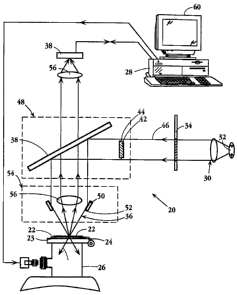
Fig. 3 is a simplified, schematic view of an apparatus 20 constructed in
accordance with the
invention. The target to be interrogated, here indicated at 22, is supported
on a substrate 23 held on
a microscope stage 24 which is selectively movable in the x-y plane under the
control of a stage stepper
motor device, indicated generally at 26, under the control of a computer 28,
which includes other
computational components of the apparatus as described below.
The target is illuminated by an optical light source 30 which directs
illuminating light, typically
light in the visible range, and at one or more selected wavelength ranges,
onto the target surface. As
will be detailed below, the light source typically includes a means 32 for
generating light of a given
wavelength or spectral frequency, one or more filters, such as filter 34, for
producing a desired
frequency band of illuminating light, and a lens system 36 for focusing the
light onto the target, in a
manner to be detailed below.
Spectral emission light from the target, in this case light scattered from the
target, is directed
through lens 56 to an optical detector 58. The optical detector functions, in
a manner to be detailed
below, to detect one or more spectral emission characteristics of the
individual PREs in the illuminated
portion of the field. The detector is typically a CCD (Charge Coupled Device)
array which operates
to generate and store an array of optical intensity values corresponding to
the array pixels, as will be
detailed below.
An image processor contained within computer 28 is operatively connected to
the detector to
receive values of light intensity at each of the detector array positions,
under each selected illumination
condition, e.g., different wavelength or polarization state. The image
processor functions to construct
a computer image of the positions and values of one or more spectral emission
characteristics measured
by the detector. Typically, this is done by treating each pixel in the
detector array as a position point
18
............._...., .... ~
CA 02280794 1999-08-12
WO 98/37417 PCT/US98102995
in the illuminated field, and assigning to each pixel "position" the light
intensity value recorded by that
pixel. The image generated by the image processor may be a matrix of stored
numbers, e.g., position
coordinates and associated spectral emission characteristic value(s), or an
actual map in which position
are represented, for example, in an x-y plane, and each measured spectral
emission value, represented
as a quantity along the z axis, for each pixel location.
A discriminator 42 in the apparatus, also forming part of computer 28,
functions to discriminate
PREs with a selected spectral signature, i, e. , a selected range of values of
one or more selected spectral
emission characteristics, from other light-scattering entities in the computer
image. Examples of the
operation of the discriminator will be given below.
C 1. Substrate
As indicated above, the target is supported on a substrate which is mounted on
a microscope
stage. Suitable substrates include standard glass slides, cover slips, clear
polystyrene, and clear mica
as examples. Other suitable transparent substrates are those associated with a
TEM grid. including for
example, formvar, carbon and silicon nitride. These TEM-associated substrates
are all optically
transparent at the thicknesses used. Conducting, semiconducting, and
reflecting substrates are also
suitable for PRE applications.
Another suitable substrate for use in the present invention are those which
may initially appear
opaque to the spectral wavelengths of interest for PRE observation, but which
can be rendered suitable
by the application of a suitable fluid or vapor. An example is white
nitrocellulose "paper" as used for
the transference of biological samples of interest in diagnostic techniques
such as "Southerns",
"Northems", "Westerns", and other blotting, spotting, or "dip stick" tests.
Once the materials of
interest have been transferred and fixed as desired, the PRE's can be applied
as preformed entities, or
one can apply PRE nucleation entities and enhance as described below. The
white nitrocellulose at this
stage may typically present significant non-specular light scattering which
makes it difficult to visualize
the PREs. However, if a suitable treatment which results in a significant
reduction of the non-speculai
scattering is used, for example, allowing acetone vapor to encompass the
nitrocellulose substrate, while
monitoring the PREs, the substrate can become much less opaque, and permit
efficient observation of
the PREs.
Silicon is a preferred substrate for many PRE detection applications because
it can be made very
smooth and free of defects, resulting in very little non-specular scattering
under darkfield illumination.
One example of a particularly preferred silicon substrate is the highly
polished, etched, and defect free
surfaces of silicon wafers commonly used in the manufacture of semiconductors.
The nearly complete
absence of contaminants and surface imperfections of such a substrate produces
excellent contrast of the
PRE scattering under darkfield illumination conditions. However, it should be
appreciated that such
19
CA 02280794 1999-08-12
WO 98/37417 PCT/US98I02995
silicon wafers typically have a thin layer of SiO~ present on their surface as
a result of the various
processing steps. It may be mentioned that silicon substrates with
approximately 100nm or more of SiOz
on their surface produce some of the most intense, high contrast PRE spectra
so far observed from a
solid substrate, and it may be advantageous to intentionally grow a sub-micron
layer of Si02 on the
silicon wafer surface.
If the oxide layer is removed from the silicon surface in a manner that
prevents rapid re-growth
of an oxide layer, for example, by etching in HF acid and passivating the
surface with hydrogen, the
optical image of the "point-source" PREs has been observed to be torus-shaped,
rather than the usual
Airy ring pattern with a bright central region. This "doughnut" phenomenon
most likely arises as a
result of damping of the transverse driving electric fields (those parallel to
the silicon surface), leaving
only the perpendicular driving fields which can excite a plasmon mode that
radiates well, but not at all
directly along the normal. This property of bare silicon substrates can be
useful in determining whether
a particular PRE is closely bound to the surface of the silicon substrate, or
is bound via a tether
molecule or system that has placed it further from the surface, thereby
changing the dipole component
scattering ratios.
C2. Light source and detector
With continued reference to Fig, 3, light-generating means 32 in the light
source may suitably
be a mercury, xenon, or equivalent arc; or a Quartz-tungsten halogen bulb, of
approximately 20 to 250
watts, which provides incident light in a frequency band corresponding to
wavelengths from
approximately 350nm to 800nm, for visible light PRE scattering, or a
conventional UV source for
lower-wavelength PRE scattering.
Filter 34 typically includes a set of pre-selected narrow bandwidth filters,
allowing manual or
computer controlled insertion of the respective filters. The bandwidth for
such filters is typically S-10
nm.
Other methods of illuminating a target with a series of selected bandwidths
include the use of
light sources such as lasers of all types where one may utilize very narrow
bandwidths. Multiple
frequency sources are also contemplated, such as tuned lasers (i. e. Ar-ion)
to select any of the
characteristic defined strong "line" sources. Alternatively a grating or prism
monochrometer can be
used. All the light sources can be either of continuous or pulsed variety, or
a suitable light amplitude
modulation device (not shown) can be inserted in the incident path to vary the
intensity level in a
prescribed temporal manner. The polarization of the light to be incident upon
the sample can be varied
by the insertion of suitable filters or other devices well known to the art.
The microscope in Fig. 3 is illustrated to be configured with an epi-
illumination system,
whereby the collimated light from the source following filtering as desired
impinges onto a half silvered
CA 02280794 1999-08-12
WO 98/37417 PCT/C1S98/02995
mirror 38, and is reflected downwards towards the DarkfieldlBrightfield
(DFIBF) lens 40. In this
particular type of DFIBF application, the incident light that would have had
rays passing through the
objective lens is physically blocked by an opaque circle 42, which is
suspended by very fme webs 44,
so as to allow only a concentric band of light to pass such as bounded
radially and illustrated by the rays
46. The unit comprising mirror 38 and opaque circle 42 may be built into an
adjustable block 48 that
can be manually (or robotically) moved thereby converting the microscope from
DF/BF to alternate
forms of operation.
Light reflected from the mirror may in turn be refracted or reflected (by a
suitable circular lens
element 50, fixed to the objective lens mount into a hollow cone of incident
light 52, converging toward
a focus at the sample plane of the target. As previously noted, the specular
reflection of such rays
causes them to return along the lines of the incident cone trajectories, where
they are ultimately
absorbed or otherwise removed from the optical system.
In this darkfield system illustrated in Fig. 3, the angle between the optic
axis and the incident
rays illuminating the sample is larger than the largest angle between the
optic axis and the rays scattered
by the PREs which is accepted into the objective lens element 45, which is
illustrated to be of the
refractive form. Also incorporated in the total optical microscope, although
not shown, is the ability
to divert the light rays away from detector 38 to other ports whereby the
image may be observed
visually through standard binocular eyepieces, or to yet another port, for
example, for photographing
the illuminated field.
It has been found to be suitable to use a Nikon DF/BF lens model CF Plan BD
ELWD with
magnification 100X and numerical aperture (N.A.) 0.8 as the lens system 54,
and also a model CF Pian
BD ELWD with magnification 20X and N.A. 0.4. In that case, the rays entering
the objective element
of the lens may be rendered parallel and incident upon the 50% mirror 38, and
into a relay lens 56
(typically magnification of 2X or SX) that focus the rays to an image plane on
detector (image capture
device) 58, where the detection is performed by a suitable CCD camera system.
The optical system, including lens 56, is preferably constructed to project
the field being viewed
into an area corresponding to the array of the detector, so that each pixel in
the array is reading light
from a defined region of the field.
Various image capture devices known in the art may be used, including fiber
coupled photo-
diode arrays, photographic film, etc. One exemplary device is a
thermoelectrically cooled CCD array
camera system, model CH250, manufactured by Photometrics, of Tucson AZ. This
device utilizes a
CCD chip model KAF1400, having a 1032 by 1037 pixel array.
21
CA 02280794 1999-08-12
WO 98/37417 PCT/US98/02995
It will be appreciated that the detector serves to detect a spectral emission
characteristic of
individual PREs and other light-scattering entities in the field, when the
field is illuminated by the light
source, simultaneously at each of the regions in the field corresponding to
array pixels.
C3. Image Processing, discrimination and output
Where the detector is used, for example, to detect spectral peak wavelength,
peak intensity,
and/or half width of the spectral peak, the detector measures light intensity
at each of a plurality of
different illuminating light frequencies, simultaneously for each of the field
regions corresponding to
a detector array pixel.
The emission (scattering) values measured at each frequency are stored,
allowing spectral
emission curves for each region to be constructed after a full spectrum of
illumination. From these
curves, peak wavelength, peak intensity, and width at half intensity are
calculated for each region.
Similarly, the peak halfwidth in the image plane can be measured with a CCD
array as described above.
The detector may be supplied with comprehensive software and hardware that
allows timed
exposures, reading out of the pixels into suitable files for data storage,
statistical analysis, and image
processing (as one of the functions of computer 28). This capability serves as
an image processor for
constructing from signals received from the detector, first the values of the
spectral emission
characteristics) being determined, and then a computer image of these values
and the corresponding
associated field positions.
The image constructed by the image processor may be a matrix of stored points,
e.g., a matrix
of associated values of each field position (regions in the field) and values
for one or more measured
spectral characteristics, or may be an actual map of field positions, e.g. ,
in the x-y plane, and associated
spectral emission values in the z plane.
The computer in the apparatus also provides discriminator means for
discriminating PREs with
a selected spectral signature from other light-scattering entities in the
computer image. The basis for
this discrimination is noted above in the discussion of various spectral
emission characteristics and their'
correlation with physical properties of light-scattering entities.
Thus, for example, to discriminate PREs with a selected spectral peak
wavelength and peak
width at half intensity, the computer image generated could provide a matrix
of all field regions and the
associated spectral peak wavelength and width values. The discriminator would
then selected those
regions containing PREs whose spectral signature meets certain ranges of these
two spectral emission
values. Depending on the particular values chosen, the discriminator could
classify light-scattering
entities in the field in a number of ways, including distinguishing:
1. PREs with a selected spectral signature from all other light-scattering
entities in the field;
2. PREs from non-PRE light scattering entities in the field;
, ,.
CA 02280794 1999-08-12
WO 98/37417 PCTIUS98/02995
3. For a selected type of PREs, those selected PREs which are interacting with
one another and
those which are not; and
4. One selected type of PRE from another selected type of PRE in the field.
In each case, the basis for the discrimination may be based on detected
values, for each light-
scattering entity in the field, of peak position, peak intensity, or peak
width at half intensity of the
spectral emission curve, peak halfwidth in the image plane, and polarization
or angle of incidence
response. Other spectral characteristics mentioned above are also
contemplated. In particular, where
the PREs have surface-localized fluorescent molecules or Raman-active
molecular entities, the detecting
may detecting plasmon-resonance induced fluorescent emission or Raman
spectroscopy emission from
one or more of said molecules or entities, respectively, and these values are
used as a basis of
discriminating such PREs from other light-scattering entities. Fig. 11 shows a
typical Raman spectrum
of a Raman-active molecule carried on the surface of a PRE.
The information obtained from the discriminating step is then used to provide
information about
the field. Various types of information available are discussed in Sections IV-
VI below. Among these
1 S are:
1. The total number of PREs of a selected type in a field. Here the
discriminating step includes
counting the number of PREs having a selected range of values of a selected
spectral emission
characteristic in the constructed computer image;
2. Determining a spatial pattern of PREs having a selected range of values of
a selected spectral
characteristic in the field. Here the discriminating includes constructing an
image of the relative
locations of PREs with those spectral-characteristic values;
3. The distance between two adjacent PREs, particularly where this distance is
less than the
Rayleigh resolution distance. Here the detecting includes exposing the field
with light of one
wavelength, to obtain a diffraction image of PREs in the field, exposing the
field with light of a second
wavelength to obtain a second diffraction image of PREs in the field, and
comparing the distance'
between peaks in the two diffraction patterns;
4. Interrogating a change in the environment of the field. Here the
discriminating includes
comparing the values of the detected spectral characteristic of a PRE in the
field before and after the
change, e. g. , change in the dielectric of the field;
S. Detecting motion of PREs in the field. The detecting here includes
detecting the centers of
the diffraction patterns of the PREs in the image plane, as a function of
time.
C4. Other embodiments
Simultaneous imaging of even 100 PRPs or more in the illuminated field may be
readily and
efficiently accomplished, using the apparatus just described. Alternatively,
the apparatus may be
23
CA 02280794 1999-08-12
WO 98/37417 PCT/US98/02995
designed to "read" a spectral characteristic of each PRE in a field by
sequentially scanning each region
in a field with a focused-beam light source, andlor sequentially detecting
light scattering from each
region in the field, by moving the microscope stage through a small
interrogation region defined by
stationary optics, sequentially interrogating each region to determine values
of a selected spectral
characteristics, according to above-described methods.
For detecting fluorescent images, the light source is filtered appropriately
for the excitation
spectrum of the desired fluorophore, and a suitable filter (not shown) is
placed in the region between
mirror 38 and relay lens 56. This filter is chosen to substantially block the
excitation light and permit
passage of light in a band matching the emission spectrum of the
fluorophore(s) of interest.
The value of the ability to make comparison of the multiple images of
darkfield PRP treated,
brightfield dyed, and/or fluorescent stained samples such as cells and other
entities of interest to
biological and medical researchers and clinical applications can be readily
appreciated.
There are several suitable means for bringing in the incident light so as to
establish effective
darkfield conditions in conjunction with suitable means for preferentially and
efficiently observing the
light scattered by the PREs. For transparent substrates the incident light may
be brought in either in
transmittance through the substrate, reflectance from the "objective side
surface", or via TIR (total
internal reflection) at the interface near which the PREs are situated, as
shown in Fig. 3 and described
in more detail below. In the latter case the evanescent tail of the TIR light
may also be used to excite
the PREs, if they are directly outside the reflecting interface, even though
such light field distributions
do not radiate directly to the objective lens.
For non-transparent substrates, the light must be incident in a manner that
results in as near
specular reflection as possible, with a minimum of such light reaching the
objective. There are several
means for accomplishing this condition. As one example, incident light may be
routed to the field of
view through one or more optical fibers. The fibers may be oriented such that
light reflects off the
substrate at a glancing angle and does not enter the objective lens of the
microscope system used to
image the PREs. It is also advantageous to use commercially available DF/BF
objective lenses.
Examples are those sold by the Nikon Company, Long Island N.Y. An example of
the way such a
DF/BF lens may be used is illustrated in Fig. 3.
The objective lens of system in the apparatus can be made as either a
reflecting or refracting
lens. For certain PRE applications, especially those requiring the most
accurate and rapid focusing of
the objective lens as a function of Iight wavelength, reflecting lenses may be
preferred because
chromatic aberrations can be greatly reduced. Refracting lenses, even those
carefully made to
compensate for chromatic aberrations when observing light emitted by PREs, can
still exhibit significant
deviation in their frequency dependence of the focal length. The fact that the
PREs near the peak of
24
CA 02280794 1999-08-12
WO 98/37417 PCT/US98/02995
their plasmon resonance can be of such extraordinary brightness for a sub-
wavelength sized source
allows them to be utilized for evaluation of optical components, whereby one
can observe a variety of
deviations from the component's ideal "point source" response.
The use of particular lens types which enhance the numerical aperture of the
objective is also
contemplated. PREs can be imaged with a standard "solid-immersion" lens having
a spherical top and
flat lower surface. Another such contemplated lens is a liquid analog to a
solid immersion lens (SIL)
having a fluid between the lower surface of a truncated solid immersion lens
and a substrate which has
an approximately equal index of refraction as the lens material.
Such a lens is illustrated in Fig. 4. The lower flat surface 80 of the lens is
cut shorter than a
standard solid immersion lens. With the index matched fluid 82 between the
lower surface 80 and a
substrate 84, the focal plane of the lens is at the usual r/n (where n is the
index of refraction of the lens
material) location, which is now inside the index matched fluid. PRPs in the
fluid at this focal plane
are thus imaged with this system, allowing focusing on non-substrate bound
PREs in solution. Such a
lens can readily be added to a commercial DF/BF microscope lens by one of
ordinary skill in the art,
thereby resulting in an improved numerical aperture and increased
magnification for viewing samples
in a liquid matrix. In particular, this modification results in increased
brightness and clarity in
visualization of the PREs. This configuration allows the user to vary the
focal position in a liquid
sample while retaining a large numerical aperture and removes the usual
requirement of total internal
reflection-based illumination as is used for the SIL. This system therefore
allows the monitoring of
movement of a particular PRE in solution.
In other preferred embodiments, darkfield optics are chosen to optimize the
signal to noise ratio
of the PRE signal. This often includes improving the contrast by reducing the
background (non-PRE)
scattered light to a minimum, or to a minimum relative to the amount of PRE
scattered light observed.
For example, using ordinary 1 " x 3" glass microscope slides, it was
determined that the Nikon DF/BF
lenses of the extended working distance class had a better contrast for
observation using the 0.8 NA lens
compared to the 0.9 NA lens at the same magnification level ( 100X), despite
the fact that the higher the
NA of a lens the more scattered light is collected from the PRE. This may be
attributed to the change
in reflecting properties of the bare glass substrate since the angle of
incidence of the darkfield
illumination light is changed when the NA of the objective is changed.
Polarized incident light can also be used at specific angles (i. e. the
Brewster angle) to reduce
the amount of reflected light from the surface of the substrate. This improves
contrast by reducing the
amount of non-PRE scattered light which enters the objective lens. When
imaging non-spherical PREs
such as ellipsoidal PREs as described above, the response to plane polarized
incident light can also be
used to distinguish different PRE populations.
CA 02280794 1999-08-12
WO 98137417 PCT/US98/02995
In another embodiment, a transparent substrate is used which is sufficiently
thick so as to reduce
the amount of light scattered from its bottom surface that reaches the
detector. If particulates are
present on the bottom surface of a thin glass substrate, some of the scattered
incident light will re-enter
the detection system and increase background. By increasing the thickness of
the substrate, one
displaces the region of the spurious scattering further from the optical axis,
thereby reducing the amount
of non-PRE scattered light that enters the detector.
Total internal reflection (TIR) may also be used to illuminate PREs in a
transparent substrate
from beneath. The evanescent tail of such light can be effectively used to
excite the PRE located near
that interface. There are several methods for exciting PREs with totally
internally reflected light, such
as using an optical fiber whose dimensions and indices of refraction of the
inner core and outer layer
are chosen so that there is sufficient evanescent field at the fiber outside
surface to excite PREs placed
thereon. The light emitted from the PRE can be transferred back to the fiber,
forming a reflected source
of light which can be observed by standard methods.
In another embodiment, illustrated in Fig. 5, a Dove prism 70 is illuminated
from the side. A
standard glass slide 72 is placed on the top surface of the Dove prism 70,
with a suitable index matching
oil 74 in between. The incident light 76 is brought in parallel to the major
surface of the prism 70 and
is refracted up toward the slide 72. The angle of incidence at the slide is
selected to be greater than the
critical angle, and results in total internal reflection at the upper surface
of the slide 72. The light then
exits the other side of the Dove prism, again parallel to the major face of
the prism 70. Evanescent
electromagnetic fields excite PREs bound to the upper surface of the slide,
and emit the usual plasmon
resonant scattered light into an objective lens located above the slide.
This Dove prism geometry is convenient for bringing light from diverse sources
such as laser,
quartz halogen, arc lamp and the like via an optical fiber or lens to impinge
upon the side of the Dove
prism, and filters may be conveniently interposed therebetween to further
control the nature of the
incident light.
The DARKLIGHT(TM) light source from Micro-Video of Avon, MA can also be used
in a
total internal reflection illumination system, although it has been found
generally inferior to the Dove
prism embodiment described with reference to Fig. 5. This totally internally
reflecting slide illuminator
takes light from a halogen source into an optical fiber, then into the edge of
a glass slide. The light
undergoes total internal reflection numerous times while spreading down the
slide, exciting PREs with
evanescent fields as with the embodiment of Fig. 5. This system is described
in detail in U.S. Patent
No. 5,249,077. Additionally, PREs can be observed via the illumination
described in U.S. Patent No.
3,856,398.
26
CA 02280794 1999-08-12
WO 98137417 PCTIUS98I02995
Oil immersion lens systems can also be used in conjunction with TIR
illumination. In these
systems, though, the index of refraction of the medium for internal reflection
must be greater than that
of the oil being used. Accordingly, flintglass, having an index of refraction
of 1.7, is one preferred
prism material.
Fig. 6 illustrates in cross section a DF/BF objective lens system comprising a
reflective
objective lens 90 combined with a suitable enclosing darkfield illumination
ring element 92 as an
alternative illumination scheme. Although the use of such a two component
reflective lens reduces the
intensity at the center of the diffraction pattern, the fact that it also
substantially reduces the chromatic
aberration compared to the refractive lenses described in the prior art, makes
it a preferred embodiment
in conjunction with the means for obtaining the simultaneous frequency
dependent scattering data for
a multiplicity of PREs and for the other non-PRE scattering entities within
the field of observation that
may be imaged and which need to be distinguished and rejected for many
applications.
In some applications, optical microscopy methods must be tailored to both
optically image and
analyze the PREs and also to observe the same PREs and associated sample
material with additional
instruments such as one or more forms of electron microscopy. In these cases,
darkfield optical
microscopy must be performed with substrates suitable for electron microscopy
as well. Because the
electrons must pass through the sample and substrate, the substrate must be
very thin, typically well
under 1 ~.m. Common substrates include formvar and/or carbon deposited upon a
supporting grid.
Background scattering is reduced from the grid boundaries or "bars" by
arranging for the field of
application of the incident darkfield illumination to be within the spacing of
the grid "bars" and/or to
restrict the field of view of the collecting objective light for the part of
the sample under observation.
For the BF/DF objective and microscope system, grids with a spacing of up to
400 bars per inch and
silicon nitride membranes are especially suitable. Alternative forms of
darkfield illumination include
a separate optical fiber or lens with darkfield illumination from below the
grid substrate and observation
of the scattered PRE light from above with a BF objective lens.
IV. PRP Compositions
The invention further includes a suspension of plasmon resonant particles
(PRPs) having one
or more populations of PRPs. The composition has four distinguishing features:
(i) the PRPs in each
population have a spectral width at halfheight of less than 100 nm; (ii) the
PRPs in a single population
are all within 40 nm, preferably 20 nm of a defined spectral emission peak
wavelength; (iii) at least 80%
of the PRPs in the composition are in one or more of the populations and have
a spectral emission
wavelength in one of the three ranges > 700 nm, 400-700 nm; and < 400 nm; and
{iv) each population
has at most a 30% overlap in number of PRPs with any other population in the
composition.
27
CA 02280794 1999-08-12
WO 98137417 PCTIUS98102995
The first feature addresses the quality of the PRPs, high-quality emitters
being characterized by
a relatively narrow frequency range of scattered light. The second feature
provides homogeneity of
spectral emission properties for all PRPs within a given population.
Specifically, PRPs within a given
population all have a peak wavelength within 40 nm of a defined wavelength.
The third requirement
provides that a large majority of the PRPs (at least 80% by number) are in one
of three different
wavelength ranges. The fourth requirement defines the uniqueness of the
populations, assuming that
each population has a distribution of spectral peak wavelengths within 40 nm
of a given peak
wavelength. The two distribution curves can be no closer than the distance at
which 30 number percent
of the particles in one population fall within the distribution curve defined
by an adjacent population.
Typically, the PRPs in the composition are in one or more of the populations,
all in the 400-700
nm wavelength range. The PRPs may be homogeneous, e.g., all blue particles, or
may be in one of
more populations, e.g., discrete populations of red, green, and blue
particles, collectively making up
80 % of the PRPs in the composition.
Particles in this spectral range may be formed, as described below, as solid
silver particles, silver
particle with a gold core, or particles with a dielectric core and an outer
silver shell of at least about
Snm.
In one general embodiment, particularly for use in a variety of diagnostic
applications, the
particles have localized at their surfaces, (i} surface-attached ligands
adapted to bind to iigand-binding
sites on a target, (ii} fluorescent molecules, and (iii) Raman-active
molecular entities. The ligands are
one of the members of a conjugate pair that can include antigen/antibody,
hormone/receptor,
drug/receptor, effectorlreceptor, enzyme/substrate, lipid/lipid binding agent
and complementary nucleic
acids strands, as examples.
The PRPs in the composition may have different surface-localized molecules on
different groups
of PRPs. These different groups may be different PRP populations, that is,
PRPs with different spectral
peak wavelengths, or may be localized on PRPs in a homogeneous PRP population.
For use in identifying a target having first and second ligand-binding sites,
the different surface
localized molecules may be different ligands effective to bind to different
ligand-binding sites, such as
two different-sequence oligonucleotides that bind at different sequence
regions of a common target
polynucleotide, or two different ligands that bind to different ligand-binding
sites on a macromolecular
target.
PRPs may be formed made by a variety of known methods, including colloidal
chemistry,
soluble gel, evaporationlannealing, nucleationlgrowth via an enhancer,
autoradiography, and
photoreaction in silver halides and other materials via electromagnetic energy
in the form of light, X-
28
, t ..
CA 02280794 1999-08-12
WO 98137417 PCT/US98102995
rays, or other incident wavelengths. In addition, lithography,
electrodeposition, electroplating, and
sputtering with a scanning tunneling microscope (STM) tip can be used.
Where the PRPs have surface localized, e.g., attached iigands, the PRPs are
able to bind
selectively to a target of interest which carries the other half of the
ligand/ligand-binding conjugate pair.
As indicated above, it is also advantageous to produce population of PRPs
having different
defined peak wavelength values. When defined populations of PRPs are combined
with specific binding
characteristics, a new class of sub-microscopic probe is created which has
significant advantages over
all currently used labeling techniques. The PRP formation techniques described
below may be used to
create such probes.
A. Formation of the PRPs By Metal Enhancement
In this method for forming PRPs, a nucleation center, typically a metal
nucleation center in the
1-20 nm size range, is placed at a targeted location, followed by in situ
development (enhancing) of the
full conductive body of the PRP. In some applications, a spatially pre-
specified position is designated
for the placement of the nucleation center. This is in contrast with other
applications where nucleation
centers are used to probe a matrix for a particular substance or feature whose
position is not known.
Pre-specified placement may be advantageous in metrology and/or
instrumentation applications which
are further described below. For example, it may be desired to place a single
PRP on the end of an
optical fiber or a scanning microscope tip. Also, it may be desired to create
a pattern of PRPs in a
particular geometric configuration.
Because it is often desirable to create PRPs with controlled spectral
characteristics, the
enhancing may advantageously be performed while being monitored, stopping the
process when a PRP
having some pre-defined spectral characteristic is formed. This is especially
convenient when
enhancement chemistry which is not affected by light is used. It is also
possible to monitor PR
formation by periodically terminating the enhancement process, observing the
characteristics of the entity
or entities formed, and re-initiating the enhancement process if one or more
spectral characteristics such
as color, for example, are not within a desired range.
The nucleation center is typically a gold particle 1-10 nm in diameter, and
the metal used for
enhancing this nucleating site to PRP size is silver. However, other elements
including, for example,
platinum, nickel, and palladium, and macromolecules, such as polynucleotide
molecules, are also
contemplated as nucleation centers for the subsequent enhancement process. Non-
metallic materials may
also be used as nucleating centers, such as protein and nucleic acid.
The configuration of the nucleating center can be controlled so as to produce
a PRP having a
desired shape or characteristic. For instance, triangular or ellipsoidal
regions of nucleating material can
be formed. The deposition process may involve many metal deposition techniques
known in the art,
29
CA 02280794 1999-08-12
WO 98!37417 PCT/US98/02995
such as vapor deposition, sputtering, Ga-focused ion beam (FIB) milling of the
thin film prepared of
the desired material, and electroplating into nanopores. Particularly well
controlled placement of
nucleating material is possible by discharging nucleating material from the
metal tip of a scanning
tunneling microscope. Techniques have also been developed whereby individual
metal atoms are picked
up with the tip of a scanning tunneling microscope, moved, and put down at a
desired location.
Individual atoms may also be "pushed" to a desired location with a scanning
tunneling microscope tip.
This technique may be used to place, for example, 10 gold atoms at a spatially
pre-specified position
for use as a nucleation center.
In one embodiment, PRPs or PRP nucleating centers are placed at a desired
location by
introducing the PRP or PRP nucleating center into a drawn micropipette tip,
moving the PRP or PRP
nucleating center to a desired location and depositing the PRP at this
location using standard micro-
manipulation techniques. The pipette tips are filled with the desired material
in solutions of varying
viscosity (i.e. with gelatin or other matrices), then the tip is manipulated
to a selected location while
observing under the microscope. By applying pressure to the solution in the
pipette, very small
quantities can be deposited onto a desired substrate. Other micromanipulation
techniques are also
possible.
Because the PRP material will have a positive dielectric constant at
frequencies above the
resonant frequency, the metal particles can be optically trapped in a focused
light beam of appropriate
wavelength using principles similar to currently practiced optical trapping of
plastic particles. The PRP
can thus be placed at a spatially pre-specified position by manipulating the
light beam in which it is
trapped. Channels of gel may also be created in order to route PRPs to
spatially pre-specified positions
electrophoretically. If the channel is configured to pass proximate to a
designated location, a PRP in
the channel will move under the influence of an electric field so as to be
guided to the location.
Electrostatic bonding techniques can also be used to removably attach PRPs to
spatially pre-specified
positions.
In one embodiment, an array of one or more conductive pads of approximately 10
to 100
microns in diameter may be created using standard integrated circuit
lithographic techniques. Each pad
may further be selectively connected to a voltage source. As PRPs in solution
can be negatively
charged, applying a suitable potential to a pad will attract a PRP from
solution and onto the conductive
pad. Removing the voltage and reversing the sign of the applied potential may
free the PRP if the
surface binding forces between the PRP and the pad are weak, i.e.,
approximately one picoNewton.
Conjugated nucleating centers and/or conjugated PRPs can also be placed at a
spatially pre-
specified position by immobilizing the other half of the conjugate pair at the
spatially pre-specified
__. ,.._ -. __ . , , ,
CA 02280794 1999-08-12
WO 98137417 PCT/US98/02995
position and binding the conjugate on the nucleating center or PRP to the
other half of the conjugate
pair.
Individual nucleating centers (or fully formed PRPs) may also be deposited by
ejecting droplets
of metal particle containing fluid to a substrate and having the fluid
evaporate away by techniques
analogous to those used in ink jet printing. In this technique, one or more
metal particles can be
delivered to a specific location defined by the drop position. In some
embodiments, the concentration
of PRPs or nucleating centers in the fluid is chosen so that it is
statistically likely for one or no PRP
or nucleating center to be contained in each drop. If nucleation centers are
deposited, they may be
subsequently silver enhanced to form PRPs. It may further be noted that these
techniques can be used
to create a desired geometric pattern of PRPs. When making such a pattern,
drops can be redeposited
at those locations where no PRP was ejected during the first pass. Articles
with such patterns of PRPs
can be useful in object identification, semiconductor mask registration, and
other uses as are described
in more detail below.
As defined herein, the term "in situ" indicates that the PRP is bound to a
substrate, immersed
in a solution or suspended in a matrix. The definition of "substrate" is
discussed below and includes
any entity with which a PRP can associate such as tissue sections, cells, TEM
grids made from, for
example, formvar, silicon (including silicon nitride and silicon dioxide),
mica, polystyrene, and the like.
These nucleation centers are typically colloidal gold preparations, and
suitable populations of nucleation
centers are commercially available either in free form or attached to various
biological molecules.
The silver enhancement of gold nucleating centers to produce larger silver
masses which can
be visible under an electron andlor a light microscope is currently practiced,
and some procedures for
imaging biological systems using silver enhanced gold particles have been
extensively developed.
Reagents for performing the enhancement, as well as the nucleating centers
themselves, are accordingly
commercially available. Antibody bound gold nucleation centers are available
from several sources,
including E-Y Laboratories of San Mateo, CA, and Amersham of Arlington
Heights, IL. Furthermore,
a large body of literature describes a variety of suitable methods for
performing such enhancement (see,
for example, M.A. Hayat, Ed., Immunogold-Silver Staining - Principles,
Methods, and Applications,
CRC Press, 1995, the disclosure of which is hereby incorporated by reference
in its entirety, most
particularly Chapters 2 and 7, which describe several experimental techniques
for silver enhancing gold
nucleation centers and a discussion of the silver masses formed thereby).
The enhancement process is preferably performed by mixing gold nucleation
centers with a
solution of a silver salt such as silver acetate, silver lactate, or silver
nitrate, and a reducing agent such
as hydroquinone in a citrate buffer at a pH of approximately 3.5 to 3.8. It
has been suitable to provide
a concentration of approximately 6 mM for the silver salt and approximately 33
mM for the
31
CA 02280794 1999-08-12
WO 98/37417 PCT/US98/02995
hydroquinone. Commercially available silver enhancement solutions containing
appropriate quantities
of silver and reducing agents are also commercially available from, for
example, BBI of the United
Kingdom. Those of skill in the art will appreciate that choice of buffering
system, silver salt, reducing
agent, and other enhancement parameters are preferably optimized for the local
environment, target
locations, and the like, and that such optimization can be performed without
undue experimentation.
In accordance with one aspect of the present invention, PRPs useful in the
invention are
prepared by silver enhancing nucleating centers until the particles possess
the properties of plasmon
resonant particles. Most preferably, the silver enhancement parameters are
controlled such that the
PRPs created have pre-defined spectral characteristics, such as appearing a
particular desired color when
viewed with darkfield microscopy. During enhancement, spectral characteristics
may be observed for
one individual evolving PRP or simultaneously for a plurality of individual
PRPs. Thus, PRPs having
specific physical properties can be made and placed at a desired location. In
addition, PRP nucleating
centers can be placed at a desired location, either in situ, in vitro or in
vivo, followed by silver
enhancement to produce a PRP at the specific location.
B. Specific Examples of PRP (or PRE) Formation by Metal Enhancement
PRPs were prepared by silver enhancement of a gold nucleating center as
described in the
following examples.
Example 1
Placement of Qold colloids on a substrate
The Alcian Blue method is one method for attaching gold colloid nucleating
centers to a
substrate by chemically treating one or more spatially pre-specified positions
of the substrate. Alcian
blue is a positively charged dye which promotes adhesion of the negatively
charged gold colloids by
charge interactions. Portions of the substrate at which PRPs are undesired may
be coated with a
blocking agent such as BSA. A 100 microliter drop of 100:1 dilution of 5 %
acetic acid and 2 % alcian
blue was placed onto a carefully cleaned glass slide for ten minutes. The
active site of the substrate was
then immersed in doubly distilled water, rinsed, and dried.
The gold colloids were diluted to the desired concentration. The solution was
placed on the
alcian blue treated region of the substrate and incubated for 10 minutes. The
substrate was then rinsed
with distilled water.
The gold colloid attached to the substrate was enhanced as described in
Example 2.
Example 2
Silver enhancement of individual yoid nucleating centers
32
CA 02280794 1999-08-12
WO 98/37417 PCTIUS98/02995
One ml of a 0.1 mg/ml solution of gelatin was mixed with 50 ~.1 initiator and
SO ~.1 enhancer
in an eppendorf tube. The initiator and enhancer were obtained from BBI
International (United
Kingdom) silver enhancement kit, light microscopy (LM) version, catalog No.
SEKL15. The substrate
was immediately covered with the enhancer solution and timing was started. The
substrate was then
viewed under darkfield illumination to determine whether PRs were present. The
approximate
enhancement time for colloids on glass, silicon or TEM substrates was about
one minute, while the
approximate enhancement time for conjugate colloids attached to biological
substrates was about seven
minutes. These times were determined by continuously observing the scattered
light from an individual
evolving PRP under darkfield microscopy while the enhancement was taking
place. It can be
appreciated that precise enhancement durations can be utilized to control the
scattering response, and
therefore the color, of the PRPs created. Enhancement was stopped by rinsing
thoroughly with distilled
water.
Example 3
Generation of PRPs in solution
For uncoated colloids, 100 ~cl stock gold colloid (BBI International, United
Kingdom) was added
to 20 ml gelatin solution (0.1 mg/ml). For protein conjugated colloid, 100 wl
stock conjugated gold
colloid was added to 20 ml doubly distilled water with 1 % bovine-serum-
albumin (BSA) to block the
surface. Conjugated colloids used were bovine serum albumin, goat anti-biotin
and rabbit anti-goat IgG.
Typical gold colloid concentrations are:
For uncoated colloid:
3 nm stock - -- 3 x 10'3 particleslml
10 nm stock - - 5.7 x 10''- particles/ml
20 nm stock - ~ 7 x 10" particles/ml
For protein conjugated colloid:
1 nm stock - ~ 2 x 10'5 particles/ml
5 nm stock - -1.7 x 10'° particleslml
10 nm stock - -1.7 x 10'3 particles/ml
20 nm stock - ~ 2 x 10'2 particles/ml
Three drops (150 ~,1) initiator was then added. Ten ~,1 increments of enhancer
were then added
while stirring, until the amount corresponding to the desired PRP scattering
peak wavelength was added.
The PRP resonance was checked by darkfield measurement or by absorption
spectroscopy as described
previously.
C. PRP and PRE Formation With Litho~raphy and Illumination
33
CA 02280794 1999-08-12
WO 98137417 PCT/US98/02995
Lithographic techniques can be used to specify where a PRP (or PRE) will be
formed and to
control its shape, shape, morphology and composition. Both positive and
negative resist methods can
be utilized to lay down either nucleation centers or fully formed PRPs. In the
positive resist method,
portions of a layer of resist is removed in order to form molds into which
metal is deposited. After
such deposition, resist and extra metal is lifted off, leaving a nucleating
center or a fully formed PRP
behind. In the negative resist method, a chosen layer of silver or other
suitable metal is covered with
a layer of resist. Portions of this resist layer are then polymerized. When
nucleation centers are formed
with this technique, the characteristic size of this polymerized region may
advantageously be
approximately 5-20 nm. For laying down PRPs whose peak wavelengths are in the
optical spectrum,
the characteristic sizes of the polymerized regions are advantageously 40-125
nm. Silver and un-
polymerized resist are then etched away, leaving the metal nucleation centers
or fully formed PRPs
under the polymerized portions of the resist. As another alternative, metal
forms can be produced which
are larger than desired, and material may be etched away by ion milling until
a PRP of desired
characteristics is formed. All of these techniques are used in the electronics
and other industries and
are well understood by those of skill in the art.
In addition, metal salts and halides (i.e. in film) can be irradiated to
obtain nucleation centers
or entire particles. Enhancement can be performed with the techniques set
forth above, or can also be
performed by thermally annealing the metal particles, or may be performed as
is done in photographic
development processes, wherein a film of photochemical metal salts or metal
halides is locally irradiated
with light until PRPs are produced, and the film is then fixed and developed.
There are several ways
a localized light spot may be produced for forming nucleating centers or PRPs
at desired locations. In
one embodiment, a very localized light spot can be generated using a metal
side-coated tapered fiber
"near-field" scanning tip to help confine the nucleation to a suitable small
size or to grow the silver
grains directly. Alternatively, a pre-made special optical fiber tip of
preferably approximately 150 nm
diameter having a PRP on its end concentrates the light at a desired location
to "write" these nucleation
centers onto a photosensitive surface material. In another embodiment, a solid-
immersion lens may be
utilized to focus the light onto the metal salts or halides. The solid-
immersion lens may include a PRP
at its focal point in order to further intensify the local radiation of the
substrate.
Photochemical silver salts or halides are also sensitive to electron and ion
beam irradiation, as
well as irradiation from radioactive elements. It will be appreciated that
these photographic methods
can also be used to produce arrays or patterns of PRPs of desired
configuration.
Whether the developing PRPs are in solution, or bound to a substrate, the
enhancing process
can be observed in situ with darkfield microscopy and the process stopped once
the PRP has reached
the desired size which corresponds to a particular color. During light
sensitive enhancement procedures,
34
CA 02280794 1999-08-12
WO 98/37417 PCTIUS98/02995
the progress of the enhancing process can be observed by washing out the
enhancer, observing the light
scattering properties of the particles created, and re-initiating enhancement
until PRPs with desired
spectral characteristics are obtained. In an alternative embodiment, a
relatively light insensitive
enhancer can be used and the enhancing process can be observed under
continuous darkfield illumination
and scattering data collection. Of course, once specific protocols have been
developed which indicate
enhancer amounts, incubation times, etc., to produce PRPs with given
properties, observation of the
enhancement process becomes unnecessary.
D. Formation of a ConiuQated PRPs
As is shown in Example 3 above, it is possible to enhance nucleating centers
which are bound
to a biological macromolecule such as an antibody. This enhancement of
conjugated gold antibodies
can be successfully performed even when the conjugated goldlantibody is in
aqueous solution and has
not been previously bound to an antigen in a cell or cell organelle.
Surprisingly, it has been found that
after silver enhancement of free conjugated gold nucleating centers to create
PRPs, an appreciable
fraction of the conjugate molecules originally present on the gold colloid are
surface bound to the
resulting PRPs. Furthermore, the biological molecule can retain biological
activity after the
enhancement process.
In conjunction with this method, the amount of bound conjugate on a given PRP
can be
controlled by controlling the size of the conjugate bound nucleation center.
For example, a
commercially available 1 nm diameter nucleation center may have only one
conjugate molecule, and be
silver enhanced to form a PRP with that conjugate molecule attached on the
outermost surface thereof.
A 20 nm nucleation center will have a correspondingly larger number of
conjugates attached, some of
which will end up bound to the surface of the silver enhanced PRP. During
silver enhancement, the
conjugate bound PRPs can be incubated with a blocking agent such as bovine
serum albumin (BSA) to
reduce the presence of non-specific bonding sites on the surface of the PRP.
In another embodiment, a conjugate is added to a PRP after the PRP is made.
The conjugate
associates with the PRP either covalently or noncovalently. For example, a
fully formed PRP is coated
with gelatin, agar, polytetrafluoroethylene (TeflonT"'), PVP, or latex to
prevent non-specific charge
interactions, followed by covalent attachment of one or more functional groups
thereto by well known
methods, thus generating a PRP attached to a first half of a particular
conjugate pair. The reagents
required to couple conjugates to immunogold nucleating centers or formed PRPs
are all commercially
available.
This method of forming conjugate bound PRPs also allows control of the number
of conjugate
molecules (i.e. "first half" conjugate pairs) bound to the surface of the PRE.
In this case, one can
incubate bare PRPs in a solution of conjugate and a blocking agent such as
BSA. The relative
CA 02280794 1999-08-12
WO 98137417 PCT/US98102995
concentrations of conjugate and BSA can be adjusted to produce, on average,
the desired amount of
conjugate on each PRP. For PRPs of unusual shape, or comprising concentric
shells of different
materials, coating with conjugates after formation is typically more
convenient. Conjugate bound PRPs
which are approximately spherical, however, can conveniently be produced from
commercially available
conjugate bound gold colloid nucleating centers and silver enhancers as
described above.
Conjugates or other molecules may be bound directly to the metal surface of
the PRP. The
surface chemistry involved in such binding is complex, but it is currently
exploited extensively in many
non-PRP immuno-gold silver staining techniques. Alternatively, the PRPs may be
coated with a shell
of plastic material such as latex prior to the binding of additional molecules
such as conjugate.
Techniques for binding molecules to latex are also well known. The molecules
bound to the metal
directly or plastic shell may be the conjugate itself, or may be other
intermediate reactive groups such
as sulfides, amides, phosphates, aldehydes, carboxyl, alcohol, or others to
which conjugate or other
molecules of interest may be bound. Conjugates or other molecules of interest
may be synthesized onto
such a reactive base with known techniques of combinatorial chemistry.
E. Formation of PRP Populations With Desired Characteristics
The differences in emission spectra for the two separate PRPs as shown in Fig.
1 can arise from
a number of factors. One significant factor is size, particles of larger size
having resonance peaks at
longer wavelengths, and also having spectral shapes with increased half-
maximum widths. Therefore,
control of the size of PRPs being produced results in control over some
spectral characteristics.
It can thus be appreciated that with the addition of a controlled amount of
enhancer, a
population of PRPs with a narrow range of diameters, and therefore a
correspondingly narrow range
of resonant peak frequencies, may be produced, such as is illustrated in Fig.
2 for four types of PRPs.
In some advantageous PRP production methods, the particles can be observed
during the enhancement
process with a suitable microscope. These methods use enhancement chemicals
such as are described
herein which are relatively unaffected by incident light needed to observe
development of the PRP
during the enhancement process. Thus, PRP development can be observed and
halted when it has
reached a desired end-point. For those applications in which it may be
desirable to use a light sensitive
enhancement process, or if development outside the microscope is desired,
timed sequential enhancement
is performed. The samples are rinsed after each application and the status of
PRP light scattering is
determined. One can continue with as many sequential enhancement steps as
desired.
As indicated above, the PRP composition of the invention includes one of more
PRP populations
having peak wavelengths 40 nm of a defined wavelength. Such homogeneity in PRP
population is
possible by stepwise addition of enhancer coupled with darkfield observation
of the PRP creation as
36
CA 02280794 1999-08-12
WO 98/37417 PCT/US98102995
described above. Because the width of a plasmon resonance peak is typically 20
to 40 nm, it is
generally unnecessary to further reduce the variance in resonance peaks of the
PRP population.
Populations of PRPs having uniform spectral characteristics can alternatively
also be prepared
by purifying non-homogeneous populations. PRP conjugates and free PRPs can be
separated by
conventional biochemical methods including column chromatography,
centrifugation, electrophoresis and
filtration. Because PRPs with surface localized molecules or entities can have
a significantly different
mobility than do free PRPs of the same size, they elute from gel filtration
columns at a different rate
than do free PRPs. Because PRPs are charged particles, they migrate in an
electric field. Thus, PRPs
can be manipulated by and even observed during electrophoresis.
PRPs having certain desired characteristics can also be separated based on
their Zeta potentials.
Zeta potential separation equipment suitable for this use is commercially
available (Coulter Corp,
Florida). Radiation pressure may also be used to force PRPs through a matrix
at different rates
depending on their structural properties. If bound and free PRPs are subjected
to electrophoresis in,
for example, an agarose or acrylamide gel, the free PRPs migrate faster than
do the bound PRPs.
Likewise, PRP conjugates may be preferentially retained by filters.
Purification can alternatively be
performed by centrifugation. Thus, with these methods, an original population
of PRPs having a wide
range of spectral characteristics can be separated into subpopulations which
have a narrower range of
spectral characteristics.
Individual populations of PRPs, may be prepared separately and later mixed
according to a
desired combination of PRP property, e.g., color, in desired amounts, each
labeled with the same or
different biological macromolecules or unlabeled depending on the application.
In such compositions,
it is preferable for the resonance peaks of the different populations of PRPs
to be substantially non-
overlapping, as defined above. In some preferred embodiments, the variance in
peak location of one
population of PRPs is controlled to be within approximately 20 nm of one
defined wavelength, and the
variance in peak location of another population of PRPs in the mixture is
controlled to be within'
approximately 20 nm of a second defined wavelength. To avoid significant
overlap, it is preferable to
ensure that the two peak wavelengths are at least 30 to 40 nm apart, and most
preferably 50 or more
nanometers apart.
Applying the above methods, new types of molecular probes can be produced by
binding
selected conjugates to selected PRPs. As mentioned above, a PRP population
with a first spectral
characteristic can be bound to a first conjugate, and a PRP population with a
second spectral
characteristic can be bound to a second, different conjugate to produce two
differentiable populations
of PRPs with different preferential binding properties. Such pre-defined
mixtures of PRPs are especially
37
CA 02280794 1999-08-12
WO 98/37417 PCT/US98/02995
useful in improving the accuracy of detection of low abundance molecules as
will be explained further
below.
F. Isolated Non-Spherical and Composite PRPs
The emission spectra of PRPs is further affected by the details of their
structure. Ellipsoidal
PRPs offer additional parameters for identification and discrimination. Each
ellipsoidal PRP may have
two or three plasmon resonant peaks, depending on whether there is one
isotropy axis or three different
principal axis dimensions, respectively. Ellipsoidal PRPs having one isotropy
axis show peaks
corresponding to two orthogonally polarized emissions, one associated with
plasmon excitation along
the major axis of the ellipse, the other associated with plasmon excitation
along the minor axis. The
distinct plasmon resonance peaks occur at maximal intensity when the
polarization of incident light is
along the corresponding principle axis. Thus, the response of a fixed
ellipsoidal PRP to polarized light
may vary with the direction of incidence.
A process for making ellipsoidal silver particles consists of pulling on a
glass matrix containing
spherical PRPs at a temperature such that the viscosity results in a
stretching of the PRPs into prolate
ellipsoidal particles having a desired aspect ratio. Conditions have also been
described for "pushing"
on such particles such that they form oblate ellipsoidal particles. One
ellipsoidal PRP containing matrix,
PoiarcorT"' (Corning Company, Corning, NY), consists of aligned ellipsoidal
PRPs in a glass matrix.
This composite material is an effective polarizer for certain optical
frequencies, principally in the red
and above. Individual ellipsoidal PRPs contained within such a glass matrix
can be isolated by
dissolving the matrix in such a manner so as to not disturb the PRPs. By
preparing ellipsoidal gold
particles of the correct aspect ratio and size in a suitable matrix, then
dissolving the matrix in a manner
that does not disturb the particles, large quantities of ellipsoidal of PIZPs
having approximately
equivalent plasmon resonance scattering properties can be prepared for use in,
for example, liquid
reagent preparations for biochemical assays as described in detail below.
Other methods can be used to prepare ellipsoidal PRPs, which may be produced
via
photoreaction of silver halides or with appropriate lithographic molds.
Alternatively, PRPs which are
originally formed to be substantially spherical can be pressed or rolled
between two surfaces to flatten
them into a desired oblate or prolate ellipsoidal configuration.
Non-spherical and non-ellipsoidal PRPs are observed to have characteristic
spectral signatures
which are different than spherical and elliptical particles, and which are
useful in creating plasmon
resonant probes with advantageous size and scattering properties. It has been
found that PltPs which
scatter strongly in the blue region may be conveniently produced by making
spherical silver particles.
As discussed above, increases in particle diameter will red shift the resonant
peak. However, for
spherical particles, the peak intensity of the scattered light begins to drop
off as the peak is shifted into
38
CA 02280794 1999-08-12
WO 98/3?417 PCTIUS98/02995
the red, and accordingly strong red scatterers are much more difficult to
produce with spherical particles
than are strong blue scatterers. Particles of other geometric shape, however,
can produce strong
scattering at longer wavelengths. Three such particles are triangular, square,
and approximately
hexagonal in cross-section. Triangular, square, and hexagonal silver particles
may, for example, be
produced via photoreaction of silver halides or with appropriate lithographic
molds. Isolated hexagonal
particles of a similar size as a blue spherical particle will typically have a
green plasmon resonance
peak. The isolated triangular particles, which may have 50-150 nm
characteristic dimension, are of
particular interest because they often exhibit a resonance peak in the red
part of the visible spectrum.
It has been found that production of triangular PRPs is one suitable method of
obtaining PRPs which
appear red. A specific example of a red triangular particle and a blue
spherical particle are discussed
below with reference to Figs. 7 and 8. It is also possible to bind small
spherical metal particles into
pairs or other conglomerates to form a variety of plasmon resonant particle
shapes.
In addition, PRPs having concentric shells of dielectric and conductive
material can be prepared.
In these embodiments of PRPs, the peak of the piasmon resonance can be tuned
to a desired frequency.
Specifically, PRPs can be made with the addition of dielectric material as
either the core or external
shell tends to red shift the resonant peak and produces a comparably strong
scatterer. For this reason,
red PRPs may advantageously be produced with the inclusion of such a
dielectric shell or core.
Particles having a dielectric core and a shell of aluminum have been found to
have a piasmon resonance
peak in the ultra-violet, at approximately 240-280 nm.
Particles of three layers comprising a dielectric corelmetal shellloutside
dielectric shell may be
useful for further flexibility in changing the peak response and the
scattering strength. In addition, a
PRP with multiple concentric conductive shells can be created. Because each
shell will have a different
diameter, complex scattering spectra often containing several separate peaks
can be produced. These
peaks can be shifted with variations in the dielectric material separating the
conductive shells. As will
be explained in more detail below, a dielectric outer shell, comprising latex,
teflon, or other polymer
coating, is also useful as a substrate suitable for binding macromolecules of
interest to the outside of
the PRP.
The production of PRPs having multiple shells of conductive material and
dielectric can be more
complex, but these may be manufactured with various film deposition techniques
including chemical
vapor deposition or sputtering. Other methods for fabricating mufti-shelled
PRP embodiments are
described in U.S. Patent 5,023,139 to Birnboim et al., mentioned above.
A PRP having a dielectric core and an outer metal shell can also be made with
electroless
plating techniques. In this process, core particles, made, for example, from
latex, have their surfaces
activated with metal atoms which may be platinum atoms. Using enhancement
procedures as described
39
CA 02280794 1999-08-12
WO 98/37417 PCT/US98/02995
above, these platinum atoms comprise nucleation centers for silver enhancement
and the formation of
a shell around the latex core.
PRP compositions which may, but do not necessarily include all of the
limitations (i)-(iv) in the
PRP composition just described are also contemplated herein, as novel PRP
compositions for use in a
variety of applications discussed herein, including the general method
disclosed in Section III.
Two-li~and composition. The composition contains two populations of PRPs, each
having a
different ligand species carried on the PRP surface. The two ligands are
designed to bind to different
ligand-binding sites on a target. The two populations PRPs may have different
spectral properties.
Fluorescent-reporter composition. The composition includes PRPs having surface
attached
ligands, for binding to the ligand-binding sites of a target. The composition
is a very sensitive, "one
site" reporter, in that fluorescence emission excited by the plasmon resonance
spectral emission of the
associated particle acts to focus excitation light at the site of the
fluorescent molecules. The composition
may also be sensitive to the target environment, if such is designed to
contain fluorescence quenching
or fluorescence energy transfer molecules.
Fluorescent Ouenchin~ or Energy Transfer. This composition includes two
populations of
PRPs, each having a surface-attached ligand (which may be the same or
different) for binding to two
proximate sites of a target. Each population contains surface-localized
florescent molecules which either
produce fluorescence quenching when proximately disposed, or which contain
donor and acceptor
fluorescent molecules for non-radiative energy transfer when proximately
disposed. The composition
is useful, for example, in a homogeneous assay for detecting a target with
first and second proximate
ligand binding sites.
PRPs with Raman-active entities. The composition includes a plurality of PRP
populations,
each with a different Raman-active entity localized on the PRP surfaces. Each
population may contain
additional surface-localized molecules, e.g., oligonucleotides with different
base sequences or
combinatorial library molecules, where the identity of each surface-localized
molecule is associated with
a Raman-active entity, e.g., molecule, with a known, unique Raman spectrum
signature. The
composition is used, for example, to identify combinatorial library compounds
that are (i) formed on
the PRPs according to standard bead-synthesis methods, and (ii) identified as
having a desired compound
activity.
As another example, the composition is used for chromosome mapping, where the
relative
spatial positions of known sequence regions, e.g., ESTs or SSTs, are
determined by (i) attaching to each
PRP with a unique Raman spectral signature, an oligo sequence fragment
complementary to one of the
chromosome sequences, (ii) hybridizing the probes with the chromosomal DNA,
and (iii) identifying
from the unique spectral signature of each PRP, the relative position of the
PRPs bound to the DNA.
. ..,..... _.._..,........_~__.._....~..__...... r... ..r...........
CA 02280794 1999-08-12
WO 98137417 PCT/US98/02995
By placing the DNA in an extended condition, as above, the mapping distances
separating the sequences
can also be determined.
Fig. 13 shows the binding of an DNA-sequence labeled PRE to a Drosophila
polytene
chromosomes, illustrating the ability to localize PREs in a chromosome region.
V. Diagnostic Methods and Compositions
The diagnostic method of the invention is intended for use in determining the
presence of, or
information about, a target having a molecular feature of interest. The method
is preferably practiced
in accordance with the method and apparatus described in Section III, and
preferably employing the
PRPs described in Section IV, where the PRPs have surface localized ligands.
ZO In practicing the method, the target is contacted with one or more PREs
having surface localized
molecules, to produce an interaction between the molecular feature and the
localized molecules. This
interaction may include (A) binding of a PRE to a target binding site, for
example, through a
ligandlligand-binding interaction, to produce a PREltarget complex, {B)
binding of two PRPs to closely
spaced target sites, to produce a spectral characteristic evidencing a PREIPRE
interaction, (C) cleavage
of a linkage between two PREs, to produce unlinked PREs, (D) binding of a PRE
to a target, e.g.,
through a ligandlligand-binding interaction, to alter the Raman spectrum of
Raman-active molecules on
a PRE in a detectable fashion, (E) binding of a PRE to a target, e.g., through
a ligand/ligand-binding
interaction, to alter, e.g., quench or enhance the intensity of the
fluorescence emission of fluorescence
molecules on a PRE in a detectable fashion, and {F) formation of a linkage
between PREs to produce
coupled PREs.
The target is illuminated with an optical light source, in a manner which
allows one or more
selected plasmon resonance spectral emission characteristics to be determined,
as detailed in Section III.
The presence of or information about the target by is then determined by
detecting a plasmon resonance
spectral emission characteristic of one or more PREs after such interaction
with the target.
The PREs employed in the method are preferably PRPs constructed as above to
contain a
surface-localized molecule that is one of a ligand/ligand-binding site
conjugate pair, such as
antigen/antibody, hormone/receptor, drug/receptor, effector/receptor,
enzymelsubstrate, lipid/lipid
binding agent and complementary nucleic acids strands. Where the spectral
emission characteristic
detected is related to a shift or change in Raman or fluorescence spectral
characteristic, the PRPs also
contain surface-localized fluorescent or Raman-active molecules of entities,
respectively. The PRPs
employed may have the quality and homogeneity attributes of the PRP
composition disclosed in Section
IV, or may have less stringent uniformity attributes.
Because PRE probes are extremely sensitive (as noted above, one can observe
and spectrally
analyze a single PRE), the method may be made very sensitive to amount of
target analyte (down to one
41
CA 02280794 1999-08-12
WO 98/37417 PCT/US98/02995
event). This allows earlier detection of pathogens in bodily fluids, including
earlier detection of HIV,
tumor markers and bacterial pathogens, detection from smaller fluid volumes,
and the ability to
miniaturize many existing diagnostic tests.
A. Binding of a PRE to a target binding site
In this general embodiment, PREs with surface ligand molecules are contacted
with a target
under conditions that lead to PRP-bound ligand binding to ligand-binding sites
on the target, forming
one or more PRE/target complexes with the target. Typically, the spectral
emission characteristics)
being measured are unchanged by complex formation. That is, neither PRPIPRP
proximity spectral
emission effects or changes in spectral emission characteristics caused by PRP
interactions with the
target are observed.
Typically in this embodiment, the target being analyzed is immobilized or
competes for an
immobilized binding site. After PRP binding to the solid phase, immobilized
surface, the solid phase
is washed to remove non-bound PRPs before illuminating the target and
detecting a plasmon resonance
spectral characteristic of the target complex(es). The PREs contacted with the
target may include two
or more populations, each with different ligands, and preferably each with
different spectral signatures
associated with different ligands, e.g., blue particles for one ligand, and
red particles for another. As
will be detailed below, this embodiment has applications for:
(i) detecting the presence of an target analyze, where the analyte is either
immobilized, competes
with an immobilized binding agent, or can be separated from unbound PRPs in
the contacting mixture;
(ii) in situ hybridization of PRP-oligonucleotide conjugates with a DNA
target, to isolate PRPs
at the site of sequence hybridization;
(iii) mapping spatial features of the target, for example, the arrangement of
a specific binding
site on a target cell or tissue, or in a DNA target, for chromosome mapping;
and
(iv) In situ labeling of a target, for example, in a Southern blot, directly
binding probe-labeled
PRPs to a DNA fractionation gel, to identify separated DNA bands.
The following examples illustrate various assays in which PRPs with surface
attached ligand
molecules are bound to immobilized target tissue, for purposes of detecting
spatial features of the
binding sites, and/or the density of binding sites.
Example 4
Labeling of rvanodine receptor in chicken muscle with PREs
Frozen chick intercostal muscle fixed in paraformaldehyde was cut in 2-3 ~.m
sections and
transferred to prepared coverslips (Cell Tak coated spots in Pap pen wells).
Tissue sections were
washed three times for 5-10 minutes each time with PBS. Nonspecific binding
sites were blocked by
incubation in 3 % normal goat serum, 1 % gelatin, 0.01 % Triton X-100 in PBS
for 20 min. Coverslips
42
~ ~.
CA 02280794 1999-08-12
WO 98/37417 PCT/US98/02995
were washed for 5 min with a 1:3 dilution of blocking buffer (working buffer),
then incubated in a 1:5
dilution of mouse anti-ryanodine monoclonal antibody (34C) in working buffer
for one hour. Coverslips
were then washed 6 times for 3-5 minutes each time with working buffer,
followed by incubation with
a 1:40 dilution of 5 or 10 nm gold particles conjugated to goat anti-mouse IgG
(AuroProbe EM,
Amersham) for 30 minutes. Coverslips were washed 3 times for 3-5 minutes each
with working buffer,
then 3 times for 3-5 min with PBS. Samples were washed 3 times for 2 min in
doubly distilled water,
then silver enhanced for 8 min using 50 ~I initiator, 50 ~.1 enhancer (IntenSE
M Silver Enhancement
kit, Amersham) and 1 ml 0.1 mg/ml gelatin. Samples were washed three times for
3-5 min with doubly
distilled water, covered with Gelvatol anti-fade media and visualized under
darkfield microscopy.
Individual PREs were observed regularly spaced along the Z-lines of the muscle
which contain the
ryanodine receptors. The results are shown in Fig. 12.
Example 5
Binding of PREs to DNA on nitrocellulose
A 1 cm x 3 cm piece of nitrocellulose membrane was cut and the top surface was
marked at one
end with a pencil to identify the DNA surface. Biotinylated DNA (100 ng) was
pipetted onto one end
of the surface of the marked nitrocellulose. Non-biotinyiated DNA (100 ng) DNA
was applied to the
other end as a control. The nitrocellulose was placed in a desiccator to dry
the DNA spots, then cross-
linked with ultraviolet light. The nitrocellulose, DNA surface up, was placed
in a small parafilm dish
and incubated for at least 4 hours in blocking solution (50 mM sodium
phosphate, 150 mM NaCI, 2%
BSA, 1 % Tween-20) at room temperature in a 100 % humidity chamber to prevent
evaporation. A goat
anti-biotin immunogold conjugate (Nanoprobes, Inc., Stony Brook, NY; about 1 x
lOs colloids), was
added and the incubation continued for 2 hours at room temperature. Immunogold
conjugates were
removed, blocking solution (500 ~cl) was added, and the nitrocellulose was
washed for one hour at room
temperature. Blocking solution was removed and the filter thoroughly rinsed
with doubly distilled
water. Double distilled water (2 ml) was added to the filter which was washed
for 30 min. After
removal of the water, one ml enhancer solution ( 1 ml 0.1 mg/ml gelatin, 50
~,1 initiator, 50 ~cl enhancer;
enhancer and initiator were from BBI International, United Kingdom, Ted Pella
Catalog ~SEKL15) was
placed on the nitrocellulose for 8 minutes. The enhancer solution was then
washed away thoroughly
with distilled water. The nitrocellulose was placed on filter paper, DNA side
up, in a desiccator to dry
for one hour, then placed on a glass slide, DNA side up, and covered with a
glass coverslip. The slide
was placed in an acetone fume chamber for 20 minutes or until the
nitrocellulose became transparent.
PREs were viewed using a darkfield microscope. A high density of PREs were
observed where
biotinylated DNA was spotted compared to a low density of PREs elsewhere on
the nitrocellulose,
43
CA 02280794 1999-08-12
WO 98/37417 PCT/US98/02995
including where non-biotinylated DNA was spotted. The same experiment also
successfully detected
biotinylated DNA when fully formed PREs conjugated to goat anti-biotin were
used.
Example 6
Binding of PREs to DNA in polvstvrene cell culture dish
Doubly distilled water (150 ~,l) and 1 M NaHC03 (17 ~,1) were added to one row
of wells in
a 48 well culture dish. Neutravidin (10 ~.g) was added to each dish followed
by incubation overnight.
All incubations were performed at 4°C, 100% relative humidity.
Neutravidin was removed and the
wells were thoroughly rinsed with doubly distilled water. 150 ~cl of blocking
buffer (50 mM sodium
phosphate, 150 mM NaCI, 2 % BSA, 1 % Tween-20) was added to each well followed
by a 4 hour
incubation. Beginning with the second well, 1 ~g biotinylated DNA was added to
the blocking buffer.
In subsequent wells, 10-fold dilutions of biotinylated DNA were added (O.I
beg, 0.01 ~.g, ...) and the
plate was incubated for 4 hours. Biotinylated DNA was removed and the wells
were thoroughly rinsed
with doubly distilled water. Blocking buffer (150 ~,I of the buffer described
above) and approximately
10g goat anti-biotin conjugated immunogold colloids was added to each well and
incubated for 4 hours.
The immunogold colloid was then removed, wells were thoroughly rinsed with
doubly distilled water,
and colloids were enhanced by applying 100 ~.1 enhancer solution to each well
for 7 minutes. Enhancer
solution was removed, wells were thoroughly rinsed with doubly distilled water
and wells were dried
with dust-free compressed air. PRE concentration in each well was determined
using darkfield
microscopy. The PRE concentration was correlated with the DNA concentration in
each well. The
same experiment also successfully detected biotinylated DNA when fully formed
PREs conjugated to
goat anti-biotin were used.
Fluorescent in situ hybridization (FISH) may be performed with PREs (PRISH)
instead of
fluorescent labels. In this method, a PRE-labeled oligonucleotide is incubated
with a DNA molecule
of interest. If complementary sequences exist on the PRE bound
oligonucleotides, the bound PREs may
be observed. Alternatively, two PREs, each attached to a different
oligonucleotide, are incubated with
a DNA molecule of interest. If a genetic deletion associated with a particular
disorder is present and
the PREs bind on either side of the deleted region, they will be much closer
together in the deletion
versus the wild type. In this method, bound pairs of PREs may be detected by
alterations in scattering
parameters, or by observing correlated pairs of PREs of either the same or
different spectral
characteristics. PRISH allows detection of a smaller defect or selected
genomic region due to gains in
localization. With appropriately conjugated PREs, bound PREs may be observed
at several locations
along a strand of nucleic acid, providing information about several sites at
one time. Distance
measurements can also be made, as discussed in detail above. As illustrated by
Figs. 7 and 8, a 230
nm distance between PREs, which corresponds to approximately 640 base pairs,
can be easily resolved
44
_._.. ._._ . _ _ _ , ~ .
CA 02280794 1999-08-12
WO 98/37417 PCT/US98102995
to an accuracy of only tens of nanometers or less with optical microscopy. In
addition, the use of PCR
or other enhancement steps is unnecessary in contrast to FISH in which
enhancement is usually required,
although PCR enhancement can also be used in conjunction with PRE
hybridization tests. Genetic
deletions and mutations can also be detected using a ligase to join two
adjacent strands of PRE coupled
nucleic acid. If the PEE coupled strands hybridize in a precisely adjacent
manner, denaturing will result
in free strands of nucleic acid coupled to a pair of PREs. These bound pairs
may then be observed as
described above. If the strands hybridize at locations which are too close or
too far apart, the ligase
reaction will not occur, and bound pairs will not form. Bound pairs of PREs
may also be produced with
PCR methods if PREs are coupled to hybridizing strands of nucleic acid, and
standard PCR techniques
are used to amplify the quantity of target nucleic acid present.
In all of these procedures, the PREs can be conjugated to the oligonucleotides
either before or
after the oligonucleotides are bound to the target nucleic acid. Furthermore,
such tests can be
performed in vitro, or in a cell. Selective PRE hybridization is also
advantageously applied to screening
multiple nucleic acid containing sample wells comprising a library of
different nucleic acid sequences.
Such sample wells may be provided on library chip arrays or standard multiwell
dishes.
PREs can also be coated with antibodies for use in assays analogous to enzyme-
linked
immunosorbent assay (ELISA) detection of various macromolecules. In one
advantageous embodiment,
multi-well dishes (i.e. 96-well microtiter plates) are coated with an antibody
specific for a molecule of
interest. A biological fluid to be tested is then placed in the wells
containing the immobilized antibody.
A PRE-labeled secondary antibody which binds a different region of the
molecule than does the
immobilized antibody is added to the wells. The plate is then read with a
plate reader compatible with
darkfield optical detection. The presence and level of PRE binding indicates
the presence and amount
of molecule in the biological fluid.
In another embodiment, a particular sample can also be visualized using
multiple populations
of PREs, each having a distinct spectral signature, and conjugated to separate
antibodies which recognize
different binding sites on a target molecule, or which recognize different
target molecules.
Alternatively, the PREs are coupled to a polyclonal antibody which recognizes
a plurality of epitopes
on the same target protein. The presence of two spectrally distinct PREs at
the same location indicates
a positive signal, while the separate presence of either particle would
constitute an incomplete
identification and would be rejected. This approach significantly reduces
false positive signals in clinical
diagnostic assays.
Additional advantages of PRE immunoassays include the fact that the ability to
detect one PRE
with a good signal to noise ratio obviates the need to amplify the signal by
using secondary antibodies
or enzymes and their substrates. This further eliminates non-specific
background. Moreover, the ability
CA 02280794 1999-08-12
WO 98/37417 PCTIUS98/02995
to analyze the sample during processing by optical microscopy allows real time
correction of incubation
and wash conditions so as to further optimize signal to noise. PRE assays may
be conveniently
employed with essentially any target substance and various binding partners in
direct, sandwich, and
other widely used test formats, some of which are described in more detail
below. Substances tested
S for and conjugated to PREs include proteins, nucleic acid, ligands,
receptors, antigens, sugars, lectins,
enzymes, etc.
Example 7
PRP Assay of Goat-Antibiotin
The wells of a polystyrene mufti-well dish were coated with biotinylated BSA.
Regular BSA
was added to block any remaining non-specific binding sites in the wells.
Samples of goat-antibiotin
antibodies ranging in concentration from 0.06 to 600 picograms {pg) were added
to individual wells.
A control sample having no goat-antibiotin antibodies was also assayed. PRPs
bound to rabbit-antigoat
antibodies were then added to each well and incubated. Unbound PRPs were
washed from the wells,
and bound PRPs in each well were observed with a darkfield optical microscope.
Light sources in the
field of view were analyzed according to the discrimination techniques
described above, and the
remaining scattering sites were individually counted in each well. The results
of this test are shown in
Fig. 9. The control sample had one count remaining after image processing, and
is illustrated as the
dark bar in Fig. 9. The number of counted PREs over the concentration range
tested varied from 4 at
0.06 pg analyte, to over 1000 at 600 pg analyte.
Because it is advantageous to perform these assays with one or more
populations of PREs
having approximately uniform spectral characteristics, it is advantageous to
form the PRE labels first
under conditions which are conducive to forming such approximately uniform
populations. As
mentioned above, the binding of nucleation centers to binding sites, followed
by metal enhancement and
optical observation has been described, but this technique provides very
little control over the spectral
characteristics of the particles thus created. And even when these techniques
have been performed, no
effort to use the PRE scattering characteristics to discriminate background
and make a highly sensitive
assay has been made or proposed. Accordingly, PRE assays performed by first
labeling target species
with nucleation centers, and then metal enhancing them to form PREs, (rather
than forming the PREs
prior to binding) are still improved when light discrimination between PRE
scatterers and background
is performed. Furthermore, assays which use pre-formed optically observable
sub-wavelength light
emitters of any kind have not taken advantage of the technique of individually
counting particles to
create a sensitive assay. When spectral and spatial discrimination of
background is performed, such
counting can be useful for fluorescent or luminescent bead labels in addition
to PRE labels. As will be
46
CA 02280794 1999-08-12
WO 98/37417 PCT/US98/02995
discussed below, fluorescence can be enhanced by local plasmon resonance, and
thus PRE enhanced
fluorescent beads provide an additional sub-wavelength light emitting label
useful for such assays.
It can also be appreciated that many variations of these types of assays may
be performed with
PRE labels. All of the various types of immunosorbent assays which are
currently performed using
fluorescent molecule labels may be performed with PREs instead. Sandwich and
competition assays,
for example, may be performed with PREs. In the first case, an entity such as
an antibody having
affinity for a target substance to be detected may be immobilized on the
bottom of an assay well. A test
sample including the target substance is added to the well, and the target
substance binds to the first
entity. A second entity, having affinity for a different portion of the target
substance, may then be
added to the well, wherein it binds to the target species. Finally, PREs bound
to a third entity having
affinity for the second entity are added to the well, which bind in turn to
the second entity. After
rinsing, it can be appreciated that PREs will only be bound to sites where the
target substance has been
previously bound. This test is very useful when the first and second entities
mentioned above are
antibodies having affinity to different epitopes on an antigen being assayed.
The third entity, bound to
the PREs, may then be an anti-species antibody, rather than being a specific
binding partner of the target
substance.
In a competition assay, a first entity may be immobilized in an assay well,
and both PRE
coupled second entities and target substances are added to the well, wherein
the second entity and the
target substance compete for binding to the first entity. When unbound PREs
are separated, the number
of PREs remaining in the well indicates the extent to which the target
substance was able to occupy
binding sites. In this type of assay, the PRE bound second entity may be the
same as the target
substance, or may be a different substance which also has an affinity for the
first entity.
Those of skill in the art will recognize that PRE labels may be used to bind
to a wide variety
of molecular complexes in a wide variety of ways to produce a sensitive assay.
As additional examples,
the conjugate on the PRE label may be a specific binding partner of the
analyte being tested for. It may
be a specific binding partner of an immobilized analytelantibody complex. As
another alternative, PRE
may bind to an immobilized antibody, but only if that immobilized antibody has
previously bound an
analyte molecule. Each of these various techniques may be especially suitable
in a given assay,
depending on the chemical nature of the analyte being tested for.
Furthermore, it will be appreciated that assays for multiple analytes can be
performed
simultaneously using populations of PREs having different spectral signatures.
Populations of PREs
different color or different polarization responses can be conjugated so as to
recognize different target
substances. When introduced into a matrix containing unknown concentrations of
several different
47
CA 02280794 1999-08-12
WO 98/37417 PCT/US98/02995
analytes, all of the assays set forth herein could be performed on several
target substances at once by
separately counting the PREs associated with each distinctive spectral
characteristic.
PRE probes can also be used to screen in vitro combinatorial libraries. In
some conventional
versions of this technique, a drug receptor is labeled with a fluorophore then
mixed with beads, the
collection of which constitutes the combinatorial library, and spread out on a
slide. The presence of
a fluorescent bead indicates receptor binding and the presence of a potential
drug bound to the bead.
In one embodiment of the invention, the fluorescent receptor is replaced with
a PRE-labeled receptor
which increases the sensitivity and photostability of the assay, thereby
allowing for the possible
production of the original combinatorial library on smaller beads and the
ability to synthesize and screen
larger chemical libraries.
The libraries may also be synthesized on microchips, where the presence of a
PRE probe
indicates receptor binding. Recent applications of combinatorial libraries for
improved drug discovery
may thus be enhanced by using PRE probes as a method of detection of potential
candidates. Selectively
attached PRE increase the resolution and sensitivity of bio-chip detection
schemes.
In all of these assays, PRE calibration is conveniently performed using PREs
of different
spectral characteristics than are used to detect the target entities. In
essence, the assays are calibrated
by introducing a predetermined quantity of PREs having a selected spectral
characteristic to create a
control population of PREs which can be detected and measured in conjunction
with the PREs used for
the assay function. As one specific sandwich assay calibration example, red
PREs may be conjugated
to the target entity being tested for, and a known amount (but of course much
lower than a saturating
amount) of this PRE conjugated target entity is added to the well along with
the sample, either
sequentially or simultaneously. After rinsing away unbound conjugated red
PREs, antibodies to the
target entity are added. After rinsing unbound target entity antibodies, blue
PREs (for example) which
are conjugated to an anti-species antibody are added which bind to the
antibody to the target entity.
After rinsing, both red and blue PREs are counted, and the red PRE count
provides a calibration count.
In an alternative to this sandwich format, a direct binding assay calibration
may also be utilized, wherein
different immobilized antibodies are provided on the bottom of the sample
well, and the calibration
PREs are conjugated to a specific binding partner to one of the immobilized
antibodies.
Assays with PREs can also be performed in cells. Conjugated PREs can be bound
to both fixed
or free sites in cells and their locations individually observed. Well known
techniques exist for
placement of particles into cells, including high pressure bursts which cause
the particles to perforate
the cell membrane and electro-perforation in which high voltage discharges are
used for the acceleration
process (the PRE is typically charged prior to the electro-perforation
techniques). Apparatus for
performing these techniques are available from BioRad Laboratories of
Hercules, CA. PREs and PRE
48
CA 02280794 1999-08-12
WO 98/37417 PCT/US98/02995
conjugates may also be introduced into cells by conventional transfection
techniques including
electroporation. PREs can also be placed into cells directly by piercing a
cell membrane with a
micropipette, and directly injecting one or more PREs into the cell. In a
preferred embodiment, the PRE
is coated (i.e. latex) by well known methods to protect it from biochemical
damage.
In some advantageous embodiments of PRE assays within living cells, two
populations of
differently conjugated PREs are inserted into one or more cells. The separate
conjugates associated with
each separate population may be selected to bind to a different epitope on a
target substance being
manufactured in the cell. After injection into the cell, presence of the
target substance will be indicated
by PRE pairing, which is detected using the techniques described above.
Depending on the nature of
the target substance, it may be desirable to have PREs with similar, or
disparate spectral characteristics
associated with each conjugate.
It is advantageous to prepare wells for use with PRE assays which are suitable
for observation
with darkfield microscopy. For the multi-well plates to include a substrate
suitable for darkfield
microscopy, the well bottoms are advantageously manufactured with particular
emphasis on uniformity,
smoothness, and cleanliness so as to hinder the formation of light scattering
imperfections. Such care
is currently not taken in the production of standard 96 well dishes. In
addition, the outside surface
under the wells should also be relatively clean and smooth, as the outside
surface also provides a light
scattering surface which can introduce undesired background signals. In some
advantageous
embodiments, the surfaces of the wells have less than approximately 100, or
even less than
approximately 10, light scattering imperfections therein. As an additional
method of increasing signal
to noise ratios in these assays, the location of imperfections in a well can
be documented, and a
scattering signal from those locations can be ignored when the assay is
performed with that particular
well.
Typically, the field of view of the optical microscopes used in these assays
comprises all of or
portions of the bottom of the well. Thus, when low levels of analyte are being
detected, it can be
important to ensure that a minimum amount of analyte stick to the walls of the
well, rather than to the
bottom. It is accordingly advantageous to include a blocking agent on the
walls of the well during
production. To make such a well, a dish may be inverted and placed on a
solution including a blocking
agent such as BSA. If the dish is pushed down into the solution, or some of
the air trapped in the wells
is removed by sucking it out with a pipette or capillary, the BSA solution can
be made to contact the
walls of the well without touching the bottom of the well. After this step,
the desired antibody or other
binding agent is immobilized on the bottom of the well, and then additional
blocking agent may be
added to block remaining nonspecific binding sites on the well bottom.
49
CA 02280794 1999-08-12
WO 98/37417 PCT/US98/02995
Assay methods according to the invention can also be automated, employing, for
example, the
method 'and apparatus of the invention described in Section III. Automated
plate readers are currently
used for conventional assay techniques, and the principles for a robotic PRE
plate reader are in some
ways similar. As with currently available plate readers, a robotic sample
loader may or may not be
provided. A robotic PRE assay plate reader would advantageously include sample
wells and a
microscope for observing all or portions of the bottom of the wells. In some
embodiments, a very small
objective lens, which may be approximately 2 mm in diameter, is lowered down
into the well and close
to the well bottom to obtain a high numerical aperture while imaging a portion
of the well bottom. In
these embodiments, the PREs may be illuminated from the bottom with total
internal reflection off the
bottom of a transparent well bottom. As the light is gathered by the
objective, light emitting entities
can be detected and discriminated from background using automated image
analysis techniques as
described above. Counting the remaining discriminated particle sources can
also be automatically
performed. In some embodiments, the field of view of the microscope may be a
portion of the assay
well bottom, and the reader may be configured to discriminate and count
particles in several regions of
the same assay well until a certain predetermined count is read. Only after
this count is reached will
the reader move to a different assay well. This technique will save time by
moving quickly from well
to well when a large signal is present, and will take the time required to
obtain adequate count statistics
when low numbers of bound PREs are present in the well. The reader may also be
configured to
perform additional levels of discrimination depending on the count received.
For example, a first
discrimination based on the spatial deviation from the expected point spread
function may be performed
for all fields of view, but an additional spectral deviation measurement will
be made when a low count
value is obtained. All of the thresholds for performing various levels of
discrimination can be
preprogrammed into the reader, again insuring that wells having low PRE counts
are analyzed to
maximize signal to noise ratios, while time is saved on wells having a large
number of counts, where
signal levels are already high.
It will also be appreciated that mercantile kits including ingredients for
performing assays
described herein may be created having novel combinations of ingredients.
Advantageously, such kits
may include a container of PREs having approximately equal spectral
characteristics. The PREs may
be conjugated to selected biological molecules such as antibodies or other
types of specific binding
partners for selected substances. They may be coupled to reactive groups for
custom formation of
conjugate at a later time. Washing and blocking solutions may also be
provided. A second container
of PREs may also be provided for calibration or multiple assays as described
above.
B. Binding of two PRPs to closel~paced tar eg t sites
CA 02280794 1999-08-12
WO 98137417 PCTIUS98/02995
As discussed above, the spectral characteristics of light emitted by PREs is
dependent on their
proximity to other PREs. Changes in observed peaks in emitted frequencies,
e.g., peak wavelength,
spectral width at half intensity, the appearance of more than one peak, and
changes in response to
polarized light, etc., can all be observed as PREs approach and move away from
one another. These
features can even be used to determine the approximate distance between PREs,
by measuring the extent
of their interaction.
Agglutination and aggregation-dependent immunoassays are thus performed using
PRE probes,
and have the capability of single molecule detection. In one embodiment, two
antibodies are each
attached to a PRE probe having either the same or distinct spectral
signatures. These antibodies bind
to the same biomolecule of interest, but at non-competitive sites. The
distance between the two binding
sites will place the PRE probes in close proximity which are directly detected
via narrow band
illumination if the two PRE have separated plasmon resonance frequencies or if
they have the same
plasmon resonance frequency, by a unique spectral signature as a result of
their interaction. For
example, blood serum is added to a tube containing PRE probes which have been
coated with antibodies
specific for a particular serum component. After incubation, the sample is
spread on a glass slide and
the frequency of aggregated (i.e. close proximity) PRE probes is determined.
This is compared to
control slides on which the serum would either contain or not contain the
molecule of interest. This
technique has application to the mufti-PRE labeling and consequent detection
of peptides, nucleic acid
oligomers or genes, as well as portions of or whole cells or viruses.
The measurement of binding constants between two entities is currently
performed by several
procedures. Macroscopic binding can be measured directly by, for example,
isothermal titration
calorimetry. Less direct methods include absorbance, fluorescence or changes
in circular dichroism
associated with complex formation. One problem associated with these methods
is that a high
concentration of material is required to observe a detectable change in
signal, and at these high
concentrations the sample may be essentially 100 % complexed, thus preventing
the measurement of a
binding constant under these conditions. In a preferred embodiment, the two
entities are labeled with
PRE probes, equilibrium is reached, and the ratio of free to bound allows
calculation of a binding
constant.
The ability to detect when two PREs are adjacent is also important for assays
of molecular
association and dissociation. If two PREs are associated with suitable
conjugate pairs and are mixed
together, they will bind to form a pair or, if not restricted, higher
complexes. As one example, PREs
conjugated to oligonucleotides will form such pairs or complexes if the
oligonucleotide sequences on
different populations of PREs contain complementary sequences, or if the PRE
bound oligonucleotide
sequences are complementary to separate regions of a target oligonucleotide
also present in the matrix.
51
CA 02280794 1999-08-12
WO 98/37417 PCT/US98102995
C. Cleavage of a linkage between two PREs
In this embodiment, a PRE is linked to another PRE thorough a cleavable
linker, e.g., a
peptide, oligonucleotide, oligosaccharide or other chemically or enzymatically
cleavable linker. The
aim of the linked composition is to detect single chemical or enzyme cleavage
events, on the basis of
an observable spectral change resulting from linked PREs becoming individual,
unlinked PREs, in
accordance with the Part B embodiment.
More generally, linked pairs of PRPs, are distinguished and, if the binding is
disrupted, by, for
example, enzymatic degradation of a peptide linker between the PREs or
denaturation, this is reflected
by the changes in the paired or complex PRE spectra. Operation of an enzyme
may be monitored by
this technique by observing an increased rate of complex formation or
disassociation in the presence of
the enzyme. One advantageous application of these methods includes monitoring
the operation of a
signal transduction cascade in a cell. Conjugated PREs are selected such that
the presence of a product
of a signal transduction cascade either disassociates previously bound PREs or
binds disassociated PREs.
The initiation of the cascade can thus be observed with optical microscopy in
a living cell by observing
association or disassociation of PREs in the cell.
Each PRE can be coated in such a way to result in a high probability of bound
pairs by coupling
with a linker such as a peptide or DNA molecule. As discussed herein, when two
PREs with the same
PR peak frequency are very close to each other, frequency shifts, additional
resonances and polarization
effects occur. If one wishes to determine whether a specific enzyme is present
in solution, a linker is
used which is susceptible to degradation by that enzyme. For example, serine
proteases can be assayed
by using a peptide linker containing a protease recognition site therein.
After proteolysis, the spectra
of the bound PREs changes dramatically as the PREs separate. In some cases,
the PREs may be
spatially separated far enough apart when linked such that they do not
interact appreciably and retain
essentially independent scattering spectra both when linked and unlinked. In
this case, pair formation
and disassociation can still be observed and measured by evaluating PRE
positions with a CCD detector,
and observe pairs of PREs having relative motion which indicates that they are
linked.
VI. Additional Compositions and Applications of PREs
A. Monitoring Local Dielectric Environment
When a PRE in air is surrounded with a medium having a dielectric constant
different from that
of air, scattering parameters may change relative to the scattering parameters
characteristic of the
particle in air. This effect is reversible if the dielectric medium is
removed. Such parameters include,
for example, a change in intensity or shift in wavelength of the resonant
peak, changes in the PREs
response to polarized light, and a change in the number of resonant peaks.
Shifts in the resonant peak
and intensity are observed following the addition of liquids of different
indices of refraction surrounding
52
,.
CA 02280794 1999-08-12
WO 98/37417 PCT/US98/02995
the PRE, and after they are removed by suitable washing steps, the PREs
exhibit their prior
characteristics. For many materials which exhibit plasmon resonance, raising
the index of refraction
of the surrounding medium will red shift the resonant peak to a longer
wavelength.
The presence of specific substances of interest or other perturbations in a
sample being tested
may therefore be detected by noting the spectral response of PREs to
substances which interact with the
PREs. For example, a suitable sample can be prepared having PRE bound to a
substrate. Selected
molecules may be bound to the PRE surface. The optical scattering parameters
(intensity, polarization
dependence, angular dependence, wavelength dependence, etc.) of each such PRE
are recorded. The
sample is then treated with material which includes molecules of interest that
selectively bind to the
surface of the PRE in such a manner that after initial treatment and/or
subsequent further treatments,
the PRE scattering parameters have changed, confirming the local absorption of
additional material or
desorption of the additional or initial material, or other changes in the
local dielectric environment. It
can be appreciated that the initial PRE sample may be prepared as a test
"library" or used to screen
an "applied " library of proteins, antibodies, etc. These peak
(D) Shift in Fluorescence Spectrum
shifts and intensity changes can also be used to detect molecular
perturbations such as association and
dissociation due to changes in the PRE local dielectric environment.
Information concerning the properties of a subject matrix can also be obtained
by observing the
spectral dependence on the relative positions of a PRE and a nearby substrate
such as a smooth Si
surface. For example, having made a record of a PRE location and spectral
signature in a given
sample, one could add an enzyme or photolyze a bond, resulting in movement of
the PRE from the
substrate, thereby changing the PRE spatial and/or spectral signature. Indeed,
if a pair of such PRE
were bound together, and one moved while the other remained bound to the
surface, the resulting
spectral signatures would clearly indicate this event. Coatings on substrates
can also be used to provide
further flexibility in creating detection and analysis systems utilizing PREs.
For example, a coating can
be applied to a substrate which will bind a desired polypeptide or
polynucleotide or a blocking coating
can be applied which will block non-specific binding of the PRE conjugates.
One suitable coating
comprises, for example, one or more layers of dielectric materials which
produce anti-reflection
properties. The coating may also comprise one or more layers of dielectric
material which will produce
an enhanced radiation by the PRE of the light that enters the observing
optical system. The coating
can also be selected so as to displace the PRE a distance away from the basic
substrate surface. A given
polarization of the light scattered by the PRE will be inhibited or enhanced
depending on the distance
from a reflecting surface. For example, if a suitable spacer layer of SiOz is
placed on the silicon, a nice
point source image peaked in the center is observed as expected. If the SiO~
layer is adjusted or another
53
CA 02280794 1999-08-12
WO 98/37417 PCT/US98102995
dielectric substance is used, conditions can be found related to the PRE
resonant wavelength and the
dielectric thickness and material where there is destructive and constructive
interference of the PRE due
to superposition of the light reflected from the substrate (interface) and the
top of the dielectric layer.
By using silicon or conducting surfaces, a noticeably different spectral
signature is obtained than if the
PRE moves away from contact with that surface or if a dielectric layer
intervenes.
B. Monitoring Motion
Three dimensional PRE motion may be directly visualized using two
observational lenses at
right angles to each other, each yielding a two axis motion in the plane
perpendicular to their respective
optical axis. This is particularly suited to motions that are small compared
to the depth of field of the
objective lens. If the motion to be observed has a component which is large
compared to the depth of
focus of the objective lens, only one lens is used for three dimensional
motion, whereby the "depth"
direction is determined via a feedback signal that keeps the PRE intensity in
focus on the image plane.
The other two dimensions are determined in the usual manner. PRE distance from
the substrate surface
can also be monitored in TIR illumination systems by measuring the intensity
of the light scattered by
the PRE as it changes position. As the excitation electric field drops off
exponentially with distance
from the reflecting interface, PRE intensity will decrease as it moves away
from the substrate surface.
Because PRE probes are so bright and so small they can be used for real-time
determination of
velocity and relative motion. For example, PREs may be used to monitor dynamic
cellular processes
including motor proteins (i.e. kinesin), cell division, vesicle transport,
etc. PRE probes are particularly
useful for in vivo temporal experiments over a broad range of timescales
because they do not
photobleach. PREs or precursor gold nucleating centers are attached to lipids
which become
incorporated into cell membranes. Specific PRE conjugates are designed to bind
to their pair on cell
surface receptors associated with the cell membrane. This method allows
monitoring of, for example,
ion channel openings. PREs may also be used to monitor movement of actin and
myosin within muscle
cells. PREs bound to or coated with conjugates can be introduced into cells.
The conjugate will then
bind to its binding partner within the cytosol, nucleus or on various
organelle membranes. Activation
of cell receptors, for example, by a particular drug, can lead to
morphological changes in cell structure.
PREs within or on cells can thus be used as an optical assay system for drug
discovery or receptor
activation. Once bound, the PRE can be localized and its motion observed. PREs
may also be used
to assay macroscopic motion. For example, a blood cell may be labeled and
observed in circulation.
Alternatively, the flow of blood or other liquid may induce a corresponding
motion of the PRE. PREs
can also be introduced into cells by a Biolistic device (BioRad Inc, Richmond,
CA) or by
electroporation.
54
...
CA 02280794 1999-08-12
WO 98/37417 PCT/US98/02995
By labeling any entity of interest with a PRE, the motion of that entity may
be monitored using
the detection process described herein by incorporating a suitable real time
data acquisition and analysis
system. Such a system may determine motion in a three dimensional sense and,
if the movement is
confined to a plane, in a two dimensional sense. Not only is precise
information available about the
motion in a specific system of interest, but also observable are changes in
molecular motion after drug
treatment or other changes in the physical and chemical environment such as
alterations in temperature,
pH, illumination, electric or magnetic field strength, or a change in
concentration of any compound of
interest.
PREs can also be used to monitor physical motion of more macroscopic objects.
For example,
a single PRE placed on an insect feeler could be used to sense its motion
which could be regular or in
response to an external molecule. This is particularly useful in detecting
molecular responses to smell
and pheromones. PREs are also ideal tools for allowing analysis of mechanical
motion on a microscopic
or sub-microscopic scale. By binding PREs to the components of so called
"nanobearings" or other
micron sized machine parts, three dimensional motion can be visualized and
analyzed on nanometer
scales. In addition to the added expense of electron microscopes, motion is
difficult to capture via
electron microscopy as the electron microscope is a scanning device, and the
field of view is therefore
generated over time with sequential scans, rather than viewed in real time as
is possible with optical
microscopy. Furthermore, with electron microscopy, extensive sample
preparation is required, in
addition to an evacuated measurement area. These factors severely limit the
potential application of
electron microscopy to real time motion analysis.
C. Near-Field Effects
The applications of PREs discussed above have focused on the far-field
observation of light
scattered by the PREs. However, because PREs also generate intense, non-
radiative short-range electric
fields, they may be used to affect the physical, chemical, and spectroscopic
properties of adjacent
molecules in useful ways. The spectroscopic technique of Surface Enhanced
Raman Scattering may be
extended to include the specific enhancement of only those materials in the
immediate vicinity of the
enhancing PRE. For example, PREs may be conjugated to bind to a target having
a known Raman
signature. Successful binding can be detected by observing the surface
enhanced Raman spectra of the
target. They can also be useful for locally enhanced excitation and modified
emission of nearby
fluorophores. Surprisingly, PREs can produce enhanced emission from even high
quantum efficiency
fluorophores if the surface of the PRE is placed from approximately 1 to 5 nm
away from the
fluorophore. In contrast, it is generally thought that the presence of a metal
quenches fluorophore
emission of high quantum efficiency fluorophores. This fact can be used to
create fluorescent labels
having a much higher brightness or a changed lifetime, compared to when not so
associated. A label
CA 02280794 1999-08-12
WO 98/37417 PCT/US98/02995
which includes a plasmon resonant conductive core (such as a silver particle
of 40-100 nm diameter)
and a non-conductive shell, made, for example, from latex, may be created,
wherein the shell has
fluorescent or Raman-active molecules embedded on or within it. Preferably,
the peak of the plasmon
resonance has a significant overlap with the efficient excitation band for the
fluorophore or Raman active
molecule. When the label is illuminated, the plasmon resonance excitation of
the core will greatly
enhance the observed fluorescence. In accordance with the above discussion,
the thickness of the non-
conductive shell is preferably less than or equal to approximately 5 nm in
order to produce fluorescence
enhancement. The plasmon resonant core, selected to resonate at a chosen
wavelength, thus
dramatically improves label performance over the fluorescent latex particles
currently commercially
available.
Ellipsoidal PRE responses can also be advantageously employed in conjunction
with
fluorescence spectroscopy. Because ellipsoidal particles simultaneously permit
resonance excitation at
two or three distinct frequencies, they are particularly effective for
localized excitation of a selected
fluorophore by one such plasmon resonance, and then simultaneously effective
for effecting the radiation
(i.e. emission) of the excited fluorophore at wavelengths corresponding to the
other plasmon resonance.
Thus, targeted PREs can induce very localized spectroscopic effects, again
improving the
collection of information about submicroscopic systems. Similar to the case of
fluorescent resonance
energy transfer (FRET), clustering of PREs gives rise to new optical
properties including localized and
Photonic Band Gap modes, which can be used to advantage in making highly
responsive PRE-based
detectors of molecular binding events.
D. Metrology and Instrumentation
Excited PREs can produce localized heating, and an individual PRE can be used
to write to a
polycarbonate substrate. As individual, highly localized light sources, PREs
can be useful in precision
lithography, photochemistry, or for inducing light activated chemical
reactions.
PREs can also be used as markers in conjunction with all other non-optical
forms of very high
resolution microscopy, including electron, atomic force, and scanning
tunneling microscopy. In these
applications, a macromolecule of interest, such as a segment of DNA, is marked
with one or more
optically observable PREs. Preferably, the high resolution microscope is also
equipped with darkfield
optical microscopy apparatus for optically observing the portions of the
surface to be imaged with the
non-optical microscope. The PRE's bound to the molecule can be optically
observed, and the relevant
portion of the high resolution microscope, such as the atomic force or
scanning tunneling probe tip, can
be immediately positioned at a location of interest on the molecule to be
observed. This can increase
the efficiency of the use of high resolution microscopes, saving excess
scanning time normally used to
locate the object to be imaged. Atomic force, scanning tunneling, or any other
type of scanning high
56
..
CA 02280794 1999-08-12
WO 98!37417 PCT/US98102995
resolution microscope can advantageously be constructed to incorporate
darkfield microscopy systems
in order to utilize this feature of PREs.
Industrial applications requiring high precision alignment or registration may
also benefit from
the use of PREs. One such application is the semiconductor manufacturing
process, where lithographic
masks must be precisely aligned with the semiconductor wafer being processed.
Because PREs can be
localized to a precision of a few manometers or even less, a comparison of the
position of one or more
PREs on the semiconductor wafer with the position of one or more PREs on the
lithographic mask can
determine the location of the mask relative to the wafer at the manometer
level. As only relative
positioning is important, either random or controlled PRE patterns on the mask
and the wafer may be
used.
Another application of the present invention takes advantage of the fact that
PREs are essentially
point sources of optical frequency light, having a diameter much less than the
emission wavelength.
Thus, they produce only the point-spread-function pattern characteristic of
the instrument through which
they are viewed, and not an image of their structure. This point spread
function can be analyzed to
detect imperfections in the optical system used to create it. As one example,
an angular variation in the
intensity of the circular fringes indicates a lens in the viewing system which
has a circumferential
asymmetry. Localizing the center of the Airy pattern at two or more emission
wavelengths also
evaluates a lens systems for chromatic aberrations.
The point source nature of PREs can also be used to test an optical system for
its resolution.
Using techniques described above for the placement of individual PREs at
specific locations, a
calibration set of PRE pairs can be created with varying distance between the
PREs. It can then be
determined how close two PREs must be before the central peaks of their
respective point spread
functions overlap to produce a single non-differentiated peak. To some extent,
the same measurement
can be performed by measuring the width of the peak of a single PRE in the
focal plane with the lens
system of interest.
PREs may also be used to profile the intensity distribution of focused light
beams, thereby
gathering information concerning the properties of lenses and other optical
systems used to produce such
beams. As illustrated in Figs. l0A and B, a focusing lens 100 produces a light
beam 102 focused to
a narrow beam in the lens focal plane. As is well known in the art, the beam
is not focused to a point
at the focal plane, but the intensity has an approximately Gaussian intensity
as a function of distance
away from the center of the focused beam. The details of the intensity as a
function of position in the
focal plane will depend on the characteristics of the optical system which
produced the focused beam.
Referring now to Fig. 10A, a thin transparent plate 104 may be placed in the
beam 102 at the
focal plane. The transparent plate 104 includes a PRE mounted thereon.
Preferably, of course, the
57
CA 02280794 1999-08-12
WO 98137417 PCTNS98/02995
peak of the plasmon resonance response of the PRE is selected to approximately
match the predominant
frequency band of the incident light beam 102. It can be appreciated that the
intensity of the light
scattered by the PRE will depend on the intensity of the illumination.
Accordingly, if the plate 104 or
beam 102 is moved such that the PRE moves to different locations in the focal
plane, the intensity as
a function of position can be determined by collecting scattered light with a
suitably placed objective
lens of an observing microscope. As with other darkfield techniques described
above, the objective of
the observing microscope may be placed so that it collects light emitted by
the PRE but does not collect
light transmitted through or specularly reflected by the plate 104. This
system may be used to test the
characteristics of solid-immersion-lenses, lasers, and other optical systems.
E. Object Identification
Still another application of the present invention is the labeling and
identification of paper or
plastic items subject to forgery such as paper currency or credit cards, or
identification badges. Either
random or pre-defined patterns of PREs may be bonded to the surface of the
item. In advantageous
embodiments, the PREs are coated with a protective layer or film. Later
observation of the proper PRE
pattern on the item with microscopy techniques as described above can be used
for authentication
purposes. Such authentication can be implemented via a pattern recognition
system on a computer,
allowing for real time authentication at point-of sale terminals, facility
entry locations, and the like.
Alternatively, a magnetic strip, bar code, or other data storage media may be
placed on the item {e.g.,
a credit card) in addition to the PRE arrangement. A coded version of the PRE
array is also stored in
this media, and a match indicates that the item was validly created. Of
course, a cryptographic
algorithm which produces a matching magnetic code based on the PRE array that
cannot feasibly be
deduced from the array itself should be used, and such algorithms are well
known in the cryptographic
art.
G. Forensics
The robustness and easy visibility of PREs also makes them ideal for several
forensic
applications. Bodily oils, fluids, DNA, etc. which is present in fingerprints
can bind PREs, making the
fingerprint easily visible under appropriate illumination. Many different
goods may also be labeled with
PREs to provide traceability. PREs having different scattering characteristics
can be mixed with
explosives, food, drugs, poisons or other toxins, etc. The particular PRE
could provide source
identification. PREs are ideal for this application because of their
resistance to degradation and the
ability to detect even single individual PREs in a sample.
H. Identi ink small molecules in combinatorial libraries by Raman spectrum
PREs
PREs can also be differentiated by the characteristics of molecules which are
attached to their
surface which may be provided in addition to the one or more conjugate
molecules. Surface enhanced
58
_.... ._.. r ~. .
CA 02280794 1999-08-12
WO 98137417 PCT/US98/02995
Raman scattering from Raman active molecules adjacent to individual PREs has
been reported (Nie and
Emory, Science, Volume 275, 1102-1106, 1997). If molecules with different
Raman spectra are
attached to different populations of PREs, PREs from different populations may
be identified by their
different Raman scattering signatures. Given the wide variety of Raman
molecules available, a large
number of differentiable probes are possible which may be particularly useful
in conjunction with
combinatorial library techniques. The use of Raman markers may also be used as
an alternative way
(in addition to four different plasmon resonance wavelengths) to produce four
differentiable PRE
populations, which would be useful in DNA sequencing techniques which use four
differentiable
markers, one for each base. Fluorescent molecules may also be bound to PREs to
provide an additional
marker, as can oligonucleotides, which are distinguished by their preferential
hybridization properties
rather than spectrally. If desired, PREs having a conductive resonant core and
a non-conductive
dielectric shell such as latex may include embedded fluorescent molecules in
the dielectric shell. This
label embodiment is discussed in more detail below. It can be appreciated as
well that combinations of
different resonant scattering characteristics, different fluorescent markers,
and different Raman markers
can be utilized to prepare hundreds or even thousands of spectrally
differentiable probes. For example,
a library may include four different plasmon resonance signatures, four
different fluorescent signatures,
and ten different Raman signatures, thereby producing 160 different
distinguishable probes by different
combinations of the available spectral signatures. Accordingly, populations of
PREs may be
distinguished based on differences in size or shape, or by differences in
material bound thereto.
Furthermore, known techniques of combinatorial chemistry can be used to
simultaneously
synthesize a marker molecule and a conjugate molecule onto PREs in a
simultaneous series of molecular
assembly steps. In same embodiments, this process would start with a label
precursor entity which
comprises a PRE having one or more reactive groups bonded to it which may form
a base on which
combinatorial chemical synthesis may initiate. The reactive groups may
include, for example, a
phosphates, aldehydes, carboxyls, alcohols, amides, sulfides, amino acids, or
nucleic acid bases. Foi:
example, a selected Raman active molecule could be synthesized simultaneously
with an oligonucleotide
conjugate. Alternatively, a library of drug candidate compounds may be
synthesized simultaneously
with identifying oligonucleotide markers.
I. Cell Sorting
PRE probes are also suitable for cell sorting, analogous to fluorescent
activated cell sorting
(FACS). A mixed cell population is analyzed for one cell type expressing a
particular surface antigen
using a particular PRE probe. In addition, several cell types are isolated by
simultaneously using
multiple PRE probes because of the number of uniquely identifiable PRE probes
with distinct spectral
signatures that can be made. It is contemplated that all of the PRE detection
and localization methods
59
CA 02280794 1999-08-12
WO 98/37417 PCT/US98102995
described herein can be fully automated to produce, among other items, cell
sorters. With a PRE cell
sorter, it is advantageous to pass the cell population of interest
substantially one at a time into the field
of view of a darkfield microscope. The detection, discrimination, and analysis
techniques described in
detail above can be used in the cell sorting context to identify PRE labelled
cells.
Many different cell routing schemes may be used in such an apparatus. In one
advantageous
embodiment, the cells are deposited into a stream of fluid, such as water,
which is constrained to move
within the confines of a surrounding shell of a second fluid, such as an oil,
which is substantially
immiscible in the first fluid. This forces the cells to remain confined to a
small region for darkfield
viewing as they pass through the field of view of the microscope. Preferably,
the indices of refraction
of the two fluids are approximately equal, to minimize reflections of incident
light at the interface
between them.
In addition, PRE labeling can be used in addition to, rather than as a
substitute for, fluorescent
labeling in a cell sorting technique. In this case, fluorescent labels and PRE
labels are made to bind to
the same target cells. The cell sorting may be done based on an observation of
the fluorescent marker.
If a portion of the sorted cells are saved as an archival record of the result
of the sorting process, the
PRE can be used to verify successful sorting in the future. This is more
effective than observing the
fluorescence of stored samples, due to the stability and non-photobleaching
properties of the PRE.
A further application of the same technology is performed in vivo or ex vivo.
In this technique,
cells are permeabilized and PRE probes) attached to antibodies against a
cellular biomolecule of interest
are introduced into the permeabilized cells. The cells are then incubated with
a combinatorial chemical
library. The viable cells are spread out on a slide and the cells are selected
which have been "affected"
by the chemical library. "Affected" could indicate a change in localization or
distribution of PRE
probes due to a change in localization of the attached biomolecule, or it
could indicate a clustering of
PRE probes leading to a new spectral signature. Because any entity of interest
(i.e. cell, DNA,
organelle) can be labeled with a PRE, it can then be optically detected
because the collection of PRE
can be observed moving as a unit.
J. Clinical Applications of PREs
PREs can also be used in a wide variety of clinical applications. One
significant area is in the
diagnosis of different conditions in animals, including humans, which can be
identified by the selective
binding of conjugate to specific organs in the animal. In this technique, PREs
having selected scattering
characteristics may be injected into the bloodstream or ingested by the
animal. These PREs may further
be bound to an antibody or other conjugate to target or identify the presence
of a particular substance
in the animal. Tissue may then be removed form the animal and tested for the
presence of PREs under
a microscope. If desired, control PREs which are not bound to the specific
binding conjugate can also
~ . ~ ,.
CA 02280794 1999-08-12
WO 98/37417 PCT/US98/02995
be injected or ingested to determine the non-specific binding background.
These techniques have been
developed with colored latex particles as the probe, and reagents for
performing these tests with the
latex particles are commercially available from, for example, Triton
Technologies of San Diego, CA
and Molecular Probes of Eugene, OR. The use of PREs, due to their brightness,
biocompatibility, and
resistance to degradation will improve the sensitivity of such tests.
Cell modification and therapy techniques such as gene therapy may also be
enhanced with PREs.
In this case, cells having the desired genetic characteristics are labeled
with PREs and selected with a
cell sorter using the techniques set forth above. Selected cells are then
placed in a patient. If desired,
the PRE can be disassociated and removed prior to placement in the patient.
Selective heating and drug delivery is also possible with PREs. If PREs are
localized in a
selected tissue or region of a patient, they can be illuminated so as to
locally heat the tissue or region
without significant affect on neighboring areas of the body. The
administration and activation of light
activated drugs is also enhanced with PREs. Light activated drugs can be
activated with far less total
light energy by being bound to a PRE where the electric field will be
enhanced. The use of light
activated drugs to treat breast cancer has received recent attention, and may
be improved by binding the
drugs to PREs to enhance their activation at locations deeper in the tissue.
The application of optical PRE detection and analysis to biochemical systems
is considered to
provide many advantages over current labeling techniques, and appears to
comprise an area where PRE
analysis can have a large impact. Other areas, however, may also benefit from
the PRE detection and
spectral analysis of the present invention.
From the foregoing, it will be appreciated how various objects and features of
the invention
have been met. The method and apparatus of the invention are ideally suited to
a variety of target-
interrogation tasks that have been difficult or impossible heretofore,
including, as representative
examples:
1. detecting single molecule events;
2. resolving sub-wavelength distance relationships in a biological target in a
natural hydrated state;
3. direct spatial mapping of selected target sites on a biological target,
such as direct mapping of
selected sequences in a chromosome for purposes of chromosome mapping; and
4. optical reading of microencoded information;
The method and apparatus can further be applied to a wide variety of
diagnostics applications,
to achieve improved sensitivity, spatial and distance information, ease of
sample preparation, and
flexibility in the type of target sample that can be interrogated.
Although the present invention has been described with respect to particular
methods,
compositions, and devices. It will be appreciated that various changes and
modifications can b made
without departing from the invention.
61