Note: Descriptions are shown in the official language in which they were submitted.
CA 02281474 2005-11-07
DOSAGE FORMS EXHIBITING MULTIPHASIC
RELEASE KINETICS AND
METHODS OF MANUFACTURE THEREOF
Field of the Invention
The invention relates to methods of controlled drug delivery. More
specifically the
invention relates to dosage forms incorporating one or more than one
pharmaceutically active
material.
Background of the Invention
One of the problems with the current technology for drug delivery is the lack
of
precision and resulting lack of quality control. This in turn causes a lack of
precision in the
release rates of the encapsulated drug and requires that patients take the
drug at specified
times throughout the day. Oftentimes, especially for complex dosage regimes,
patient
I S compliance is well below acceptable levels, resulting in diminished
therapeutic effect.
Construction of drug delivery devices which could release drugs according to
complex
prescribed temporal patterns could have broad application for delivery of
bioeffecting agents
by both oral and implantable routes. For example, implants to areas of the
body not easily
accessed, such as the ocular cavity, can be designed for prolonged drug
delivery. Dosage
forms in which release of active materials coincides with circadian rythms are
also possible.
In addition, patient compliance problems can be obviated by reducing the
number of times a
patient must self administer drug.
U.S. Patent No. 5,490,962 teaches the preparation of dosage forms using solid
free-
form fabrication (SFF) methods. These methods can be adapted for use with a
variety of
different materials to create dosage forms with defined compositions,
strengths, and densities,
through the use of computer aided design (CAD). Examples of SFF methods
include stereo-
lithography (SLA), selective laser sintering (SLS), ballistic particle
manufacturing (BPM),
fusion deposition modeling (FDM), and three dimensional printing (3DP) to
precisely
position bioactive agents(s) within a release matrix to control the rate of
release and allow
either a pulsed or constant release profile.
The macrostructure and porosity of the dosage forms of the '962 patent can be
CA 02281474 2005-11-07
2
manipulated by controlling printing parameters, the type of polymer and
particles size, as well
as the solvent and/or binder. Porosity of the matrix walls, as well as the
matrix per se, can be
manipulated using SFF methods, especially 3DP. Structural elements that
maintain the
integrity of the devices during erosion can also be incorporated so that more
linear release of
incorporated material is obtained. Most importantly, these features can be
designed and
tailored using computer aided design (CAD) for individual patients to optimize
drug therapy.
Despite the significant advances in drug delivery systems (DDS) described by
U.S. Patent No. 5,490,962, there is room for improvement in implementing 3DP
to produce
suitable dosage forms. For example, the treatment of various disorders with
multiple drug
therapy may require different release rates for each drug. A single dosage
form combining the
multiple drugs would require separate domains for drug release at the
different rates. Drugs
having high potency and/or toxicity require special handling for both safety
reasons and
consistency in dose level. Other drugs may have low solubility in bodily
fluids, requiring that
they be modified for proper absorption. Certain drug therapies may require
pulsatile release
over prolonged periods.
The present invention addresses these needs.
Summary of the Invention
It is accordingly an aspect of the invention to provide a multiphasic dosage
form
capable of providing delivery of multiple drugs having different release
characteristics.
It is another aspect of the invention to provide a multiphasic dosage form, as
above,
which provides pulsatile release for one drug and continuous release for
another drug.
It is yet another aspect of the invention to provide a multiphasic dosage form
incorporating a small, precisely measured amount of a high potency and/or high
toxicity drug.
It is yet another aspect of the invention to provide a multiphasic dosage form
which
provides adequate absorption of a drug which is sparingly soluble in bodily
fluid.
It is another aspect of the invention to provide a method for making the above
dosage
forms.
These objects and others set forth more fully hereinbelow, are achieved by a
method
for forming a multiphasic dosage form containing one or more than one
pharmaceutically
active material. The method comprises the steps of (a) preparing a first layer
of
CA 02281474 2005-11-07
3
pharmaceutically acceptable particulates on a platform; (b) forming a first
pattern of adhered
particulates in the first layer by applying a binder to selected portions of
the first layer, the
first pattern incorporating one of the pharmaceutically active materials; (c)
preparing a second
layer of pharmaceutically acceptable particulates over the first layer; (d)
forming a second
pattern of adhered particulates which is the same or different from the first
pattern, by
applying a binder to selected portions of the second layer the second pattern
incorporating a
second pharmaceutically active material and being adhered to the first pattern
along an
interface thereof to thereby produce a three dimensional dosage form.
Brief Description of the Drawing
For a full understanding of the invention, the following detailed description
should be
read in conjunction with the drawings, wherein:
Fig. 1 is a schematic drawing of one embodiment of the process of the
invention;
Fig. 2(a) is one embodiment of a microdose dosage form of the invention;
Fig. 2(b) is another embodiment of a microdose dosage form; and
Fig. 2(c) is a dosage form of the invention showing total encapsulation.
Detailed Description of the Preferred Embodiments
Solid free-form fabrication methods offer several unique opportunities for the
construction of dosage forms. These dosage forms can be constructed with a
specified drug
composition gradient and structure so that the dosage regimes can be much more
complex
than currently practiced and tailored for the needs of individual patients.
SFF methods can be
used to selectively control composition within the build plane by varying the
composition of
printed material. This means that unconventional microstructures, such as
those with
complicated porous networks or unusual composition gradients, can be designed
at a CAD
terminal and built through an SFF process such as 3DP.
Three dimensional printing is described by Sachs, et al., "CAD-Casting: Direct
Fabrication of Ceramic Shells and Cores by Three Dimensional Printing:
Manufacturing
Review 5 (2), 117-126 (1992) and U.S. Patent No. 5,204,055. Suitable devices
include both
those with a continuous jet stream print head and a drop-on-demand (DOD) print
head. A
continuous jet head provides for a fluid that is pressure driven through a
small orifice.
CA 02281474 2005-11-07
4
Droplets naturally break off at a frequency that is a function of the fluid's
properties and the
orifice diameter. Initial prototype dosage forms were built using a single jet
head. Multiple jet
heads are preferred.
A DOD printhead utilizes individual solenoid valves that run at frequencies up
to 1.2
S kHz. Fluid is also pressure driven through these valves and a small orifice
is downstream of
the valves to ensure accurate and repeatable droplet size.
Both raster and vector apparatuses can be used. When using DOD a raster
apparatus
provides that the printhead goes back and forth across the bed with the jet
turning on and off.
A continuous jet is always on, and a vector apparatus is used similar to an x-
y printer.
3DP is used to create a solid object by ink jet printing a binder onto
selected areas of
sequentially deposited layers of powder or particulates. In the following
description, the terms
"powder" and "particulates" are used interchangeably. Each layer is created by
spreading a
thin layer of powder over the surface of a powder bed. In a preferred
embodiment, a
moveable powder piston is located within a cylinder, with a powered roller to
deliver
dispensed powder to a receiving platform located adjacent to the powder feeder
mechanism.
Operation consists of raising the feed piston a predetermined amount for each
increment of
powder delivery. The roller then sweeps across the surface of the powder
feeder cylinder and
deposits it as a thin layer across the receiving platform immediately adjacent
to the powder
feeder. The powder feeding piston is then lowered as the roller is brought
back to the home
position, to prevent any back delivery of powder.
The powder piston and cylinder arrangement can also consist of multiple
piston/cylinders located in a common housing, which would be used to dispense
multiple
powders in the following sequence:
1. Line up the first desired powder cylinder with the rolling/delivery
mechanism
2. Increment the movable position piston up to deliver an incremental
amount of powder
3. Activate roller to move powder to receiving platform
4. Lower the powder piston driving mechanism
5. Laterally slide the powder feeder housing such that the next desired
powder cylinder is lined up with the delivery mechanism
CA 02281474 2005-11-07
6. Repeat steps 2, 3, 4 and 5
7. Continue for as many different powders and/or powder layers as
required.
This method of powder feeding can be controlled manually or be fully
automated.
5 Cross contamination of different powders is minimized since each powder is
contained in its
own separate cylinder. One of the advantages to this method is that only one
piston
raising/lowering mechanism is required for operation, regardless of the number
of powder
cylinders. By raising the powder for delivery rather than dropping it from
above, problems
associated with gravity based delivery systems such as "ratholing", incomplete
feed screw
filling/emptying and "dusting" with the use of fine powders is eliminated or
minimized since
only enough energy is introduced to move the powder up an incremental amount.
The powder
feeder housing, with its multiple cylinders and pistons, can also be designed
as a removable
assembly, which would minimize changeover times from one powder system to
another.
The powder bed is supported by a piston which descends upon powder spreading
and
printing of each layer (or, conversely, the ink jets and spreader are raised
after printing of
each layer and the bed remains stationary). Instructions for each layer are
derived directly
from a computer-aided design (CAD) representation of the component. The area
to be printed
is obtained by computing the area of intersection between the desired plane
and the CAD
representation of the object. The individual sliced segments or layers are
jointed to form the
three dimensional structure. The unbound powder supports temporarily
unconnected portions
of the component as the structure is built but is removed after completion of
printing.
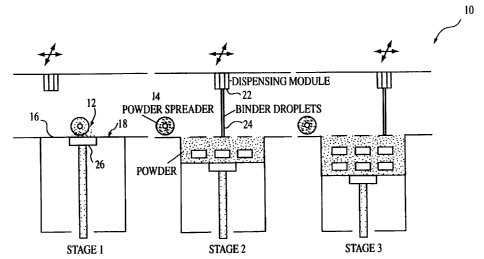
The 3DP process is shown schematically in Fig. 1, wherein a 3DP apparatus is
indicated generally by the number 10. Powder 12 is rolled from a feeder source
(not shown)
in stage 1 with a powder spreader 14 onto a surface 16 of a build bed 18. The
thickness of the
spread layer is varied as a function of the type of dosage form being
produced. Generally the
thickness of the layer can vary from about 100 to about 200 ~,m. The printhead
22 then
deposits the binder (fluid) 24 onto the powder layer and the build piston 26
is lowered one
layer distance. Powder is again rolled onto the build bed 18 and the process
is repeated until
the dosage forms axe completed (stages 2 and 3 of Fig. 1). The droplet size of
the fluid is from
about 50 to about 500 ~m in diameter. Servo motors (not shown) axe used to
drive the various
actions of the apparatus 10.
CA 02281474 2005-11-07
6
While the layers become hardened or at least partially hardened as each of the
layers is
laid down, once the desired final part configuration is achieved and the
layering process is
complete, in some applications it may be desirable that the form and its
contents be heated or
cured at a suitably selected temperature to further promote binding of the
powder particles. In
either case, whether or not further curing is required, the loose unbonded
powder particles are
removed using a suitable technique, such as ultrasonic cleaning, to leave a
finished device.
As an alternative to ultrasonic cleaning, water soluble particulates may be
used.
Fabrication of structures with designed pore structures is a challenging task
even with
additive manufacturing processes such as 3DP. Cylindrical structures with
radial pores of
hundreds of microns in diameter can be fabricated, however, the removal of
loose powder
from the narrow channels requires a cumbersome manual clean up process. One
solution is to
employ mixtures of water soluble particulates (sodium chloride) with polymers
used to
fabricate specimens. The small particles then leach out to reveal an
interconnected porous
structure. While this technique is useful in fabricating a network of pores,
control of pore
architecture is lost. An improvement on this technique is to selectively
deposit the soluble
phase to form internal soluble patterns prior to building any external
features. Water soluble
materials such as polyethylene glycol) can be deposited on a flat surface
prior to spreading a
new layer of powder. This enables the process to build walls of soluble
material. Loose
powder can be spread after completion of the patterning. The external or
insoluble features of
the specimen can then be built by printing with binder solution. Following the
requisite
iterations of the patterning and printing processes produces a dosage form
that has intricate
internal features that can be dissolved easily when immersed in an appropriate
solvent. This
concept can be used to fabricate components with controlled internal pore
channels. These
soluble patterns can also be used to create drug delivery devices with
prescriptive release.
Devices that are relatively insoluble in physiological fluids can be designed
and fabricated
with controlled soluble channels within. Upon ingestion or implantation,
dissolution of the
channels will expose the active materials that are isolated until the removal
of the soluble
phase in the channels.
Construction of a 3DP component can be viewed as the knitting together of
structural
elements that result from printing individual binder droplets into a powder
bed. These
elements are called microstructural primitives. The dimensions of the
primitives determine
CA 02281474 2005-11-07
7
the length scale over which the microstructure can be changed. Thus, the
smallest region
over which the concentration of bioactive agent can be varied has dimensions
near that of
individual droplet primitives. Droplet primitives have dimensions that are
very similar to the
width of line primitives formed by consecutive printing of droplets along a
single line in the
powder bed. The dimensions of the line primitive depend on the powder and the
amount of
binder printed per unit line length. A line primitive of 500 ~m width is
produced if an ink jet
depositing 1.1 cc/min of methylene chloride is made to ravel at 8"/sec over
the surface of a
polycaprolactone (PCL) powder bed with 45-75 ~m particle size. Higher print
head velocities
and smaller particle size produce finer lines. The dimensions of the primitive
seem to scale
with that calculated on the assumption that the liquid binder or solvent needs
to fill the pores
of the region in the powder which forms the primitive.
Finer feature size is also achieved by printing polymer solutions rather than
pure
solvents. For example, a 10 wt.% PCL solution in chloroform produces 200 ~m
lines under
the same conditions as above. The higher solution viscosity slows the
migration of solvent
1 S away from the center of the primitive.
The solvent drying rate is an important variable in the production of polymer
parts by
3DP. Very rapid drying of the solvent tends to cause warping of the printed
component.
Much, if not all, of the warping can be eliminated by choosing a solvent with
a low vapor
pressure. Thus, PCL parts prepared by printing chloroform have nearly
undetectable amounts
of warpage, while large parts made with methylene chloride exhibit significant
warpage. It
has been found that it is often convenient to combine solvents to achieve
minimal warping
and adequate bonding between the particles. Thus, an aggressive solvent can be
mixed in
small proportions with a solvent with lower vapor pressure.
There are two principle methods for incorporation of bioactive agent (e.g., a
drug). In
the first method, a layer of dispersed fine polymer powder is selectively
bound by ink jet
printing a solvent onto the polymer particles which dissolves the polymer.
This process is
repeated for subsequent layers to build up the cylinder, printing directly on
top of the
preceding layer, until the desired shape is achieved. If it is desired to
design a constant rate
release matrix, the drug is dissolved or dispersed (e.g., micellular) in the
solvent, yielding
drug dispersed evenly through the matrix. The printing process for this case
would then be
continued layer by layer until the desired shape was obtained.
CA 02281474 2005-11-07
In the second method, devices for pulsed release of drugs are prepared by
constructing
drug-rich regions within the polymer matrix. In this case, multiple printheads
are used to
deposit drug containing solvent in selected regions of the powder bed. The
remaining volume
of the desired device is bound with pure solvent deposited by a separate
printhead. The
printing process is repeated layer by layer to yield a device which gives a
pulsed release of
drug. For example, a cylindrical device could contain a cylindrical annulus
region which is
enriched with a drug.
Significant amounts of matter can be deposited in selective regions of a
component on
a 100 ~m scale by printing solid dispersions or solid precursors through the
ink jet print
heads with hundreds of jets that can be incorporated into the process. The
large number of
individually controlled jets make high rate 3DP construction possible.
A specific embodiment of the invention, the dosage form, can incorporate a
solubility
or stability enhancer. Suitable materials in this regard are cyclodextrins,
cyclodextrin
derivatives and/or substances that spontaneously form micelles as
solubility/stability
enhancers to facilitate the dispensing procedure, as well as the releasing
pattern of
poorly/sparingly soluble or unstable drugs in the fabrication of 3DP drug
delivery systems
(i.e. tablets, implants, etc.). Cyclodextrins (CDs) and their derivatives are
commonly used
complexing agents (CA). When incorporated in the fabrication of 3DP dosage
forms, CDs
can be used as follows:
1. to prepare aqueous solutions of sparingly soluble drugs so they can be
dispensed in
sufficient concentration through the nozzle, thus avoiding the use of
suspensions and
minimizing the need for extensive solvent removal or drying,
2. to increase drug stability by preventing labile groups/molecules from
interacting with
solvent,
3. to form a drug complex in situ, so that wetting and solublilzation are
enhanced when in
contact with GIT fluids (oral DDS) or subcutaneous fluids (implantable DDS).
This
substantially improves the rate of delivery leading to a desirable fast onset
of therapeutic
activity, and,
4. as a corollary of 3 above, when CA is placed at the bottom of a reservoir
(designed within
the dosage form) it will act as a carrier that facilitates/assists the release
of remaining drug,
which in turn leads to the desired fast offset of activity and prevents
undesirable leaching out
CA 02281474 2005-11-07
9
of sub-therapeutic drug levels.
By properly combining 3 and 4 above, a desirable pulsing pattern can be
achieved.
By combining the properties of drug-complex systems with the 3DP fabrication
process the three scenarios and any combination/variation of them can be
produced/modeled
to provide a solution to a particular drug release profile to be achieved.
Surface finish of the dosage forms of the invention is governed by the
physical
characteristics of the materials used as well as the build parameters. These
factors include
particle size, powder packing, surface characteristics of the particles and
printed binder (i.e.
contact angle), exit velocity of the binder jet, binder saturation, layer
height, and line spacing.
Interaction of the binder liquid with the powder surface, in particular, can
be controlled
carefully to minimize surface roughness. In a case where the binder gets
wicked out in a large
area, the feature size control becomes difficult, resulting in a rough
surface.
In one embodiment, the invention circumvents this problem in cases where no
substitute material combinations can be found. An intermediary material can be
deposited on
a powder bed to form a wetting barrier for the binder material. These
intermediaries are
deposited in such as fashion that spreading of the subsequently printed binder
is hindered by
the presence of the "outlining" intermediary region. An extreme example will
be the printing
of an oil around the specimen to limit wicking of a water-based binder.
A number of materials are commonly used to form a matrix for bioactive agent
delivery. Unless otherwise specified, the term "polymer" will be used to
include any of the
materials used to form the bioactive agent matrix, including polymers and
monomers which
can be polymerized or adhered to form an integral unit. In a preferred
embodiment the
particles are formed of a polymer, such as a synthetic thermoplastic polymer,
for example,
ethylene vinyl acetate, poly(anhydrides), polyorthoesters, polymers of lactic
acid and glycolic
acid and other a hydroxy acids, and polyphosphazenes, a protein polymer, for
example,
albumin or collagen, or a polysaccharide containing sugar units such as
lactose. The polymer
can be non-biodegradable or biodegradable, typically via hydrolysis or
enzymatic cleavage.
Non-polymeric materials can also be used to form the matrix and are included
within the term
"polymer" unless otherwise specified. Examples include organic and inorganic
materials such
as hydoxyapatite, calcium carbonate, buffering agents, and lactose, as well as
other common
excipients used in drugs, which are solidified by application of adhesive
rather than solvent.
CA 02281474 2005-11-07
Erodible bioactive agent delivery devices are one of the simplest medical
devices that
can be constructed. These types of bioactive agent delivery devices can be
used in an oral or
implantable form depending on the desired method for delivering the specific
bioactive agent.
They differ in the time period over which the bioactive agent is delivered and
excipients used
5 in the device construction. Erodible bioactive agent delivery systems are
constructed by
dispersing the desired bioactive agent in a matrix chosen so that it dissolves
or decomposes in
the presence of a body fluid. Oral erodible systems, for example, begin to
dissolve when they
contact with body fluid. In principle, release of the bioactive agent in both
cases is controlled
both by the rate at which the excipient reacts with the fluid and the rate of
bioactive agent
10 diffusion out of the device. This is true only if the surface of the device
erodes in a uniform
manner and its internal structure remains unchanged by prior reaction at the
surface.
Photopolymerizable, biocompatible water-soluble polymers include polyethylene
glycol tetraacrylate (Ms 18,500) which can be photopolymerized with an argon
laser under
biologically compatible conditions using an imitator such as triethanolamine,
N-vinylpyrollidone, and eosin Y. Similar photopolymerizable macromers having a
polyethylene glycol) central block, extended with hydrolyzable oligomers such
as
oligo(d,l-lactic acid) or oligo (glycolic acid) and terminated with acrylate
groups, may be
used.
Examples of biocompatible polymers with low melting temperatures include
polyethyleneglycol 400 which melts at 4°-8°C., PEG 600 which
melts at 20°-25°C, and PEG
1500 which melts at 44°-48°C., and stearic acid which melts at
70°C. Other suitable polymers
can be obtained by reference to The Polymer Handbook, 3rd edition (Wiley, N.Y.
1989). The
material for construction of the devices is selected based on the mechanism of
drug transport
and compatibility of their processing technology with the stability of the
bioactive agent.
The binder can be a solvent for the polymer and/or bioactive agent or an
adhesive
which binds the polymer particles. Solvents for most of the thermoplastic
polymers are
known, for example, methylene chloride or other organic solvents. Organic and
aqueous
solvents for the protein and polysaccharide polymers are also known, although
an aqueous
solution is preferred if denaturation of the protein is to be avoided. In some
cases, however,
binding is best achieved by denaturation of the protein.
The binder can be the same material as is used in conventional powder
processing
CA 02281474 2005-11-07
11
methods or may be designed to ultimately yield the same binder through
chemical or physical
changes that take place in the powder bed after printing, for example, as a
result of heating,
photopolymerization, or catalysis.
The selection of the solvent for the bioactive agent depends on the desired
mode of
S release. In the case of an erodible device, the solvent is selected to
either dissolve the matrix
or is selected to contain a second polymer which is deposited along with the
drug. In the first
case the printed droplet locally dissolves the polymer powder and begins to
evaporate. The
drug is effectively deposited in the polymer powder after evaporation since
the dissolved
polymer is deposited along with the drug. The case where both the drug and a
polymer are
dissolved in the printed solution is useful in cases where the powder layer is
not soluble in the
solvent. In this case, binding is achieved by deposition of the drug polymer
composite at the
necks between the powder particles so that they are effectively bound
together.
Aggressive solvents tend to nearly dissolve the particles and reprecipitate
dense
polymer upon drying. The time for drying is primarily determined by the vapor
pressure of
the solvent. There is a range from one extreme over which the polymer is very
soluble, for
example, 30 weight percent solubility, which allows the polymer to dissolve
very quickly,
during the time required to print one layer, as compared with lower
solubilities. The degree to
which the particles are attacked depends on the particle size and the
solubility of the polymer
in the solvent. Fine powder is more completely dissolved than powder with
larger particle
sizes.
There are essentially no limitations on the bioactive agents that can be
incorporated
into the devices, although those materials which can be processed into
particles using spray
drying, atomization, grinding, or other standard methodology, or those
materials which can be
formed into emulsifications, microparticles, liposomes, or other small
particles, and which
remain stable chemically and retain biological activity in a polymeric matrix,
are preferred.
Those bioactive agents which can be directly dissolved in a biocompatible
solvent are highly
preferred. Bioactive agents also include compounds having principally a
structural role, for
example, hydroxyapatite crystals in a matrix for bone regeneration. The
particles may have a
size of greater than or less than the particle size or the polymer particles
used to make the
matrix.
Examples generally include proteins and peptides, polysaccharides, nucleic
acids,
CA 02281474 2005-11-07
12
lipids, and non-protein organic and inorganic compounds, referred to herein as
"bioactive
agents" unless specifically stated otherwise. These materials have biological
effects including,
but not limited to anti-inflammatories, antimicrobials, anti-cancer,
antivirals, hormones,
antioxidants, channel blockers, growth factor, cytokines, lymphokines, and
vaccines. It is also
possible to incorporate materials not exerting a biological effect such as
air, radiopaque
materials such as barium, or other imaging agents.
Example 1 : Intraocular device capable of delivering an anti-inflammatory and
antiproliferative drug
Anti-proliferative and anti-inflammatory agents are used to treat a number of
ocular
diseases, including traction retinal detachment, that often result in
blindness. Traction retinal
detachment can develop in proliferative retinal diseases, such as
proliferative diabetic
retinopathy or after penetrating ocular trauma. The antiproliferative, 5-
fluorouracil (5-FU),
and the anti-inflammatory, diclofenac, are used to construct dosage forms
using 3DP with the
objective to contemporaneously deliver 5-FU in a pulsatile manner and
diclofenac at a
constant rate from the same device. The dosage form has its application in the
treatment of
the proliferation and inflammation resulting from traction retinal detachment,
especially after
trauma.
Anit-proliferatives like 5-FU can be extremely toxic; in such cases, pulsed
intraocular
delivery could produce the same therapeutic benefts as continuous release
while reducing
side effects, toxicity in normal cells, and the risk of multiple drug
resistance (MDR) in
fibrous cells, thereby enhancing the efficacy of the treatment. Diclofenac, on
the other hand,
is less toxic and is effective when delivered at a constant rate.
The first step of the procedure is to optimize prescriptive release rates of 5-
FU and
diclofenal independently and thereafter combine the two substructures into one
device. The
latter process is accomplished by 3DP fabrication during a single
manufacturing process.
Methods and results
The implant that can be divided into two portions. The top portion consists of
the 5-
FU chambers and the lid layers that encapsulate the actives. These caps are
designed to
degrade at different rates to cause the drug, to release at predetermined lag
times. The lower
CA 02281474 2005-11-07
13
portion of the implant releases diclofenac at zero-order kinetics throughout
the therapy.
Different portions of the intravitreal implant mandate distinct
characteristics that cannot be
achieved from a monolithic structure. Internal structure and composition at
each portion of
the implant device are controlled individually to meet the release
characteristics criteria.
Polymeric film degradation experiments are conducted to quickly identify
candidate
materials for constructing the intraocular implant devices. The initial
polymer selection is
limited to products that are approved by the United States FDA for use in
humans. In
addition, some of these polymers such as polyanhydrides are not widely
available
commercially. The polymers tested are further limited to those commercially
available and
those that could be prepared in powder form, however, other products may have
characteristics which are suitable in some but not all of these criteria and
are included within
the scope of the present invention.
Different copolymers of the polyactides and polyglycolides of a wide range of
molecular weights are studied. These include polylactide-co-glycolide (PLGA)
with varying
1 S lactide: glycolide ration (7S:2S, SO:SO) and molecular weights ranging
from 1 S KDa to 60
KDa. Among the low molecular weight polyactides tested are poly (l-lactide) 2
KDa. A
number of different polyanhydrides are also evaluated for fast eroding lids.
These include
polysebacic acid (PSA), polyfatty acid dimer-sebacic acid (PFAD:SA; SO:SO, S1
KDa),
polyricinoleic acid maleate-sebacic acid (PRAM:SA, SO:SO, 34 KDa).
PLGA is chosen to form the slow eroding walls of the implant based on the film
degradation study. Polyanhydrides, especially P(FAD:SA) exhibit fast erosion
characteristics.
This makes P(FAD:SA) an ideal system to be used in construction of the S-FU
caps. The
surface erosion mechanism of polyanhydrides also suggests that different
thickness films can
be used to control the lag time.
2S
Pulsatile Release Implants
A number of prototype intraocular implant devices are fabricated by 3DP. One
implant has four chambers containing S-FU. Walls of the implant are fabricated
by printing
chloroform into thinly spread PLGA powders. Only the printed region became
dense while
the PLGA powder from the unprinted region remains unbound. A scanning electron
micrograph (SEM) taken from the center of the device is used to confirm that
the
CA 02281474 2005-11-07
14
microstructures desired, formation of four distinct compartments, during 3DP
fabrication
process are achieved.
Two orthogonal walls form the separation between the four chambers of the
implant
devices fabricated. Two different devices are constructed by printing 8 lines
side by side in
one and 4 lines side by side in the other. Visible evidence from scanning
electron
micrographs demonstrates that the resulting wall thickness increases as the
number of
printing lines increases. Differences in the release characteristics from
these implant devices
are also a function of printing line number and therefore wall thickness.
Prototype implant devices are manually loaded with 160~,g 5-FU and polymeric
caps
are constructed on top of the chambers to encapsulate the active agent.
P(FAD:SA) powders
are used to build caps of different thicknesses. The PLGA walls are saturated
with chloroform
to enhance bonding to the P(FAD:SA) layer. Prototype implant devices that are
fabricated
with the presaturation steps do not exhibit any immediate dose dumping.
Another design
feature implemented to avoid premature dumping of SFU is an increase in the
side wall
thickness. This feature serves a dual purpose. The increased wall thickness
effectively
decreases the chance of 5-FU permeation through the side walls. At the same
time, the
contact surface area between the side walls of the chamber and the top lid is
increased, thus
minimizing 5-FU release from the PLGA and P(FAD:SA) interface.
Drug release is analyzed by immersing the dosage in 10 ml of phosphate
buffered
saline (PBS) solution kept at 37°C. Samples are taken at predetermined
intervals (at least 5
per assay) and analyzed using quantitative HPLC. Approximately 90% of the drug
is released,
approximately 146~g in separate bursts. A number of different prototype
implant device
designs are tested and yield distinctively different release characteristics.
The release profile from four sets of different prototype designs are measured
using an
HPLC method. The first profile labeled as Prototype 3 is taken from implant
devices with
porous and loosely attached P(FAD:SA) lid layers. These implants showed
complete 5-FU
release within the first 24 hours of the study. This demonstrates that the 5-
FU in the implant
devices will pulse out rapidly from the microchambers when there are enough
pores to allow
channeling of the release medium. Modifications made in the processing
conditions for
Prototype 4 result in pore-free lid layers as was discussed earlier. The
release profile of
Prototype 4 shows a significant difference from that of Prototype 3. A short
lag time of ~6
CA 02281474 2005-11-07
1S
hours with peak release at 14 hours is observed with Prototype 4 devices.
These implant
devices exhibit imperfections at the PLGA and P(FAD:SA) interface to which
could be
attributed the relatively quick release of S-FU. Further improvements in the
fabrication
sequence and increased side wall thickness resulted in improved bonding
between the PLGA
S walls and the P(FAD:SA) lid layers. Release profiles of Prototype S clearly
demonstrated lag
times of 24 hours or 36 hours, depending on the number of lid layers. The
number of
P(FAD:SA) lid layers also affect the release rates. A peak release rate of Sag
per hour is
achieved at 43 hours for the implants with 2 lid layers while implants with 3
lid layers reach a
peak of 2.S pg per hour in S6 hours.
These data demonstrate that modifications in process parameters and implant
design
may be used to achieve pulsatile release of drugs from implants. Close
examination of the
pulses from the prototype implants suggest that once the implant configuration
and material
system is optimized, multiple pulses form a single implant may be achieved.
In addition to the material systems investigated, other material systems that
would
1 S erode faster without allowing significant diffusion can be used for
achieving pulsatile release.
A further embodiment of the present invention is a device in which the device
design is
modified in order to allow sequential exposure of the lid layers. In the
proposed
configuration, sequential inner S-FU chambers are exposed to the release
medium and only
the contents from the first chamber are exhausted.
Continuous Release Implants
Before fabrication of implant devices can be achieved, the optimal 3DP
parameters
are determined. Polyesters are used as the polymer phase, which axe soluble in
chloroform.
Diclofenac is insoluble in chloroform but readily soluble in methanol. The
solubility of
2S diclofenac in different ratios of methanol and chloroform is investigated
in order to optimize
the balance of high drug concentration and polymer dissolution ability of the
binder solution.
In addition, the ability of these solvent combinations to dissolve polyesters
is examined. It is
determined that a 20:80 mixture of methanol:chloroform is optimal for
dissolving the
polymer while delivering a high concentration of drug to the device. A
homogeneous implant
is achieved by incorporating 24 mg/ml of diclofenac into the binder solution,
which is
deposited on a bed of polyester polymer.
CA 02281474 2005-11-07
16
Several diclofenac-containing prototypes are successfully fabricated using the
3DP
technology and tested for drug release in static phosphate buffered saline
solution at 37°C. An
HPLC assay is developed and tested for linearity, precision, specificity, and
sensitivity prior
to quantitative analysis of diclofenac.
a. Diclofenac prototype 1
Six disks from the first prototype batch are exposed to liquid COZ, to remove
residual
chloroform and determine if this process affects the diclofenac content.
However, the amount
of diclofenac in the disks exposed to liquid COZ is 7.20.2 mg (n=3), which is
the same as
disks not subjected to liquid COz. The amount of residual chloroform in disks
exposed to
liquid COZ is 1.910.3% (n=3), compared to 5.50.3% (n=3) for control devices
not exposed to
liquid COZ. Thus, exposure to liquid COZ under these conditions reduces the
amount of
chloroform by 65% but does not affect diclofenac content.
These disks are homogenous with diclofenac distributed throughout the entire
disk.
The actual implant will contain an additional portion for the pulsatile
release of separate drug
(i.e., 5-FU), thus, only one major face of the diclofenac section will be
directly exposed to the
external environment. As a result, subsequent prototypes contain a placebo
section to mimic
the pulsatile section of the final implant device.
b. Diclofenac, prototype 2
Initial prototypes of the diclofenac intravitreal implant exhibit high rate of
release for
2 days (0.8 and 0.6 mg per day on days l and 2, respectively) which is
attributable to the large
drug containing surface area of the implant exposed to the dissolution medium.
Thereafter,
diclofenac release is continuous at measurable but sub-therapeutic rates
(greater than 0.08 but
less than 0.36 mg per day) for an additional 14 days.
c. Diclofenac prototype 3
The second generation of prototypes is fabricated with a large fraction of the
diclofenac-containing component capped with placebo polymer layers of
different thickness.
This results in a initial drug release that tapers and then gradually exceeds
the target rate of 80
ug per day after 10 days. Furthermore, complete drug release was not observed
within the
desired sixteen days.
d. Diclofenac prototype 4
This batch is similar to Prototype 3 except that the placebo cap is fabricated
with 30%
CA 02281474 2005-11-07
17
sodium chloride to create holes in the cap to increase the initial release of
diclofenac. This
technique increases diclofenac release during the first day, but for the next
13 days, the
release rate is low, similar to prototype 3.
e. Diclofenac prototype 5
The previous prototypes exhibit drug release rates that vary from high to low
release
or low to high release. To achieve zero-order release, the interconnectivity
of the diclofenac
particles in the polymer phase is increased without altering the drug loading
by using sodium
chloride as an inert filler. 'Thus, prototype 5 disks are fabricated with a
blend of PLGA
polymer with sodium chloride. The addition of 35% (w/w) sodium chloride
ensures that the
combined loading of diclofenac and sodium chloride is above the minimum
necessary to
create interconnected particles. A void volume or drug occupancy of 35% is
recognized as
above the minimum necessary to establish complete interconnection of all pores
and channels
within a porous structure and is well known by those skilled in the art as
"percolation theory."
These prototypes are also covered with a placebo polymer coating to inhibit
initial dose
dumping by the implant. The results of this study indicate that Prototype Sb
exhibits the target
diclofenac release rate of 80~g/day.
~ Solvent Extraction
Preliminary experiments on post-fabrication exposure of the diclofenac
prototypes to
liquid COZ for 5, 30, and 60 minutes indicate that the procedure reduces the
amount of
residual solvent in the implant devices without significantly affecting
diclofenac content. The
results are summarized in Table 1.
Table 1. Effect of liquid COZ exposure time on diclofenac content in devices
manufactured by 3DP using polylactide-co-glycolide as the polymer and
chloroform as the
binder.
Prototype COi Diclofenac Control Residual
# Exposure (mg) Diclofenac Chloroform
(min) (mg) (wt%)
1 5 7.30 7.30.2 2.19
1 5 7.36 7.30.2 1.67
1 5 7.07 7.30.2 1.83
3 30 1.014 1.230.02 0.35
CA 02281474 2005-11-07
18
3 60 1.038 1.230.02 0.39
3 60 1.007 1.230.02 0.17
4 30 0.994 1.020.09 0.12
4 60 0.995 1.020.09 0.18
These results demonstrate that the fabrication of a implantable device or oral
dosing device
using 3DP can be manipulated to produce a single device exhibiting both
pulsatile and
continuous active release over periods as long as days or weeks.
Example 2: Contraceptive Implant Containing 17-DAN
A contraceptive implant device was designed for cyclic release of 17-
diacetylnorgestimate ( 17-DAN). Biodegradable polyesters of different types
and molecular
weights including poly-1-lactic acid (PLLA) and poly-epsilon-caprolactone
(PCL) are used to
construct different regions of prototype devices in a single contiguous
process. The regions
formed included a slow-degrading outer framework as a drug release restraint,
a drug-
carrying core, and a diffusion layer for drug delivery rate control. The drug
is incorporated in
the implant core surrounded by three impermeable walls and one permeable wall.
The
printing parameters are also optimized to minimize the presence of defects.
A device that in final dimension is 1.5 cm X 1.5 cm X 3.5 cm and containing
200 pg
of 17-DAN in a central core is fabricated. The releasing layer is composed of
either PCL or
PLLA printed with 20% of low molecular weight PCL/chloroform. The non-
releasing layer
is composed of PLLA printed with 2.5% PCL/chloroform. In one of the
experiments, the
permeable wall is replaced by an impermeable wall. The implant shows no drug
release,
clearly demonstrating the ability to fabricate biodegradable surfaces that are
impervious.
Exam In a 3
Fabrication of microdose oral dosage forms by conventional tablet pressing
technique
presents many content uniformity issues and safety hazards. 3DP processing can
be used to
build highly accurate dosage forms by depositing metered amounts of
medicaments(s) in the
center portion of the dosage form. Since the medicament can be completely
encapsulated
therein, the content uniformity will not be altered by subsequent handling,
packaging, or
CA 02281474 2005-11-07
19
during storage. Furthermore, it will be recognized that the safety hazards due
to exposure to
the medicament being used to persons involved in the manufacture of the dosage
forms axe
immediately reduced. The framework for micro-dosage tablets built by 3DP
further allows
the release rate of the medicament from the center portion to be controlled by
the methods
described herein and as previously set forth in U.S. Patent No. 5,490,962.
In one particularly preferred embodiment, an oral dosage form of the present
invention is constructed to release hormones in submilligram quantities. The
combination of
norethindrone acetate and ethinyl estradiol is one such combination
contemplated. The active
component may be 500 ~g of norethindrone acetate and 2.5 ~g of ethinyl
estradiol. The
powder material used may be selected from microcrystalline cellulose, lactose,
arabinogalctans, starch, or super disintegrant. The binder solution
composition may contain
arbinogalactans or Eudragits (e.g. a methacrylate). The architecture of the
dosage form may
be of single chamber type or include multiple active chambers in order to
effect the desired
release profile of both agents from the dosage form.
Example 4
Materials
Sure-Jell~ (Kraft General Foods, Inc., White Plains, NY, Lot 6-032-P-0659-4,
exp. Feb
1999) powder is used to build the framework for the microdose tablets (dosage
forms). Fruit
pectin is the major ingredient of Sure-Jell. Fumaric acid is also present to
assist gelling of the
powder. As-received powder is fractionated by sieving through 100 mesh and 325
mesh
screens to remove large agglomerates and fines. The tablets are built by using
only the
powder that is left on the 325 mesh screen (d+45~150~.m). Deionized water is
used to bind
the Sure-Jell powder.
The active ingredient used in this set of microdose tablets is an
antibacterial drug
called nitrofurantoin (Sigma Specialty Chemicals, St. Louis, MO, Lot
115H1012). An
ethanolic (Aldrich, Milwaukee, WI, Lot 15013 HQ) solution of nitrofurantoin is
deposited.
The concentration of the solution is 0.0188 mg/ml.
CA 02281474 2005-11-07
Fabrication
The 3DP parameters used to build the framework for microdosage forms are
explained below. Sure-Jell powder is spread into 170 ~m layers by manual
spreading, using a
stainless steel rod. The flow rate of the binder, distilled water, is dept at
1.2 cc/min. Speed of
5 the fast axis is 1.5 m/sec and each line is separated by 170 wm from the
others. A spring steel
stencil (Rache Corp, Camarillo, CA) with an 11 x 11 array of 1.04 cm circular
openings is
used to define the shape of the dosage forms. Ten 170 ~m base layers are built
with Sure-Jell
and water and active solution is deposited on the top surface of the tenth
layer.
The flow rate of the nitrofurantoin solution is kept at 1.0 cc/min and raster
speed is
10 kept at 1.6 m/sec. The active solution is dispensed throughout the entire
surface of the tenth
layer to achieve the dosage level of 1 ~.g per tablet. Total 5.32 ~L of
nitrofurantoin solution is
delivered per tablet. These parameters were calculated based on the line
spacing of 170 ~.m
and the estimated total line segment of 51.02 cm to cover the circular surface
area of each
dosage form.
15 After drying the active solution by waiting for 60 minutes, another 170 ~m
layer of
Sure-Jell is spread on the top of the dosage forms and bound to the drug
containing layer with
distilled water. This top layer is designed to cover the active-containing
region and prevent
the loss of active ingredient during handling processes.
The tablets are allowed to air dry for 30 minutes, then the powder bed is
removed
20 from TheriForm Alpha-0 machine and kept under vacuum (-760 mmHg) for 30
minutes to
facilitate the removal of residual moisture. Each dosage form is removed from
the powder
bed manually.
Analytical Methods
An ethanol extraction method is used to prepare the assay samples for UV-
analysis
(BioSpec1601TM, Shimadzu, Princeton, NJ). Each tablet is ground to powder form
using an
agate mortar and pestle. Caution is taken not to lose parts of the dosage form
during the
grinding process. The ground powder form of the tablet is transferred into
vials. Five ml
aliquots of ethanol are added to each vial using micro-pipettes (Eppendorf,
Brinkmann
Instruments, Westbury, NY) and mixed for 15 minutes on an orbital shaker
(Environ Shaker,
Lab-Line Instruments, Melrose Park, IL). Ethanol dissolves only two major
ingredients of the
CA 02281474 2005-11-07
21
dosage form: nitrofurantoin and fumaric acid. Nylon AcrodiscsTM (Gelman
Sciences, Ann
Arbor, MI) were used to filter out particulates from the samples in order to
minimize
interference. UV-maximum for nitrofurantoin is at 356 nm while that of fumaric
acid is
around 270 nm. The UV-absorbance at 356 nm was taken to estimate the amount of
nitrofurantoin in each of the sample dosage forms. The assay concentration of
nitrofurantoin
was in a well defined linear response region. A total of 18 tablets were
tested for content
uniformity.
Results
The UV-absorbance and corresponding nitrofurantoin content per individual
dosage
form is summarized in Table 2.
Table 2
Content Uniformity of Nitrofurantoin Microdosage Forms
Sample UV- NitrofurantoinSample UV- Nitrofurantoin
ID absorbance per tablet ID absorbance per tablet
(pg) (~,g)
1 0.0112 0.936 10 0.0114 0.956
2 0.0139 1.160 11 0.0111 0.926
3 0.0142 1.190 12 0.0129 1.078
4 0.0118 0.987 13 0.0118 0.987
5 0.0129 1.078 14 0.0117 0.977
6 0.0106 0.883 15 0.0140 1.170
7 0.0131 1.099 16 0.0118 0.987
8 0.0105 0.875 17 0.0120 1.007
9 0.0096 0.804 18 0.0140 1.170
The lowest dose is 0.804 ~g while the highest was 1.190 fig. Average of the 18
specimens
was 1.01 ~g per tablet with a standard deviation of 0.113 ~g (RSD=11.13%).
The above procedure demonstrates the ability to build dosage forms with very
small
amount of drug. Average content of 1.015 pg is only 1 % off from the intended
dose. USP
requirements for content uniformity mandates that dosage units fall within the
range of 85%
CA 02281474 2005-11-07
22
to 115% and the RSD less than or equal to 6.0%. The variability between dosage
forms can
be reduced by fine tuning the process. Figure 2 (a) illustrates one embodiment
which may be
susceptible to edge losses. Minor modifications can be made to the tablet
design so that the
active-containing region is less prone to edge losses during handling as shown
in Figure 2 (b).
An even more robust and safer architecture will encapsulate the active
materials completely
as shown in Figure 2 (c). This architecture has an advantage of eliminating
the danger of
exposing the highly potent pharmaceutical agents to the hands of the workers
and patients,
container walls, and neighboring tablets.