Note: Descriptions are shown in the official language in which they were submitted.
CA 02282379 1999-08-27
WO 99/33402 PCT/US98/27782
METHOD AND APPARATUS FOR ATTACHING OR LOCKING
AN IMPLANT TO AN ANATOMIC VESSEL OR HOLLOW ORGAN WALL
BACKGROUND OF THE INVENTION
FIELD OF THE INVENTION
The present invention relates to an apparatus and a method for repairing an
anatomic vessel wall or the wall of a hollow organ, such as the esophagus,
particularly in the human body. In particular, the present invention relates
to an
attaching or locking element for attaching an implant, such as stent or stent
graft,
to a vascular or hollow organ wall from the outside of the wall, as well as an
instrument for positioning and inserting the attaching or locking element into
the
body. The present invention also encompasses a method for attaching or locking
an implant to a vessel or hollow organ wall.
DESCRIPTION OF RELATED ART
An Abdominal Aortic Aneurysm ("AAA") is a weakening of the wall of the
aorta in the abdominal area. AAAs pose a significant health problem and over
160,000 AAAs are diagnosed annually in the United States. A full 25% of AAAs
will go on to eventually rupture; in spite of numerous advances in acute
medical
care, medical transport and resuscitation, ruptured AAAs continue to have a
50%
mortality rate.
Figure l shows an infrarenal AAA A' located in the torso T of a patient P,
below the heart H and kidneys K and above the point of bifurcation B of the
aorta
A into the iliac arteries IA. As may be seen by comparing Figures 2 and 3, a
CA 02282379 1999-08-27
WO 99/33402 PCT/US98/27782
normal aorta A (Figure 2) exhibits non-bulging walls above the point of
bifurcation
B, while an aorta A which includes an AAA A' (Figure 3) bulges outwardly from
its normal condition. This bulging is the result of weakening of the aortic
vessel
walls.
The traditional surgical technique for treating AAAs involved excision of
the aneurytic tissue and replacing that tissue with either a synthetic graft
or a graft
from another section of the patient's body. This approach required a large
abdominal incision and total bowel displacement and large disruption of the
retroperitoneum, followed by excision of the aneurytic tissue and attachment
of the
replacement graft to the vessel ends. Disadvantages of this prior art surgical
technique include hypothermia, coagulation problems, prolonged ileus, a risk
of
sexual disfunction and significant pain and disfigurement. As a result of
these
significant disadvantages attendant to the traditional surgical technique,
alternative
techniques for AAA repair have been investigated and used.
In 1992, Juan Parodi, a surgeon, first described the placement of a
percutaneous vascular prosthesis or stent in the abdominal aorta using
interventional
radiological techniques in Transfemoral Intraluminal Graft Implantation for
Abdominal Aortic Aneurysms, Ann. Vasc. Surg. 1991: 5:491-499. The prosthesis
or scent effectively excluded, i.e., provided support for, the aneurysm sac,
while
avoiding a major access incision in the abdomen. This prior art technique
required
only a small groin incision, through which the stent was inserted and lead to
the
aneurysm site with appropriate stmt guidance and deployment tools. Upon
reaching the aneurysm site, the stent was deployed and grafted to the vascular
walls
of the aorta at the aneurysm site. A stent S deployed at an aneurysm A' is
shown
in dotted lines in Fig. 3. The use of stent grafts, as in this prior art
technique,
decreased patient morbidity, and because of the Iess invasive nature of the
technique used to insert and deploy the graft, significantly reduced the
problems
with the traditional surgical technique for repairing AAAs.
Despite the advantages attendant the stent graft technique, difficulties in
2
CA 02282379 1999-08-27
WO 99/33402 PCT/US98/27782
passing the stmt to the aneurysm site, untimely opening of the stmt, and
complications, including emboli at the proximal and distal ends of the
aneurysm,
intimal damage, perforation, and thrombosis, have occurred. In addition,
unsatisfactory methods and devices for proximal stmt end fixation in order to
prevent stent migration, as well as persistent endoleaks, have limited the
effectiveness of stmt grafts. The proximal stent end is the end of the stmt
nearest
to the heart; this end needs to be fixed to the aorta in order to prevent the
stmt
from migrating from its initial deployed position to a position where it does
not
fully exclude and support the AAA. This movement or migration can also cause
endoleaks (L, Fig. 3), in which blood passes between the stent S and the
aneurysm
A', putting pressure on the aneurysm which can result in rupture.
When inserting implants, such as stents or stent grafts, into vessels or
hollow organs, in particular when repairing an aneurysm using a stmt graft, it
is
necessary that the stmt introduced into the vessel or hollow organ be attached
at
both its distal and proximal ends tightly and permanently to the vessel wall
surrounding the stent, in order to ensure that the stent does not migrate in
the
vessel and to ensure that the stmt seals off the aneurysm, thus reinforcing
the
weakness in the vessel. Prior art stems used for repair of AAAs have used a
variety of mechanisms for attaching the stmt to the vessel wall. One mechanism
used to attach a stent to a vessel wall is hook-shaped projections at the
proximal
and distal ends of the stent, which hook-shaped projections are pressed
against the
vascular wall from the inside of the vessel. The hook-shaped projections
mechanically grip the vessel walls to secure the stent or stmt graft to the
vessel
wall. In a similar attachment method, disclosed in U.S. Patent No. 5,527,355,
the
stent or stent graft is secured in position against the vessel wall from the
inside
using hook-shaped retaining elements; these hook-shaped retaining elements are
inserted into bands and surround the vessel externally.
PCT Publication No. WO 97/09008 to Medtronics shows a tubular implant
used for repairing aneurysms. In this implant, a sealing layer is disposed at
least at
the ends of the implant, in order to reduce endoleaks. The implant described
in
3
*rB
CA 02282379 1999-08-27
WO 99/33402 PCT/US98l27782
this publication, however, does not include any mechanism for securely
fastening
the implant to the vessel wall to prevent migration.
U.S. Patent No. 5,342,393 to Richard Stack of Duke University shows a
device for repairing a perforation in a vascular wall. The device of that
patent is
not disclosed for any use in securing implants into an anatomic vessel or
hollow
organ. Furthermore, the device of that patent uses a large-diameter catheter
or
sheath which is inserted through a large perforation in the vessel wall. This
device
is therefore not suitable for securing an implant to a vessel or hollow organ
wall,
where large perforations are to be avoided in deploying the implant.
U.S. Patent No. 4,669,473 to Acufex Microsurgical describes a surgical
fastener used for fastening two or more sections of tissue to one another.
This
fastener is not disclosed or used for fastening of any type of anatomic vessel
or
organ, in particular any type of hollow anatomic vessel or organ, and the
thick bar-
like head of that fastener is specifically designed to be embedded within the
tissue
to be fastened. The head of the fastener also includes at least one pointed
end for
embedding in tissue. Furthermore, the fastener of that device is not used to
clamp
two walls together, nor is that fastener used to attach an implant to a vessel
or
hollow organ.
SUMMARY OF THE INVENTION
Prior art methods for affixing an implant, such as a stent or stmt graft, to a
vessel or hollow organ wall have not always been reliable. In addition, many
of
these prior art methods could only be employed using open surgical techniques
requiring large incisions. These prior art methods, and the apparatuses used
with
these methods, have not been amenable to less-invasive techniques.
An object of the present invention is to provide an attachment or locking
apparatus which can effectively secure an implant, such as a stent or stent
graft, to
a vessel or hollow organ wall and which allows less invasive techniques, such
as
laparotomy with a markedly reduced incision, and minimally-invasive
techniques,
4
CA 02282379 1999-08-27
WO 99/33402 PCTNS98/27782
such as laparoscopy or endoscopy, to be used to attach the implant.
A further object of the invention is to provide a method and device for
deploying an attachment or locking apparatus for securing an implant, such as
a stent
or stmt graft, to a vessel or hollow organ wall which achieves minimal yet
reliable
penetration of the vessel or hollow organ wall, as well as a method or device
for
manipulating the attaching or locking apparatus in the body.
A still further object of the invention is to provide an attachment or locking
apparatus and a method and device for deploying the apparatus for fixation of
a stent
graft to a the wall of the aorta in the repair of AAAs which prevents stmt
migration
and persistent endoleaks.
These and other objects of the present invention are achieved using a
locking element which is inserted through the vessel or hollow organ wall and
the
implant from the outside of the vessel or hollow organ wall. The stmt lock of
the
present invention preferably includes a thin retaining element. The retaining
element has a clamping element joined flexibly at one end of the retaining
element. A fixing element is secured in position on the retaining element to
secure
or attach the implant in place.
In, for example, the attachment of a stent or stent graft to a vascular wall,
a
thin cannula may be inserted into the patient's body, such that the cannula
penetrates both the vascular wall and the stent or stent graft inserted within
the
vascular wall. The retaining element may then be inserted through the cannula
so
that the end with the clamping element is deployed on the inside of the stent
or
stent graft, and the other end projects through the stent or stent graft and
the vessel
wall. When withdrawing the end of the retaining element opposite the clamping
element from the vessel, the clamping element, because of the flexible
connection
to the retaining element, tilts into a position transverse to the insertion
opening.
As a result, the clamping element is positioned against the inner wall of the
stent or
stmt graft, so that the clamping element abuts against the interior of the
vessel and
5
CA 02282379 1999-08-27
WO 99/33402 PCT/US98/27782
the implant. The fixing element, already placed upon the retaining element or
attachable upon the retaining element, is pushed from the outside against the
vascular wall. In this manner, the distal and proximal ends of the stent or
stmt
graft are locked together with the vessel wall and the stmt or stent graft and
the
vessel wall are held between the clamping element and the fixing element. The
fixing element can be fixed in position on the clamping element, after
providing the
necessary tension in both the stmt or stent graft and the retaining element,
by
crimping or deformation, for example, or by a snap fit.
It is particularly advantageous if the clamping element is connected to the
retaining element in the mid-section of the clamping element, so that when the
retaining element is withdrawn from the opening in the vessel or hollow organ
wait, the clamping element is positioned on both sides of the opening in the
vessel
or hollow organ wall across an equal contact area of the implant.
The clamping element must be designed so that it is insertable through a
very small opening in the vessel or hollow organ wall and deployable into the
interior of the vessel or hollow organ, and so that in the interior thereof,
it
nevertheless lies over a sufficiently large area against the implant so that
the
clamping element is reliably prevented from being withdrawn again through the
opening in the wall. This can be ensured, for example, by the manner in which
the
clamping element is introduced into the interior of the vessel or hollow
organ. The
clamping element may be folded, bent or rolled into an elongated shape and
inserted into the vessel or hollow organ, and thereafter may open, flex or
expand
therein. In a particularly preferred specific embodiment the clamping element
may
be pin-shaped. Thus, it becomes possible to insert the clamping element in the
longitudinal direction of the cannula, with a very small cross-sectional
puncture
area, into the interior of the vessel or hollow organ. Once in the vessel or
hollow
organ, the clamping element flexes or spreads out into a transverse position,
in
front of the insertion opening, and thus prevents the clamping element from
pulling
out of the insertion opening. It is advantageous if the clamping element
flexes or
expands out radiatly from the retaining element, in a plane transverse to the
6
CA 02282379 1999-08-27
WO 99/33402 PCT/US98/27782
longitudinal direction of the retaining element, thus providing a suitable
contact
area for the clamping element against the implant. The clamping element and
retaining element may be integrally formed, or the retaining element may be
embedded into, or otherwise connected to, the clamping element.
In one preferred embodiment, the clamping element can be curved in its
transverse direction, so that it adapts or fits to the curve or shape of the
implant or
vessel or hollow organ wall. The clamping element preferably has ends which
are
not sharp, and may be smooth, preventing the ends of the clamping element from
piercing or penetrating the vessel or hollow organ wall, thereby preventing
damage.
One particularly preferred specific embodiment provides for the retaining
element and the clamping element to be formed in one integral piece. The
flexible
joining of these two parts is then effected, for example, by manufacturing the
1 S retaining element and the clamping element integrally from a suitable
polymeric or
metallic material.
In another specific embodiment, the clamping element is tubular, and the
retaining element is formed by a suture or thread whose two ends are
introduced
from opposite sides into the tubular clamping element and emerge together from
the clamping element through the opening in the vessel or hollow organ wall.
It is beneficial if the fixing element is a permanently compressible sleeve
through which the retaining element is passed. Initially, this sleeve is
freely
movable or slidable on the length of the retaining element. The fixing element
can be brought forward closely against the outside of the vessel or hollow
organ
wall so that the wall and the implant, such as a stent or stent graft, are
sufficiently
compressed between the clamping element and the fixing element. When this
condition is reached, the sleeve is crimped or deformed and thus secured in
position
with respect to the clamping element. As an alternative, the fixing element
may be
made of a resilient material, and may be snapped into an appropriate position
using
beads or other protuberances along the length of the retaining element.
7
CA 02282379 1999-08-27
WO 99/33402 PCT/US98/27782
At an end of the retaining element opposite the clamping element, an
enlargement can be provided which prevents the fixing element, when it is not
yet
secured in position, from sliding off the retaining element. This feature
prevents
the fixing element from dislodging from the retaining element during the
insertion
procedure.
In another preferred specific embodiment, a pressure element, having a
pressure surface which is arranged approximately transverse to the
longitudinal
direction of the retaining element and approximately parallel to the clamping
element, is supported on the retaining element between the vessel or hollow
organ
wall and the fixing element. The pressure element preferably is freely movable
along the retaining element. In particular, this pressure element can be a
disk or
band having a center through which the retaining element is passed. This
pressure
element is held by the fixing element against the outside of the vessel or
hollow
organ wall, and ensures that the force holding the vessel or hollow organ and
the
implant together is introduced over a large surface area on the outside of the
vessel
or hollow organ.
In addition, between the pressure element and the vessel or hollow organ
wall, a large-area pressure-distribution element can be supported on the
retaining
element in a manner that it is moveable during insertion. This pressure-
distribution
element further distributes the pressure against the vessel or hollow organ
wall. It
is advantageous if the pressure-distribution element is elastically
compressible, so
that it positions itself over a large surface area against the vessel or
hollow organ
wall, thus reducing pressure peaks on the wall. In one preferred embodiment,
the
pressure-distribution element can have the shape of a cylinder, and the
retaining
element may pass transversely through the pressure-distribution element.
The individual parts of the locking element are made of materials well
tolerated by the body, in particular the retaining element, the clamping
element, the
fixing element, the pressure element and/or the pressure-distribution element
can be
made of a non-absorbable plastic material. In other embodiments, it is also
8
CA 02282379 1999-08-27
WO 99/33402 PCT/US98/27782
possible for the retaining element, the fixing element and/or the pressure
element to
be made of titanium, or possibly of another metal alloy well tolerated by the
body.
In another preferred specific embodiment of the invention, the pressure-
S distribution element can be made of a foam or a non-woven fabric, so that it
is
elastically compressible and positions itself gently against the outer surface
of the
vessel or hollow organ wall.
It is also the object of the invention to provide a positioning or insertion
instrument for inserting and locking in place the locking element of the
present
invention. This objective is fulfilled according to the present invention by a
positioning instrument that includes a hollow needle, trocar or cannula into
which
at least the clamping element of the locking element is insertable, and an
ejector to
push the locking element out of the hollow needle, trocar or cannula. This
apparatus may be large enough to be manipulable outside the body, or may be
made to be inserted within the body so as to manipulable using appropriate
endoscopic tools. Using such a positioning instrument, the locking element is
accommodated in the hollow needle, trocar or cannula which can be inserted
into
the patient percutaneously, via laparoscopic, laparotomic or endoscopic
techniques,
and thereafter through the vessel or hollow organ wall and the implant. The
clamping element is then pushed out of the hollow needle, trocar or cannula by
an
ejector. The ejector can extend out of the body cavity, to be actuated by hand
by
the surgeon, or could be located within the abdominal cavity, to be actuated
using
suitable endoscopic tools. After withdrawing the hollow needle, trocar or
cannula
from the vessel or hollow organ, the small opening caused by the hollow
needle,
trocar closes elastically around the retaining element which projects
outwardly
through the closed opening. The clamping element may be pressed against the
inner wall of the implant by pulling on an opposite end of the retaining
element.
Thereafter, the pressure-distribution and/or pressure elements may be pressed
against the outside of the vessel or hollow organ wall, and then the fixing
element
secured in place to lock the implant in place.
9
CA 02282379 1999-08-27
WO 99/33402 PCT/US98/27782
It is beneficial if the hollow needle, trocar or cannula is beveled at the end
inserted into the patient, thereby tapering to a sharp point. It is also
advantageous
if the hollow needle, trocar or cannula has an elongated slot open toward the
end
inserted into the patient for receiving the clamping element. This is
especially
beneficial when the clamping element flexes or expands radially from the
retaining
element and therefore would be impeded by the inner wall of the hollow needle,
trocar.
Depending on the location at which the hollow needle, trocar or cannula is
to be inserted into the patient, the hollow needle, trocar or cannula can be
optionally straight or curved. The hollow needle, trocar or cannula is
preferably
very thin, sharp and rigid, so that it may readily penetrate calcified vessel
tissue,
without creating a large puncture subject to weakening or rupture.
In the method of the present invention, as exemplified by its use in the
treatment of an AAA, a first percutaneous incision is made at a location near
the
aneurysm site. A second percutaneous incision is made preferably in the groin
or
pubic area, to gain access to a femoral or iliac artery or the distal end of
the
abdominal aorta. Using standard interventional techniques the stent or stent
graft is
guided to the aneurysm site and then deployed. Access is gained to the
exterior of
the aorta at the aneurysm site through the first incision. The first incision
may be a
laparotomy incision, followed by suitable procedures to gain open access to
the
aorta, or may be a small incision as part of a laparoscopic procedure in which
additional small incisions are made to deploy additional instruments into the
abdomen. In either case, the positioning device, with an attached locking
element
is inserted through the first incision, guided to the aneurysm site, and then
the
hollow needle, trocar or cannula punctures the outside wall of the aorta and
is
inserted within the interior of the aorta and the stent or stmt graft. The
clamping
element is then ejected, using an ejector, into the interior of the aorta and
stent or
stent graft. The hollow needle, trocar is then withdrawn. The locking element
is
pulled tight, and pressure-distribution andlor pressure elements are then slid
down
the locking element toward the aorta wall. The fixing element is then slid
against
CA 02282379 1999-08-27
WO 99/33402 PCTIUS981277$2
the pressure-distribution and/or pressure elements, and secured into place by
crimping, deforming, or by a snap fit. The free end of the locking element may
then be cut off near the fixing element. The process of inserting and securing
the
locking element may be repeated until a sufficient number of locking elements
are
in place to securely hold the stmt or stmt graft to the aorta wall.
BRIEF DESCRIPTION OF THE DRAWINGS
The following description of preferred specific embodiments of the
invention, in conjunction with the drawings, serves to explain preferred
embodiments of the invention more precisely.
Figure 1 is a view of an abdominal aortic aneurysm in a patient.
Figure 2 is a detail view of an aorta without an abdominal aortic aneurysm.
Figure 3 is a detail view of an aorta with an abdominal aortic aneurysm and
an implant.
1 S Figure 4 is a perspective view of a vessel with a number of applied
locking
elements.
Figure 5 is a longitudinal cross-sectional view through the hollow needle or
cannula of a positioning instrument with an undeployed inserted locking
,element.
Figure 6 shows a plan view of the hollow needle or cannula of the
positioning instrument of Figure S.
Figure 7 shows a view similar to Figure 5, with the ejector pushed forward
and the clamping element partially pushed out.
Figure 8 shows a first embodiment of a locking element.
Figure 9 shows a second embodiment of a locking element having a tubular
clamping element.
Figure 10 shows the embodiment of Fig. 8 mounted in an infra-abdominal
cannula and ejection tool manipulated by an endoscopic tool.
Figure I 1 shows a plan view of a positioning tool of the present invention,
including a locking element.
Figs. l la-l lb show first, second and third embodiments of the cannula of
the present invention.
Figure 12 shows a third embodiment of a locking element.
11
CA 02282379 1999-08-27
WO 99/33402 PCT/US98/27782
Figure 13 shows a fourth embodiment of a locking element.
Figure 14 shows a fifth embodiment of a locking element.
Figure 15 shows a sixth embodiment of a locking element.
Figure 16 shows an seventh embodiment of a locking element.
Figure 17 shows an eighth embodiment of a locking element.
Figure 18a shows a ninth embodiment of a locking element.
Figures 18b and 18c show the embodiment of Figure 18a inserted in a
cannula.
Figure 19 shows a perspective view of a fourth embodiment of a hollow
needle or cannula.
Figure 20 shows a perspective view of a tenth embodiment of a locking
element used with the hollow needle or cannula of Figure 19.
Figure 21 a and Figure 21 b show, respectively, perspective and elevation end
views of a trocar of a positioning instrument with an undeployed inserted
locking
element.
Figure 22 shows a plan view of a patient's body, demonstrating the manner
in which the method of the present invention is implemented in the treatment
of an
AAA and some of the surgical tools used.
Figure 23 shows a plan view of an eleventh embodiment of a locking
element.
Figure 24 shows an elevation, partially cross-section, view of the locking
element of Figure 23 used with the insertion tool of Figure 10;
Figure 25 illustrates a twelfth embodiment of a locking element;
Figure 26 is an enlarged view of the locking element of Figure 25 in place
through a vessel.
DESCRIPTION OF THE PREFERRED EMBODIMENTS
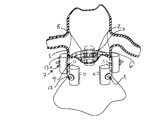
Figure 4 shows an anatomic vessel, in this case a section of the aorta,
having a wall 1 into which a tubular implant 2 is inserted, so that implant
wall 3 is
in contact with vascular wall 1. In one embodiment of the present invention,
the
tubular implant 2 may be a stent or stent graft for repairing an Abdominal
Aortic
12
CA 02282379 1999-08-27
WO 99/33402 PCTNS98/27782
Aneurysm ("AAA"). In the exemplary embodiment shown in Figure 4, a suitable
known interventional tool such as a catheter is used to guide and deploy the
tubular
implant 2 using a minimally-invasive percutaneous incision. A free annular
space 6
is formed in the interior of the vascular wall 1.
Vascular wall 1 and implant wall 3 -- lying flat against the vascular wall 1
-- are locked together by one or more locking elements 7. The locking elements
7
include a clamping element 8, which in a preferred embodiment may be
pin-shaped, a thin retaining element 9 joined to the clamping element 8, and
an
enlargement 11 (Fig. 8) provided at a free end 10 of retaining element 9. It
is
possible, for example, to form the enlargement 11 using a sleeve slid onto
free end
10 and pressed or crimped down at the free end 10, or the enlargement 11 may
be
molded, knotted or otherwise formed integrally with free end 10. The
enlargement
should be larger than an opening in the fixing element 12, to prevent the
fixing
element from sliding off of the free end 10. A fixing element 12, that may be
in
the form of a sleeve and have an opening through which the retaining element 9
passes, surrounds retaining element 9 and is initially freely moveable or
slidable on
the retaining element 9. The fixing element 12 may be captured between the
clamping element 8 and the enlargement 11. The fixing element 12 may
preferably
be made of a deformable polymeric material which retains a deformed condition
upon deformation or crimping, for example UHMWPE (Ultra High Molecular
Weight Polyethylene). The fixing element 12 may alternatively be formed of a
resilient material which allows the fixing element 12 to be snap fit over
beads or
protuberances on the retaining element 9. In each of the embodiments of the
clamping elements described below the ends of the clamping element are
preferably
not sharp and may be smooth, so as to prevent ruptures or punctures of the
implant
2 or vessel wall 1.
Between the clamping element 8 and fixing element 12, the retaining
element 9 is passed transversely through a pressure-distribution element 13,
which
in a preferred embodiment may be cylindrical, and also through a central
opening
in a pressure element 14, which in a preferred embodiment may be in the form
of a
13
CA 02282379 1999-08-27
WO 99/33402 PCT/US98/27782
flat, circular disk or a band. Both the pressure-distribution element 13 and
the
pressure element 14 may be freely movable or slidable on retaining element 9.
Clamping element 8 is very thin so that it may be inserted into the vessel
from the outside with minimal puncturing of the vessel; for example, the
clamping
element 8 may have a diameter of approximately 0.01-1 mm. The length of
clamping element 8 is preferably only a fraction of the circumference of the
vessel
into which the locking element 7 is inserted, but should be long enough so
that it is
not pulled out of the vessel once inserted; for example the length of the
clamping
element may be approximately 8-20 mm. The clamping element 8 may be made of
titanium or a titanium allow such as TIAL6V4, of any other metal alloy well
tolerated by the body, or of a plastic material, in particular a non-
absorbable plastic
such as SURGILENE 2/0 USPTM polypropylene, manufactured by B. Braun, or a
woven polyester, and may be in the form of either a polyfiliment or a
1 S monofiliment.
In the exemplary embodiment shown in Figure 4, retaining element 9 is
embedded into clamping element 8 and emerges radially from the clamping
element
8 at its lengthwise center. Retaining element 9 may be secured to clamping
element 8 by any known securing technique such as molding or deformation.
Retaining element 9 is preferably thin or thread-like, flexible and has a
very small diameter. The retaining element 9 is preferably made of a non-
absorbable polymeric material, such as SURGILENE 2/0 USPTM polypropylene,
manufactured by B. Braun, and may be fashioned from a surgical suture
material,
and may be of a diameter approximately that of conventional suture materials.
The
elements that are freely movable on the retaining element 9 -- fixing element
12,
pressure-distribution element 13 and pressure element 14 -- are safeguarded
from
being unintentionally pushed off the retaining element 9 by the enlargement 11
at
free end 10 of the retaining element 9, which enlargement is larger than the
opening in the fixing element 12, pressure-distribution element 13 and
pressure
element 14 through which the retaining element 9 passes. As a result, the
locking
14
CA 02282379 1999-08-27
WO 99/33402 PCT/US98/27782
element 7 forms a unitary structure which may be inserted into the body
without
the risk of individual parts becoming dislodged.
The retaining element 9 is preferably made of a material well tolerated by
S the body such as a metal wire or a plastic thread; in particular, the
retaining
element 9 can be made of non-absorbable plastic such as SURGILENE 2/0 USPTM
polypropylene, manufactured by B. Braun. The retaining element 9 may also be
formed integrally with, and of the same material as, retaining element 8, if
desired.
The pressure-distribution element 13 may be elastically compressible and
can be made, for example, of a non-woven fabric or a foam such as PTFE
(polytetrafluoroethylene) fleece; this element can also be manufactured from a
non-
absorbable plastic material.
1 S To join vascular wall 1 and implant wall 3, it is preferable that a number
of
locking elements 7 are arranged on the vessel along its periphery; preferably,
the
locking elements 7 are approximately equally spaced circumferentially around
the
vessel periphery. The locking element 7 may be attached at the proximal end of
the implant 2, the distal end, or both ends. The locking elements 7 are
preferably
attached to the vessel using a positioning instrument as is shown in Figures S-
7, 10,
11, l la-l lc, 21 and 22.
As shown in Figs. S-7, a preferred embodiment of the positioning
instrument of the present invention includes a hollow needle or cannula 17
having a
2S beveled tip 18, and has, in a side wall, an axial slot 19 open toward the
free end of
the hollow needle or cannula I7. The hollow needle or cannula 17 should be of
a
relatively small outer diameter, for example 0.25-1.0 mm, and should be
fashioned
of a relatively strong material so that it may easily penetrate a vessel wall
1 which
may be calcified. The beveled tip 18 should also be very sharp, so as to more
readily penetrate the vessel wall 1 and the implant 2. An ejector 20 is
supported in
a manner so that it is longitudinally movable in the interior of hollow needle
or
cannula 17. Ejector 20 can be pulled back so that a clamping element 8 can be
1S
___._. -.____ .__.. _ ,
CA 02282379 1999-08-27
WO 99/33402 PCT/US98/27782
completely inserted into hollow needle or cannula 17 in front of ejector 20,
as
shown in Figure 5. In this inserted state, retaining element 9 enters
longitudinal
slot 19 and is then arranged on the outer side of hollow needle or cannula 17.
The
retaining element 9 could alternatively be retained within hollow needle or
cannula
17, between the hollow needle or cannula 17 and the ejector 20.
In the state shown in Fig. 5, hollow needle or cannula 17 can be inserted
from the outside of the vessel -- using laparoscopic, endoscopic or open
surgical
techniques described in pertinent detail below -- through vascular wall 1 and
implant wall 3, into the vessel, tip 18 then arriving in annular space 6. As
soon as
the clamping element 8 is completely within the annular space 6, ejector 20
may be
pushed forward so that clamping element 8 is pushed out of hollow needle or
cannula 17 (Fig. 7) and is now free in annular space 6. Hollow needle or
cannula
17 is subsequently withdrawn from the vessel, and the opening formed by said
hollow needle or cannula 17 is closed by the elasticity of vascular wall 1 and
implant wall 3. However, retaining element 9 projects outwardly through this
opening, and the vascular wall 1 and implant wall 3 close around the retaining
element 9.
By pulling on the retaining element 9, the pin-shaped clamping element 8
flexes or expands to a position approximately transverse to the retaining
element 9
and approximately parallel to the vascular wall 1 and implant wall 3, and
becomes
positioned against implant wall 3. The clamping element 8 thus is anchored
against
the inner side of implant 2. While continuing to keep retaining element 9 taut
by
pulling on the free end 10, the surgeon next pushes pressure-distribution
element
13, pressure element 14 and fixing element 12 along the length of the
retaining
element 9 in the direction toward vascular wall 1, until the vascular wall 1
and
implant wall 3 are clamped between clamping element 8 on one side and pressure-
distribution element 13 and/or pressure element 14 on the other side, and thus
are
pressed flat against each other. In this state, fixing element 12 is slid down
the
retaining element 9 and then secured in position on retaining element 9, for
example, by pressing or crimping the sleeve together using a suitable tool or
16
CA 02282379 1999-08-27
WO 99133402 PCT/US98/27782
instrument. Alternatively, the fixing element 12 can be snap fit in position.
In this
manner, a locking element 7 has been secured on the vessel which holds
vascular
wall 1 and implant wall 3 flat against one another, as shown in Figure 4. The
end
of the retaining and locking element 9 passing beyond the fixing element I2
can
then be cut off using a suitable tool or instrument.
Locking elements 7 of the present invention are placed in number in the
circumferential direction around the vessel. In this manner, a number of
fixing
points are produced along the periphery, which lock implant wall 3 and
vascular
wall 1 permanently and imperviously together (Fig. 4).
A locking element T is shown in the exemplary embodiment of Figure 9
which differs from that of Figures 4-8 only in that clamping element 8' is
formed
from a short hollow tube, preferably of titanium alloy such as TIAL6V4 or a
non-
absorbable polymeric material. In the embodiment of Figure 9, retaining
element
9' is in the form of a loop; the free ends 15 of the retaining element 9' are
introduced from opposite ends into tube-shaped clamping element 8' and emerge
radially through an opening 16 in the lengthwise center wall area of the
clamping
element 8'. The short hollow tube of the clamping element 8' may be
manufactured by laser-drilling a thin titanium alloy or polymeric rod or pin.
The clamping element of the present invention may preferably be
manufactured to be of a slightly curved shape, or to flex or expand into a
slightly
curved shape after insertion into the vessel, so as to better conform with the
curved
inner wall 3 of the implant 2.
Fig. 10 shows a first embodiment of a positioning instrument 33 of the
present invention, which is used intra-abdominally. The positioning instrument
33
includes a hollow needle or cannula portion 34 and an ejector portion 35. The
ejector portion slides within the interior of cannula portion 34, so that in a
retracted
position (shown in Fig. 10), a clamping element 8 is inserted within the
hollow
needle or cannula portion 34, and in an ejected position (similar to the
position
17
CA 02282379 1999-08-27
WO 99/33402 PCT/US98/27782
show in Fig. 7), clamping element 8 is ejected from the hollow needle or
cannula
portion 34. The positioning instrument is of a size comparable to conventional
infra-abdominal needles or cannulas used in endoscopic surgical techniques,
and is
inserted into the abdominal cavity, and manipulated within that cavity once
inserted, by a suitable endoscopic grasping tool T or other endoscopic tools.
Fig. 11 shows a view of a second embodiment of a positioning instrument
25 of the present invention. As discussed in detail above, the positioning
instrument includes a hollow needle or cannula 17 having a beveled tip 18, and
has, in a side wall, an axial slot 19 open toward the free end of the hollow
needle
or cannula 17. An ejector 20 is supported in a manner that it is
longitudinally
movable or slidable in the interior of hollow needle or cannula 17 and the
interior
of the instrument body 23, and can be pulled back so that a clamping element 8
can be completely inserted into hollow needle or cannula 17 in front of
ejector 20.
The ejector 20 may be made of a flexible material so that it can be
accommodated
in a curved hollow needle or cannula 17a, 17b (Figs. 11 b, 11 c) and may
include a
knob or handle 21 at one end which allows the surgeon to eject the clamping
element from the positioning instrument into the vessel. The instrument body
23
may include finger grips 22 to assist in ejecting the locking elements using
the
ejector 20.
Figures 12-18a show various alternative embodiments of the locking
element of the present invention. It is to be understood that Figures 12-18a
show
only the retaining and clamping element portions of the locking elements, and
that
the other components of the locking elements are not shown for clarity.
Figure 12 shows an embodiment of the locking element 37 in which the
retaining element 39 and the clamping element 38 are integrally molded or
manufactured in an approximately "T" shape. This locking element 37 could be
manufactured either of a metallic material or a polymeric material. In the
embodiment of Fig. 13, the retaining element 60 is attached to the clamping
element 62 through a hinge 66, which allows these two elements to pivot
relative
18
CA 02282379 1999-08-27
WO 99/33402 PCT/US98/27782
to one another. In the embodiment of the locking element 47 in Figure 14, the
retaining element 48 is a wire or thread with a knot 41 at one end. The
clamping
element 49 is formed by a flat spring of a metallic or polymeric material,
which
has a. hole 42 through which the wire or thread of the retaining element 48
passes.
S The knot 41 prevents the spring of the clamping element 49 from dislodging
from
the retaining element 47. In a variation of the embodiment of Figure 14 shown
in
Figure 15, the knot 41 may be replaced by a protuberance or bead 42.
Figure 16 shows an embodiment of a locking element 57 in which the
clamping element 59 is disk-shaped in a flexed or expanded condition, and
which
assumes a conical or hemispherical configuration 59' when retained contracted
or
folded inside the hollow needle or cannula 17 for insertion into the vessel.
The
retaining element 58 may include a series of protuberances or beads 53 along
its
length. These beads 53 may be used to fix a fixing element 12 in place without
deformation or crimping. The fixing element 12 would be slid down the length
of
the retaining element 58, snapping over the beads 53, until the fixing element
12
reaches a position in which it securely clamps the clamping element 59 against
the
implant wall 3. The bead 53 above the fixing element 12 would prevent that
element from thereafter slipping back away from the vessel, thereby loosening
the
locking element 57. In this way, the fixing element 12 is secured by a snap
connection or fit along the length of the retaining element 58.
Fig. 17 shows an embodiment of a locking element 77 in which the
clamping element 79 is integrally formed with the retaining element 78. The
retaining element 78 includes teeth 79 along its length, which interact with
teeth 76
on fixing element 72, allowing only one-way movement of fixing element 72
along
retaining element 78. The fixing element 72 would be slid down the length of
the
retaining element 78, snapping over the teeth 79, until the fixing element 72
reaches a position in which it securely clamps the clamping element 79 against
the
implant wall 3. The interaction of teeth 79 and 76 would prevent fixing
element
72 from thereafter slipping back away from the vessel, thereby loosening the
locking element 77. In this way, the fixing element 72 is secured along the
length
19
CA 02282379 1999-08-27
WO 99/33402 PCT/US98/27782
of the retaining element 78.
In Figs. 18a-18b, the locking element 27 is formed of an integrally
manufactured wire or thread of a resilient or shape-memory material, which
could
either be metallic or polymeric. The retaining element 29 joins the clamping
element 28 by way of a series of two bends or hinges 30, 31. As shown in Fig.
18a, the locking element 27 is manufactured to expand or flex into a shape in
which the clamping element 28 is approximately perpendicular to the retaining
element 27. During insertion through hollow needle or cannula 17 (Figs. 18b
and
18c), the locking element 27 may be deformed into one of two configurations,
shown in Figs. 18b and 18c, which allow insertion through hollow needle or
cannula 17.
Figures 19 and 20 show an alternative configuration of a hollow needle or
cannula 17' and an alternative configuration of a locking element 7"' used
with
that hollow needle or cannula 17'. The hollow needle or cannula 17' of Figure
19
differs from the hollow needle or cannula 17 of Figures S-7 in that the hollow
needle or cannula 1T of Figure 19 include an additional slot 19' opposite the
slot
19. The locking element 7"' includes, on the clamping element 8 a ridge 61,
which ridge 61 fits into and slides along the additional slot 19'. The
interaction
between ridge 61 and additional slot 19' helps to guide the clamping element 8
into
a proper position within the vessel.
Fig. 21 is an illustration of an alternative insertion tool 17", in which a
trocar tip 18' is used instead of the hollow needle or cannula. In all other
respects,
however, the embodiment of Fig. 21 is identical to the embodiment of Figs. 5-
7.
The trocar tip 18' can be used with either the embodiment of the insertion
tool as
shown in Figs. 10 or 11.
Fig. 22 is an illustration of a patient P and the manner in which the method
of the present invention is used to repair an AAA. A first percutaneous
incision h
is made, at a location near the site of the AAA or the site where the implant
1 is to
CA 02282379 1999-08-27
WO 99/33402 PCTNS98/27782
be retained. A second percutaneous incision h is made, preferably in the groin
or
pubic area near one of the femoral arteries F or one of the iliac arteries IA.
Alternatively, the second percutaneous incision IZ could be made at the distal
end
of the abdominal aorta A, if the femoral arteries F or iliac arteries IA are
too small
or obstructed, and therefore inappropriate for the use of standard
interventional
techniques. Suitable interventional techniques known in the art, such as those
described by Parodi, are used to transfer an implant through the incision I2
and into
the aorta A until the stmt or stent graft is in position at the location of an
AAA.
Suitable known techniques can be used to properly position the implant.
Thereafter,
as known in the art, the stmt or stmt graft is deployed or expanded so that
the wall
3 of the implant 2 contacts the vascular wall I. The positioning tool 25 is
inserted
through a port P,, which has been positioned through first incision I,, along
with a
locking element (of any of the above-described embodiments) -- including one
or
more of the fixing element I2, pressure-distribution element 13 and pressure
element 14 contained on the retaining element. Using an endoscope E inserted
through a port P3 which has been positioned through a third incision I3, the
positioning tool 25 is guided so that the hollow needle or cannula enters the
vascular wall 1 at the location of an end of the stent or stent graft 2, and
so that
the hollow needle or cannula punctures and passes through both the vascular
wall 1
and the stmt or stent graft 2 (Figures 5-7). The ejector 20 is then pushed
down the
length of the hollow needle or cannula to thereby eject the clamping element
within
the vessel (Figure 7). The hollow needle or cannula is then pulled out of the
vessel, and the end of the retaining element (which preferably is held outside
the
patient's P body) is then pulled until it is tight, using, for example,
grasping
forceps or other suitable tools. Thereafter, the pressure-distribution element
13 and
pressure element 14 may be slid down the retaining element until they are
pressed
against the outside of the vessel wall, and the fixing element 12 may then be
slid
down the retaining element and thereafter deformed, crimped or snapped in
place.
The pressure-distribution element 13 and pressure element 14 may be slid down
the
retaining element using suitable endoscopic suturing or grasping forceps or
tools,
and the fixing element 12 may be slid down the retaining element using similar
tools. If it is desired to deform or crimp the fixing element 12 in place, a
suitable
21
CA 02282379 1999-08-27
wo 99r~3ao2 rc~r~s9sn~7si
endoscopic forceps may be used for this purpose. This procedure may be
repeated
as many times as necessary to put into place a number of locking elements
needed
to secure the stent or stmt graft 2 in place. The ends of the retaining
elements
may thereafter be cut, at a position near the fixing element, using suitable
S endoscopic cutting tools. Suitable surgical tools T,, TZ may be deployed
through
ports P4, PS in incisions I4, IS and used as part of the procedure for
securing the
stmt or stmt graft in place. Such tools T" TZ could include, but are not
limited
to, various clamps, graspers, forceps, scissors, needleholders, trocars,
endoscalpels,
dissecting spatulas, suction devices, rummels, containers or endracks.
Fig. 23 is an illustration of an additional embodiment of the locking element
86 of the present invention. The clamping element 88 is configured as a
flattened
loop of a metallic material, preferably PhynoxTM wire of 0.2 mm diameter.
Similarly, the retaining element 89 is configured as a flattened loop of a
metallic
material, preferably PhynoxTM wire of 0.2 mm diameter. The two loops of
clamping element 88 and retaining element 89 are interlinked, at one end of
retaining element 89 and at the center of clamping element 88. At the other
end of
the retaining element 89, a loop of security thread or suture material 87 can
pass
through the loop of the retaining element 89.
Fig. 24 shows the embodiment of the locking element 86 of Fig. 23 used
with the positioning instrument 33 of Fig. 10. As shown in Fig. 24, the
clamping
element 88 pivots relative to retaining element 89 to assume a elongated
position
when inserted in positioning instrument 33. This pivoting is a result of the
interlinking of the loops of clamping element 88 and retaining element 89.
Depression of ejector portion 35 toward beveled tip 18 causes a ejector end
35' to
push clamping element 88 out of beveled tip 18, similar to the manner shown in
Figs. 5-7. Once the clamping element 88 has been ejected from the positioning
instrument 33, the clamping element 88 pivots into a position transverse to
the
retaining element 89. It is to be understood that the locking element 86 could
also
be used with the positioning instrument 25 of Fig. 11.
22
CA 02282379 1999-08-27
WO 99/33402 PC'f/US98/27782
An alternate embodiment of a locking element 335 of the present invention
is shown in Fig. 25.
The embodiment of Fig. 25 is fabricated from a biocompatible material, like
those disclosed above for the prior embodiments, and is a few millimeters in
length
and width once in place.
The locking element 335 can include a fine flexible retaining element 337,
having an axis 343, of a length comparable to the length of the positioning
instrument, so that it can be maneuvered from the exterior of the patient's
body.
As an example, the retaining element 337 can have a length of 10 to 15 cm.
Near
its distal end, the retaining element 337 includes a series of beads or
protuberances
339 and terminates near distal end in a clamping element in the form of a
conical
crown 341 which can be lodged inside the positioning instrument, while also
being
capable of radial displacement.
The locking element 335 is also includes a fixing element 347 of a diameter
d2, preferably provided with radial feet 349 and mounted so that slides along
the
length of beads or protuberances 339, which define axial positioning notches
for
the fixing element 347.
While pulling retaining element 337 backward from outside the patient's
body, the surgeon displaces fixing element 347 toward the distal end, until
the wall
P anatomic vessel or hollow organ V is sufficiently compressed between the
fixing
element 347 and the expanded conical crown 341. The conical crown 341 thereby
acts to secure a stmt or stmt graft 100 to the anatomic vessel or hollow organ
V
wall P. The stent or stent graft 100 can include a stent portion 105 having
stages
105a and a synthetic, biocompatible sleeve 108 connected to the stmt portion
105
using sutures 161.
Once fixing element 347 is held and kept in position by one of the beads or
protuberances 339, a clamp can, for example, be used to cut the retaining
element
23
*rB
CA 02282379 1999-08-27
WO 99/33402 PCTNS98I27782
337 between the beads or protuberances 339 located behind the fixing element
347,
thus providing locking element 335 the appearance it has in Fig. 26. The
surgeon
can then pull on locking element 337 to remove it.
Although in a preferred embodiment minimally-invasive techniques are used
to insert and secure the locking elements into the hollow vessel or organ, it
is to be
understood that conventional open surgical techniques could also be used to
open
the area around the hollow organ or vessel and thereafter to secure the
locking
elements in place. For the repair of an AAA, for example, a laparotomy --
through
a markedly reduced midline incision h' above or near the umbilicus U, of, for
example, 10 cm -- would be performed and a self retaining retractor would be
used
to displace the small bowel and transverse colon and gain access to the area
of the
aneurysm site.
Thus, there is shown and described a unique design and concept for
attaching a stent to a vascular wall. It is to be understood that the present
invention could be used in other applications, for example, attaching any
implant to
the wall of any vessel or hollow organ, and is not limited to aortic stents.
While
this description is directed to a particular embodiment, it is understood that
those
skilled in the art may conceive modifications and/or variations to the
specific
embodiments shown and described herein. Any such modifications or variations
which fall within the purview of this description are intended to be included
as part
of the invention. It is understood that the description herein is intended to
be
illustrative only and is not intended to be limitative. Rather, the scope of
the
invention described herein is limited only by the claims.
24