Note: Descriptions are shown in the official language in which they were submitted.
CA 02282488 2006-07-24
WO 98/38912 PCTIUS98/04574
1
OVER-THE-WIRE EP CATHETER
RELATED APPLICATIONS
BACKGROUND OF THE INVENTION
This invention generally relates to an intravascular catheter for ablating
tissue
of a patient's heart and particularly to an ablation catheter.which can be
advanced
through the patient's coronary arteries or cardiac veins to treat arrhythmia
from
within the blood vessel.
Prior methods for treating a patient's arrhythmia include the use of
antiarrhythmic drugs such as sodium and calcium channel blockers or drugs
which
reduce the Beta-adrenergic activity. Other prior methods include surgically
sectioning the origin of the signals causing the arrhythmia or the conducting
pathway
for such signals. More frequently, however, to terminate the arrhythmia the
heart
tissue which causes the arrhythmia is destroyed by heat, e.g. applying a laser
beam
or high frequency electrical energy (RF or microwave) to a desired location on
the
patient's endocardium.
In the latter instance, the location of the tissue site causing or involved
with
the arrhythmia must be accurately know in order to e able to contact the
desired
location with a tissue destroying device . A major problem with ablating the
site of
the origin of the signals or a conductive pathway is to accurately determine
the
location of the site so that an excessive amount of healthy tissue is not
damaged or
destroyed along with the arrhythmogenic site, while at the same time ensuring
that
the arrhythmia does not return. For example, the average arrhythmogenic site
consists of an area of about 1.4 cm2 of the endocardial tissue, whereas a re-
entrant
site might be much larger. RF ablation techniques produce lesions about 0.5
cm2
in area, so several lesions may be necessary to completely ablate an area of
interest. If the arrhythmogenic or re-entrant site is not accurately mapped,
much
healthy tissue surrounding the site will be unnecessarily damaged or
destroyed.
CA 02282488 2006-07-24
WO 98/38912 PCT/US98/04574
2
A variety of prior methods have been used to detect electrical activity within
a
patient's heart to facilitate the mapping of electrical activity causing the
arrhythmia.
A number of these prior methods are disclosed in U.S. Patents which use
elongated
intravascular signal sensing devices with one or more electrodes on a distal
portion
of the device which are advanced through the patient's vasculature until the
distal
portions of the sensing devices are disposed within one or more of the
patient's
heart chambers with one or more electrodes in contact with the endocardial
lining.
While this procedure is widely used, it does not always allow the site of the
arrhythmogenic signals to be accurately determined.
In U.S. Patent No. 5, 509,411 (Littmann et al.) which issue on April 23, 1996,
reference is made to intravascular devices which are advanced through a
patient's
coronary arteries or cardiac veins to desired locations in the patient's
epicardium
where eiectrical activity is detected by means of eiectrodes on the distal
ends of the
devices to locate arrhythmogenic sites or conductive pathways causing or
involved
with the arrhythmia. _
While these prior devices provided many advantages, there were no ways of
controlling the temperatures of the emitting electrodes.
SUMMARY OF THE INVENTION
This invention is directed to an elongated intravascular device for creating a
lesion in tissue of a patient's body and particularly tissue adjacent a
patient's blood
vessel from within a patient's blood vessel. The device preferably has means
for
detecting electrical activity in adjacent tissue from within the blood vessel
to facilitate
accurate placement of the device within the blood vessel to ensure creating an
effective lesion. The device is particularly suitable for creating a lesion in
a patient's
heart which terminates the electrical activity causing an arrhythmia.
The intravascular device of the invention comprises an elongated shaft with
proximal and distal ends, a port in the distal end and a guidewire lumen
extending
through at least the distal section of the shaft to the guidewire port in the
distal
CA 02282488 1999-08-30
WO 98/38912 PCT/US98/04574
3
section. The distal section of the shaft is configured so as to be advanceable
through the desired blood vessel or other desired body lumen, such as the
patient's
coronary arteries or cardiac veins. The device may also be used in blood
vessels or
other body lumens in other parts of the patient's body.
In accordance with the invention, distal shaft section is provided with at
least
one emitting electrode which is electrically connected by means of a conductor
which extends through the shaft to a high frequency electrical energy source
exterior
to the patient. The emitting electrode on the distal shaft section preferably
forms the
distal tip of the elongated shaft and has an inner lumen extending to the port
in the
distal end which is a continuation of the lumen extending within the shaft.
This
allows the intravascular device to be advanced over a guidewire to the desired
location within a patient's body where the ablation is to occur.
To form an effective lesion in the tissue adjacent to a body lumen without
causing unnecessary tissue damage, the temperature of the emitting electrode
should be controlled during emission between about 70 C and 100 C and
preferably
about 75 C - 85 C.
To effectively cool the electrode, it is preferably provided with one or more
fluid directing passageways which extend radially or longitudinally to
facilitate
passage of cooling fluid when the emitting electrode is in operation.
Alternatively,
the emitting electrode may be provided with a sheath on the exterior thereof
which
directs cooling fluid along the outer surface to control surface temperatures.
The
emitting electrode may be provided with a proximal tubular extension which is
secured by a suitable adhesive within the inner lumen extending within the
shaft.
In one presently preferred embodiment, a plurality of sensing electrodes are
also provided on the distal shaft section proximal to the emitting electrode
so that
electrical activity can be detected in tissue adjacent to the body lumen to
ensure
accurate placement of the emitting electrode within the body lumen and
effective
lesion formation. The sensing electrodes may be electrically configured for
monopolar or multipolar operative modes. Up to 15 or more sensing electrodes
may
be provided along the distal shaft section. The sensing electrodes may have
constant or variable electrode spacings and they may be arranged in a first
array of
sensing electrodes with a compact spacing and a second array of sensing
CA 02282488 1999-08-30
WO 98/38912 PCTIUS98/04574
4
electrodes with a much greater spacing than that in the first array. In this
latter
embodiment, the second array of sensing electrodes may be used to detect the
general location of electrical activity, such as an arrhythmogenic site or
pathway,
and then the first array may be utilized to more accurately pinpoint the area
of
interest based upon the general location detected by the first array of
sensing
electrode means. The interelectrode spacing in the second array of electrodes
should be between about 0.25 and about 2 mm, preferably between about 0.5 and
about 1.5 mm, and the interelectrode spacing between the electrodes in the
first
array may be about 1 to about 10 mm. When a bipolar or multipolar mode of
sensing is to be used, the spacing between a pair of bipolar electrodes may be
much less than the spacing between pairs of bipolar electrodes.
The shaft of the intravascular device is preferably formed of a plurality of
individually insulated electrical conductors braided or wound into an
elongated
tubular member with the inner lumen extending therein. However, not all of the
braided strands which make up the tubular member need be electrical
conductors.
Some may be high strength fibers such as nylon, Kevlar and the like. The
insulation on individual electrical conductors is exposed adjacent to each of
the
electrodes to facilitate an electrical connection with the electrode and the
electrode
may be secured to the exposed conductor by means of a suitable solder or
brazing
material. The sensing electrodes may be secured by their inner periphery to
the
underlying tubular member formed of electrical conductors by a suitable
adhesive to
further ensure maintenance of electrical contact between the electrodes and
the
exposed conductors.
In another embodiment of the invention where the emitting electrode on the
distal end of the device is in the form of a helical coil formed of conducting
metallic
material, a supporting tube is provided within the helical coil forming the
electrode
which is formed of braided strands, preferably metallic, which has an open
weave
construction so as to allow fluid passing through the shaft of the device to
pass
through the braided strands, contacting and thus cooling the helical coil of
the
emitting electrode.
CA 02282488 1999-08-30
WO 98/38912 PCT/US98/04574
The sensing electrodes may be circular bands about 0.25 to about 1 mm in
width (the longitudinal dimension when on the device) and are preferably made
from
conducting material such as gold which is biocompatible with the body fluids.
A plastic jacket, preferably a lubricous polymer such as a thermoplastic
5 fluoropolymer, Pebax or a polyethylene may be provided on the exterior of
the shaft
with a slight overlap of the jacket over the edges of the individual
electrodes formed
of bands to prevent exposure of a sharp metallic edge which can cause damage
when the elongated device is advanced through blood vessels. The entire
exterior
of an electrode need not be exposed. For example, the plastic jacket may be
disposed about the distal shaft section on which the electrodes are mounted
and
holes may be made in the jacket to expose small portions of the underlying
electrodes. The proximal ends of the electrical conductors connected to the
electrodes are electrically connected to one or more multi-pin connectors on
the
proximal end of the shaft which may be configured to be connected to a
receiving
member in electrical communication with a video unit which can display
representations of the electrical activity sensed.
When using the intravascular device of the invention, a guiding catheter is
first introduced into the patient's vasculature and advanced therein until the
distal tip
of the guiding catheter is seated within the ostium of the coronary sinus or
the
ostium of a coronary artery. A guidewire is then advanced through the guiding
catheter out the distal end thereof and then directed to a desired venous or
arterial
branch. The intravascular device of the invention is advanced over the
guidewire to
the desired location where the lesion is to be formed. The sensing electrodes
on the
distal section of the intravascular device are used to detect the electrical
activity
causing or involved with the arrhythmia. Once located, the position of the
intravascular device can be adjusted to the extent necessary to place the
emitting
electrode on the distal tip of the device within the vessel as close as
possible to the
tissue causing or involved with the arrhythmia so, when the lesion is formed
by
emitting high frequency electrical energy, the tissue in question is within
the lesion.
With the device of the invention, the arrhythmogenic site is accurately
detected and the lesion formed is large enough to encompass the site with
little
damage to tissue not involved with the arrhythmia so as to effectively and
CA 02282488 1999-08-30
WO 98/38912 PCTIUS98/04574
6
permanently terminate the arrhythmia. These and other advantages of the
invention
will become more apparent from the following detailed description of the
invention
and the accompanying exemplary drawings.
BRIEF DESCRIPTION OF THE DRAWINGS
Fig. 1 is an elevational view of an intravascular device having features of
the
invention wherein an emitting electrode is provided on the distal end of the
device
for the delivery of high frequency electrical energy.
Fig. 2 is a transverse cross-sectional view of a distal portion of the
intravascular device shown in Fig. 1 taken along the lines 2-2.
Fig. 3 is a longitudinal cross-sectional view of a distal portion of an
alternative
embodiment of the invention wherein a plurality of radially extending
passageways
are provided in the emitting electrode to allow for the passage of cooling
fluid.
Fig. 4 is a transverse cross-sectional view of the embodiment shown in Fig. 3
taken along the lines 4-4.
Fig. 5 is a longitudinal cross-sectional view of a distal portion of another
alternative embodiment of the invention wherein a plurality of longitudinally
extending passageways are provided in the emitting electrode to allow for the
passage of cooling fluid.
Fig. 6 is a transverse cross-sectional view of the embodiment shown in Fig. 5
taken along the lines 6-6.
Fig. 7 is an elevational view, partially in section, of another alternative
embodiment of the invention wherein a portion of the emitting electrode is
provided
with an insulating sheath.
Fig. 8 is a transverse cross-sectional view of the catheter shown in Fig. 7
taken along the lines 8-8.
Fig. 9 is an elevational view, partially in section, of another alternative
embodiment of the invention wherein a sheath is positioned on the exterior of
the
proximal end of the emitting electrode to direct cooling fluid onto the
outside of the
electrode.
~ _ ~,
CA 02282488 1999-08-30
WO 98/38912 PCT/US98/04574
7
Fig. 10 is a transverse cross-sectional view of the catheter shown in Fig. 9
taken along the lines 10-10.
Fig. 11 is an elevational view, partially in section, of another alternative
embodiment of the invention wherein an expandable balloon is provided on one
side
of the distal section of the device so when it is inflated, the emitting
electrode will be
urged against the interior of the body lumen.
Fig. 12 is a transverse cross-sectional view of the catheter shown in Fig. 11
taken along the lines 12-12.
Fig. 13 is a longitudinal cross-sectional view of another alternative
embodiment of the invention wherein the distal section of the device is
provided with
an emitting electrode formed of a coiled wire.
Fig. 14 is a transverse cross-sectional view of the catheter shown in Fig. 13
taken along the lines 14-14.
Fig. 15 is a longitudinal cross-sectional view of an embodiment similar to
that
shown in Figs. 13 and 14 but with separate guidewire and fluid lumens.
Fig. 16 is a transvetse cross-section of the catheter shown in Fig. 15 taken
along the lines 16-16.
Fig. 17 is a longitudinal cross-sectional view of the distal section of
another
embodiment of the invention.
Fig. 18 is a transverse cross-sectional view of the embodiment shown in Fig.
17 taken along the lines 18-18.
Fig. 19 is an elevational view, partially is section, of another embodiment of
the invention which has a braided internal tubular support member extending
within
a helical coil which forms the emitting electrode on the distal end of the
device.
Fig. 20 is a transverse cross-sectional view of the device shown in Fig. 19
taken along the lines 20-20.
Fig. 21 is a transverse cross-sectional view of the device shown in Fig. 19
taken along the lines 21-21.
Fig. 22 is a transverse cross-sectional view of the device shown in Fig. 19
taken along the lines 22-22.
CA 02282488 1999-08-30
WO 98/38912 PCT/US98/04574
8
DETAILED DESCRIPTION OF THE INVENTION
Reference is made to Figs. 1-2 which schematically illustrate an embodiment
of the invention wherein the elongated intravascular device 10 includes shaft
11 with
a distal section 12 and a proximal section 13 and an inner lumen 14 extending
within
the shaft. The shaft 11 has a braided tubular member 15 formed of a plurality
of
electrical conductors 16. All the strands forming the tubular member 15 need
not be
conductors 16, some may be formed of polymer materials such as nylon or
Kevlar0.
The distal section 12 of the shaft 11 is provided with an emitting electrode
17 at the
distal tip and a plurality of sensing electrodes 18 located proximal to the
emitting
electrode.
The emitting electrode 17 has a proximal tubular extension 19 which extends
within the inner lumen 14 and is secured by suitable adhesive to the interior
surface
of the braided tubular member 15. One or more individual insulated electrical
conductors 16 are electrically connected by solder 20 to the emitting
electrode 17.
Individual insulated electrical conductors 16 are also electrically connected
to the
sensing electrodes 18 by solder (not shown). The conductors 16 extend to the
proximal end of the shaft 11 where they are bundled and formed into cable 21
leading to multiple pin electrical connector 22 where each electrical
conductor is
connected to a separate pin (not shown). The proximal extremity of the
conductor or
conductors electrically connected to the emitting electrode 17 are
electrically
connected through the pins to a source of high frequency electrical energy (RF
or
microwave) and the proximal extremities of the conductors electrically
connected to
sensing electrodes 18 are connected through the pins to a display system (not
shown) where representations are presented on the signal received by the
sensing
electrodes.
Preferably a safety wire 23 extends within the wall of the shaft 11 and is
secured by its distal end to the emitting electrode 17 to prevent its loss
within the
patient. The distal extremity 24 of the safety wire 23 is coiled within the
shaft wall
proximal to the emitting electrode 17 and is bonded by suitable adhesive 25 to
the
proximal extension 19. The proximal end of the safety wire may be secured to
the a
band (not shown) in the shaft 11 spaced proximal to the emitting electrode 17.
_
t T
CA 02282488 1999-08-30
WO 98/38912 PCT/US98/04574
9
A conventional adapter 27, which is secured to the proximal end of the shaft
11, has a central arm 28 for entry of a guidewire into the inner lumen 14 and
a side
arm 29 also in fluid communication with the inner lumen 14 for delivery of
flushing or
cooling fluid to the emitting electrode 17 on the distal section of the shaft.
An 0-ring
may be provided in the proximal hub of the central arm 28 to prevent the
escape of
fluid.
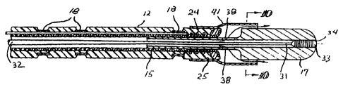
The embodiment shown in Figs. 3 an 4 is essentially the same as the
embodiment shown in Figs. 1 and 2 (and is similarly numbered) except that a
plurality of radially extending passageways 30 extend between the inner lumen
14
and the exterior of the electrode 17. The guidewire 31, having a core 32 and a
coil
33 on the distal extremity of the core, is slidably disposed within the inner
lumen 14
and the coil on the distal end of the guidewire extends beyond the passageways
30
and to a significant extent occludes the inner lumen 14 and reduces
considerably
the passage of fluid through the port 34 in the distal tip of the emitting
electrode 17.
Fluid flowing through the inner lumen 14 will then be forced to flow through
the radial
passages 30 thereby cooling the emitting electrode 17.
Another embodiment is shown in Figs. 5 and 6 where the emitting electrode
17 has longitudinally disposed passageways 35 for directing cooling fluid from
the
inner lumen 14 through the electrode and out the ports 36 in the distal tip of
the
electrode. A tubular sheath 37 formed of a high strength polymer material,
such as
polyimide, extends between the body of adhesive 25 securing the coiled distal
extremity of the safety wire 24 to the tubular extension 19 of the emitting
electrode
17 to the proximal end of the electrode to direct fluid which passes from the
inner
lumen 14 through the ports 38 in the tubular extension 19 to the passageways
35 as
indicated by the arrows shown in Fig. 5. The intravascular device shown is
otherwise essentially the same as the prior devices and is similarly numbered.
A
guidewire 31 may be used to occlude inner lumen 14 as in the prior embodiment
to
ensure an adequate flow of cooling fluid through passageways 35 to maintain
the
temperature of the emitting electrode 17 at a desired level.
Figs. 7 and 8 illustrate yet another embodiment of the invention wherein an
arcuate insulating sheath 40 is secured about an exterior portion of the
emitting
electrode 17 to ensure a more focused emission of high frequency electrical
energy
CA 02282488 1999-08-30
WO 98/38912 PCT/US98/04574
from a smaller exposed portion of the electrode toward the tissue to be
treated to
control the size of the lesion formed. This device is for the most part the
same as
the previously discussed embodiments, except for insulation sheath 40, and is
therefore similarly numbered.
5 Another embodiment is depicted in Figs. 9 and 10 wherein a fluid control
sheath 41 which is secured by its proximal extremity to the adhesive 25 and
extends
over the exterior of the emitting electrode 17. The inner diameter of the
distal end of
the sheath 41 is slightly larger than the outer diameter of the electrode 17
to provide
an annular gap 42 therebetween which directs cooling fluid along the exterior
10 surface of the electrode as indicated by the arrows. The cooling fluid
passes from
the inner iumen 14 through the ports 38 in the tubular extension 19 and
through the
annular gap 42. In this embodiment a guidewire 31 is disposed within the inner
lumen 14 with the coil 33 at least partially occluding the distal portion of
the inner
lumen so that an adequate flow of cooling fluid passes along the exterior of
the
electrode 17 to ensure sufficient cooling thereof.
In larger blood vessels, it frequently is difficult to maintain contact
between
the emitting electrode 17 and the blood vessel wall. 'To overcome this
problem, it is
desirable to provide an expandable positioning member, such as an inflatable
balloon 43, which when inflated ensures contact between a desired portion of
the
blood vessel wall 44 and the emitting electrode 17 as shown in Figs. 11 and
12. An
inflation lumen 45 extends through the shaft 11 from its proximal end to a
location
within the interior of the balloon 43. To accommodate for the extra lumen a
three
arm adapter (not shown) is secured to the proximal end of the shaft. While
only one
sensing electrode 18 is shown in the drawings, a plurality of sensing
electrodes may
be provided proximal to the balloon 43. The maximum transverse dimension of
the
balloon 43 as measured from the opposite side of the shaft 11 may range from
about 0.5 to about 5 mm, preferably about 1.5 to about 4 mm.
Figs. 13 and 14 represent another embodiment where the emitting electrode
50 is a helical coil on the distal end of the shaft 11. The proximal end of
the coil 51
is secured by solder 52 to the distal end of the shaft 11 shown in Fig. 13 to
facilitate
an electrical connection with the conductors 16 in the shaft 11 and the distal
end of
the coil is secured by adhesive to the enlarged distal end 53 of the lining
54.
T T
CA 02282488 1999-08-30
WO 98/38912 PCT/US98/04574
11
Perfusion holes 55 are provided in lining 54 to allow fluid passing through
inner
lumen 14 to contact and thus cool the coil 51.
In the embodiment shown in Figs. 15 and 16 the inner lumen 14 is disposed
within the inner tubular member 60 which extends to the distal tip 61. Annular
lumen
62 extends between the interior surface of braided tubular member 15 and the
exterior surface of inner tubular member 60. Electrode coil 63 is secured by
its
proximal end to the shaft 11 by solder 64 and is electrically connected to a
conductor of the braided tubular member 15. The distal end of the coil 63 is
secured
to the distal tip 61 by a suitable adhesive or by fusing the distal tip about
the distal
end of the coil. In this embodiment the delivery of cooling fluid through the
annular
lumen 62 is independent of a guidewire (not shown) in lumen 14.
Figs. 17 and 18 illustrate the distal portion of yet another embodiment of the
invention where an emitting coil electrode 70 is secured to the distal tip of
shaft 11
by means of adhesive or solder. A safety wire 71, which extends through the
shaft
11 as in the previous embodiments, is soldered to the distal tip of the
emitting coil
electrode 70. Sensing electrodes 18 are provided on shaft 11 proximal to the
emitting electrode coil 70 as in the previous emboditnents. The details of
shaft 11
are the same as shown in the prior embodiments.
Figs. 19-22 schematically illustrate another embodiment of the invention
wherein the device 100 has an elongated shaft 101 with a distal shaft section
102,
proximal shaft section 103 and a first inner lumen 104 extends through the
shaft. An
inner tubular member 105 is disposed within the first inner lumen 104 of the
shaft
101 and defines a second inner lumen 106 that extends to the port 107 in the
distal
tip 108 of the shaft 101. The distal tip 108 is preferably formed of
relatively soft
polymeric material such as Pebax 4033 and is secured by a suitable adhesive to
the
distal end of the inner tubular member 105. An emitting electrode 109 in the
form of
an expanded coil is provided on the distal shaft section 103 spaced a short
distance
from the distal tip 108 and a plurality of sensing or mapping electrodes 110
in the
form of coils are longitudinally spaced along a length of the distal shaft
section
spaced proximal to the emitting electrode.
The tubular structure of shaft 101 is formed in part by braided strands of
insulated electrical conductors 111, insulated thermocouple wires 112 and
metallic
CA 02282488 1999-08-30
WO 98/38912 PCT/US98/04574
12
ribbon 113, likewise preferably insulated, which are embedded in polymer
jacket
114. The distal ends of individual electrical conductors 111 are electrically
connected by solder 116 (or other suitable means) to the sensing electrodes
110
and one or more of the electrical conductors are electrically connected by
their distal
ends to the emitting electrode 109 by solder 117. The proximal extremities of
the
electrical conductors 111 are bundled together and provided with a polymeric
jacket
to form a connector lead 118 extending out of the adapter 119. The proximal
ends
of the conductors 111 are electrically connected to a multi-pin electrical
connector
119 for connection to a high frequency electrical source (not shown). The
distal
ends of thermocouple wires 112 are connected to thermocouple 120 (e.g. Type T,
Copper-Constantan) provided within the interior of emitting electrode 109. The
proximal extremities of the thermocouple wires 112 are likewise bundled and
jacketed to form a lead 121 with the proximal ends of the thermocouple wires
connected to the connector 122 which is configured to be electrically to a
temperature read out device (not shown). The leads 118 and 121 may also be
bundled and provided with a jacket upon exiting from the adapter 119 as shown
in
Fig. 19.
The braided insulated metallic ribbons 113 extend through the interior of the
emitting electrode 109 and are secured to the proximal end of the emitting
electrode
by solder 117 and to the distal end of the emitting electrode by solder 123.
The
braid of metallic ribbons 113 is of open weave construction so that cooling
fluid such
as saline passing through the first inner lumen 104 can pass through the open
weave construction and cool the expanded coil of the emitting electrode 109 as
shown by the arrows in Figs. 19 and 20.
The coil forming the emitting electrode 109 generally has a central or
intermediate section which has exposed uninsulated turns 124 and proximal and
distal sections which have exposed insulated turns 125 and 126 respectively.
The adapter 118 is provided with a central arm 127 with a guidewire port 128
which is in fluid communication with the second inner lumen 106 of the inner
tubular
member 105 and which is preferably configured to receive guidewires having
diameters of about 0.01 to about 0.018 inch (0.25-0.46 mm), preferably about
0.01
to about 0.015 inch (0.25-0.38)and to guide such guidewires to the second
inner
~
I
CA 02282488 2006-07-24
WO 98/38912 PCTIUS98/04574
13
lumen. Cooling fluid can be introduce into first inner lumen 104 through side
arm
129 of adapter 119.
The overall length of the intravascular devices of the invention may range
from about 80 to about 300 cm, typically about 120 to about 175 cm for
delivery
through the femoral artery or vein and about 80 to about 120 cm for delivery
through
the brachiocephalic artery or internal jugular vein. Because the intravascular
device
is to be advanced over a guidewire, the guidewire must be longer than the
catheter
by about 20 to about 60 cm. The outer diameter of the shaft of the
intravascular
device should be not greater than about 0.08 inch (1 mm) and the distal
sections 12 and 102 about 0.035-0.072 inch (0.89-1.8 mm). The inner lumen
within
the inner tubular member has a diameter of about 0.02 to about 0.04 inch (0.5-
1
mm) to facilitate the reception and advancement of a guidewire therethrough,
which
is typically about 0.010 to about 0.018 inch (0.25-0.46 mm) in outer diameter.
The
diameter of the inner lumen through the emitting electrode may be smaller than
the
diameter of the inner lumen in the more proximal portions of the shaft 11. The
distal
section 12 of the shaft is about 3 to about 20 cm in length. An intermediate
section
having an intermediate stiffness may be provided between the proximal section
13
and the distal section 12 with a length of about 5 to about 40 cm in length,
typically
about 20 cm in length. The radial passageways 30 are typically about 0.02 inch
(0.5
mm) in diameter and the iongitudinaf passageways 35 are typically about 0.01
inch
(0.25 mm). The emitting electrode is generally longer than about 2 mm. For
solid
electrodes the length is generally less than about 10 mm, but for an emitting
electrode in the form of helical coil the length may be about 2 to about 30
mm,
preferably about 2 to about 10 mm.
To the extent not previously described, the materials of construction of the
intravascular device of the invention may be formed of conventional materials.
The
electrical conductors 16 may be electrical grade copper wire about 0.003 inch
(0.08
mm) in diameter which are provided with a thin insulated jacket or coating of
polyimide or other suitable insulator. The outer jacket may be a thermoplastic
polyurethane such as PBAX which is available from Eif Atochem Polymers of
Philadelphia, Pennsylvania. The jacket of the proximal section is preferably
Pebax
1147 or 6833, the jacket of the intermediate section is preferably Pebax 6333
and
CA 02282488 2006-07-24
WO 98/38912 PCT/US98/04574
14
the jacket of the distal section is Pebax 4033 or 3533. The sensing and
emitting
electrodes are preferably formed of an alloy of platinum and iridium, e.g. 90%
Pt and
10% Ir (wt.%) or of Gold (100%). If the emitting electrodes are in coil form,
the wire
forming the coil is typically about 0.005 inch ( 0.13 mm) in diameter. The
safety wire
23 may be a stainless steel wire about 0.003 inch (0.08 mm) in diameter with a
polyimide coating. The preferred solder used to join the electrical conductors
to the
various electrodes is 95% Sn - 5% Ag or 80% Au - 20% Sn.
One presently preferred method of using the elongated intravascular device
includes first advancing a guiding catheter through the patient's vascular
system
until the distal tip of the guiding catheter is seated within the coronary
sinus ostium
or the ostium of one of the coronary arteries. The guiding catheter is torqued
by its
proximal extremity which extends out of the patient to guide the distal tip
into the
selected ostium. Once the distal end of the guiding catheter is seated, the
intravascular device of the invention with a guidewire slidably dispos'ed
within the
inner lumen thereof are advanced through the guiding catheter and out the
distal
end thereof. The guidewire is first advanced into the target vein or artery
and the
intravascular device of the invention is advanced over the guidewire into the
target
blood vessel. The sensing electrodes 18 or 110 on the intravascular device of
the
invention are used to detect electrical activity which allows the physician or
operator
to determine the location of the arrhythmogenic focus. When the focus is
located,
the intravascular device is moved within the blood vessel, as required, to
position the
emitting electrode 17 or 109 as close as possible to the focus. High frequency
electrical-energy, preferably in the RF range, is directed through the
electrical
conductors 16 or 111 connected to the emitting electrode 17 or 109 to form the
desired lesion which encompasses the arrhythnogenic focus. Energy levels of
about
5 Watts to about 100 Watts, preferably about 30 Watts to about 70 Watts are
suitable to terminate most arrhythmias. Typical lesions formed are about 3 mm
to
about 20 mm in diameter and about 3 mm to about 20 mm in length. In some
instances, where the site of the arrhythmic activity is detected by other
means, an
intravascular device may be utilized which does not have sensing electrodes.
For
example, the guidewire utilized to advance the intravascular device of the
invention
into the desired blood vessel may be provided with sensing electrodes for
detecting
CA 02282488 2006-07-24
WO 98/38912 PCT/US98104574
the electrical activity of interest.
While there are several means described herein to cool the emitting
electrode, a wide variety of means can be used to control the temperature of
the
5 emitting electrode. For example, the electrical energy to the emitting-
electrode can
be controlled so as to maintain the temperature thereof. A thermistor or other
temperature sensing device can be employed to monitor the electrode
temperature
and the temperature sensed is used to control in a conventional feedback
arrangement the electrical power delivery.
10 Although individual features of one embodiment of the invention may be
described herein and shown in one or more of the drawings and not in others,
those
skilled in the art will recognize that individual features of one embodiment
of the
invention can be combined with any or all the features of another embodiment
of the
invention. Various modifications and improvements may be made to the invention
15 without departing from the scope thereof.