Note: Descriptions are shown in the official language in which they were submitted.
CA 02282855 1999-09-02
WO 98/38904 PCT/GB98/00702 -
1
..
1 Biolocrical Measurement System
2
3 This invention relates to apparatus for use in non-
4 invasive in vivo monitoring of physiological substances
such as blood and the like.
6
7 One particular, but not exclusive, application of the
8 present invention is in the monitoring of blood
9 glucose, for example in the management of diabetes
mellitus. It is accepted that the management of
11 diabetes can be much improved by routine monitoring of
12 blood glucose concentration and clinicians suggest that
13 monitoring as often as four times per day is desirable.
14
The monitoring technique currently available for use by
16 patients involves using a spring loaded lancet to stab
17 the finger to obtain a blood sample which is
18 transferred to a glucose test strip. The concentration
19 is derived either by reading the test strip with a
reflectance meter or by visual comparison of colour
21 change against a colour scale. Many diabetics find the
22 testing onerous as the technique is painful,
23 inconvenient, messy, potentially embarrassing and
24 offers a site for the transmittance and acceptance of
infection.
CA 02282855 1999-09-02
WO 98/38904 PCT/GB98100702
2
1 Techniques have also been developed for non invasive
2 measurement using transmittance or reflectance
3 spectroscopy. However the required instruments are
4 expensive and it is difficult to obtain accurate and
repeatable measurements.
6
7 There are also known various types of in vivo chemical
8 sensors. These rely on implanting minimally invasive
9 sensors under the skin surface, but such sensors have
ZO poor long term reproducibility and bio-compatibility
11 problems.
12
13 There is therefore a need for improved means for
14 routine monitoring of blood glucose in a manner which
is simple and straightforward to use.
16
17 The present invention makes use of photoacoustic
18 techniques. The fundamentals of photoacoustic
19 techniques are well known per se. A pulse of light,
typically laser light, is applied to a substance
21 containing an analyte of interest in solution or
22 dispersion, the wavelength of the applied Light being
23 chosen to interact with the analyte. Absorption of the
24 light energy by the analyte gives rise to microscopic
localised heating which generates an acoustic wave
26 which can be detected by an acoustic sensor. These
27 techniques have been used to measure physiological
28 parameters in vitro.
29
US Patents 5348002 and 5348003 (Caro) propose the use
31 of photoacoustics in combination with photoabsorption
32 for the measurement of blood components in vivo.
33 However, the arrangement proposed by Caro has not been
34 demonstrated as a workable system and may suffer from
interference to a degree which would preclude useful
36 acoustic signals, and since they would also suffer from
CA 02282855 1999-09-02
WO 98/38904 PCT/GB98/00702
3
1 interference and resonance effects from hard structures
2 such as bone.
3
- 4 It has also been proposed by Poulet and Chambron in
Medical and Biological Eng~ineering~ and Computi,~cr,
6 November 1985, Page 585 to use a photoacoustic
7 spectrometer in a cell arrangement to measure
8 characteristics of cutaneous tissue, but the apparatus
9 described would not be suitable for measuring blood
analytes.
11
12 Published European Patent Application 0282234A1
13 (bowling) proposes the use of photoacoustic
14 spectroscopy for the measurement of blood analytes such
as blood glucose. This disclosure however does not
16 show or suggest any means which would permit the
17 required degree of coupling to body tissues for use in
18 vivo.
19
Accordingly, the present invention provides a sensor
21 head for use in photoacoustic in vivo measurement,
22 comprising a housing shaped to engage a selected body
23 part, light transmission means terminating in said
24 housing so as to transmit light energy from a light
source to enter the body part along a beam axis, and
26 acoustic transducer means mounted in the housing to
27 receive acoustic waves generated by photoacoustic
28 interaction within the body part, the acoustic
29 transducer means being disposed in the housing to
receive said acoustic wave in a direction of high
31 acoustic energy.
32
33 The expression "direction of high acoustic energy" is
34 used herein to denote a direction other than the
forward direction of the light beam. Preferably, the
36 transducer means is disposed so as to intercept
CA 02282855 1999-09-02
WO 98/38904 PCT/GB98/00702
4
1 acoustic energy propagating at right angles to the
2 optical beam axis, or at an angle to the optical beam
3 axis which may be down to about 20°, typically about
4 45°.
6 An exact measure of the angle of high acoustic energy
7 can be worked out but is dependent upon the specific
8 geometry of the light source, the properties of the
9 tissue and the absorption coefficient of the tissue.
IO One model for understanding the propagation of the
11 acoustic energy in any homogenous media was developed
12 by Huyghens and is called the principle of
13 superposition. In this model each volume element that
14 is illuminated by the light generates an acoustic
pressure wave that radiates outward in a spherical
16 manor. The magnitude of the pressure wave at each
17 volume element depends on the intensity of the optical
18 beam at that location, the absorption coefficient of
19 the material at that location, the wavelength of light
and on several other physical properties of the
21 material such as the speed of sound and the specific
22 heat. The signal measured at the detector is just the
23 superposition of all pressure waves from all points
24 that are illuminated by the source light. An
analytical solution for the pressure wave has been
26 worked out for a few cases in aqueous material. The
27 analytical case that best matches the in-vivo
28 measurements is that of a cylindrical optical beam
29 propagating in a weekly absorbing material. In this
case the direction of highest acoustic energy is
31 perpendicular to the optical axis. The base detector
32 location is with the plane of the detector
33 perpendicular to the acoustic energy, or parallel to
34 the optical axis. This is because the acoustic
detector has the highest sensitivity when the acoustic
36 energy strikes the detector perpendicular to the plane
CA 02282855 1999-09-02
WO 98/38904 PCT/GB98/00702
1 of the detector. This analytical model is not
2 completely accurate for the in-vivo measurement case
3 because of scattering of the tissue and because the
- 4 tissue absorbs more than the model predicts. These
5 differences indicate that a different position for the
6 detector will be optimal. A detailed numeric model is
7 required to determine the best detector location and. is
8 dependent upon the beam properties (focused to a point,
9 colligated, etc.), body site (finger, earlobe, arm
etc.) and wavelength. One skilled in the art can
11 readily develop an appropriate mode. However, suitable
12 locations for a detector will generally be at an angle
13 to the optical axis. Angles between 40 and 90 degrees
14 should be suitable.
16 In one preferred arrangement, the acoustic transducer
17 means is arranged parallel to the optical beam axis.
18 This arrangement is particularly suitable for use where
19 the selected body part is the distal portion of a
finger, in which case the housing may include a
21 generally half-cylindrical depression in which the
22 finger may be placed with the light transmission means
23 aimed at the end of the finger.
24
Preferably, the acoustic transducer means comprises a
26 piezoelectric transducer which most preferably is of a
27 semi-cylindrical shape. This transducer may be
2B provided with a backing of lead or other dense
29 material, and the backing may have a rear surface
shaped to minimise internal acoustic reflection.
31
- 32 Alternative transducer means include a capacitor-type
33 detector, which is preferably small and disk-shaped; an
34 integrated semiconductor pressure sensor; and an
optical pressure sensor, for example based on an
36 optical fibre.
CA 02282855 1999-09-02
WO 98/38904 PCT/GB98/00702
6
1 In an alternative arrangement, the plane of the
2 transducer may be arranged to be perpendicular to the
3 optical axis to detect the acoustic wave which is
4 propagating in a direction opposite to the direction of
the light beam. For example, the acoustic transducer
6 means may be part-spherical with an aperture to allow
4
7 access for the light beam. This may be particularly
8 suitable for engagement with a body part other than the
9 finger, for example the back of the arm.
11 The generation of a surface acoustic wave is an
12 inherent aspect of the in vivo pulsed photoacoustic
13 generation in tissue and may be used to characterize
14 tissue properties such as density. A surface wave
detector may be provided in the sensing head assembly.
16
17 Preferably means are provided for ensuring a consistent
18 contact pressure between the selected body part and the
19 acoustic transducer means. In the case where the
selected part is the distal portion of the finger, said
21 means may be provided by mounting the portion of the
22 housing engaged by the finger in a resiliently biased
23 fashion against the remainder of the housing, and
24 providing means to ensure that measurement is effected
when the predetermined force or pressure is applied by
26 the subject against the resilient bias. In the case
27 where the selected part is the earlobe, said means may
28 be provided by placing the ear between two plates and
29 applying pressure to the ear with springs or weights or
other force method. The two plates holding the ear may
31 contain a removable insert. The two plates may be fiat
32 or may be of another shape to optimally position the
33 detector with respect to the beam axis.
34
In addition, the present invention provides a sensor
36 head for use in photoacoustic in-vivo measurements,
T
CA 02282855 1999-09-02
WO 98138904 . PCT/GB98100702
7
1 comprising a housing shaped to receive a removable
2 insert, a removable insert that engages a selected body
3 part, the insert being fitted to an individual,
4 allowing for a range of sizes of body parts to be used,
. 5 and further comprising light transmission means
6 terminating in or near said removable insert so as~to
7 transmit light energy from a light source or sources to
8 enter the body part along a beam axis, and an acoustic
9 transducer means mounted in the housing or in the
removable insert to receive acoustic waves generated by
11 photoacoustic interaction within the body part to
I2 receive said acoustic waves in a direction of high
13 acoustic energy.
14
From another aspect the present invention provides an
16 in vivo measuring system comprising a sensor head as
17 hereinbefore defined in combination with a light source
18 coupled with the light transmission means, and signal
19 processing means connected to receive the output of the
acoustic transducer means and to derive therefrom a
21 measurement of a selected physiological parameter.
22
23 Preferably, the light transmission means is a fiber
24 distribution system where each light source is
connected to an individual fiber and when multiple
26 light sources are used the multiple fibres are joined
27 by some standard fiber combining method, such as a
28 wavelength division multiplexer or a fiber coupler.
29 The fiber that comes from the light source, or contains
the combined light for a multiple source system, is
31 then terminated in proximity to the body part being
32 measured. The fiber could be in contact with the body
33 part or alternatively standard optics, such as lenses,
34 beamsplitters and such, could be employed to convey the
light from the end of the fiber to the body part. A
36 reference detector or several reference detectors and
CA 02282855 1999-09-02
WO 98/38904 PCT/GB98/00702
8
1 beamsplitters can be added to the optical distribution
2 system to determine the energy of the light entering
3 the body part.
4
Alternatively, the optical distribution system may
6 contain mechanical holders, lenses and such to convey
7 the light from the source, or sources, to a location in
8 proximity to the body part being measured. A reference
9 detector or several reference detectors and
beamsplitters can be added to the optical distribution
11 system to determine the energy of the light entering
12 the body part.
13
14 The acoustic signal from the detector contains
information in both time and frequency, and there may
16 be information from several sources. The processing
17 means is preferably a multi-dimensional processing
18 method, such as Classical Least Squares (CLS) or
19 Partial Least Squares (PLS). Alternatively the
processing method may be more flexible, such as a
21 Neural Network. In addition to these methods the
22 signals may be analysed for their frequency content
23 using such techniques as Fourier Analysis or Frequency
24 Filtering In addition techniques may be employed that
use time information such as the time delay from source
26 trigger. Techniques that combine both frequency and
27 time information may be employed, such as Wavelet
28 analysis.
29
The light source is preferably a laser light source and
31 is most suitably a pulsed diode laser, but may utilise
32 a set of such lasers or utilise a tunable laser source.
33 In a particularly preferred form, suitable for use in
34 measuring blood glucose concentration, a laser diode is
used with a wave length in the range of approximately
36 600 nm to 10,000 nm and a pulse duration of the order
T..
CA 02282855 1999-09-02
WO 98/38904. PCT/GB98/00702
9
1 of 5 to 500 ns.
2
3 The delivery to the measurement site may be either
4 directly or by optical fibre with a suitable optical
element to focus the beam into the tissue.
6
4 _
7 Preferably means are provided for time multiplexing
8 multiple sources when multiple sources are used. Each
9 source is switched on, and it generates an optical
pulse, or a set of optical pulses. This pulse, or set
11 of pulses, generates an acoustic signal that is
12 detected by the detector. Each source is pulsed in
13 sequence until all sources have been used to generate
14 their own signal.
16 The measuring system may conveniently be in the form of
17 a self contained system including a power supply and a
18 readout, which may be carried on the person and used at
19 any convenient time.
21 It is also possible for such a self contained system to
22 incorporate, or to be provided with facilities for
23 connection to, a cellular telephone, two-way pager or
24 other communication device for routine transmission of
measurements taken to a central data collection point.
26
27 In addition the measuring system may have provision for
28 manipulating the body part under measurement and for
29 performing additional measurement of the tissue to get
other information about the state of the physiology of
31 the issue. It is well-known in the art that squeezing
32 a section of tissue to increase the pressure and then
33 releasing the pressure will cause changes in the total
34 blood volume in the measurement site. The present
invention may allow for this type of manipulation
36 including the squeezing of a body part, such as an
CA 02282855 1999-09-02
WO 98/38904 PCT/GB98/00702
1 earlobe, and making photo acoustic measurements at
2 several different pressures. The present invention may
3 also allow for the measurement of the temperature of
4 the body site and to apply a correction to the
5 measurements based upon the temperature of the body
6 site.
7
8 Another type of physiological manipulation is body
9 temperature. It is known in the art that several
10 parameters involved in the detection of the photo
11 acoustic signal, such as the speed of sound, are
12 dependent upon the temperature of the medium the signal
13 is propagating through (the tissue}. Also the
14 profusion of the blood in the small capillaries is
dependent upon the temperature of the tissue.
16 Additional information about the tissue can be obtained
17 if the photo acoustic measurement is made at several
18 temperatures, both higher and lower than ambient
19 temperature. This additional information is used to
better eliminate interferences to the determination of
21 the analyte under investigation. These are only two
22 examples of manipulating the body site and are not
23 intended to be an exhaustive list, and they can be used
24 in combination with other manipulation techniques.
26 The in-vivo measuring system may comprise a means for
27 storing calibration coefficients or operation
2g parameters or both calibration coefficients and
29 operational parameters, in order to calibrate the
instrument and to set critical operational parameters.
31
32 Another aspect of the present invention provides a
33 means for adjusting the calibration coefficients and
34 operational parameters to be specific to a particular
person and may be used to adjust for such things as
36 body part size, skin color, skin condition, amount of
T___ _ T.
CA 02282855 1999-09-02
_ WO 98/38904 PCT/GB98/00702
11
1 body fat, efficiency of the detector and efficiency of
2 the source(s).
3
4 In addition the present invention may provide for
having the specific calibration coefficients and
6 operational parameters be contained in a storage site _
7 located in the removable insert. This allows for the
8 system to be both mechanically and operationally
9 configured to a particular individual. Additionally
the invention may allow for the calibration
11 coefficients and operational parameters to be stored in
12 two locations, one in the non-removable housing and one
13 in the removable insert with some of the coefficients
14 and parameters stored in each location. This allows
for reader system coefficients to be stored in the
16 reader and coefficients specific to an individual to be
17 stored in the removable insert for that person,
18 enabling many people to use the same reader.
19
Another aspect of the present invention provides means
21 for connecting the non-invasive measuring system to an
22 invasive measuring system for the purpose of
23 calibrating or adjusting the operational parameters of
24 the non-invasive measuring system. Such connection may
be accomplished, but is not limited to, communication
26 by a wire, IR link or radio waves.
27
28 Another aspect of the present invention provides a
29 method for removing instrument drift from the
measurement comprising the steps of:
31
32 1. Placing a standard in the reader in place of the
33 body part.
34
2. Measuring the signal from the standard for each
36 wavelength and storing the values in the
CA 02282855 1999-09-02
WO 98/38904 PCT/GB98/00702
12
1 calibration storage location.
2
3 3. Before making a measurement of a body part,
4 placing the calibration standard in the reader.
6 4. Measuring the signal from the standard for each
7 source.
8
9 5. Comparing the just measured standard values to the
stored calibration values.
11
12 6. Calculating correction factors for each source
13 wavelength.
14
7. Removing the standard and placing the body part in
16 the reader.
17
18 8. Measuring the signal from the body part for each
19 source.
21 9. Adjusting the measured values using the calculated
22 correction factors.
23
24 In addition to the signal correction factors a
correction factor can be calculated for the instrument
26 temperature. This can be applied to each signal with a
27 different correction coefficient.
28
29 The invention further provides a method of measuring a
biological parameter in a subject, the method
31 comprising the steps of:
32
33 directing one or more pulses of optical energy
34 from the exterior into the tissue of a subject
along a beam axis, the optical energy having a
36 wavelength selected to be absorbed by tissue
CA 02282855 1999-09-02
WO 98/38904 PCT/GB98/00702
13
1 components of interest, thereby to produce a
2 photoacoustic interaction;
3
- 4 detecting acoustic energy resulting from said
photoacoustic reaction by means of a transducer
' 6 positioned to intercept acoustic energy
4 -
7 propagating in a direction other than the forward
8 direction of said beam axis; and
9
deriving from said detected acoustic energy a
11 measure of the parameter of interest; and a
12 corresponding apparatus.
13
14
Embodiments of the invention will now be described, by
16 way of example only, with reference to the accompanying
17 drawings in which:-
18
19 Figs. lA,lB and 1C are side views illustrating the
principle of operation of one embodiment of the
21 present invention;
22
23 Fig. 2 is a schematic perspective view showing a
24 sensor head for use in carrying out the
measurement illustrated in Fig. 1;
26
27 Fig 3. is a cross section view of the sensor head
28 of Fig. 2;
29
Fig. 4 is a side view of the sensor head of Fig.
31 2;
32
33 Fig. 5 is a schematic perspective view of an
34 apparatus incorporating the sensor head of Figs. 2
to 4;
36
CA 02282855 1999-09-02
WO 98/38904 PCT/GB98/00702
14
1 Fig. 6 is a perspective view illustrating an
2 alternative form of sensor head;
3
4 Fig. 7 is a schematic end view showing another
form of sensor head;
6
7 Figs. 8a and 8b are a cross-sectional side view
8 and a plan view, respectively, of a further sensor
9 head;
11 Fig. 9 is a cross-sectional side view of one more
12 embodiment of sensor head;
13
14 Fig. 10 is a perspective view of one type of ear
interface apparatus;
16
17 Fig. 11 is a schematic of a multiple laser optical
18 distribution system using lenses, mechanical
19 mounts and a reference detector;
21 Fig. 12 is a schematic of a multiple laser optical
22 distribution system using fiber optic cables and a
23 fiber Wavelength Division Multiplexer (WDM), a
24 beam splitter and a reference detector;
26 Fig. 13 is a perspective view of a finger
27 interface apparatus with removable inserts that
28 are moulded to fit one individual;
29
Fig. 13A shows part of the apparatus of Fig. 13 in
31 greater detail;
32
33 Fig. 14 is a schematic of a semi-spherical
34 detector that contains a hole for the light beam,
with a vacuum system and a fiber distribution
36 system;
.T....... ..... T...
CA 02282855 1999-09-02
WO 98/38904 PCT/GB98/00702
1
2 Fig. 15 is a perspective view showing one form of
3 the instrument utilizing the vacuum body
4 interface, a semi-spherical detector and the
5 multiple laser source with lenses and mechanical
6 housing;
7 ,,
8 Fig. 16 is a perspective view showing one form of
9 the instrument using an ear lobe body interface,
10 with the added feature of being able to manipulate
11 the pressure on the ear lobe; and
12
13 Figs. 17, 18 and 19 are graphs illustrating an
14 example.
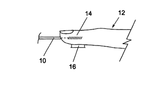
16 Referring to Fig 1, an important feature of the present
17 invention lies in introducing light energy along an
18 axis into an area of soft tissue and detecting the
19 resulting acoustic response transverse to that axis.
Accordingly, in the arrangement of Fig lA light energy
21 from a diode laser (not shown) is transmitted via a
22 fibre-optic guide 10 to the tip of a finger 12. The
23 photoacoustic interaction occurs in an approximately
24 cylindrical region indicated at 14 from which acoustic
energy is radiated in a generally cylindrical manner
26 and is detected by a transversely arranged acoustic
27 transducer 16.
28
29 In Figs 1B and 1C, the principle is similar. The
finger 12 is pressed against a support with force F.
31 In Fig 1B, the incident light beam indicated at L is
32 directed as in Fig lA, and the transducer 16 is at an
33 angle of 45 degrees thereto. In Fig 1B, the angle is
34 90 degrees as in Fig lA, but the incident beam is
directed differently into the fingertip.
36
CA 02282855 1999-09-02
WO 98/38904 PCT/GB98/00702
16
1 In the present embodiment, the laser wavelength is
2 chosen to achieve high degree of absorption by glucose
3 present in the blood. A suitable wavelength is in the
4 range approximately 1000 to 3000 nm. The laser pulse
duration is chosen to be short, typically of the order
6 of 5 to 500 ns, in order to minimise thermal diffusion
a .
7 and thus to optimise the acoustic waveform. For the
8 same reasons, it is desirable to use a spot size which
9 is sufficiently small to minimise thermal diffusion,
typically a spot size of the order of 0.05 mm to
11 0.50 mm.
12
13 The efficiency of the photoacoustic detection is also
14 influenced by the positioning and dimensions of the
acoustic transducer in relation to the characteristic
16 extinction length of the tissue at the principal
17 wavelengths chosen for measurement. in the fingertip
18 arrangement of Fig. 1, the system efficiency will be
19 improved by optimising the length of the transducer
crystal parallel to the axis of the finger, but the
21 length should not be so great as to give rise to
22 undesired signals which would occur at the point of
23 entry of the optical energy into the finger and by
24 reason of interaction of the acoustic energy with bone
or other hard tissue.
26
27 A second limit on the size of the acoustic detector
28 derives from the wavelength of the acoustic wave in the
29 tissue. Again making use of Huyghens principal of
superposition we view each point of tissue, that is
31 illuminated by the incoming light, as a point source
32 that generates a spherical pressure wave. The signal
33 measured at the detector is just the superposition of
34 all pressure waves from all points that are illuminated
by the source light. Normally if the size of the
36 detector is increased then the signal should also
T _. __ _.___._ ~~
CA 02282855 1999-09-02
WO 98/38904 PCT/GB98/00702
17
1 increase because more energy is received by the
2 detector. However if the acoustic detector is too
3 large then a pressure wave generated from a tissue
- 4 element will create a pressure wave that will strike
the both ends of the detector. If the paths length
6 from the tissue element to the first end of the
7 detector is different than the path length to the 4
8 second end of the detector and if this difference in
9 path length is about one half of the acoustic signal
wavelength then the signal will destructively interfere
11 with itself and will reduce the magnitude of the
12 measured signal.
13
14 Referring to Fig 2, one manner of carrying out the
arrangement shown in Fig 1 makes use of a sensor head
16 having a finger rest 18 which is slidably moveable
17 within housing 20 closed by a front plate 22. The user
18 inserts his finger in a semi-cylindrical depression 24
19 in the finger rest 18 with the finger tip engaged
against an end surface 28 which includes an exit face
21 26 of the optical fibre 10. The finger is then pressed
22 downwardly against a resilient bias to enable a
23 standardised contact to be obtained between the skin
24 and the acoustic transducer. The finger tip may first
be dipped in water or coated with an aqueous gel to
26 improve the acoustic coupling.
27
28 Referring to Figs 3 and 4, in this preferred
29 arrangement the acoustic transducer comprises a semi-
cylindrical piezoelectric transducer 30. The
31 transducer 30 is provided with a backing member 32 of
32 lead or another dense substance, the rear face 34 of
33 which is shaped in irregular curves. The use of the
34 semi-cylindrical transducer 30 maximises the area for
reception of acoustic energy from the finger, while the
36 use of a dense backing material minimises ringing
CA 02282855 1999-09-02
WO 98/38904 PCT/GB98/00702
18
1 effects within the transducer. Additionally, the rear
2 face 34 is shaped as shown to reduce reflection of
3 acoustic energy back towards the piezo crystal.
4
Fig 3 also shows the finger rest biased upwardly by the
6 use of constant tension springs 38.
a -
7
8 Fig 5 illustrates schematically the apparatus of Figs.
9 2 and 3 embodied in a self-contained, portable blood
monitoring apparatus including a user readout 40. An
11 apparatus of this nature allows a diabetic to monitor
12 blood glucose concentration in a convenient manner, as
13 frequently as may be desired, and in a painless and
14 discreet manner.
16 Other forms of photoacoustic sensor head are possible
17 within the scope of the present invention. For
18 example, Fig. 6 shows an arrangement in which a light
19 guide 50 and an acoustic transducer 52 are applied to a
finger 54 by means of a hinged clamp member 56. Fig. 7
21 shows a finger 60 engaged by a light guide 62 and an
22 acoustic transducer 64 which are carried on a moveable
23 assembly 66 with the finger 60 being trapped between
24 the moveable assembly 66 and a fixed anvil 68.
26 It is also possible to arrange the sensor head to co-
27 operate with a soft tissue surface of the body, for
28 example a soft part of the abdomen. Figs. 8a and 8b
29 show an arrangement in which a cup shaped member 70,
suitably of rubber, causes a light guide 72 and an
31 acoustic transducer 74 to be contacted with a bulge of
32 soft tissue 76 which may for example be drawn into
33 contact by means of a partial vacuum within the member
34 70 caused by suction through a conduit 78, or by other
mechanical or adhesive means.
36
_ _ T _ _ ____ T _
CA 02282855 1999-09-02
WO 98/38904 PCT/GB98/00702
19
1 A somewhat similar arrangement is shown in Fig. 9 in
2 which a planar mount 80 carrying a light guide 82 and
3 acoustic transducer 84 is secured to a soft area of
4 body by means of surgical adhesive 86.
6 Referring to Fig. 10, one method of performing
7 measurement on an ear lobe involves placing the ear
8 lobe between a fixed plate 87 and a movable plate 88.
9 The acoustic detector 89 is mounted partially
perpendicular that is at an acute angle, to the beam
11 axis defined as line going from the center of a lens 90
12 to the center of a window 91. It has been found that
13 the system works satisfactorily with the detector 89 at
14 an angle or 45° to the beam axis. The window 91 and
the detector 89 are placed in direct contact with the
16 ear and the opposite plate 88 places pressure on the
17 ear using a suitable mechanism (not shown). This
18 particular embodiment of the ear interface apparatus
i9 incorporates an alignment ring 92 which is temporarily
attached to the ear and fits over the window housing 91
21 to aid in aligning ear into the same location every
22 time.
23
24 Referring to Fig. 11, one method of combining light
sources into the instrument is to use a mechanical
26 housing 93 with several holes used to align lenses 95
27 and laser diodes 94. The housing shown uses a
28 hexagonal array of seven holes. The sources and lenses
29 are arranged in such a way that they all focus to the
same location 96 which could be on the surface of the
31 body part. This design does not show the inclusion of
32 beamsplitters and reference detectors but they can be
33 added in an alternative arrangement.
34
An alternative method of combining several sources into
36 one beam is shown in Fig. 12. Several laser diodes 97
CA 02282855 1999-09-02
WO 98/38904 PCT1GB98J00702
1 are shown coupled to individual fiber optic cables 131.
2 These cables 132 are combined using a fiber Wavelength
3 Division Multiplexer (WDM) 98. Alternative combination
4 methods exist including couplers and multi-fiber
5 bundles. The combined light exits the WDM 98 in a
6 single fiber 104 and terminates at the focal point of a
7 lens 131. This end of the fiber is imaged to the end
8 of the finger 103 to a spot 102 using another lens 130.
9 Some of the light is split off the main beam using a
10 beam splitter 100 and focused onto a reference detector
11 101 using another lens 99. Additional reference
12 detectors and/or beamsplitters can be added to the
13 distribution system without changing its function.
14 Alternatively a reference detector could look directly
15 at the body part to measure the light reflecting off
16 the surface, as a measure of the overall light energy
17 entering the body part.
18
19 Referring to Fig. 13, another method of using a finger
20 as the body part and including removable inserts is
21 shown. A finger 105 is inserted into an insert 106
22 that is used to customize the finger holder to a
23 particular finger. The moulded insert 106 is placed
24 into a housing 107. The finger 105 is placed against a
semi-cylindrical acoustic detector in a module108 which
26 is also attached to the housing 107. A cover 109 for
27 the housing 107 contains a mechanism 111 to apply
28 constant force to the finger 105. The light beam 110
29 is introduced into the finger 105 using a suitable
optical distribution system (not shown). Fig. 13A shows
31 the module 108 in greater detail. A base 200 carries a
32 part-cylindrical piezo transducer 202 on a support 204.
33 206 indicates a coaxial connector to communicate the
34 transducer signal.
36 Fig. 14 shows a schematic of an alternative to the
T __ I
__ _._ __ __ _ _ .
CA 02282855 1999-09-02
- WO 98/38904 PCT/GB98/00702
21
1 vacuum arrangement shown in Figs. 8 and 9. In this
2 system a photoacoustic reader 121 is placed against the
3 skin 113 with a semi-spherical detector 112 in contact
4 with the skin 113. A vacuum pump 115 and vacuum seal
116 create a negative pressure and pull the skin 113
6 against the detector 112. Processing electronics 119
7 energizes light sources 118 and an optical distribution
8 system 117 routes the light to the body part through a
9 hole in the top of the semi-spherical detector 112.
The optical distribution system 117 directs a small
11 portion of the light to a reference detector 114. The
12 processing electronics 119 measures the signal from the
13 acoustic detector 112 and the reference detector 114
14 for each optical source 119 and calculates the glucose
value. The value is displayed on a display 120.
16
17 Fig. 15 shows a similar system 125, only using another
18 type of optical distribution system 127. Again a
19 vacuum pump 123 creates a negative pressure which draws
the skin up to an acoustic detector 122. Processing
21 electronics 124 signals light sources in optical
22 distribution system 127 to illuminate and a signal is
23 generated at acoustic detector 122. The processing
24 electronics 124 calculates the proper value and
displays it on a display 126.
26
27 Fig. 16 shows an alternative arrangement of a photo-
28 acoustic reader. In this system 128, the vacuum system
29 is replaced with an ear squeeze mechanism 129 which
applies pressure to the ear. An acoustic detector 130
31 detects the signals from the ear lobe.
32
33 In the most straightforward forms of the invention, a
34 single analyte such as glucose in blood can be measured
by using light of selected wavelengths and by measuring
36 the area or the amplitude of the received acoustic
CA 02282855 1999-09-02
_ WO 98/38904 PCT/GB98/00702
22
1 pulse. It is preferable to make each measurement by
2 using a train of pulses, for example about 100 pulses,
3 and averaging the results in order to minimise the
4 effects of noise and pulse effects in the blood flow.
6 The accuracy of the detection system is governed, in
7 part, by the Signal to Noise Ratio (SNR) of the system.
8 Variations in the intensity and duration of the light
9 ~ source can cause the acoustic signal to contain
variations. A normalization technique, such as taking
11 the ratio of the acoustic signal to the optical signal,
12 can significantly reduce the effect of the source
13 variations, thereby improving the signal to noise ratio
14 of the system. The optical signal can be measured with
a reference detector, or several reference detectors,
16 one far each source or one for a wavelength range. An
17 equation describing this type of normalization follows:
18
19 Acoustic Signal
Normalized Signal -
21 Optical Signal
22
23 In some cases the relationship between the optical
24 signal land the acoustic signal changes with wavelength
and light intensity. When this is the case the
26 accuracy of the measurement can be further enhanced by
27 determining the energy dependence of the photoacoustic
28 signal. This may be determined by establishing the
29 specific relationship between the photoacoustic signal
land the incident energy from a set of measurements and
31 using this relationship to compensate for the non
32 linear response. An equation describing this type of
33 normalization is as follows:
34
Acoustic Signal
36 Normalized Signal -
T _.__ __..____.__.
CA 02282855 1999-09-02
- WO 98/38904 , PCT/GB98/00702
23
1 Scaling Factor *Optical Signal +
2 Offset
3
4 Other normalization methods can also apply. The time
interval between the optical pulse and the detection of
6 the acoustic signal may be used to characterise
7 physical properties such as the velocity of sound in
8 the tissue. In addition, in another embodiment of the
9 device the damping of the acoustic oscillations may be
used to monitor the elastic properties of the tissue
11 and, in particular, the compressibility. Both of these
12 aspects may be used in the person to person calibration
13 of the photoacoustic response.
14
More complex analysis of the received acoustic energy
16 is possible. For example, a time-gating technique may
17 be used to derive measurement at varying depths within
18 the tissue being examined. Alternatively, an array of
19 detectors can be employed to determine the profile of
the absorption of the acoustic signal at different
21 depths and locations. This depth profile will change
22 with the absorption coefficient and could be used as
23 additional information to determine the analyte
24 concentration. It is also possible to derive
information relating to a number of analytes of
26 interest by more sophisticated analysis of the received
27 acoustic energy wave forms, for example by analysis of
28 the frequency spectrum by Fourier transform or wavelet
29 analysis techniques.
31 Alternatively, or in combination with the frequency
32 techniques and multiple detectors, multiple light
33 sources can aid in the determination of the
34 concentration of a number of analytes.
36 There are a number of tissue features which may vary
CA 02282855 1999-09-02
WO 98/38904 PCT/GB98/00702
24
1 from person to person or with in the same person over
2 time which impact the photoacoustic signal observed.
3 To obtain an accurate measurement of a given analyte,
4 such as glucose, it may be helpful to also determine
the concentration of other analytes such as haemoglobin
6 which may act as interferants. One approach is to
y
7 generate several distinct photoacoustic signals using
8 excitation light of several different wavelengths. For
9 example, excitation light of a wavelength of which
haemoglobin absorbs strongly but glucose has little if
11 any absorption could be sued to obtain a measure of the
12 haemoglobin concentration with which to normalize the
13 effect of haemoglobin on measurements made on different
14 persons or on the same person at different times.
These measurements which are to be normalized might be
16 based on the photoacoustic signal generated by light of
17 a wavelength at which glucose absorbs.
18
19 It is also possible to measure the concentration of
such interferants by other means, such as infrared
21 light absorption, and thus normalize or correct the
22 photoacoustic signal representative of the desired
23 analyte for variations in these interferants. Thus,
24 for example, the photoacoustic signal representative of
glucose could be corrected for variations in
26 haemoglobin concentration determined by optical
27 absorption techniques such as those taught in US Patent
28 No 5,702,284.
29
For the reliable and reproducible determination of
31 glucose a signal to noise ratio of at least 10,000 is
32 recommended. In this regard water is typically present
33 in human tissue of a concentration of about 50 molar
34 while glucose is present at a concentration of about 5
millimolar in a normal individual.
36
r _ ____ z
CA 02282855 1999-09-02
- WO 98/38904 PCT/GB98/00702
1 Apparatus and method embodying the present invention
2 have been found to yield accurate and repeatable
3 results. In the case of blood glucose measurement, the
4 clinical range of glucose concentration is
5 approximately 5-10 m mol/1 in healthy subjects, and up
6 to 40 m mol/1 in diabetics. An analysis based on
7 simple absorption models suggests that the change in~
8 photoacoustic signal over this range might be as little
9 as 0.2~. The present invention has been found to
10 provide a change in photoacoustic signal of up to 140
11 for a change in glucose concentration of 15m mol/1.
12
13 The precise mechanisms involved are not at present
14 fully understood. It is believed, however, that
15 absorption occurs primarily in body plasma and is
16 modified by the presence of glucose, and that this
17 affects beam geometry.
18
19 Example
21 The blood glucose levels of three individuals, one
22 normal individual, one type 1 diabetic and one type 2
23 diabetic, were followed over a two hour period
24 following each individual taking about 75 grams of
glucose orally in an aqueous solution by both
26 photoacoustics and direct blood measurement. The
27 results are reported in Figures 17, 18 and 19.
28 Photoacoustic measurements were made every five minutes
29 and blood measurements were made very ten minutes. The
blood samples were venous blood samples analysed by the
31 standard glucose oxidase method using a Yellow Springs
32 instrument. The error bands for the blood measurements
33 were derived from the literature accompanying the
34 testing instrument while those for the photoacoustic
results were based on the averages taken over 1000
36 pulses. The results were obtained from a configuration
CA 02282855 1999-09-02
_ WO 98/38904 PCT/GB98100702
26
1
2
3
4
6
7
8
9
11
12
13 wavelength Average pulse Pulse width Approximate
in nm in ns
14 energy in bandwidth in
nm
microJoules
16
17 1064 2.7 600 4
18 1120 2.25 500 6
19
1176 2.0 450 8
2
0
21 1240 1.5 425 12
2 1308 0.85 400 15
2
23
1390 0.3 350 20
24
1450 0.1 350 20
2
5
2 1500 0.2 350 20
6
2 1550 0.18 360 20
~
28
29
30
31
32
33 The photoacoustic
resulting signal was
detected by
a
34 5mm
disc
transducer
with
a
lead
backing
and
fed
to
an
35 amplifier an oscilloscope.
and The transducer
was
36 generally
placed
as
16
in
Figure
1
but
was
not
similar to that illustrated in Figure 1 in which 10 was
an end of a 1 km multimvde fibre optic cable which was
placed against the finger 12. The other end received
600 nanosecond pulses of 1040 nanometer light from a Q
switched Nd:YAG laser delivering 2,7 micro joules per
pulse for each measurement. Raman interactions in the
fibre caused the production of light an additional
wavelengths as set forth in the following table:
__ T
CA 02282855 1999-09-02
- WO 98/38904, PCT/GB98100702
27
1 precisely parallel to the beam axis; its detection
2 plane was at an angle of about 20 degrees to the beam
3 axis. The photoacoustic signal was evaluated in terms
4 of the difference in voltage signal from the positive
peak of the compression to the negative peak of the
6 relaxation of the acoustic pulse.
d
8 The change in photoacoustic response correlated well
9 with the change in blood glucose concentration over the
two hour measurement period. A correlation of 0.89 was
11 achieved on samples ranging from 4 to 35 m mol/1.
12
13 Other modifications and improvements may be made to the
14 foregoing embodiments within the scope of the present
invention as defined in the claims.
16