Note: Descriptions are shown in the official language in which they were submitted.
CA 02283082 1999-08-30
WO 98/39479 PCT/US98/04041
THERMAL CYCLING OR TEMPERATURE CONTROL DEVICE AND METHOD USING ALUMINA PLATE
The polymerise chain reaction (PCR) is a technique involving
multiple cycles that results in the geometric amplification of specific
polynucleotide
sequences present in a test sample each time a cycle is completed. To amplify
the
specific nucleic acid sequences ("target sequences"), PCR reagents are
combined
with the test sample. These reagents include, for example, an aqueous buffer,
pH 8-
9 at room temperature, usually also containing approximately 0.05 M KCI; all
four
common nucleoside triphosphates (e.g., for DNA polymerise, the four common
dNTPs: dATP, dTTP, dCTP, and dGTP) at concentrations of approximately 10'5 M
to 10-3 M; a magnesium compound, usually MgClz, generally at a concentration
of
about 1 to S mM; a polynucleotide polymerise, preferably a thermostable DNA
polymerise, e.g., the DNA polymerise I from Thermos aquaticus, at a
concentration
of about 10''° to 10'8 M; and single-stranded oligonucleotide primers,
preferably
deoxyribo-oligonucleotides, usually 15 to 30 nucleotides in length, containing
base
sequences which have Watson-Crick complementary to sequences preferably on
each strand of the target sequence(s). Each primer is present at a
concentration of
about 10-' to 10-5 M.
Initially, a reaction tube containing the test sample is heated to a
temperature at which nucleic acid sequences are denatured, generally
90°C to
100°C. Then the sample is subjected to a temperature at which
oligonucieotide
primers, preferably at least two oligonucleotide primers, can anneal to
opposing
strands of the target sequence, generally 40°C to 75°C. The
polymerise then
catalyzes the incorporation of nucleoside monophosphates, beginning at the 3'
end
of the primer ("primer extension"), generally at 40°C to 75°C.
The practical benefits of PCR nucleic acid amplification have been
rapidly appreciated in the fields of genetics, molecular biology, cellular
biology,
clinical chemistry, forensic science, and analytical biochemistry. For
example, see
CA 02283082 1999-08-30
WO 98/39479 PCT/US98/04041
2
Erlich (ed.)E~Tec nolo~, Stockton Press (New York} (1989); Erlich et al.
(eds.),
Pol~~er~e ~hainBeac'tion, Cold Spring Harbor Press (Cold Spring Harbor, N.Y.)
(1989); Innis et al., ECR Protocols, Academic Press (New York) (1990); and
White
et al., Trends in tene ics 5/6: 185-189 (1989). PCR can replace a large
fraction of
molecular cloning and mutagenesis operations commonly perfornled in bacteria,
having advantages of speed, simplicity, and lower cost. Furthermore, PCR
permits
the rapid and highly sensitive qualitative and even quantitative analysis of
nucleic
acid sequences.
Although one can move PCR reaction tubes manually back and forth
between thermostatted baths in each temperature range, PCR most commonly is
performed in an automated temperature-controlled machine, known as a "thermal
cycler," in which a microprocessor is programmed to change the temperature of
a
heat-exchange block or bath containing reaction tubes back and forth among
several
specified temperatures for a specified number of cycles, holding at each
temperature
for a specified time, usually on the order of one-half to two minutes. The
total cycle
time is usually less than 10 minutes, and the total number of cycles is
usually less
than 40, so that a single, multi-cycle amplification, amplifying the targeted
nucleic
acid sequence 105 to 10~° times, normally occurs in less than seven
hours and often
less than four hours.
PCR has also been applied to amplify specific DNA segments inside
cells, without first extracting the DNA from the cells. This technique is
called in situ
PCR. The cells may be individual cells, or part of a tissue sample. Most
often, in
situ PCR is performed on cells or thin slices of tissue ("tissue sections")
mounted on
microscope slides. Cells which do not form tissues, such as leukocytes and
many
cultured cells (such as HeLa cells), are spread out upon a slide by
centrifugation,
producing a "cytospin" preparation. The cells or tissue usually have been
fixed by
treatment with formalin, or other reagents ("fixatives"), so that their
morphology is
preserved and recognizable after PCR and subsequent detection of the amplified
nucleic acid.
CA 02283082 1999-08-30
WO 98/39479 PCT/US98/04041
3
To perform in situ PCR on fixed cells or tissue samples on a glass
microscope slide, the slide is pretreated with an agent that inhibits or
prevents the
cells or tissue from being removed during the PCR process, or during the
subsequent treatments for visualization of the amplified nucleic acid. For
example,
the surface of the slide is treated so as to covalently bond 3-aminopropyl
triethoxysilane, or the surface is coated with poly(lysine) or gelatin/chrome
alum.
The area of the slide with the specimen is then covered with PCR reagents. The
slide and reagents are then cycled 10 to 40 times between temperatures
typically
between about 95°C and 68°C, but sometimes as low as
37°C, spending at least a
fraction of a minute or more at each of two or three selected temperatures
during
each cycle.
There are several important requirements that must be met during
thermal cycling for in situ PCR to be successful. One is that evaporation of
water
from the PCR reagents must be prevented. No more than about S% change from
optimum PCR reagent concentrations can be tolerated without resulting in lower
amplification yields or less specificity. Moreover, material which inhibits
the PCR
should be omitted from the process. In addition, bubbles of air or dissolved
gas
which are released by the reagents when they are heated should not disturb the
access of the liquid reagent to the entire area to be processed. Furthermore,
the
conditions employed during the thenmal cycling or subsequent processing to
visualize the amplified nucleic acid should not disrupt tissue or cell
morphology and
should result in uniform and reproducible results.
Thus, in situ PCR requires a delicate balance between two opposite
requirements of PCR in a cellular preparation: the cell and subcellular (e.g.,
nuclear) membranes must be penmeabilized sufficiently to allow externally
applied
PCR reagents to reach the target nucleic acid, yet must remain sufficiently
intact and
nonporous to retard diffusion of amplified nucleic acid out of the cells or
subcellular
compartments where it is synthesized. In addition, the amplified nucleic acid
must
be sufficiently concentrated within its compartment to give a microscopically
visible
CA 02283082 1999-08-30
WO 98/39479 PCT/US98/04041
4
signal, yet remain sufficiently dilute that it does not reanneal between the
denaturation and probe-annealing steps.
Nuovo et al. (U.S. Patent No. 5,538,87I) disclose that a
commercially available thermal cycler, designed to accommodate multiple small
plastic microcentrifuge tubes, can be modified to accommodate microscope
slides.
For example, it is disclosed that a single flat metal sample block can be
machined to
replace the top surface of a thermal cycler. It is also disclosed that the
sample block
can contain vertical slots in which the microscope slides are placed. However,
Nuovo et al. do not disclose a sample block other than a metal sample block to
perform PCR on microscope slides. Moreover, Nuovo et al. do not disclose a
means
to detect the temperature of the microscope slide during thermal cycling.
Thus, what is needed is an improved thermal cycling device for
microscope slides.
1 S he lnvention
The invention provides a thermal cycling device for regulating the
temperature of a substantially flat substrate, e.g., a microscope slide, a
cover slip, or
a nitrocellulose or nylon membrane. The thermal cycling device of the
invention
comprises a substantially flat ceramic, e.g., silica, alumina, silicon
carbide,
zirconium oxide or boron nitride, sample plate or block for holding at least
two
substantially flat substrates. One substrate, the control, is attached to a
means for
sensing the temperature of the substrate. The other substrates) ("test"
samples)
comprises a biological sample, such as a tissue section, on the upper surface
of the
substrate. The test samples are overlaid with a volume of liquid, e.g.,
reagents for in
situ PCR, and then the liquid is overlaid with a water impermeable barrier,
e.g., a
cover slip. The substrates are then thermal cycled. Preferably, the heating of
the
substrates occurs on an alumina sample plate, which is heated by direct
thermal
conductance.
The present invention outperforms currently available thermal
cycling devices because it transfers heat through a ceramic sample plate that
is at
T ~
CA 02283082 1999-08-30
J
least ~0-fold thinner than the metal, i.e., aluminum, sample plate required
for
thermoelectric units of the Peltier type. Thus, the invention provides a
device in
which a ceramic sample plate transfers heat more rapidly to a substantially
flat
substrate, which comprises a biological sample, than currently available
thermal
cycling devices. Moreover, the device of the invention measures the
temperature
of the substrate directly, in contrast to currently available devices which
measure
the temperature of the metal sample block or other heat transfer medium, or
measure the temperature of the liquid on the surface of a microscope slide.
Furthermore, the device of the invention is simpler in design and thus less
costly
to manufacture than currently available thermal cyclers.
The device is preferably contained in a housing or body, which
comprises a lower hollow enclosure or compartment and an upper hollow
enclosure or compartment, i.e., a lid or cover. The housing preferably
comprises
polystyrene, polypropylene, polyethylene, or other plastics with compatible
electrical and thermal conductances. The ceramic sample plate rests on the
uppermost edges of the sidewalk and endwalls of, or is mounted to the inner
sidewalls and/or endwalls of, the lower hollow enclosure.
In one embodiment of the invention, the sample plate comprises
an alumina sample plate which has a horizontal flat upper surface dimensioned
to hold at least two microscope slides with their largest dimensions oriented
horizontally. For example, an alumina sample plate with dimensions of about
6.5 inches ( 16.25 cm) in length, about 3.5 inches (8.75 cm) in width and
about
0.025 inches (0.0625 cm) deep can accommodate six microscope slides,
although other dimensions are within the scope of the invention. Thus, a
ceramic sample plate of the invention is about 0.002-0.125 inches (0.005-
0.3125 cm), preferably about 0.004-0.040 inches (0.01-0.1 cm), and more
preferably about 0.01-0.3 inches (0.025-0.75 cm), thick.
Alternatively, the ceramic sample plate may have at least two
recesses, or wells, suitable for holding individual flat substrates, e.g., a
rectilinear
recess for a microscope slide, a water impermeant barrier and a volume of a
vapor
AMENDED S~EET
.... , ,
CA 02283082 1999-08-30
WO 98/39479 PCT/US98/04041
6
barrier, e.g., mineral oil, which prevents drying of the liquid film which
covers the
biological sample during thermal cycling.
The invention also provides a ceramic sample plate which comprises
one or more substantially vertically oriented slots, which substantially and
closely
S enclose the substantially flat substrate, e.g., a rectilinear slot for a
microscope slide
with its largest dimensions oriented in an approximately vertical plane. Such
orientation substantially increases the number of substrates comprising
biological
samples which can be analyzed at one time.
The device of the invention also comprises a temperature sensor that
detects the temperature of a substantially flat substrate. Preferably, the
sensor is
attached or affixed to the upper surface of a control flat substrate.
The device of the invention also comprises a computer-regulated
conductive heating means so as to regulate the heat transfer from the ceramic
sample
plate to a substantially flat substrate disposed on the sample plate. The
means of
heating is preferably an etched foil heater, a kapton-insulated-etched foil
heater, a
wire wound resistive heater or a silicone rubber insulated wire wound
resistive
heater, affixed or attached, e.g., glued, to the lower surface of the ceramic
sample
plate. Preferably, the heater is electrically insulated and controlled by a
relay
switch.
In order to rapidly cool the sample plate, the device of the invention
includes a means for cooling the sample plate. The means for cooling the
sample
plate comprises a means for forcing cool, i.e., ambient, air toward the means
for
heating the sample plate and a means for dispersing air located between the
means
for cooling and the means for heating. Preferably, the means for forcing cool
air
toward the sample plate and the means for dispersing the air are the same,
i.e., an
appropriately positioned fan. Preferably, the cooling means is a fan placed
beneath
and parallel to, or at an angle to, e.g., 90° , the heating means.
Preferably, the fan is
controlled by a relay switch. Optionally, a refi~igerated means of cooling may
be
employed for lower than ambient temperatures.
T
CA 02283082 1999-08-30
WO 98/39479 PCT/US9$/04041
7
Thus, once a heating cycle is completed, the fan sweeps ambient
temperature air across the lower surface of the heater, and sweeeps hot air
out of the
device. Thus, the present invention allows heating and cooling of a sample to
take
place both quickly and uniformly.
The device of the invention also comprises a controller or computer.
The controller or computer, e.g., a commercial microcomputer or a self
contained
microprocessor, executes commands written in software so as to turn on and off
the
heating and cooling elements so that the biological sample on the
substantially flat
surface is subjected to a predetermined temperature versus time profile. These
heating and cooling cycles correspond to the denaturation, annealing and
elongation
steps in a PCR.
Therefore, the device of the invention is useful for temperature-
sensitive manipulations of nucleic acids or proteins, or cell preparations or
living
cells, that are performed on microscope slides and other substantially flat
substrates
employed in medical diagnostics, molecular biology, and cellular biology, at
temperatures that ranges from ambient to 100°C. In particular, the
device is useful
for in situ PCR of a biological sample present on the flat substrate, e.g., in
a method
to detect the presence of the nucleic acid or protein of a pathogen, such as a
virus,
bacterium or fungus, in a method to detect the presence of nucleic acid
sequences
associated with a genetic disease, nucleic acid hybridizations, e.g., Northern
and
Southern blot hybridizations, or in situ hybridization of nucleic acids. For a
review
of in situ hybridization, see Nagai et al., 1987, Intl_ J_ G~m_ Path_ 6:366-
379.
PCR amplified nucleic acid, or RNA or DNA that is present in a cell
in an amount that is detectable without amplification, can then be detected,
for
example, with a radiolabeled probe. Moreover, if the biological sample
comprises
protein, e.g., a tissue section, the sample can also be mixed with a moiety,
e.g.,
antibodies, which specifically bind to a cellular protein to form a complex,
and the
complex subsequently detected ("immunocytochemistry"). The combination of in
situ PCR and immunocytochemistry can identify the presence of a specific
nucleic
acid sequence and a specific protein in a single cell in a biological sample.
CA 02283082 1999-08-30
WO 98/39479 PCT/US98/04041
8
The device of the invention is also useful to perform a ligase chain
reaction (LCR), a cyclic two-step reaction. The first step in LCR is a
denaturation
step. The second step is a cooling step in which two sets of adjacent,
complementary primers anneal to a single-stranded target DNA molecule and are
ligated together by a DNA ligase enzyme. The product of ligation from one
cycle
serves as a template for the ligation reaction of the next cycle. LCR results
in the
exponential amplification of ligation products.
In one embodiment of the invention, a device is provided for
subjecting a plurality of biological samples disposed on at least one
substantially flat
substrate to thermal cycling. The device preferably comprises:
a thermal sensing means placed on the upper surface of one
substantially flat substrate and at least one substantially flat substrate
lacking said thermal sensing means and comprising at least one
biological sample;
a means for holding the plurality of substantially flat substrates,
wherein the means for holding comprises a ceramic sample plate,
and wherein the substantially flat substrates are disposed on the upper
surface of said holding means;
a means for heating the lower surface of the means for holding,
wherein the means for heating is positioned parallel to and in close
proximity to the means for holding;
a means for cooling the lower surface of the means for heating,
wherein the means for cooling comprises a rotating means for
dispersing air beneath the means for heating; and
a means for controlling, wherein the controlling means is
operatively connected to the means for thermal sensing, the means
for heating and the means for cooling such that the temperature of the
substrates can be rapidly and controllably increased and decreased by the
control means in response to the temperature sensed by the means for
CA 02283082 1999-08-30
WO 98/39479 PCT/US98/04041
9
sensing such that the biological sample can be subjected to rapid thermal
cycling over a temperature range of at least 40°C.
In one embodiment of the invention, the means for heating the lower
surface of the means for holding can include a plate which is positioned
parallel to
and in close proximity to the means for holding. For example, the heating
means
can include an etched foil heater that is attached to the surface of the
parallel plate.
The heater is attached to the surface of the parallel plate which is more
distal to the
sample plate. Preferably, the plate is positioned so that it is no more than
about 1 /8
of an inch from the ceramic plate. Preferred materials from which to form the
parallel plate include ceramic, e.g., alumina, as well as metals such as
aluminum and
copper, although other conductive materials are also envisioned. More
preferably,
the parallel plate is formed from alumina. The space between the parallel
plate and
the sample plate is preferably filled with air. The position of the two plates
relative
to each other is maintained by a structure such as a gasket, e.g., formed from
rubber,
or by attachment of each plate to the housing. The use of two plates provides
a
more uniform temperature distribution across the ceramic plate.
Another preferred embodiment of the invention is a thermal cycling
device useful for the amplification of nucleic acids. The device preferably
comprises:
a thermal sensing means placed on the upper surface of one
substantially flat substrate and at least one substantially flat substrate
lacking said thermal sensing means and comprising at least one
biological sample;
a means for holding the plurality of substantially flat substrates,
wherein the means for holding comprises an alumina sample plate, and
wherein the substantially flat substrates are disposed on the upper surface of
said holding means;
a means for heating the lower surface of the means for holding,
wherein the means for heating is attached to the means for holding;
CA 02283082 1999-08-30
WO 98/39479 PCT/US98/04041
a means for cooling the lower surface of the means for heating,
wherein the means for cooling comprises a rotating means for
dispersing air beneath the means for heating; and
a means for controlling, wherein the controlling means is
5 operatively connected to the means for thermal sensing, the means
for heating and the means for cooling such that the temperature of the
substrates can be rapidly and controllably increased and decreased by the
control means in response to the temperature sensed by the means for
sensing such that the biological sample can be subjected to rapid thermal
10 cycling over a temperature range of at least 30°C.
Further provided is a device for maintaining the temperature of a
plurality of biological samples which are disposed on at least one
substantially flat
substrate. The device comprises:
a thermal sensing means placed on the surface of one
substantially flat substrate and at least one substantially flat substrate
lacking said thermal sensing means and comprising at least one
biological sample;
a means for holding the plurality of substantially flat substrates,
wherein the means for holding comprises a ceramic sample plate,
and wherein the substantially flat substrates are disposed on the
surface of said holding means;
a means for heating the surface of the means for holding,
wherein the means for heating is positioned in close
proximity to the means for holding;
a means for cooling the surface of the means for heating,
wherein the means for cooling comprises a rotating means for
dispersing air; and
a means for controlling, wherein the controlling means is
operatively connected to the means for thermal sensing, the means
for heating and the means for cooling such that the temperature of the
CA 02283082 1999-08-30
WO 98/39479 PCT/C1S98/04041
11
substrates can be maintained at a particular temperature
by the control means in response to the temperature sensed by
the means for sensing such that the biological sample can be
maintained at a particular temperature over a temperature
range of at least 40°C.
In one embodiment of the invention, the substantially flat substrate is a
nylon or
nitrocellulose membrane comprising isolated nucleic acid. The membrane is
contacted with an amount of a labeled probe and a hybridization solution to
form a
mixture. Preferably, the mixture is placed in a water impermeable vessel or
container, e.g., a plastic bag that can be sealed. The mixture is then
maintained at a
particular temperature by placing the mixture on the ceramic sample plate. The
temperature is selected so as to permit Watson Crick base pairs to be formed
between the probe and a target nucleic acid sequence present in the isolated
nucleic
acid, i.e, Northern or Southern hybridization. The vessel or container is then
overlaid with a flat substrate having width and lengthwise dimensions similar
to or
greater than those of the vessel, i.e., the vessel is sandwiched between the
sample
plate and the flat substrate.
Also provided is a device useful for the in situ hybridization of
nucleic acids. The device comprises:
a thermal sensing means placed on the surface of one
substantially flat substrate and at least one substantially flat substrate
lacking said thermal sensing means and comprising at least one
biological sample;
a means for holding the plurality of substantially flat substrates,
wherein the means for holding comprises an alumina sample plate,
and wherein the substantially flat substrates are disposed on the
surface of said holding means;
a means for heating the lower surface of the means for holding,
wherein the means for heating is attached to the means for holding;
CA 02283082 1999-08-30
WO 98/39479 PCT/US98/04041
12
a means for cooling the lower surface of the means for heating,
wherein the means for cooling comprises a rotating means for
dispersing air; and
a means for controlling, wherein the controlling means is
operatively connected to the means for thermal sensing, the means
for heating and the means for cooling such that the temperature of the
substrates can be maintained at a particular temperature
by the control means in response to the temperature sensed by the
means for sensing such that the biological sample can be maintained
at a particular temperature over a temperature range of at least 30°C.
The invention also provides a device for subjecting a biological
sample to thermal cycling. The device comprises:
a housing;
a flat substrate having a thermal sensor coupled to said flat
substrate, said flat substrate having a biological samples disposed
thereon;
a holder for said flat substrate, said holder attached to said housing,
wherein said holder comprises a ceramic sample plate, and wherein
said flat substrate is disposed on the upper surface of said ceramic
sample plate;
a cooler for said flat substrate, said cooler attached to said housing;
and
a heater thermally coupled to said flat substrate.
Also provided is a device for maintaining the temperature of a
biological sample. The device comprises:
a housing;
a flat substrate having a thermal sensor coupled to said flat
substrate, said flat substrate having a biological samples disposed
thereon;
CA 02283082 1999-08-30
WO 98/39479 PCT/US98/04041
13
a holder for said flat substrate, said holder attached to said housing,
wherein said holder comprises a ceramic sample plate, and wherein
said flat substrate is disposed on the upper surface of said ceramic
sample plate;
a cooler for said flat substrate, said cooler attached to said housing;
and
a heater thermally coupled to said flat substrate.
Yet another embodiment of the invention
The invention also provides a method for thermal cycling, or
maintaining the temperature of, a biological sample on a substantially flat
surface.
One embodiment of the invention comprises a method for amplifying target
nucleic
acid. The method comprises:
(a) contacting a biological sample, which comprises nucleic acid, that is
disposed on a substantially flat substrate with an amount of PCR
reagents so as to yield a mixture;
(b) subjecting the mixture to thermal cycling in the device of the present
invention so as to yield amplified nucleic acid.
Also provided is a method for in situ PCR amplification of target
nucleic acid wherein said amplified nucleic acid is spatially confined to
individual
cells originally containing said target nucleic acid. The method comprises
(a) contacting fixed cells suspected of containing the target nucleic acid
with
an amount of PCR reagents sufficient to amplify said target nucleic acid so
as to form a mixture; and
(b) subjecting the mixture to thermal cycling in the device of the present
invention so as to yield amplified nucleic acid.
Further provided is a method for in situ hybridization of a target
nucleic acid wherein said target nucleic acid is spatially confined to a
substantially
flat surface. The method comprises:
CA 02283082 1999-08-30
14
(a) contacting the target nucleic acid with an amount of a labeled probe
comprising a preselected DNA comprising the target nucleic acid
sequence to as to form a mixture;
(b) maintaining the temperature of the mixture for a sufficient time to
S form binary complexes between at least a portion of said probe and said
target nucleic acid, wherein the temperature is maintained on the device
of the present invention; and
(c) detecting the absence or presence of said binary complexes.
Yet another embodiment of the invention is a method for in si!u
hybridization of target nucleic acid wherein said target nucleic acid is
spatially
confined to individual cells originally containing said target nucleic acid.
The
method comprises:
(a) contacting fixed cells suspected of containing the target nucleic acid
with an amount of a labeled probe comprising a preselected DNA
I S comprising the target nucleic acid sequence so as to form a mixture; and
(b) maintaining the temperature of the mixture for a sufficient time to
form binary complexes between at least a portion of said probe and said
target nucleic acid, wherein the temperature is maintained on the device
of the invention; and
(c) detecting the absence or presence of said binary complexes.
Brief Descriytion of the Figy r c
Figure 1 is a top view of a rectilinear ceramic sample plate 7, a
microscope slide 8 fitted with a temperature sensor 9, and an array of
experimental slides 10. The endwall margins 11 and the sidewall margins 12 of
the sample plate provide support for a lid, which covers the sample plate 7
and
slides. In this embodiment, the ceramic sample plate 7 is 6.5" ( 16.25 cm)
long
and 3.5" (8.75 cm) wide. Since standard microscope slides are 3" (7.5 cm) long
and 1" (2.5 cm) wide, the illustrated sample plate accommodates the slide with
a
temperature sensor 9, and five experimental slides 10.
AMENDED SHEET
IPEA/EP
CA 02283082 1999-08-30
Figure 2 is a bottom view of the ceramic sample plate 7 to which
a 6" ( 16.?5 cm) long x 3" (7.5 cm) wide etched-foil heater 14 is attached. A
solder pad corinection 13 is attached to the lower surface of the heater.
Figure 3 illustrates a cross sectional view of a fan mounting
S arrangement in which the impeller blades of a fan 17 are parallel to the
ceramic
sample plate 7. Air is drawn into the lower compartment 16 through input
openings or vents 18 and driven against the heater by the fan, and out of the
lower compartment 16 through vents 19 in the endwalls 21 a and 21 b located
perpendicular to and between the fan 17 and the heater 14. Also shown are the
10 lid 15 and outlet openings or vents 19.
Figure 4 illustrates a fan mounting arrangement in which the
impeller blades of the fan 17 are at an angle, i.e., perpendicular, to the
ceramic
sample plate 7. Air is drawn into the lower compartment, diverted 90°,
driven
against the heater, and out of the lower compartment through vents (not shown)
15 on the sidewalls.
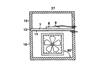
Figure 5 is a block diagram of the thermal cycler of the invention.
Shown are the thermal cycling device 27, a user's keyboard and display 22a and
16b, and a computer 23/power supply 26. Also shown are the control for cooling
32, control for heating 31, an analog to digital converter 28, a cable 30 and
a
connector 33.
Figure 6 is a graph of time versus temperature for a representative
slide. The slide was heated to 93.6°C, maintained at that temperature
for
60 seconds, cooled by active convection for 60 seconds, and then cooled by
passive convection for 60 seconds. The plot reveals that the temperature of
the
representative slide increases at a rate > 1°C/second, maintained
within 0.5°C of
the target temperature, cooled at a rate > 0.7°C/second by active
convection, and
then cooled at a rate <0.20°C/second by passive convection.
The invention provides a thermal cycling device comprising a
ceramic sample plate or block. The ceramic sample plate increases the speed
and
AMEi~~D~.~' SJ~E~'
f~--L,~'=~'
CA 02283082 1999-08-30
WO 98/39479 PCT/US98/04041
16
reliability of in situ PCR performed on a biological sample attached to a
substantially flat substrate. The invention is an improvement over
commercially
available thermal cycling devices as the sample plate of the invention
accelerates
and renders more uniform the heat transfer which occurs during thermal
cycling.
S
As used herein, a "substantially flat substrate" means a material on
which isolated nucleic acid, polypeptide or protein, or intact cells or
tissues, can be
maintained for an indefinite period of time. Thus, materials such as plastic,
glass,
nitrocellulose, nylon and the like are substantially flat substrates within
the scope of
the invention. Plastics useful as substrates include, but are not limited to,
polystyrene, polypropylene, polycarbonate, polyethylene and the like.
As used herein, the term "biological sample" includes isolated and/or
purified nucleic acid or polypeptide, or intact cells present in a specimen or
sample
obtained from any prokaryotic or eukaryotic organism, e.g., blood or a biopsy
sample from a mammal. More than one biological sample may be present on any
one substantially flat substrate. A preferred biological sample is a mammalian
tissue section. As used herein, the terms "isolated and/or purified" refer to
in vitro
isolation of a nucleic acid or polypeptide molecule from its natural cellular
environment, and from association with other components of the cell, such as
nucleic acid or protein.
As used herein, the term "ceramic" means a compression-resistant,
heat-resistant, corrosion-resistant substance prepared by firing terrestrial
minerals
such as clay, corundum and the like, which comprise one or more metals in
combination with a non-metal, generally oxygen. Ceramics within the scope of
the
invention include, silica, alumina, silicon carbide, zirconium oxide and boron
nitride. Alumina is a preferred ceramic for use in the devices and methods of
the
invention, as alumina conducts heat more efficiently than other ceramics.
"PCR" refers to a process of amplifying one or more specific nucleic
acid sequences, wherein 1) oligonucleotide primers which determine the ends of
the
CA 02283082 1999-08-30
WO 98/39479 PCT/US98/04041
17
sequences to be amplified are annealed to single-stranded nucleic acid in a
test
sample, 2) a nucleic acid polymerise extends the 3' ends of the annealed
primers to
create a nucleic acid strand complementary in sequence to the nucleic acid to
which
the primers were annealed, 3) the resulting double-stranded nucleic acid is
denatured
to yield two single-stranded nucleic acids, and 4) the processes of primer
annealing,
primer extension, and product denaturadon are repeated enough times to
generate
easily identified and measured amounts of the sequences defined by the
primers.
Practical control of the sequential annealing, extension, and denaturation
steps is
exerted by varying the temperature of the reaction container, normally in a
repeating
cyclical manner. Annealing and extension occur optimally in about the
35°C to
80°C, preferably about the 40°C to 75°C, temperature
range, whereas denaturation
requires temperatures in about the 80°C to 100°C range.
While a single primer pair is most often employed in PCR, a single
primer ("one-sided PCR"), multiple primers ("multiplex PCR"), degenerate
primers,
and nested primers may also be employed in the methods of the invention.
Moreover, in addition to amplification of DNA, the device and method of the
invention can be employed for RT-PCR, i.e., reverse transcription of an RNA
molecule to produce a single stranded cDNA with subsequent PCR of the cDNA.
PCR specificity may be increased by omitting at least one reagent
necessary for PCR until the sample temperature is between 50-80°C ("Hot
StartTM"),
the addition of a reagent which interferes with nonspecific polymerise
reactions
(e.g., SSB), or the addition of a modified nucleotide (e.g., dUTP) and the
corresponding glycosylase (e.g., UNG) into the reaction mixture. See U.S.
Patent
No. 5,538,871, the disclosure of which is incorporated by reference herein.
"Thermal cycling" commonly is automated by a "thermal cycler," an
instrument which rapidly (on the time scale of one to several minutes) heats
and
cools a "sample compartment," a partly or completely enclosed container
holding
the vessel, e.g., a microcentrifuge tube, or flat substrate, a microscope
slide, on
which nucleic acid amplification occurs and the heat-transfer medium directly
contacting the PCR vessel or flat substrate. Most commonly, the sample
CA 02283082 1999-08-30
WO 98/39479 PCT/US98/04041
18
compartment is a "sample block," which can be temperature controlled. While
conventional sample blocks are manufactured from metal and contain wells
designed to fit tightly the plastic microcentrifuge tubes in which PCR
amplification
normally is performed, the sample plate of the present invention is
manufactured
from ceramic, preferably alumina, and replaces some or all of the conical
wells in
conventional thermal cyclers with a flat surface or slots designed to optimize
heating
and cooling of a biological sample, preferably a sample on a flat substrate,
e.g., a
tissue section on a microscope slide, although it is envisioned that a ceramic
sample
plate of the invention may be manufactured to hold or support other shaped
vessels,
e.g., microcentrifuge tubes.
"PCR reagents" refers to the chemicals, apart from the biological
sample, needed to make nucleic acid amplification work. The reagents consist
of
five classes of components: ( 1 ) an aqueous buffer, (2) a water-soluble
magnesium
salt, (3) at least four deoxyribonucleoside triphosphates (dNTPs), although
these can
1 S be augmented or sometimes replaced by dNTPs containing base analogues
which
Watson-Crick base-pair like the conventional four bases, such as the analog
deoxyuridine triphosphate (dU'TP) and dUTP carrying molecular tags such as
biotin
and digoxigenin, covalently attached to the uracil base via spacer arms, (4)
oligonucleotide primers (normally two for each target sequence, with sequences
which define the 5' ends of the two complementary strands of the double-
stranded
target sequence), and (5) a polynucleotide polymerase, preferably a DNA
polymerase, most preferably a thermostable DNA polymerase, which can tolerate
temperatures between 90°C and 100°C for a total elapsed time of
at least 10 minutes
without losing more than about half of its activity.
"Southern analysis" or "Southern blotting" is a method by which the
presence of DNA sequences in a restriction endonuclease digest of DNA or DNA-
containing composition is confirmed by hybridization to a known, labeled
oligonucleotide or DNA fragment. Southern analysis typically involves
electrophoretic separation of DNA digests on agarose gels, denaturation of the
DNA
after electrophoretic separation, and transfer of the DNA to nitrocellulose,
nylon, or
CA 02283082 1999-08-30
WO 98/39479 PCT/US98/04041
19
another suitable membrane support for analysis with a radiolabeled,
biotinylated, or
enzyme-labeled probe, i.e, "Southern hybridization," as described in sections
9.37-
9.52 of Sambrook et al., supra.
"Northern analysis" or "Northern blotting" is a method used to
identify RNA sequences that hybridize to a known probe such as an
oligonucleotide,
DNA fi~agment, cDNA or fragment thereof, or RNA fragment. The probe is labeled
with a radioisotope such as'zP, by biotinylation or with an enzyme. The RNA to
be
analyzed can be usually electrophoretically separated on an agarose or
polyacrylamide gel, transferred to nitrocellulose, nylon, or other suitable
membrane,
and hybridized with the probe, i.e, "Northern hybridization,", using standard
techniques well known in the art such as those described in sections 7.39-7.52
of
Sambrook et al., supra.
"Fixed cells" refers to a sample of cells which has been chemically
treated to strengthen cellular structures, particularly membranes, against
disruption
by solvent changes, temperature changes, mechanical stresses, and drying.
Cells
may be fixed either in suspension or while contained in a sample of tissue,
such as
might be obtained during autopsy, biopsy, or surgery. Cell fixatives generally
are
chemicals which crosslink the protein constituents of cellular structures,
most
commonly by reacting with protein amino groups. Preferred fixatives are
buffered
formalin, 95% ethanol, formaldehyde, paraformaldehyde, or glutaraldehyde.
Fixed
cells also may be treated with proteinases, enzymes which digest proteins, or
with
surfactants or organic solvents which dissolve membrane lipids, in order to
increase
the permeability of fixed cell membranes to PCR reagents. Such treatments must
follow fixation to assure that membrane structures do not completely fall
apart when
the lipids are removed or the proteins are partially cleaved. Protease
treatment is
preferred following fixation for more than one hour and is less preferred
following
shorter fixation intervals. For example, a ten-minute fixation in buffered
formalin,
without protease treatment, is standard after suspended cells (e.g., from
blood) have
been deposited centrifugally on a slide by cytospin procedures standard in the
cytochemical art.
CA 02283082 1999-08-30
WO 98/39479 PCT/US98/04041
A preferred mode of fixing cell samples for in situ PCR according to
the present invention is to incubate them in 10% formalin, 0.1 M Na phosphate,
pH 7.0, for a period of 10 minutes to 24 hours at room temperature. The cells
may
be a suspension, as would be obtained from blood or a blood fraction such as
buffy
S coat, or may be a solid tissue, as would be obtained from biopsy, autopsy,
or
surgical procedures well known in the art of clinical pathology. If PCR is to
be
performed in cell suspension, suspended cells preferably are centrifuged after
formalin fixation, resuspended in phosphate-buffered saline, and re-
centrifuged to
remove the fixative. The washed, pelleted cells may be resuspended in PCR
buffer
10 and added directly to a PCR tube. If PCR is to be performed on a microscope
slide,
suspended cells preferably are deposited on the slide by cytospin, fixed 10
minutes
in buffered formalin, washed 1 minute in water, and washed 1 minute in 95%
ethanol. Alternatively, suspended cells can be pelleted in a centrifuge tube
and the
pellet can be embedded in paraffin and treated like a tissue specimen. Tissue
1 S samples may be processed further and then embedded in paraffin and reduced
to
serial 4-5 ~m sections by microtome procedures standard in the art of clinical
pathology. Histochemical sections are placed directly on a microscope slide.
In
either case, the slide preferably will have been treated with 2% 3-
aminopropyltriethoxysilane in acetone and air dried. After smears ~or sections
have
20 been applied to slides, the slides are heated at about 60°C for
about 1 hour.
Paraffin-embedded sections can be deparaffinized by 2 serial 5 minute washes
in
xylene and 2 serial S minute washes in 100% ethanol, all washes occurring at
room
temperature with gentle agitation.
"Histochemical section" refers to a solid sample of biological tissue
which has been frozen or chemically fixed and hardened by embedding in a wax
or a
plastic, sliced into a thin sheet, generally several microns thick, and
attached to a
microscope slide.
"Cytochemical smear" refers to a suspension of cells, such as blood
cells, which has been chemically fixed and attached to a microscope slide.
CA 02283082 1999-08-30
WO 98/39479 PCT/US98/04041 .._,_
21
"Vapor barrier" refers to an organic material, in which water is
insoluble, which covers a PCR reaction or preparation in a way which
substantially
reduces water loss to the atmosphere during thermal cycling. Preferred vapor
barrier
materials are liquid hydrocarbons such as mineral oil, or paraffin oil,
although some
synthetic organic polymers, such as fluorocarbons and silicon rubber, also may
serve as effective PCR vapor barriers. Waxes which are solid at temperatures
below
about 50°C and liquid at higher temperatures also make convenient vapor
barriers.
To isolate the PCR reagents from the atmosphere and from the vapor
barrier, a thin, "water-impermeant barrier" such as a plastic or glass film,
e.g., a
glass cover slip or a polypropylene cover slip, is placed over the liquid
filin which
comprises the PCR reagents. The water-impermeant barrier is generally attached
to
the microscope slide. For example, a cover slip can be placed over the liquid
filin
and sealed to the microscope slide with nail polish or a similar adhesive. See
Komminoth et al., DiaQnottic Molecular Pa h~,1(2), 85-9 (1992). The cover
slip can also be clipped to the slide. See U.S. Patent No. 5,527,510.
Alternatively,
a gasket can be placed between the cover slip and a chambered slide, which
contains
the PCR reagent, sealed with 2.5% hot agarose and the assembly covered with
saran
wrap. See, Chiu et al., Histochem_ and C o .h m_, 4Q, 333-341 (1992). However,
any other fastening mechanism may be employed to attach the cover slip to a
microscope slide, such as the use of other high temperature resistant
adhesives.
"Detection" of PCR-amplified nucleic acid refers to the process of
observing, locating, or quantitating an analytical signal which is inferred to
be
specifically associated with the product of PCR amplification, as
distinguished from
PCR reactants. The analytical signal can result from visible or ultraviolet
absorbance or fluorescence, chemiluminescence, or the photographic or
autoradiographic image of absorbance, fluorescence, chemiluminescence, or
ionizing radiation. Detection of in situ PCR products involves microscopic
observation or recording of such signals. The signal derives directly or
indirectly
from a molecular "tag" attached to a PCR primer or dNTP or to a nucleic acid
probe,
which tag may be a radioactive atom, a chromophore, a fluorophore, a
CA 02283082 1999-08-30
WO 98/39479 PCT/US98/04041
22
chemiluminescent reagent, an enzyme capable of generating a colored,
fluorescent,
or chemiluminescent product, or a binding moiety capable of reaction with
another
molecule or particle which directly carries or catalytically generates the
analytical
signal. Common binding moieties are biotin, which binds tightly to
streptavidin or
avidin, digoxigenin, which binds tightly to anti-digoxigenin antibodies, and
fluorescein, which binds tightly to anti-fluorescein antibodies. The avidin, .
streptavidin, and antibodies are easily attached to chromophores,
fluorophores,
radioactive atoms, and enzymes capable of generating colored, fluorescent, or
chemiluminescent signals.
"Nucleic acid probe" refers to an oligonucleotide or polynucleotide
containing a sequence complementary to part or all of the PCR target sequence,
also
containing a tag which can be used to locate cells in an in situ PCR
preparation
which retains the tag aRer mixing with nucleic acid probe under solvent and
temperature conditions which promote probe annealing to specifically amplified
nucleic acid.
I?evice of t-he Invention
The invention provides a thermal cycler comprising a ceramic sample
plate which is optimized for heat flow to and from biological samples attached
or
affixed to a substantially flat substrate, e.g., a microscope slide, present
on the upper
surface of the sample plate. For in situ PCR applications where very few
slides are
to be run simultaneously, the top surface is designed to create flat
horizontal areas
large enough to hold slides so that the large dimensions (height and width)
are
horizontal. These flat areas may be recessed in shallow wells, which may
optionally
hold a vapor barrier that covers the slides, or which physically isolate one
substrate
from another. For microscope slides, the area is at least about 16 mm wide and
77
mm long to fit conventional glass microscope slides. The wells are at least
about
2 mm deep to fit a slide and cover slip and optionally a vapor barrier.
For in situ PCR applications where a large number of samples each
affixed to a substantially flat substrate such as a microscope slide are to be
run
CA 02283082 1999-08-30
WO 98/39479 PCT/US98/04041
23
simultaneously, the ceramic sample plate may be designed to contain many
narrow,
deep, vertical or approximately vertical slots, sized to hold slides inserted
edgewise
with minimal space separating the slide from the ceramic surfaces facing the
top and
bottom surfaces of the slide. The intervening space normally is filled with
mineral
oil or another nonvolatile liquid to provide a vapor barrier and efficient
heat transfer
during thermal cycling. However, because the heat transfer between a flat
sample
plate and a flat substrate is more efficient, a vapor barrier may be optional
for some
applications. The plane of a slot may be inclined from the vertical by as much
as
about 45° in order to use the force of gravity to assure that one
surface of the slide
touches the ceramic of the sample plate. Slots must be about 15 mm deep, at
least
77 mm long, and at least 2 mm wide to fit a conventional slide plus a cover
slip.
This design is not compatible with manual addition of missing PCR reagents)
because it blocks rapid access to the in situ PCR preparation for cover slip
removal,
manual addition of the missing PCR reagent(s), and cover slip replacement.
The ceramic sample plate 7 can include both wells optimized for
biological samples present on a substantially flat substrate, e.g., a
microscope slide,
and wells designed to hold conventional nucleic acid amplification reaction
tubes,
e.g., 0.5 ml microcentrifuge tubes. Preferably, the reaction tube wells occupy
one or
several rows along the edges of the sample plate, reserving the central area
of the
sample plate for microscope slide wells.
It is also envisioned that the ceramic sample plate of the invention
may be prepared so as to replace the top surface of a sample plate present in
a
commercially available thermal cycler, leaving the other design features
(except
possibly plate or block thickness) substantially unchanged in order to
minimize the
impact of the invention on thermal cycler manufacture and performance. It is
also
envisioned that the ceramic sample plate of the invention is equal in mass to
the
conventional sample block of a commercially available thermal cycler, to
minimize
impact on heating and cooling kinetics.
To prepare a ceramic sample plate 7, the sample plate may be
manufactured by machining a single ceramic plate, for example with a rotary
mill,
CA 02283082 1999-08-30
WO 98/39479 PCT/L1S98/04041
24
exact dimensions, wells, and other contours needed to integrate with the rest
of the
thermal cycler. Holes for bolting the plate to the rest of the thermal cycler
may be
made with a drill press. The rectilinear shape of wells adapted to fit
microscope
slides may also be produced by stamping or machining of relatively thin sheets
of
ceramic which are bolted together to create a laminated assembly. The entire
plate
may be laminated; or just the top portion, holding the microscope slide wells,
oan be
laminated and bolted to a solid bottom portion which contains the features of
the
plate which integrate with the rest of the thermal cycler.
The device of the invention 21 is preferably enclosed in a housing or
body which comprises a lower hollow compartment 16 and an upper hollow
compartment (the lid; 9). Although the two compartments 9 and 16 may be formed
in any suitable, compatible and practical shape, they are preferably box-
shaped.
Each compartment comprises a pair of sidewalk 20a and 20b and a pair of
endwalls
21 a and 21 b. The lid 15 also comprises a substantially flat upper surface 24
attached to the sidewalls and endwalls of the lid. The lower compartment 16
comprises a substantially flat lower swface 25 the outer swface on which,
preferably, are feet. The lower swface 25 comprises an inlet opening 18 for
ambient
air intake. The lower swface 25 of the lower compartment is attached to the
sidewalls 20a and 206 and endwalls 21a and 21b of the lower compartment 16.
The
sidewalls 20a and 20b and/or endwalls 21a and 21b of the lower compartment 16
have at least one outlet opening 19.
The housing may be fabricated from any available material, e.g., a
plastic, metal, such as stainless steel, ceramic, glass or combinations of any
of the
foregoing materials. However, it is preferred that the material be plastic,
such as
polypropylene or polycarbonate or the like, so that the housing may be molded
in an
inexpensive fashion. Moreover, it is preferred that the walls of the housing,
including sidewalls 20a and 20b, endwalls 21a and 21b, lower surface 24, and
upper
surface 25, be relatively thin in dimension in order to provide a housing with
low
thermal mass. The most straightforward, but not necessarily limitative,
construction
of housing is one in which all of the walls are of the same relative
thickness.
CA 02283082 1999-08-30
WO 98/39479 PCT/LTS98/04041
The lower compartment 16 comprises a ceramic sample plate 7,
which provides mechanical support and a heat exchange element for the flat
substrates. Preferably, the ceramic sample plate comprises alumina (Hoechst
Ceramic North America Inc., Mansfield, MA; Coors Ceramics Co., Golden, CO)
5 The outer margins of the sample plate 5 and 6 may lie on the outer and
uppermost
margins of the lower compartment 16, or may be affixed, mounted or attached to
the
inner sidewalk 20a and 20b and endwalls 21a and 21b of the lower compartment
16
by, for example, a support bracket. The ceramic sample plate 7 may be
substantially
flat, or may comprise a plurality of recessed rectilinear wells for microscope
slides.
10 It is preferred that the wells in the sample plate may include sidewalls
which are
integrally formed in, and from the same material as, the ceramic sample plate
7.
Moreover, the wells are preferably configured to hold the slides, or other
substantially flat substrate, in relatively tight contact with sidewalls of
the wells, to
facilitate optimum conduction of heat to and from the slides.
15 Reference is now made to the drawings, which describe preferred
embodiments of the invention, but are not intended to limit the invention to
the
embodiments shown. As shown in Figure 1, a ceramic sample plate 7, preferably
an alumina sample plate, is dimensioned so as to accommodate 6 microscope
slides.
However, the ceramic sample plate may be fashioned so as to accommodate fewer
20 or greater than 6 substantially flat substrates. The upper surface of one
representative substantially flat substrate, e.g., a microscope slide 8, is
attached to a
thermosensor 9. The other substantially flat substrates 4 each comprise at
least one
biological sample on their upper surface. The outer edges or margins of the
surface
of the ceramic sample plate 5 and 6 are useful for placing the lower edges of
a lid 15
25 over the slides during thermal cycling.
The thermosensor 9 is an integrated circuit which provides an output
current that is directly proportional to temperature (K°) (AD592 or
AD590 from
Analog Devices, Norwood, MA). The therrnosensor 9 thus provides an electrical
input signal to the microcomputer or microprocessor 23 which corresponds to
the
temperature of the representative substantially flat substrate 2 on the sample
plate 7.
CA 02283082 1999-08-30
WO 98139479 PCT/CTS98/04041
26
Temperature monitoring during operation of the thermal cycling device of the
present invention is preferably achieved using a type K thermocouple (COI-K;
Omega Engineering, Inc., Stamford, CT) or a 100 S2 resistance temperature
device
(F3101; Omega Engineering, Inc., Stamford, CT). The controller uses this
information to regulate the heating means 9 and cooling means 11 according to
predetermined temperature versus time profiles probed therein.
Figure 2 illustrates an exemplary heating means 14 for the ceramic
sample plate 7. The heating means is preferably an etched foil type heater
(HIC
5468 893.8 L12A; MINCO Products, Minneapolis, Ml~ which is preferably glued
to the ceramic sample plate 7. However, any heating unit suitable for heating
the
ceramic sample plate may be used. The heating means is activated by an output
relay 13 attached to the microcomputer or microprocessor 23. Preferably, the
relay
is Crydom A1202 purchased from Allied Electronics, Fort Worth, TX).
Figure 3 illustrates a side view of the thermal cycling device of the
invention. The lid 9 can be opened to allow access to the ceramic sample plate
7.
To cool the ceramic sample plate 7, the heating means 9 is deactivated and the
fan
17 is activated. Air from outside the housing is drawn into the lower
compartment
16 though an inlet opening 18 by the fan 17 which is connected to a motor
shaft
driven by a motor (not shown). The fan 17 is mounted to the interior surface
of the
lower wall of the lower compartment, although other mounting arrangements are
envisioned. The lower surface has a inlet opening 18. There is at least one
other
opening 19 in the sidewall 20a or 20b or the endwall 21a or 21b of the lower
compartment 16. Thus, the present invention may have two such openings, but
the
present invenrion is not limited to two since the number of openings may vary,
depending upon the design and configuration of the housing. These openings
provide communication between interior of the housing and the outside
environment, so that air may be moved into and out of the hollow interior of
the
lower compartment, according to the present invention.
The fan assembly preferably employs a propeller type fan due to its
generally low thermal mass, or if desired, a squirrel cage type fan, the fan
preferably
CA 02283082 1999-08-30
WO 98/39479 PCT/US98/04041
27
having at least about 40, more preferably at least about 50, and even more
preferably
at least about 60 cubic feet per minute minimum capacity. The fan 17 draws
ambient temperature air through the inlet opening 18 into the hollow interior
of the
lower compartment, and forces the air against the heating means 14. The air is
S dispersed through outlet or exit openings 19 in the endwall or sidewalls of
the lower
compartment. Operation of the fan 17 allows the sample plate 7 in to be
brought to
a lower predetermined temperature as quickly as possible. Thus, due to the
minimum thermal mass of the sample plate 7, and the action of the fan 17, vast
quantities of air are forced against the heating means 14 and from there out
of the
hollow interior of the outlet openings 19 in the lower compartment 16. Thus,
rapid
cooling of substantially flat substrates on the sample plate is obtained.
Moreover,
the combination of heating and cooling means together allow the flat
substrates to
be maintained at a particular temperature.
The fan motor (not shown) is located externally of housing. It
1 S would be disadvantageous to mount the motor within the chamber which would
subject the motor to temperature variations and also would add the thermal
mass of
the motor to that which is subject to heating and cooling. For example, a
Comair
FT12M3 fan purchased from Digi-Key Corporation (Thief River Falls, MN;) can be
employed in the device of the invention, although other cooling devices and
fans
well known to the art may be employed in the practice of the invention.
Figure 4 illustrates an alternative embodiment in which the fan 17
assembly is placed at an angle to the heating means 14.
Figure S is a block diagram of the invention. A microcomputer or
microprocessor 23 can be programmed by means of input keys 16a and display 16b
to cause the substantially flat substrate on the ceramic sample plate 7 to be
cycled
through a series of temperatures over a predetermined period of time. Although
not
specifically illustrated in the drawings, it is contemplated that the device
of the
invention would include, as appropriate, timing mechanisms, electronic or
otherwise, for maintaining time intervals for each cycle, and for counting the
number of repetitions.
CA 02283082 1999-08-30
WO 98/39479 PCT/US98/04041
28
The microcomputer or microprocessor 23 is electrically attached to a
relay controller 29 by means of a transmission cable 30. This controller 29
regulates
the supply of power 20 to the heating means 14. It also regulates the supply
of
power 21 to the fan blower motor (not shown). A preferred controller is
available
S from 3BR Electronic Systems, Inc. ( Baltimore, MD; ECP2). The cable also
supplies power to the blower motor (not shown), and to the heating means 14. .
The microcomputer or microprocessor 23 also is connected to an
electronic sensing device which is an analog to digital converter 28 that is
connected
to the temperature sensor 9. A preferred converter 28 is the DAS-TEMP,
available
from Keithley Metrabyte (Taunton, MA). The microcomputer or microprocessor 23
can be any well-known type of temperature controller unit which is
programmable
to control the heating means 14 and fan motor so as to achieve predetermined
temperatures as a function of time on the substantially flat substrates
present on the
ceramic sample plate 7.
When the device of the present invention is used for cyclic DNA
amplification, repetitive cycling through a temperature versus time profile is
required. Samples containing a reaction mixture for the polymerise chain
reaction
generally must be cycled approximately 30-40 times through a temperature
versus
time profile which corresponds to the denaturation, annealing and elongation
phases
of the amplification process. Figure 6 illustrates, in graphic form, the
temperature
profile of a microscope slide undergoing thermal cycling. It can be seen that
the
slide reached a temperature of approximately 94°C on the hot cycle, and
then was
rapidly cooled down to about 44°C by active convection on the cold
cycle. It can be
seen that active convection, relative to passive convection, has a
substantially more
rapid rate of decrease of temperature on the cold cycle. As a result of use of
the
present invention, it is possible to realize temperature increases of the flat
substrate
of at least about 1.0°C/sec or greater, and temperature decreases of
the flat substrate
of at least about 0.7°C/sec or greater.
Method of he nvention
CA 02283082 1999-08-30
WO 98/39479 PCT/US98/04041
29
To amplify nucleic acid sequences in a biological sample, such as a
histochemical section or cytochemical smear attached to a microscope slide,
the
section or smear on the microscope slide is preferably covered with about 5 to
25 pl,
more preferably about 5 to 10 pl, of a PCR reagent mixture. Preferably, the
PCR
reagent mixture lacks at least one reagent, such as enzyme. Then a plastic
cover slip
is placed over the preparation, the microscope slide is placed on a ceramic
thermal
cycler sample plate. After the sample plate is brought to about 80°C
and held at that
temperature, the cover slip is lifted and 2 to 10 ul of PCR buffer containing
the
missing reagents) are distributed across the surface of the reagent mixture.
The
cover slip is replaced, and the slide is covered with enough mineral oil to
assure that
the cover slip, including their edges, is protected from the atmosphere.
Preferably,
the oil has been pre-heated, so that its addition does not transiently reduce
the
temperature of the in situ PCR preparation. Then a standard two-temperature or
three-temperature thermal cycle is run for about 40 cycles. Cycle parameters,
e.g.,
1 S number of cycles, and PCR reagent concentrations are optimized by methods
well
known to the art.
After amplification, the mineral oil is removed from the slide with an
organic solvent such as xylene, and the slides are dried with 100% ethanol or
a
graded series of ethanol concentrations. The oil-free preparation is incubated
for
approximately 15 minutes at about 50°C in 0.15 M NaCI, 0.01 S M Na
citrate,
pH 7.0 to remove unreacted PCR reagents.
The detection phase of in situ PCR employs two basic detection
strategies. The first strategy involves tagging either the PCR primers or at
least one
of the dNTPs with a radioisotope or with a binding moiety such as biotin,
digoxigenin, or fluorescein, or with another fluorophore. In this case, tag
incorporated into amplified nucleic acid can be analyzed directly, provided
that the
unreacted tagged reagent has been washed out post-PCR and provided that the
washing and drying procedure has not mobilized the amplified nucleic acid from
its
point of synthesis. The analytical validity of this simple detection strategy
requires
that the invention has increased in situ PCR specificity sufficiently that
negligible
CA 02283082 1999-08-30
WO 98/39479 PCTlUS98/04041
nonspecific products have been made which are large enough to resist washing
from
the preparation.
To test and validate this consequence of the first three aspects of the
invention, appropriate control reactions can be performed. The logically most
5 compelling control reaction is to perform the procedure on cells known to
lack the
target sequence; validation of the simplified detection strategy requires that
no
signal be generated in the control cells. Often such control cells are present
in a
histochemical or cytochemical preparation, so that the standard analysis
contains its
own control. A less compelling control is to use primers which differ
sufficiently
10 from the optimal primers for the target sequence that they will not amplify
the target
sequence under the specified annealing and extension conditions.
The second strategy involves detecting amplified nucleic acid by in
situ hybridization to a tagged nucleic acid probe: an oligonucleotide or
polynucleotide with a sequence complementary to at least part of the amplified
15 nucleic acid sequences (preferably excluding the primer sequences). In situ
hybridization, well known in the histochemical and cytochemical art, has four
basic
steps: denaturation of DNA in the test sample, annealing of probe to test
sample
nucleic acid under stringent conditions, wash of the microscope slide with a
solvent
under stringent conditions to remove unhybridized probe, and detection of the
probe
20 which has been retained on the slide.
Regardless of which detection strategy is used, the methods for
observing and recording the presence and location of tag on the microscope
slide are
the same. If the tag is a radioisotope (preferably a strong beta radiation-
emitter,
such as'2P or'ZSI), the microscope slide is coated with nuclear track emulsion
such
25 as NTB-2 from Eastman Kodak Co. (Rochester, N.Y.), incubated at 4°C
for an
interval determined by trial and error, and developed by standard methods to
leave
microscopically detectable silver grains in the vicinity of immobilized tags.
Procedures for'ZSI tagging probe or PCR product are described by Haase et al.,
Proc.
l~3ti. Acad. Sci L1~A, $2, 4971 (1990), incorporated herein by reference.
CA 02283082 1999-08-30
WO 98/39479 PCT/US98/04041
31
If the tag is a fluorophore, it may be observed directly in a
fluorescence microscope with excitation and emission filters optimized for the
particular fluorophore. This detection method is particularly suitable for
multiplex
in situ PCR with different primer pairs for different target nucleic acid
sequences.
Either different fluorophores can be attached to primers of different
specificity, or
different fluorophores can be attached to probes of different specificity.
Methods of
attaching fluorophores to oligonucleotides and polynucleotides, preferably at
their S'
ends, are well known in the nucleic acid chemistry and PCR arts.
If the tag is a binding moiety such as biotin or digoxigenin, it is
incorporated directly into PCR product (via primers or dNTPs) or into probes
by
essentially the same methods used to attach other tags. However, in this case,
signal
generation requires additional detection steps.
Preferably, the microscope slide is incubated in buffered aqueous
solvent containing a covalent conjugate of a detection enzyme and a binding
protein
1 S specific for the tag (avidin or streptavidin for biotin; an anti-
digoxigenin antibody
for digoxigenin, an anti-fluorescein antibody for fluorescein). The preferred
detection enzyme is horseradish peroxidase or alkaline phosphatase. After
unbound
enzyme conjugate is removed by washing in a buffered aqueous solvent, the
microscope slide is immersed in a solution containing a chromogenic substrate
for
the enzyme used. After an insoluble dye, product of the enzyme reaction, has
been
deposited at points on the microscope slide where enzyme conjugate has been
bound, unreacted substrate is washed away in water or buffered aqueous solvent
to
prevent the buildup of nonspecific background stain over time. The preferred
chromogenic substrates which generate insoluble products are well known in the
histochemical and cytochemical art, as are the methods for staining and for
enzyme
conjugate incubation and washing. The substrates and enzyme conjugates are
commercially available from a wide variety of sources well known to
histochemists
and cytochemists.
A preferred companion procedure in the detection steps of the present
invention is counterstaining of the microscope slide with fluorescent dyes
(for
CA 02283082 1999-08-30
WO 98/39479 PCT/US98104041 .~
32
fluorescent tags) or chromophoric dyes (for radio-autoradiographic detection
or
enzymatic generation of insoluble chromophores) which emit or absorb with
different spectral characteristics than the analyte-specific signals and which
highlight cell structures, especially in cells which lack target nucleic acid
sequence.
Especially preferred for examination of insoluble blue dye deposits by
transmission
microscopy is counterstaining by nuclear fast red, standard in the
histochemical and
cytochemical art. The methods for examining stained in situ PCR preparations
by
transmission or fluorescence microscopy are well known in the histochemical
and
cytochemical art, as are methods of recording permanently the microscopic
image
photographically or via digitized video images.
The invention will be fiuther described by the following examples.
EXAMPLE 1
A thermal cycler of the invention 27 may include the following
components. The housing, comprising a lid 15 and a lower hollow compartment
16,
is constructed from polystyrene, polypropylene, polyethylene or other plastics
having appropriate thermal and electrical conductances. The ceramic alumina
plate
1 is about 6.5" long, about 3.5" wide, and about 0.025" thick. The microscope
slides are about 3.0" long, about 1.0" wide, and about 0.125" thick. The
heater 14 is
of the etched foil type, and is electrically insulated with a thin film of
Kapton or
similar substance. The fan 17 may be powered by alternating current or direct
current. The impeller blades of the fan may be constructed from plastic or
metal.
The fan 17 and the heater 14 are controlled by electrical switches of
the relay type. The relays can be of the solid state or mechanical varieties.
The computer or controller 23 can be a commercial microcomputer
or a self contained microprocessor. A microprocessor can be incorporated into
the
control electronics of the apparatus by methods well known to the art. The
microprocessor executes commands written in software that collect user input
via
the keyboard, compare the input to actual temperatures, and turn off or on the
heating 8 or cooling 11 units as appropriate. The electronics may also include
a
CA 02283082 1999-08-30
WO 98/39479 PCT/US98/04041
33
timer, readable by the microprocessor. This allows the microprocessor to
compare
the elapsed time that the reaction mixture has been at a particular
temperature and
compare it to a desired time input by the user.
The temperature sensor 9 can be of the thermocouple type, or the
thermistor type, or the resistance temperature detector type, or the current
detector
type. In each of these devices, a change in temperature at the interface
between.the
sensor and its environment produces a change in the ability of the sensor to
conduct
electrical current. The sensors generate electrical signals that are
proportional to the
extent of the temperature change. The temperatures of the experimental slides
10
are taken by the thermosensor 9 as the temperature of the representative slide
2. A
representative time versus temperature plot for the thermal cycler described
above is
shown in Figure 6. The difference between the rates of active and passive
convection illustrates that a cooling means, e.g., a fan, is required for the
effective
performance of the invention.
1 S Communication between the computer 23 and the temperature sensor
9 is maintained by an electrical device known as an analog to digital
converter. This
device takes the electrical signal produced by the temperature sensor and
converts it
to a form that the circuitry of the computer can evaluate. Different types of
temperature sensors require different specialized types of analog to digital
converters.
Communication between the computer 23 and the relays is
maintained by switching devices. These devices respond to signals from the
computer by producing an altered electrical signal that causes a response in
the
relay.
The computer program, implemented in a combination of assembly
language and the C language, although other programming languages may be used,
causes the computer 23 to evaluate the temperature received from the
temperature
sensor 9, compare this value to the "target" temperature, and send appropriate
electrical signals to the relays controlling the heater 14 and the fan 17.
Specific
CA 02283082 1999-08-30
WO 98/39479 PCT/US98/04041 _
34
patterns of signals from computer 23 to relays provide the means by which the
representative slide is heated, cooled, or maintained at a steady temperature.
EXAMPLE 2
Cells of the stable human cervical cancer cell line, SiHa (ATCC HTB
35), containing one integrated copy of human papilloma virus (HPB) type 16 .
genome per human genome, are grown to density of about 105 cells/mL in Eagle's
minimal essential medium with non-essential amino acids, sodium pyruvate, and
15% fetal bovine serum, washed two times in Tris-buffered saline, adjusted to
an
approximate density of 10° cells/mL, and stirred overnight at room
temperature in
10% (vol/vol) formaldehyde in phosphate buffer. The formaldehyde-fixed cells
are
centrifuged at 2,000 rpm for 3 minutes, and the pellet is embedded in
paraffin.
Microtome sections (4 ~m thickness) of the paraffin block are attached to
glass
microscope slides which had been dipped in 2% 3-aminopropyltriethoxysilane
(Aldrich Chemical Co.) in acetone by floating the sections in a water bath.
After attachment, sections are deparaffinized and proteolytically
digested with reagents from the Viratype~ in situ Tissue Hybridization Kit
(Life
Technologies, Inc., Gaithersburg, Md.) following the manufacturer's
instructions.
Slides are overlaid with 5 to 10 ~.1 of PCR solution (see below). A plastic
cover slip
is placed over each in situ PCR preparation. The cover slip is anchored to the
slide
with a drop of nail polish. The slide is placed on the sample plate 7 of the
thermal
cycler described in Example 1, and covered with approximately 1 ml of mineral
oil.
The pH 8.3 PCR solution contains 10 mM TrisCl, SO mM KCI,
4.5 mM MgCh, 20 mM of each dNTP, 0.2 unit/~L of AmpIiTaq~ DNA polymerase
(Perkin Elmer Cetus Instruments, Norwalk, Conn.), and 6 pM of each primer. The
primers employed are PV 1, PV2, PV3, PV4, PVS, PV6 and PV7 (see U.S. Patent
No. 5; PV1 S' CAGGACCCACAGGAGCGACC 3' (SEQ ID NO:1); PV2 S'
TTACAGCTGGGTTTCTCTAC 3' (SEQ >D N0:2); PV3 5' CCGGTCG
ATGTATGTCTTGT 3' (SEQ ID N0:3); PV4 5' ATCCCCTGTTTTTTTTTCCA 3'
(SEQ ID N0:4); PVS S' GGTACGGGATGTAATGGATG 3' (SEQ m NO:S); PV6
t
CA 02283082 1999-08-30
WO 98/39479 PCT/US98/04041 _
5' CCACTTCCACCACTATACTG 3' (SEQ 1D N0:6); PV7 5'
AGGTAGAAGGGCGCCATGAG 3' (SEQ >D N0:7)) which result in the
production of overlapping approximately 450 by PCR products. The predicted PCR
product is a 1247 by product.
5 For the first thermal cycle, denaturation is performed for 3 minutes at
94°C, and annealing/extension are performed for 2 minutes at
55°C; the remaining
39 cycles consist of 1 minute denaturation at 94°C and 2 minutes
annealing-
extension.
After DNA amplification, mineral oil is removed by dipping in
10 xylene, the cover slip is removed, and the mounted sections are dried in
100%
ethanol. Each slide is incubated with 10 pl of a 500 ng/ml solution of
biotinylated
HPV type 16 specific polynucleotide probe (Viratype Kit, Life Technologies,
Inc.)
in 0.03 M Na citrate, 0.30 M NaCI, pH 7.0, 5% dextran sulfate, 50% formamide
at
100°C for 5 minutes and then 37°C for 2 hours; then the slide is
treated with an
15 alkaline phosphatase-streptavidin conjugate and the phosphatase substrates,
S-
bromo-4-chloro-3-indolyl phosphate (BCIP) and nitro blue tetrazolium (NBT),
according to the instructions of the supplier of the S6800 Staining Kit
(Oncor,
Gaithersburg, Md.). After enzymatic detection of biotinylated probe captured
on the
sections, the sections are counterstained with nuclear fast red for 5 minutes.
20 When the stained slides are examined by transmission microscopy
under 40-400 X magnification, single-copy HPV targets in SiHa cells are
detectable
in most nuclei.
All publications and patents are incorporated by reference herein, as
25 though individually incorporated by reference, as long as they are not
inconsistent
with the present disclosure. The invention is not limited to the exact details
shown
and described, for it should be understood that many variations and
modifications
may be made while remaining within the spirit and scope of the invention
defined
by the claims.
CA 02283082 1999-08-30
WO 98/39479 PCT/US98104041
36
SEQUENCE LISTING
(1) GENERAL INFORMATION
(i) APPLICANT: Regents of the University of Minnesota
(ii) TITLE OF THE INVENTION: ALUMINA PLATE METHOD AND DEVICE FOR
CONTROLLING TEMPERATURE
(iii) NUMBER OF SEQUENCES: 7
(iv) CORRESPONDENCE ADDRESS:
(A) ADDRESSEE: Schwegman, Lundberg, Woessner & Kluth, P.A.
(B) STREET: P.O. Box 2938
(C) CITY: Minneapolis
(D) STATE: MN
(E) COUNTRY: USA
(F) ZIP: 55402
(v) COMPUTER READABLE FORM:
(A) MEDIUM TYPE: Diskette
(B) COMPUTER: IBM Compatible
(C) OPERATING SYSTEM: DOS
(D) SOFTWARE: FastSEQ Version 1.5
(vi) CURRENT APPLICATION DATA:
(A) APPLICATION NUMBER: Unknown
(B) FILING DATE: Herewith
(C) CLASSIFICATION:
(vii) PRIOR APPLICATION DATA:
(A) APPLICATION NUMBER: 08/810,641
(B) FILING DATE: 03-MAR-1997
(viii) ATTORNEY/AGENT INFORMATION:
(A) NAME: Embretson, Janet E
(B) REGISTRATION NUMBER: 39,665
(C) REFERENCE/DOCKET NUMBER: 600.383W01
(ix) TELECOMMUNICATION INFORMATION:
(A) TELEPHONE: 612-373-6959
(B) TELEFAX: 612-339-3061
(C) TELEX: N/A
(2) INFORMATION FOR SEQ ID NO:1:
(i) SEQUENCE CHARACTERISTICS:
CA 02283082 1999-08-30
WO 98/39479 PCT/US98104041
37
(A) LENGTH: 20 base pairs
(B) TYPE: nucleic acid
(C) STRANDEDNESS: single
(D) TOPOLOGY: linear
(ii) MOLECULE TYPE: other nucleic acid
(iii) HYPOTHETICAL: NO
(iv) ANTISENSE: NO
(v) FRAGMENT TYPE:
(vi) ORIGINAL SOURCE:
(xi) SEQUENCE DESCRIPTION: SEQ ID NO: l:
CAGGACCCAC AGGAGCGACC 20
(2) INFORMATION FOR SEQ ID N0:2:
(i) SEQUENCE CHARACTERISTICS:
(A) LENGTH: 20 base pairs
(B) TYPE: nucleic acid
(C) STRANDEDNESS: single
(D) TOPOLOGY: linear
(ii) MOLECULE TYPE: other nucleic acid
(iii) HYPOTHETICAL: NO
(iv) ANTISENSE: NO
( v ) FRAGMENT TYPE
(vi) ORIGINAL SOURCE:
(xi) SEQUENCE DESCRIPTION: SEQ ID N0:2:
TTACAGCTGG GTTTCTCTAC 20
(2) INFORMATION FOR SEQ ID N0:3:
(i) SEQUENCE CHARACTERISTICS:
(A) LENGTH: 20 base pairs
(B) TYPE: nucleic acid
(C) STRANDEDNESS: single
(D) TOPOLOGY: linear
(ii) MOLECULE TYPE: other nucleic acid
(iii) HYPOTHETICAL: NO
(iv) ANTISENSE: NO
(v) FRAGMENT TYPE:
(vi) ORIGINAL SOURCE:
(xi) SEQUENCE DESCRIPTION: SEQ ID N0:3:
CCGGTCGATG TATGTCTTGT 20
CA 02283082 1999-08-30
WO 98/39479 PCT/US98/04041
38
(2) INFORMATION FOR SEQ ID N0:4:
(i) SEQUENCE CHARACTERISTICS:
(A) LENGTH: 20 base pairs
(B) TYPE: nucleic acid
(C) STRANDEDNESS: single
(D) TOPOLOGY: linear
(ii) MOLECULE TYPE: other nucleic acid
(iii) HYPOTHETICAL: NO
(iv) ANTISENSE: NO
(v) FRAGMENT TYPE:
(vi) ORIGINAL SOURCE:
(xi) SEQUENCE DESCRIPTION: SEQ ID N0:4:
ATCCCCTGTT TTTTTTTCCA 20
(2) INFORMATION FOR SEQ ID N0:5:
(i) SEQUENCE CHARACTERISTICS:
(A) LENGTH: 20 base pairs
(B) TYPE: nucleic acid
(C) STRANDEDNESS: single
(D) TOPOLOGY: linear
(ii) MOLECULE TYPE: other nucleic acid
(iii) HYPOTHETICAL: NO
(iv) ANTISENSE: NO
(v) FRAGMENT TYPE:
(vi) ORIGINAL SOURCE:
(xi) SEQUENCE DESCRIPTION: SEQ ID N0:5:
GGTACGGGAT GTAATGGATG 20
(2) INFORMATION FOR SEQ ID N0:6:
(i) SEQUENCE CHARACTERISTICS:
(A) LENGTH: 20 base pairs
(B) TYPE: nucleic acid
(C) STRANDEDNESS: single
(D) TOPOLOGY: linear
(ii) MOLECULE TYPE: other nucleic acid
(iii) HYPOTHETICAL: NO
(iv) ANTISENSE: NO
(v) FRAGMENT TYPE:
(vi) ORIGINAL SOURCE:
(xi) SEQUENCE DESCRIPTION: SEQ ID N0:6:
r
CA 02283082 1999-08-30
WO 98/39479 PCT/US98/04041
39
CCACTTCCAC CACTATACTG 20
(2) INFORMATION FOR SEQ ID N0:7:
(i) SEQUENCE CHARACTERISTICS:
(A) LENGTH: 20 base pairs
(B) TYPE: nucleic acid
(C) STRANDEDNESS: single
(D) TOPOLOGY: linear
(ii) MOLECULE TYPE: other nucleic acid
(iii) HYPOTHETICAL: NO
(iv) ANTISENSE: NO
(v) FRAGMENT TYPE:
(vi) ORIGINAL SOURCE:
(xi) SEQUENCE DESCRIPTION: SEQ ID N0:7:
AGGTAGAAGG GCGCCATGAG 20