Note: Descriptions are shown in the official language in which they were submitted.
CA 02283128 1999-08-27
WO 98/40122 PCTNS98/04980
METHOD AND APPARATUS FOR TREATING
CARDIAC ARRHYTHMIA
Field of the Invention
The present invention relates to methods and an implantable apparatus for
treating
cardiac arrhythmia, particularly ventricular fibrillation.
Background of the Invention
One object in developing implantable defibrillation apparatus has been to
lower
the shock strength produced by that apparatus so that the size of the shock
capacitor, and
hence the size of the implantable apparatus itself, can be reduced. Several
approaches to
achieving this goal have been taken. U.S. Patent No. 4,780,145 to Tacker et
al. discusses
the problem with single-pulse defibrillation systems in that the current
density between
the electrodes is not uniform throughout the ventricles. Tacker describes a
sequential-
2 o pulse, multiple current pathway defibrillation method in which two
defibrillation pulses
are delivered along different current pathways.
U.S. Patent No. 5,536,764 to Adams et al. and U.S. Patent No. 5,344,430 to
Berg
et aI. both describe implantable defibrillation systems employing two or more
successive
pulses, but again all pulses are defibrillationpulses. Similarly, U.S. Patent
No. 5,324,309
2 5 to Kallok describes successive defibrillation pulses that overlap in time.
Adams et al.
point out that, after four separate defibrillation attempts, therapy is
terminated because
conversion thresholds increase with time in a fibrillation episode, and that
patients are
likely to suffer brain damage after prolonged fibrillation. Hence, it is
extremely desirable
to increase the likelihood of successful defibrillation on an early attempt: a
goal not
3 o always consonant with that of decreasing shock strength.
Other implantable defibrillators employ pacing, or pretreatment, pulses. U.S.
Patent No. 5,366,485 to Kroll et al. and U.S. Patent No. 4,559,946 to Mower et
al. both
describe defibrillation apparatus in which pacing or pretreatment pulses are
delivered
CA 02283128 1999-08-27
WO 98/40122 PCT/US98/04980
-2-
through the same electrodes as the defibrillation pulse. U.S. Patent No.
4,693,253 to
Adams and U.S. Patent No. 5,431,682 to Hedberg both describe defibrillation
apparatus
in which pacing pulses are delivered after defibrillation. U.S. Patent No.
5,282,836 to
Kreyenhagen et al. describes an atrial defibrillator wherein pacing pulses are
delivered
through a pacing electrode prior to defibrillationpulses being delivered
through a separate
set of defibrillationelectrodes.
U.S. Patent No. 5,489,293 to Pless et al. describes an apparatus for treating
cardiac tachyarrhythmiawhich uses a lower voltage defibrillation apparatus by
providing
a rapid sequence of defibrillation shocks synchronized with sensed sequential
cardiac or
electrogram events or features during an arrhythmia.
U.S. Patent No. 5,464,429 to Hedberg et al. describes an apparatus in which a
stimulation pulse is delivered through an electrode that ordinarily serves as
a pacing
electrode, with the stimulation pulse being delivered prior to a
defibrillation pulse (the
latter being delivered through separate defibrillation electrodes). The
stimulation pulse is
of a magnitude greater than that of a pacing pulse, but less than that of a
defibrillation
pulse, and is said to produce a refractory area around the stimulation
electrode. However,
the stimulation pulse is delivered via an electrode that also serves as a
pacing electrode,
rather than an electrode specifically positioned in a weak field area of the
defibrillation
electrodes. The use of a stimulation pulse of a reverse polarity to the first
phase of a
2 0 biphasic defibrillationpulse is not disclosed.
U.S. Patent No. 5,282,837 to Adams et al. (InControl, Inc.)(see also
Divisional
application 5,282,837) describes, in Figure 1 and accompanying text, an atrial
defibrillator and method in which a lead 36 is inserted into the coronary
sinus so that a
first tip electrode 42 is within the coronary sinus adjacent the left
ventricle, a second
2 5 ring electrode 44 is within the coronary sinus beneath the left atrium,
and the third
electrode 46 within the right atrium or superior vena cava. The first
electrode serves as
a sensing electrode, the second electrode (still in the coronary sinus) serves
as both a
sensing and defibrillating electrode, and the third electrode serves as a
sensing and
defibrillating electrode (see Col. 5 line 57 to Col. 6 linel2).
3 0 U.S. Patent No. 5,433,729 to Adams et al. (corresponds to PCT W092/18I98)
is a CIP of Adams '837. Adams '729 describes, in Figure 9 and accompanying
text, a
lead system 250 configured in accordance with that described above. A first
(right
CA 02283128 1999-08-27
WO 98/40122 PCT/US98/04980
-3-
ventricle) lead 252 includes an elongate large surface area electrode 256, a
distal or tip
sense electrode 258, and a ring or proximal sense electrode 260. Sense
electrodes 258,
260 are positioned in and in contact with the wall of the right ventricle, and
elongate
electrode 256 is in the right atrium. A second (coronary sinus) lead 254
includes a tip,
or distal sense electrode 264, a ring or proximal sense electrode 266, and a
second
elongate, large surface area electrode 262. Distal and proximal sense
electrodes 264,
266 are both adjacent the left ventricle within the great vein, and elongate
electrode 262
is within the coronary sinus beneath the left atrium. The right ventricle
sense electrodes
258, 260 are coupled to inputs SOa, SOb of first sense amplifier 50; the great
vein sense
l0 electrodes 264, 266 are coupled to inputs 52a, 52b of second sense amplifer
52. This is
to provide sensing of the right ventricle and the left ventricle, and the non-
coincident
sensing of the depolarization activation waves. for synchronizing delivery of
energy to
the atria (see column 15 line 34 to column 16 line 54; column 5 lines 62-64).
U.S. Patent No. 5,014,696 to Mehra {Medtronic Inc.) describes an endocardial
defibrillation electrode system in which a coronary sinus electrode extending
from an
area adjacent the opening of the coronary sinus and terminating in the great
vein is used
in combination with subcutaneous plate electrodes and with right ventricular
electrodes.
The coronary sinus electrode 78 encircles the left ventricle cavity 86 {Col. 5
lines 50-
51; Fig SB). It is stated "it is important not to extend the electrode 78
downward
through the great vein 80 toward the apex 79 of the heart" (col. 5 lines 28-
30). U.S.
Patent No. 5,165,403 to Mehra (Medtronic, Inc.) describes an atrial
defibrillation
electrode 112 that is located "within the coronary sinus and the great cardiac
vein."
U.S. Patent No. 5,099,838 to Bardy (filed December 15, 1988; Medtronic, Inc.)
describes a defibrillation electrode in the great vein that is used in
combination with
2 5 subcutaneous plate electrodes and with right ventricular electrodes (col.
1 line 65 to col.
2 line 2). With respect to the great vein electrode, it is stated at column 5,
lines 20-33
therein: "When so mounted, the elongate defibrillation electrode 78 extends
from a
point adjacent the opening of the coronary sinus 74 and into the great vein
80. This
provides a large surface area defibrillation electrode which is generally well
spaced
3 0 from the ventricular defibrillation electrode 74 and provides good current
distribution in
the area of the left ventricle 77. It is desireable to extend the electrode 78
around the
heart as far as possible. However, it is important not to extend the electrode
78
CA 02283128 1999-08-27
WO 98/40122 PCT/I3s98/04980
_4-
downward through the great vein 80 toward the apex 79 of the heart, as this
will bring
the coronary sinus and right ventricular electrodes into close proximity to
one another,
interfering with proper current distribution. U.S. Patent No. 5,193,535 to
Bardy (filed
August 27, 1991) also describes a great vein electrode. At column 7, lines 31-
35, it is
stated: "The coronary sinus lead is provided with an elongated electrode
located in the
coronary sinus and great vein region at 112, extending around the heart until
approximately the point at which the great vein turns downward toward the apex
of the
heart."
U.S. Patent No. 5,431,683 to Bowald et al. (Siemens) describes a ventricular
defibrillation electrode system in which on electrode is placed through the
coronary
sinus into a peripheral vein of the heart. The term "peripheral vein" is
defined therein as
to encompass "the venous side of the coronary vessels running between the base
and
the apex of the heart. The [sic] include the middle and small cardiac veins,
and the
portion of the great cardiac vein which runs between the base and apex of the
heart.
The definition of "peripheral veins" used herein, therefore, excludes that
portion of the
great cardiac vein which runs along the base plane of the heart, which has
been used
[as] a site for electrode placement in prior art electrode systems." The
electrodes are in
the shape of a helix to apply pressure against the inner wall (col. 4, lines
14-17), with
blood being able to flow unobstructed through the interior of the helix
(column 4, lines
2 0 46-48)(See also U.S. Patent No. 5,423,865 to Bowald). Such stmt-type
electrodes can
be difficult to adjust or remove. Only a simple shock pattern is described in
Bowald,
and efficacious electrode configurations and shock patterns are neither
suggested nor
disclosed.
U.S. Patent No. 5,690,686 to Min et al. (Medtronic Inc.) describes an atrial
2 5 defibrillation method in which a coronary sinus/great vein electrode is
coupled to a
right atrial/superior vena cava electrode and a subcutaneous electrode in the
form of the
housing of an implantable defibrillator. The device is stated to be preferably
practiced
as a combined atrial/ventricular defibrillator (col. 2, lines 26-35).
In view of the foregoing, a first object of the invention is to provide an
3 0 implantable system for treating cardiac arrythmia that does not require
invasion of the
chest cavity for the placement of epicardial electrodes.
A second object of the invention is to provide an implantable cardioversion
CA 02283128 1999-08-27
WO 98/40122 PCT/US98/04980
-5-
system wherein the probability of successful cardioversion on administration
of the first
cardioversionpulse is enhanced, particularly in the case of
ventricularfibrillation.
A third object of the invention is to provide an implantable system for
treating
cardiac arrythmia that can enable reduction of cardioversion, and particularly
defibrillation, shock strength.
Summary of the Invention
A first aspect of the present invention is an implantable system for the
defibrillation or cardioversion of a patient's heart. The system comprises a
plurality of
primary electrodes, a power supply, and a control circuit. The plurality of
primary
electrodes are configured for delivering a defibrillation pulse along a
predetermined
current pathway in a first portion of the heart, with a first one of the
primary electrodes
configured for positioning through the coronary sinus and within a vein on the
surface of
the left ventricle of the heart. The control circuit is operatively associated
with the power
supply and the primary electrodes, and the control circuit is configured for
delivering a
defibrillationpulse through the primary electrodes.
A second aspect of the present invention is an implantable system for the
defibrillation or cardioversion of the heart of a patient in need of such
treatment. The
system comprises a plurality of primary electrodes, at least one auxiliary
electrode, a
2 0 power supply, and a control circuit. The plurality of primary electrodes
are configured for
delivering a defibrillation pulse along a predetermined current pathway in a
first portion
of the heart, the current pathway defining a weak field area in a second
portion of the
heart. The weak field area is the portion of the heart where the
defibrillation shock field
intensity is at or near a minimum. At least one auxiliary electrode is
configured for
2 5 delivering an auxiliary pulse to the weak field area. The control circuit
is operatively
associated with the primary electrodes, the auxiliary electrode, and the power
supply, with
the control circuit configured for delivering a cardioversion sequence
comprising an
auxiliary pulse sufficient to alter transmembrane potential in the weak field
area through
the auxiliary electrode, followed by a defibrillation pulse through the
primary electrodes
3 0 delivered while the electrophysiological effects imparted by the auxiliary
pulse in the
weak field area are present.
One preferred embodiment of the foregoing apparatus is an implantable system
CA 02283128 1999-08-27
WO 98/40122 PCT/US98/04980
-6_
for the defibrillation of the ventricles of the heart of a patient in need of
such treatment.
The system comprises a plurality of primary electrodes, at least one auxiliary
electrode, a
power supply, and a control circuit. The plurality of primary electrodes are
configured for
delivering a defibrillation pulse along a predetermined current pathway in a
first portion
of the heart, the current pathway defining a weak field area in a second
portion of the
heart. At least one auxiliary electrode is configured for delivering an
auxiliary pulse to
the weak field area, with the at least one auxiliary electrode configured for
positioning
through the coronary sinus and in a vein on the surface of the left ventricle
of the heart.
The control circuit is operatively associated with the primary electrodes, the
at least one
auxiliary electrode, and the power supply, the control circuit configured for
delivering a
cardioversion sequence comprising a monophasic auxiliary pulse through the
auxiliary
electrode, followed by a biphasic defibrillation pulse through the primary
electrodes, with
the defibrillationpulse delivered within 20 milliseconds after the auxiliary
pulse, and with
the first phase of the defibrillationpulse in opposite polarity to the
auxiliary pulse.
Primary electrodes and auxiliary electrodes may be carried by one or more
transvenous leads, and the implantable defibrillatorhousing may carry an
electrode on the
outer surface thereof.
In alternate embodiments of the invention, the order of the cardioversion
sequence
may be reversed, so that the sequence comprises a defibrillationpulse through
the primary
2 0 electrodes, followed by an auxiliary pulse sufficient to alter
transmembrane potential in
the weak field area through the auxiliary electrode while the
electrophysiological effects
imparted by the primary pulse in the weak field area are present. Parameters
for the two
shocks (time intervals, shock strength and polarities) are otherwise the same.
However,
when the auxiliary pulse is delivered after the primary, or defibrillation,
pulse, the
2 5 auxiliary pulse is preferably a biphasic pulse (in this case, the primary
pulse may
optionally be monophasic).
A still further object of the present invention is an electrode lead useful
for the
cardioversion or defibrillation of a patient's heart. The lead comprises an
elongate
transveneous electrode lead having a distal end portion, with the lead
configured for
3 0 positioning the distal end portion within the right atrial appendage or
the right ventricular
outflow track, and a primary electrode connected to the electrode lead and
positioned on
the distal end portion thereof.
CA 02283128 1999-08-27
WO 98/40122 PCT/US98/04980
The foregoing and other objects and aspects of the present invention are
described
in greater detail in the drawings herein and the specification set forth
below.
Brief Description of the Drawings
Figure 1 illustrates a preferred set of electrode placements in an apparatus
for
carrying out the present invention;
Figure 2 schematically illustrates the control circuitry employed in an
apparatus
of the present invention;
Figure 3 illustrates a waveform that may be used to carry out the present
invention;
Figure 4 illustrates a preferred waveform that may be used to carry out the
present
invention;
Figure 5 illustrates an alternate set of cardiac electrode placements in an
apparatus for carrying out the present invention;
Figure 6 illustrates endocardial electrodes that may be used to carry out the
apparatus illustrated in Fig. 5;
Figure 7 schematically illustrates how an apparatus of the present invention
is
modified to control the therapy delivered;
Figure 8 schematically illustrates the thirteen treatment procedures,
including
2 o control, used in Example I below;
Figure 9 provides histograms of the mean delivered energy at defibrillation
threshold for pulsing schema utilizing auxiliary shocks according to Figure 8;
Figure 10 is similar to Figure 9 above, except delivered energy is expressed
as
leading edge voltage rather than in Joules;
2 5 Figure 11 schematically illustrates transvenous electrode placement in the
closed-
chest dog model described in Example 2 below;
Figure 12 schematically illustrates seven treatment protocols used in Example
2
below;
Figure 13a illustrates a set of waveforms and electrode configurations that
may
3 0 be used to practice the present invention;
Figure 13b illustrates a set of waveforms and electrode configurations that
may
be used to practice the present invention;
CA 02283128 1999-08-27
WO 98/40122 PCT/US98/04980
Figure 13c illustrates a set of waveforms and electrode configurationsthat may
be
used to practice the present invention; and
Figure 13d illustrates a set of waveforms and electrode configurations that
may
be used to practice the present invention.
Detailed Description of the Invention
The present invention may be used to treat all forms of cardiac
tachyarrytnmias,
including ventricular fibrillation, with defibrillation (including
cardioversion) shocks or
pulses. The treatment of polymorphic ventricular tachycardia and ventricular
fibrillation
are particularly preferred.
Anatomically, the heart includes a fibrous skeleton, valves, the trunks of the
aorta,
the pulmonary artery, and the muscle masses of the cardiac chambers (i.e.,
right and left
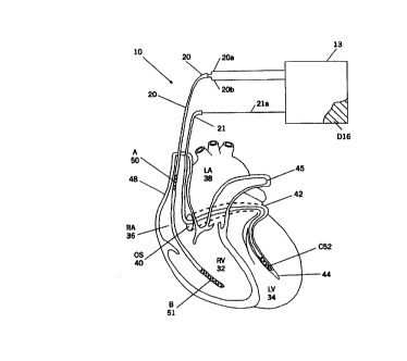
atria and right and left ventricles). The schematically illustrated portions
of the heart 30
illustrated in Figure 1 includes the right ventricle "RV" 32, the left
ventricle "LV" 34, the
right atrium "RA" 36, the left atrium "LA" 38. the superior vena cava 48, the
coronary
sinus "CS" 42, the great cardiac vein 44, the left pulmonary artery 45, and
the coronary
sinus ostium or "os" 40.
The driving force for the flow of blood in the heart comes from the active
contraction of the cardiac muscle. This contraction can be detected as an
electrical signal.
2 0 The cardiac contraction is triggered by electrical impulses traveling in a
wave
propagation pattern which begins at the cells of the SA node and the
surrounding atrial
myocardial fibers, and then traveling into the atria and subsequently passing
through the
AV node and, after a slight delay, into the ventricles.
The beginning of a cardiac cycle is initiated by a P wave, which is normally a
2 5 small positive wave in the body surface electrocardiogram. The P wave
induces
depolarization of the atria of the heart. The P wave is followed by a cardiac
cycle portion
which is substantially constant with a time constant on the order of 120
milliseconds
("ms")
Various embodiments of the present invention can be illustrated with reference
to
3 o Figure 1. The defibrillator 10 of Figure I includes an implantable housing
13 that
contains a hermetically sealed electronic circuit 15 (see Fig. 2). The housing
includes an
electrode comprising an active external portion 16 of the housing, with the
housing 13
CA 02283128 1999-08-27
WO 98/40122 PCT/US98/04980
preferably implanted in the left or right thoracic region of the patient
(e.g., subcutaneously
or submuscularly, in the left or right pectoral region, or subcutaneously or
submuscularly
in the left or right (preferably left) abdominal region; the left pectoral
region is most
preferred) in accordance with known techniques as described in G. Bandy, U.S.
Patent No.
5,292,338.
The system includes a first catheter 20 and a second catheter 21, both of
which are
insertable into the heart (typically through the superior or inferior vena
cava) without the
need for surgical incision into the heart. The term "catheter" as used herein
includes
"stylet" and is also used interchangeably with the term "lead". Each of the
catheters 20,
21 contains electrode leads 20a, 20b, 21a, respectively.
As illustrated in Figure 1, the system includes an electrode A; 50 that
resides in
the superior vena cava or innominate vein, an electrode B; 51 positioned in
the right
ventricle, and an electrode C; 52 positioned within a vein on the postern
lateral surface of
the left ventricle (e.g., in the apical third of the posterior cardiac vein or
the apical half of
the great cardiac vein). The active external portion of the housing 16 serves
as a fourth
electrode D. Designations "A" through "D" herein refer to electrodes in the
aforesaid
positions.
Electrode C may be a hollow electrode to allow the flow of blood through the
electrode (e.g., a stmt-type electrode that engages the vessel wall) when
positioned in the
2 0 vein, or may be a solid electrode configured (that is, of a shape and
size) to allow the flow
of blood around the electrode when positioned within the vein. A solid
electrode is
preferred. Electrode C may be positioned entirely within a vein on the postern-
lateral
surface of the left ventricle, or may also extend into the coronary sinus (as
in the case of
an elongate electrode).
2 5 The position of electrode C may be achieved by first engaging the coronary
sinus
with a guiding catheter through which a conventional guidewire is passed. The
tip of the
torqueable guidewire is advanced under fluoroscopic guidance to the desired
location.
The lead 21 on which electrode C is mounted passes over the guidewire to the
proper
location. The guidewire is withdrawn and electrode C is incorporated into the
lead
3 0 system. Such an electrode is considered a solid-type electrode herein.
Figure 2 illustrates one example of an implantable housing 13 containing an
electronic circuit 15, which includes one or more amplifiers (not shown) for
amplifying
CA 02283128 1999-08-27
WO 98/40122 PCT/US98/04980
-10-
sensed cardiac signals. The amplified signals are analyzed by an detector 70
which
determines if ventricular fibrillation (or other arrythmia, depending on the
specific
treatment for which the device is configured) is present. The detector 70 may
be one of
several known to those skilled in the art. Although, as illustrated, a sensing
signal is
provided by the electrode A 50, it will be appreciated by those of skill in
the art that the
sensing electrode may also be a plurality of sensing electrodes with a
plurality of signals,
such as bipolar configurations, and may also be electrodes that are positioned
in alternate
cardiac areas as is known in the art, such as for example, the CS. In this
situation, the
input line to the detector may be a plurality of lines which if providing only
sensing will
provide an input to the detector.
The defibrillation electrodes may alternately be configured to sense cardiac
cycles,
or may have smaller sensing electrodes placed adjacent thereto and thereby
provide input
to the electronics package as well as provide a predetermined stimulation
shock output to
predetermined cardiac areas as directed by the controller.
The electronic circuit 15 also includes a cardiac cycle monitor
("synchronization
monitor 72") for providing synchronization information to the controller 74.
As
discussed below, the synchronization is typically provided by sensing cardiac
activity in
the RV, but may also include other sensing electrodes which can be combined
with the
defibrillation electrodes or employed separately to provide additional
assurance that
2 0 defibrillation shock pulses are not delivered during sensitive portions of
the cardiac cycle
so as to reduce the possibility of inducing ventricular fibrillation.
Numerous configurations of capacitor and control circuitry may be employed.
The power supply may include a single capacitor, and the control circuit may
be
configured so that both the auxiliary pulse and the defibrillation pulse are
generated by
2 5 the discharge of the single capacitor. The power supply may include a
first and second
capacitor, with the control circuit configured so that the auxiliary pulse is
generated by the
discharge of the first capacitor and the defibrillationpulse is generated by
the discharge of
the second capacitor. In still another embodiment, the power supply includes a
first and
second capacitor, and the control circuit may be configured so that the
auxiliary pulse is
3 o generated by the discharge (simultaneous or sequential) of both the first
and second
capacitors, and the defibrillationpulse likewise generated by the discharge of
the first and
second capacitors.
CA 02283128 1999-08-27
WO 98/40122 PCT/US98/04980
-11- w
One defibrillationwaveform that may be used to carry out the present invention
is
illustrated in Figure 3, which shows a schematic illustration of a biphasic
truncated
exponential waveform. While a variety of different waveforms can be used, as
discussed
herein, surprisingly good results are achieved when an auxiliary pulse is
delivered prior to
the primary, or defibrillation, pulse, with the auxiliary pulse being
delivered along a
different current pathway. A particularly surprising finding was that better
results can be
achieved when the auxiliary pulse is of an opposite polarity than the first
phase of the
defibrillation pulse. Such a biphasic truncated exponential waveform primary
pulse with
a monophasic auxiliary pre-pulse is illustrated in Figure 4. The foregoing
waveforms can
l0 be modified in ways that will be apparent to those skilled in the art
(e.g., a chopped
waveform can be delivered; the waveform can be time-based or fixed tilt; etc).
The auxiliary pulse may be from .5 or 1 to 5 or 10 milliseconds in duration,
with a
2 millisecond pulse currently preferred. The time interval from the end of the
auxiliary
pulse to the leading edge of the primary pulse may be from 1 or 2 milliseconds
to 10, 15
or 20 milliseconds, with a delay of about 5 milliseconds currently preferred.
The optimal auxiliary-to-primary interval may differ depending on the type of
rhythm or condition of the myocardial tissue at the time the therapy is
applied. Therefore,
the control circuitry may also be configured to sense a characteristic of the
cardiac rhythm
(e.g., an activation interval or a dynamical pattern of consecutive activation
intervals) and
2 0 then select an optimum auxiliary-to-primary shock time interval (e.g.,
from a look up
table stored in a microprocessormemory).
The percent tilt of the primary pulse and the auxiliary pulse may each be from
10,
or 30 percent up to 50 or 60 percent. Percent tilt = (Vo-Vf x 100)No, where Vo
is the
initial voltage and Vf is the final voltage of the pulse. Vf refers to the
final voltage of the
2 5 final phase of the shock where the shock sequence has multiple phases.
In general, the control circuit is configured so that the auxiliary pulse is
not more
than 40% or SO% of the peak current and not more than 20% or 30% of the
delivered
energy (in Joules) of the defibrillationpulse. In a preferred embodiment, the
trailing edge
voltage of the auxiliary pulse is equal (~10 Volts) to the leading edge
voltage of the
3 0 defibrillation pulse. Particular voltage, current, and energy outputs will
depend upon
factors such as the condition of the tissue and the particular disorder being
treated. In
general, the auxiliary pulse may have a peak voltage of from 20 or 30 volts to
200 or 250
CA 02283128 1999-08-27
WO 98/40122 PCT/US98/04980
-12-
volts, with a peak voltage range of 50 to 150 volts preferred. The energy of
the auxiliary
pulse may be from .O1 or .OS to 1 or 2 Joules. The energy of the
defibrillation pulse may
be from 5 or 10 Joules to 30, 40 or 50 Joules. An object of the instant
invention is to
enable the reduction of the size of the implantable defibrillator, which is
made possible by
defibrillation pulse energy ranges as described. Thus, a further aspect of the
present
invention is an implantable defibrillator comprising a housing and a power
supply
contained within the housing, and a control circuit contained within the
housing and
operatively associated with the power supply. The control circuit is
configured for
delivering a cardioversion sequence as described above. Based on the ranges
above, the
maximum storage capacity of the capacitor in the power supply may be from 5.01
to 52
Joules, and is most preferably from 10 or 15 to 20 Joules. Thus the housing
for such a
power supply preferably has a volume less than 35 cubic centimeters (but
typically at
least 5 cubic centimeters) .
Without wishing to be bound to any particular theory for the preferred
waveforms
described above, it appears that the auxiliary pulse, which is of a magnitude
greater than
pacing pulses but less than a defibrillation pulse, is sufficient to
affect/substantially alter
the intrinsic patterns of recovery of excitability and thereby momentarily
yield localized
cessation of propagation by inactivating sodium ion conductance channels via
elevation
of the transmembrane potential. Importantly, the tissue portions affected by
the auxiliary
2 0 pulse is tissue in a weak field area for the primary, or defibrillation,
pulse. The weak field
area affected by the auxiliary pulse should be selected to include the weakest
field area of
the primary pulse. In a preferred embodiment, the weak field area is generally
the left
lateral aspect of the left ventricle, extending from the apex to the base
thereof.
Numerous different embodiments of the implantable system of the present
2 5 invention can be implemented with the apparatus of Figures 1 and 2 and the
waveforms of
Figures 3 and 4, depending on the specific configuration of the control
circuitry for the
use and pairing of particular electrodes. Specific examples are discussed
below.
Table 1 illustrates a first embodiment of the invention. After a
tachyarrhythmic
condition is detected and reconfirmed by algorithms running in the controller
74, therapy
3 0 in the form of an electrical shock of Figure 3 is applied to the heart by
discharging
capacitor 78. A preferred pairing of electrodes for this embodiment is
illustrated in Table
1 below. In all tables herein, a "+" indicates that the electrodes are
electrically common,
CA 02283128 1999-08-27
WO 98/40122 PCTNS98/04980
-13-
and an "->" indicating current flow (which may be reversed).
Table 1: Electrode Pairings
Primary Pulse
B+C -> A+D
Table 2 illustrates a second embodiment of the invention. This embodiment
introduces the use of an auxiliary pulse, with four possible configurations
being shown in
Figure 4. The auxiliary pulse is delivered through a different set of
electrodes than the
primary, or defibrillation,pulse.
1 o Table 2: Electrode Selection
Auxiliary Pulse Primary Pulse
C->D A->B
A->B C->D
C->A B->C
B->D C->A
C->D B->D
C->B B->A+D
In one embodiment of an apparatus configured according to Table 2, the control
circuitry is configured so that only one capacitor is employed to deliver both
pulses, and
that the different sets of electrodes are switched in and out of the discharge
circuit to
achieve the therapeutic effect. In this embodiment, the trailing edge of the
auxiliary pulse
is equal to the leading edge of the primary pulse.
In another embodiment of an apparatus configured according to Table 2, the
control circuitry is configured so that the auxiliary pulse and the primary
pulse arise from
separate capacitors. For example, if the design goal is to control the time
constant of the
CA 02283128 1999-08-27
WO 98/40122 PCT/US98/04980
-14-
capacitor discharge waveform (time constant is the product of the resistance
and the
capacitance) and assuming further that the resistance to the shock (ratio of
voltage to
current) along the auxiliary pathway is two-fold higher than along the primary
pathway,
then the capacitance of the auxiliary capacitor could be half that of the
primary shock
capacitor. Further, with a two capacitor implementation, the relative strength
of the
pulses can be made independent. In this way, the minimum auxiliary shock
strength can
be applied that produces the synergistic action between the auxiliary and
primary shocks,
thereby minimizing the shock strength requirements for effective
defibrillation.
Table 3 below and Figure 5 illustrate another embodiment of the invention,
where the beneficial effects are augmented by placing an additional electrode
E; 53 on
endocardial transvenous elongate lead 23 in the area of the heart experiencing
the weakest
electric field when electrode C; 52 is present. The weak field area in this
location is in
the region of the right ventricular conus. Specifically, the electrode E can
be located
within the right atrial appendage or the right ventricular outflow track. To
accomplish
this, the electrode should be located at the most distal portion of the lead
body. One
configuration for pairing of electrodes in this embodiment is given in Table
3:
Table 3: Electrode Pairings for Fig. 5
Auxiliary Pulse Primary Pulse
C -> E B -> A+D
Two embodiments of a suitable transvenous elongate electrode lead 23 are
illustrated in Figure 6, with 6a showing a pace/sense electrode 54 located at
the distal tip
of the lead 23, while the distal end of the primary electrode 53 is located 10
to 15
millimeters from the top so as to minimize the shock effects on sensing from
tissue very
2 5 near the pace/sense electrode. Sensing of atrial activity is accomplished
by measuring the
potential difference between the pace/sense electrode 54 and some indifferent
electrode
such as the shock coil or an electrode away from the heart such as electrode D
16. In the
embodiment of 6b, a pair of pace/sense ring electrodes 54, 54' are located
proximal to the
primary electrode 53. The primary electrode is about 15 to 25 millimeters in
length, most
CA 02283128 1999-08-27
WO 98/40122 PCT/US98/04980
-15-
preferably 20 millimeters in length, and preferably about 4 to 6 French in
diameter, the
pair of ring electrodes (2-4 millimeters in length together, with a diameter
at least equal to
that of the lead body) being positioned 10 to 20 millimeters proximal to the
primary
electrode. Pacing and sensing capability on lead 23 are particularly important
when the
system 10 is configured to monitor electrical rhythm activity in both atrial
and ventricular
chambers.
Table 4 below, taken together with the apparatus of Figure 3 implementing the
waveform of Figure 4, illustrate three additional configurations of the
present invention:
Table 4: Electrode Pairings
Auxiliary Pulse Primary Pulse
B -> C B+C -> A+D
C -> D B+C -> A+D
C -> D B -> C+A+D
Figure 7 presents a flow chart schematically illustrating how the electrodes
employed to carry out the present invention can be used to modify the therapy
delivered.
In Figure 7, electrode C permits sensing of electrical rhythm information and
furthermore,
allows the implanted device to use that information to select therapy that is
tailored to
specific rhythm characteristics. In Figure 7 electrodes C and B are
electrically common
during sensing and the combined signal is fed into a sensing module for
subsequent
feature extraction, therapy adaptation in light of the detected feature, and
therapy delivery.
For example, the time at which the shock is delivered is determined by an
algorithm that
2 0 chooses the optimum time for the defibrillation shock to produce its most
significant
electrophysiological effects. Other therapy adaptations include the coupling
interval
between the auxiliary and primary pulses. Several features that could be used
alone or in
a combined, weighted fashion include mean activation interval, negative and
positive
slope threshold. In the alternative, rather than electrodes C and D being
common,
2 5 electrograms recorded between electrodes B and C and a common indifferent
electrode
(electrodes A or D) could be separately fed into the sensing module 60. The
feature
CA 02283128 1999-08-27
WO 98/40122 PCT/US98/04980
-16-
extraction algorithm can examine features from each electrogram signal alone
or in a
differential fashion. As previously, the features extracted are then used to
guide therapy
adaptation and optimize therapy delivery.
Additional embodiments of the present invention are illustrated in Table 5
below,
taken in conjunction with the electrode placements illustrated in Figure 5 and
the
waveforms presented in Figure 13, illustrate additional configurations for
shocks and
electrodes of the present invention.
Table 5.
No. Figure Primary Pulse Auxiliary
Pulse
1 13a B->A+D C->A+D
2 13a B->D C->A
3 13b B->A+D C->A+D
4 13b B->D C->A
5 13c B->A+D C->A+D
6 13c B->D C->A
7 13d B->A+D C->A+D
8 13d B->D C->A
to
In Table 5, Current flow is indicated by the direction of the arrow from anode
(+) to
cathode (-). The most preferred configuration is currently Number 5 in Table 5
and
Figure 13C, with a biphasic primary, or defibrillation pulse, followed by a
biphasic
auxiliary pulse, with the first phase of the auxiliary pulse of opposite
polarity from the
second phase of the primary pulse, with the primary pulse delivered between a
right
ventricle electrode B and two electrically common electrodes A and D; and with
the
auxiliary electrode delivered between the left ventricle electrode C and two
eletrically
common electrodes A and D.
In alternate embodiments of the invention, the order of the primary pulse and
2 0 auxiliary pulse for the embodiments set forth in Tables 2 through 5 may be
reversed.
Systems as described above may be implanted in a patient by conventional
surgical techniques, or techniques readily apparent to skilled surgeons in
light of the
disclosure provided herein, to provide an implanted defibrillationor
cardioversion system.
CA 02283128 1999-08-27
WO 98/40122 PCT/US98104980
Additional features can also be added to the invention without affecting the
function of the invention and result thereof. Such additional features
include, but are not
limited to, safety features such as noise suppression or multiple wave
monitoring devices
(R and T), verification checking to reduce false positive, precardioversion
warning,
programmed delayed intervention, bipolar configured sensing electrodes,
intermittently
activated defibrillation detector to reduce energy drain, a switching unit to
minimize lines
from the pulse generator, etc.
Although the system has been described above as an implantable system, it will
be
appreciated by those of ordinary skill in the art that the invention could
also be
l0 incorporated into an external system which employs catheters to position
the electrodes
for a short time within a patient's heart.
The present invention is explained further in the following non-limiting
examples.
EXAMPLE 1
Sub-threshold,Critically-timed,Monophasic
Epicardial Pre-shock Significantly Reduces Transvenous
Binhasic Defibrillation Threshold in Swine
This example shows that the strength and temporal prematurety of the
monophasic auxiliary shock significantly affects the strength of the
defibrillation
2 0 threshold of the biphasic primary shock.
Animal model preparation. Domestic farm swine (30-35 kg) were tranquilized
via an intramuscular injection of ketamine (20 mg/kg). After about 15 minutes,
anesthesia was induced with an intravenous bolus injection of sodium
pentobarbital (30
mg/kg) through a 20 gauge needle placed in a prominent ear vein. An
endotracheal tube
2 5 was inserted and the cuff was inflated to provide closed circuit
ventilation.
Electrocardiographicmonitoring leads were placed on cleaned and shaved
portions of the
fore limbs and hind limbs. The animal was placed in dorsal recumbence and
secured to
the table with limb restraints. A deep surgical plane of anesthesia was
maintained with
continuous intravenous infusion of sodium pentobarbital (0.05 mg/kg/min).
Skeletal
3 0 muscle paralysis was induced with intravenous succinylcholine ( 1 mg/kg)
and maintained
with a dosage of 0.25 to 0.50 mg/kg each hour. Additional intravenous
injections of
sodium pentobarbital (10-20 mg) were given to titrate the anesthesia to an
appropriate
CA 02283128 1999-08-27
WO 98/40122 PCT/US98/04980
-18-
level. Sterile 0.9% saline solution was infused (2-5 ml/kg/hr) through a
central venous
catheter placed in an internal jugular vein. A femoral artery was surgically
exposed and
isolated through an inguinal cutdown. A 4 French polyurethane catheter was
inserted and
its tip was advanced into the descending aorta. Central arterial pressure was
continuously
displayed on a monitor (Hewlett Packard Corp.). Anesthesia level was routinely
monitored by testing cardiac reflex response to intense pedal pressure, jaw
tone and basal
heart rate and pressure. Both arterial blood electrolytes (K+, HC03 and Ca+),
blood
gasses p0z, pCOz) and pH were measured every 30-60 minutes. Abnormal values
were
corrected by adding electrolytes to the hydration fluids and by adjusting
ventilation rate
and tidal volume. Esophageal temperature was continuously monitored. Heated
water-
circulating mats were used to maintain a normothermia (36°-38°
C).
The chest was opened through a median sternotomy. A retractor was installed to
improve exposure of the heart and surrounding organs. The pericardium was
carefully
incised along an axis connecting the base and apex of the heart. A pericardial
cradle was
fashioned to elevate the heart to a closed-chest position within the chest
cavity.
Throughout each experiment, the surface of the heart was kept moist and warm
by
flushing its surface with normal saline and covering the chest cavity with a
sheet of
plastic.
Defibrillation electrode placement. Four defibrillation electrodes were used
in
2 0 this study; two for the primary shocks and two for the monophasic
auxiliary shocks.
Defibrillation electrodes mounted on a commercially available lead system
{ENDOTAK~ model 0094, CPI/Guidant Corp., St. Paul, MN) were introduced through
a
right jugular venotomy. The distal coil electrode (4.0 cm length) was advanced
under
fluoroscopic guidance to the right ventricular apex. The proximal coil (6.8 cm
length)
2 5 was positioned with its distal tip 1 to 2 cm cephalid to the junction of
the right atrium and
superior vena cava using fluoroscopic guidance. The distal and proximal
catheter
electrodes were used to deliver all the biphasic shocks.
To deliver the monophasic auxiliary shocks, an epicardial electrode formed by
concentric ellipses fashioned from platinum coated titanium coils 2 mm in
diameter was
3 0 sutured to the lateral, apical aspect of the left ventricular free wall.
This coil-patch
electrode circumscribed about 15 cmz and extended from the apex to about two-
thirds
the distance from the apex to base. The return electrode, a 6 French titanium
coil
CA 02283128 1999-08-27
WO 98/40122 PCT/US98/04980
-19-
electrode, 6.8 cm in length, was positioned in the left j ugular vein.
After the electrodes were inserted, margins of the incised pericardium were
opposed by crossing the cradle tethers and applying gentle traction. The chest
retractor
was removed, but the chest was not surgically closed. The chest wound was
covered
with an impermeable plastic drape to keep the heart warm and moist.
Test procedures. The defibrillation threshold was determined in randomized
order for each of thirteen experimental treatments in each animal.
Fibrillation. Ventricular fibrillation was induced with 60 Hz alternating
current
(50-100 mA peak to peak) applied to the pacing tip electrode of the
endocardial lead
1 o positioned in the right ventricle. In all episodes, fibrillation was
allowed to persist for at
least 10 seconds but not more than 12 seconds prior to delivery of the
defibrillation test
shock. When the test failed to defibrillate, the heart was immediately
defibrillated with a
rescue shock given through the transvenous catheter lead system. The animal
was
allowed to recover at least four minutes between each test shock.
Defibrillation waveforms. External defibrillators were used to deliver the
monophasic and biphasic truncated exponential shocks over two different
current
pathways. The monophasic shock is referred to as the "auxiliary" pulse and the
biphasic
shock as the "primary" pulse herein. When delivered simultaneously, the
leading edges
of both pulses are temporally coincident. When the shocks are given
sequentially, the
2 0 auxiliary primary coupling interval is defined as the time between the
trailing edge of the
auxiliary pulse and the leading edge of the primary pulse.
All biphasic shocks were delivered by the VENTAK~ external cardioverter
defibrillator (model 2815, CPI/Guidant Corp., St Paul, MN). This device
delivers shocks
having an overall fixed-tilt of 80%. The capacitance is 140 ~F. Total waveform
duration
2 5 varies with shock impedance. Phase one was always 60% of the total
duration. Leading
edge voltage could be adjusted in 1-volt steps.
The monophasic shocks were delivered by a research defibrillator. The research
defibrillator delivers fixed-duration shocks ( 1-20 ms) with an effective
capacitance of 150
p,F. In this study, the monophasic auxiliary pulses were always 5 ms in
duration. The
3 0 initiation of capacitor discharge for both shock generating devices could
be externally
triggered using a low-amplitude (1-5 volts) pulse. We used a commercially-
available
current source (Bloom Stimulator, Bloom & Assoc., Reading, PA) to generate 1
ms
CA 02283128 1999-08-27
WO 98/40122 PCT/US98/04980
-20-
trigger pulses on two independent output channels that were used to control
the relative
timing between the auxiliary and primary pulses.
The polarity of the defibrillation electrodes was controlled in each
experiment
since it has been shown that defibrillation can be affected by electrode
polarity. The left
ventricular electrode was always connected to the anodic terminal (positive)
of the
defibrillator output circuit, while the right ventricular defibrillation coil
electrode was
always connected to the cathodic terminal (negative).
Experimental protocol. In general, each experiment consisted of multiple
episodes of electrically-induced ventricular fibrillation that were
intentionally terminated
with test shocks. by applying an established set of rules to the observed
outcome of each
defibrillation trial, shock strengths were selected that permitted the
definition of a
defibrillation threshold for each experimental treatment. We used the modified
Purdue
technique to determine defibrillation thresholds. In brief, the strength of
the test shock is
adjusted according to the outcome (success or failure). The first
defibrillation test shock
for each treatment in the first animal was 400 V. In all subsequent
experiments, the initial
test shock strength was adjusted to the mean from the previous animals. If the
first test
shock failed the next shock voltage was increased 80 V and decreased 80 V if
it
succeeded. After the first reversal of outcome on successive trials (success
to failure or
failure to success), the shock strength step was reduced to 40 V. Trials
continued until a
2 0 second outcome reversal was encountered, after which the strength was
increased 20 V
for a failure and decreased 20 V for a success. The lowest shock strength that
defibrillatedthe ventricles was defined as the defibrillationthreshold.
In this study, we investigated the influence of two variables on the
defibrillation
threshold of the primary shock: 1 ) peak voltage of the auxiliary pulse and 2)
auxiliary-
2 5 primary pulse coupling interval. The primary pulse given alone was used as
the control
treatment. Three monophasic auxiliary pulse strengths were tested: 50 V, 100 V
and 150
V. Each auxiliary pulse strength was tested in combination with an auxiliary-
primary
pulse coupling interval. Four auxiliary-primary pulse coupling intervals,
defined as the
time between the trailing edge of the auxiliary pulse and the leading edge of
the primary
3 0 pulse, were tested: - S ms (simultaneous delivery), 1 ms, 20 ms and 40 ms.
The
combination of the two variables and the control yields thirteen treatments as
shown in
Figure 9. The experimental treatments were tested in randomized order in each
animal.
CA 02283128 1999-08-27
WO 98/40122 PCTIUS98104980
-21-
Data acquisition. Defibrillation threshold measurements are more accurate and
precise when shock strength measurements are made directly across the
defibrillation
electrodes. Therefore, the current and voltage during the defibrillation
pulses were
measured through 4:1 and 200:1 dividers by a waveform analyzer (model 6100,
Data
Precision, Inc., Danvers, MA). The analog current and voltage signals were
digitized at
20 kHz and stored in a buffer. The digitized waveforms were displayed after
each
defibrillation attempt to permit visual inspection. Custom analysis software
was used to
define the time and amplitude of the leading and trailing edges and to compute
the shock
impedance and total energy delivered in each pulse. Peak voltage, peak
current, shock
impedance and energy delivered was recorded for each test shock.
Analysis and results. The mean and standard deviation of peak voltage, peak
current, delivered energy and shock impedance for each pulse at defibrillation
threshold
for each treatment were calculated for the eight animals. For the treatments
utilizing an
auxiliary pulse, the mean total delivery energy values include the energy
delivered in the
monophasic pulse. The mean peak current and peak voltage values always reflect
the
strength of the biphasic primary pulse.
Repeated measures analysis of variance with the Student Newman-Keul's test was
used to compare peak voltage, peak current, delivered energy and shock
impedance
among the treatments. Differences among the means were considered significant
when
2 0 P<0.05. All reported values are mean + SD unless noted otherwise.
The mean energy delivered at defibrillationthreshold for each of the
experimental
treatments is presented in Figure 9. The mean defibrillation threshold for the
control
treatment was 24 + 10.4 J. The defibrillation thresholds were significantly
lower (~SO%)
when a monophasic auxiliary pulse was delivered simultaneously with the
biphasic
primary pulse. The mean energy delivered in the 50, 100 and 150 V monophasic
pulses
was 0.09 J, 0.38 J and 0.87 J, respectively. However, there was no significant
differences
among the simultaneous treatments. Similarly, the defibrillation thresholds
for the
treatments with a 1 ms auxiliary -primary pulse coupling interval were
significantly
lower than control, and unlike the simultaneous treatments, there was a trend
suggesting
3 0 that the strength of the monophasic pulse affected the amount of
defibrillation threshold
reduction.
Peak voltage requirements at defibrillation threshold followed trends very
similar
CA 02283128 1999-08-27
WO 98/40122 PCT/US98/04980
-22-
to the trends for energy delivered. Figure 10 shows mean peak voltage of the
primary
pulse at defibrillation threshold with and without the auxiliary pulse. When
the auxiliary
and primary pulses were applied simultaneously,the peak voltage defibrillation
threshold
was reduced about 25%. For auxiliary-primary coupling intervals of 20 ms and
40 ms,
the defibrillation thresholds were not different than control for auxiliary
shocks of 50 V
and 100 V. However, the defibrillationthreshold for the 150 V auxiliary shock
with a 20
ms auxiliary-primary coupling interval was significantly lower than the
control treatment
(P<0.05).
1 o EXAMPLE 2
Single Capacitor Implementation of Dual Shock
Defibrillation Method in Closed-Chest Do s
This example demonstrates the feasibility of the dual shock defibrillation
therapy
demonstrated in Example I above with a single capacitor implementation, and
with a
transvenous lead system.
Animal model preparation. A total of six animals were studied. Methods of
preparation were essentially equivalent for each animal. Mixed-breed canines
(26-36 kg)
were tranquilized via an intramuscular injection of ketamine (10 mg/kg), if
necessary.
After about I S minutes, anesthesia was induced with an intravenous bolus
injection of
2 0 sodium pentobarbital (30 mg/kg) through a catheter placed in a cephalic
vein. An
endotracheal tube was inserted and the cuff was inflated to provide closed
circuit
ventilation. Electrocardiographic monitoring leads were placed on the cleaned
and
shaved portions of the fore limbs and hind limbs. The animal was placed in
dorsal
recumbence and secured to the table with limb restraints. A deep surgical
plane of
2 5 anesthesia was maintained with continuous intravenous infusion of sodium
pentobarbital
(0.05 mg/kg/min). Skeletal muscle paralysis was induced with intravenous
succinylcholine ( 1 mg/kg) and maintained with a dosage of 0.25 to 0. SO mg/kg
each hour.
Additional intravenous injections of sodium pentobarbital (10-20 mg) were
given to
titrate the anesthesia to an appropriate level prior to performing any
surgical procedures.
3 0 Sterile 0.9% saline solution was infused (2-5 ml/kg/hr) through a central
venous catheter
placed in an internal jugular vein. A femoral artery was surgically exposed
and isolated.
A 4 French polyurethane catheter was inserted and its tip was advanced into
the
CA 02283128 1999-08-27
WO 98/40122 PCT/US98104980
-23-
descending aorta. Central arterial pressure was continuously displayed on a
monitor
(Hewlett Packard Corp.). Anesthesia level was routinely monitored by testing
cardiac
reflex response to intense pedal pressure, jaw tone and basal heart rate and
blood pressure.
Both arterial blood electrolytes, blood gasses, as well as pH were measured
every 30-60
minutes. Abnormal values were corrected by adding electrolytes to the
hydration fluids
and by adjusting ventilation rate and tidal volume. Esophageal temperature was
continuously monitored. Heated water-circulating mats were used to maintain a
normothermia (36°-38°C).
The chest was opened through a median sternotomy. A retractor was installed to
1 o improve exposure of the heart and surrounding organs. The pericardium was
carefully
incised along an axis connecting the base and apex of the heart. A pericardial
cradle was
fashioned to elevate the heart to a closed-chest position within the chest
cavity. When the
chest was open during the initial stages of the study, the surface of the
heart was kept
moist and warm by flushing its surface with normal saline and covering the
chest cavity
with a sheet of plastic.
Defibrillation electrode placement. Four defibrillation electrodes were used
in
this study; two for the primary shocks and two for the monophasic auxiliary
shocks (see
Figure 11). Defibrillation electrodes mounted on a commercially available lead
system
(ENDOTAK~ model 0094, CPI/Guidant Corp., St. Paul, MN) were introduced through
a
right jugular venotomy. The distal coil electrode (4.0 cm length) was advanced
under
fluoroscopic guidance to the right ventricular apex. The proximal coil (6.8 cm
length)
was positioned with its distal tip 1 to 2 cm cephalid to the junction of the
right atrium and
superior vena cava using fluoroscopic guidance. The distal and proximal
catheter
electrodes were used to deliver all the biphasic shocks.
2 5 We elected to simulate a transvenous introduction of the left ventricular
electrode
used to deliver the monophasic auxiliary shocks. The approach was taken
because we
wanted to control the position of the left ventricular electrode. Closed chest
introduction
and positioning of the left ventricular electrode using fluoroscopic guidance
alone is not
trivial. Improper positions could have severely impacted the results of this
study.
3 o Therefore, efforts were made to simulate a closed-chest model. To
accomplish this goal,
the left ventricular coil electrode (3 French, 3 cm length, tri-filar platinum
coated
titanium) was inserted into the posterior cardiac vein. In addition, the chest
was closed
CA 02283128 1999-08-27
WO 98/40122 PCT/US98/04980
-24-
and evacuated after the left ventricular electrode was positioned. This
procedure assured
that the volume conductor characteristics of a closed chest were present at
the time that
defibrillation trials were conducted. The 3 French coil electrode was inserted
into the
posterior cardiac vein by elevating the apex of the heart to expose the
postern-lateral Left
ventricle. A short segment of an 18 gauge catheter was partially inserted so
that about 1
cm was outside the vein. Back flow of venous blood confirmed proper location.
The
specially designed tip of the defibrillation coil was allowed to engage the
catheter which
acted as a micro-introducing sheath. Both the introducing catheter and
defibrillation
electrode were carefully advanced into the vein and secured with a single
stitch. this
l0 technique was successfully used to position the left ventricular
defibrillation electrode
within the posterior cardiac vein in all of the six animals.
The return electrode for the monophasic auxiliary shocks, a 6 French titanium
coil
electrode, 6.8 cm in length, was positioned in the left jugular vein. See
Figure 11.
After the electrodes were inserted, margins of the incised pericardium were
opposed by crossing the cradle tethers and applying gentle traction. The chest
retractor
was removed and the chest was surgically closed in three layers. A chest tube
was
inserted and continuous suction was applied to evacuate the thoracic cavity.
Test procedures. The defibrillation threshold was determined in randomized
order for each of seven experimental treatments in each animal.
2 0 Fibrillation. Ventricular fibrillation was induced with 60 Hz alternating
current
(50-100 mA peak to peak) applied to the pacing tip electrode of the
endocardial lead
positioned in the right ventricle. In all episodes, fibrillation was allowed
to persist for at
least 10 seconds but not more than 12 seconds prior to delivery of the
defibrillation test
shock. When the test failed to defibrillate, the heart was immediately
defibrillated with a
2 5 rescue shock given through the transvenous catheter system. The animal was
allowed to
recover at least four minutes between each test shock.
Defibrillation waveforms. External defibrillators were used to deliver the
monophasic and biphasic truncated exponential shocks over two different
current
pathways. The monophasic shock is referred to as the "auxiliary" pulse and the
biphasic
3 0 shock as the "primary" pulse herein.
Three pulsing schema were tested in this study and are shown in Figure 12.
Unidirectional shocks served as control treatments. Bidirectional and
sequential shocks
CA 02283128 1999-08-27
WO 98/40122 PCT/US98/04980
-25-
served as the test treatments. Unidirectional shocks were given using the
conventional
transvenous shock vector (RV -> SVC). Bidirectional shocks were applied to
electrodes
in the shock vector RV+LV -> SVC. Sequential shocks were given in a manner
similar to
that described in the previous chapter. However, in this study the auxiliary-
primary
coupling interval (defined as the time between the trailing edge of the
auxiliary pulse and
the leading edge of the primary pulse) tested were 1 ms, 5 ms, 10 ms and 20
ms.
For all sequential shock treatments a single capacitor waveform was emulated.
Thus, the trailing edge of the auxiliary pulse was set equal (~10 V) to the
leading edge of
the primary pulse.
Biphasic primary shocks were delivered by the VENTAK~ external cardioverter
defibrillator as described in Example 1 above or by a research defibrillator.
The research
defibrillator was programmed to emulate a single capacitor truncated
exponential
waveform. The first phase duration was 4 ms and the second phase duration was
3 ms.
The trailing edge of phase one was equal (~10 V) to the leading edge voltage
of phase
two.
All of the monophasic shocks were delivered by a research defibrillator. The
research defibrillator delivers fixed-duration shocks (1-20 ms) with an
effective
capacitance of 150 ~F. In this study, the monophasic auxiliary pulses were
always 5 ms
in duration. The initiation of capacitor discharge for both shock generating
devices could
2 0 be externally triggered using a low-amplitude ( 1-5 volts) pulse. We used
a commercially-
available current source (Bloom Stimulator, Bloom & Assoc., Reading, PA) to
generate 1
ms trigger pulses on two independent output channels that were used to control
the
relative timing between the auxiliary and primary pulses.
The polarity of the defibrillation electrodes was controlled in each
experiment
2 5 since it has been shown that defibrillation can be affected by electrode
polarity. The left
ventricular electrode was always connected to the anodic terminal (positive)
of the
defibrillator output circuit, while the right ventricular defibrillation coil
electrode was
always connected to the cathodic terminal (negative). When bidirectional
shocks were
given the left ventricular electrode was connected along with the right
ventricular
3 o electrode to the cathodic terminal of the external defibrillator.
Experimental protocol. In general, each experiment was carried out as
described
in Example 1 above. The lowest shock strength that defibrillated the
ventricles was
CA 02283128 1999-08-27
WO 98/40122 PCT/US98/04980
-26-
defined as the defibrillationthreshold.
Data acquisition. Data acquisition was carried out in essentially the same
manner
as described in Example 1 above. Analysis and results. Data analysis was
carried out in
essentially the same manner as described in Example 1 above.
As shown with reference to Figure 12 delivered energy requirements at the
defibrillation threshold were significantly lower for the dual shock
treatments 4, 5, 6 and
7 (P<0.05). Differences among the mean energy delivered at the defibrillation
threshold
for unidirectional shocks (treatments 1 and 3) and bidirectional shocks
(treatment 2) were
not statistically significant. Additionally, none of the differences among the
mean energy
delivered at defibrillation threshold for the sequential shocks (treatments 4,
5, 6 and 7)
were statistically significant, although there was a strong trend suggesting
that the
sequential shocks having a 20 ms coupling interval required more energy for
defibrillation
than sequential shocks having a 1 ms coupling interval (15.4+7.2J vs.
10.2+4.1J,
P=0.076).
EXAMPLE 3
Effect of Varying Preshock and Postshock Tilt on Efficacy of
Seduential Waveform Defibrillationlncorporatingan LV Electrode
In this example, sequential waveform optimization was tested in ten swine
using a
four-electrode configuration incorporating a left ventricular electrode (LVA).
Nine left
ventricle (LV) preshock/right ventricle (RV) postshock waveforms were tested,
with the
tilts of the pre-and postshocks being varied across a large range (20-60%).
TRIAD~'~"'
apparatus (available from Guidant Corporation Cardiac Pacemakers (CPI), 4100
Hamline
Avenue North, St. Paul, MN 55112-5798) and an RV preshock/LV postshock
waveform
2 5 were used as controls.
Methods. The swine were pre-anesthetized with a 2.5 ml IM injection of
Telazol,
ketamine and xylazine mixture (50 mg/ml tiletamine, 50 mglml ketamine, 50
mg/ml
xylazine), then were anesthetized with sodium pentothal (50 mg/kg) injected
through a
cannulated ear vein. They were then intubated with a cuffed endotracheal tube
and placed
3 0 on a ventilator, where then were maintained on an oxygen/isoflurane
mixture.
Under fluoroscopy, an ENDOTAK~ lead (available from Guidant Corporation
Cardiac Pacemakers (CPI})) was inserted via a jugular venotomy into the right
ventricle.
CA 02283128 1999-08-27
WO 98!40122 PCT/US98104980
-27-
A subclavicular, subcutaneous pocket was made on the left thorax for insertion
of a MINI
II "active can" emulator (can). An arterial line was placed in the carotid
artory to monitor
blood pressure.
A 3 cm DBS electrode was used as the LVA Iead in this study. To implant the
LVA lead, first a median sternotomy was performed. The exposed pericardium was
then
incised and the electrode was sutured to the epicardium in a position
approximating the
path of the lateral coronary vein. The pericardium was then sutured closed.
The LVA
lead was brought out through the chest wall at the fifth intercostal space. A
chest tube
was added for drainage. The sternotomy was then closed and the chest
evacuated.
Fifteen ohms of external resistance was connected to the LVA lead to simulate
a
prototype LVA lead. The RV vector for preshocks and postshocks was RV ->
superior
vena cava (SVC) + can. The LV vector for preshocks and postshocks was LV->SVC
+
can. The protocol had eleven test configurations:
1. TRIAD (RV -> SVC + can (control))
2. LV preshock, 20% tilt preshock/20%tilt postshock
3. LV preshock, 20% tilt preshock/40%tilt postshock
4. LV preshock, 20% tilt preshock/60% tilt postshock
5. LV preshock, 40% tilt preshock/20% tilt postshock
6. LV preshock, 40% tilt preshock/40% tilt postshock
2 0 7. LV preshock, 40% tilt preshock/60% tilt postshock
8. LV preshock, 60% tilt preshock/20% tilt postshock
9. LV preshock, 60% tilt preshock/40%tilt postshock
10. LV preshock, 60% tilt preshock/60% tilt postshock
11. RV preshock, 5 ms fixed duration preshock/40% tilt postshock (control).
2 5 The LV preshock test waveforms (numbers 2-10 above) corresponded to the
waveform of
Figure 13c number 5 and Table 5 number 5, and were consistently a fixed tilt
biphasic,
60:40 duration ratio, truncated exponential preshock, followed by a 5 ms
delay, and then a
fixed tilt, 60:40 duration ratio, biphasic, truncated exponential postshock.
The RV
preshock waveform ( 11 ) was a 5 ms fixed duration monophasic preshock
followed by a 5
3 0 ms delay and a 40% fixed tilt, 60:40 biphasic postshock.
Simulated capacitance was 225 ohms for all sequential test configurations.
Waveforms were delivered using an AWAG arbitrary waveform generator. Voltage,
CA 02283128 1999-08-27
WO 98/40122 PCT/US98/04980
-28-
current and energy data were collected with an automated data collection
system.
Fibrillation was induced by two 9 volt batteries placed in series across the
shock
coils. Fibrillation was confirmed by disorganizationof the surface ECG and
loss of blood
pressure. Fibrillation was allowed to run ten seconds before a test shock was
attempted.
In the event of a failure, the animal was rescued using a 2815 ECD. Leading
edge current
of the preshock was increased ten percent after failures, decreased ten
percent after
successes. In either instance, animals were allowed to recover two minutes
between
fibrillation induction attempts. The up-down procedure was continued until
three
reversals were observed.
Results. The results are summarized in Table 6 below.
TABLE 6.
Preshock
voltage,
stored and
total delivered
energies
shown for
all
configurations.
ConfigurationVoltage of FirstStored Energy Total Delivered
Pulse Energy
1 47420# 16.01.4# 15.21.4#
2 32112* 11.70.9* 7.30.6*
3 31713 * 11.50.9* 9.50.8*
4 29912* 10.20.8* 10.50.9*
5 3 5423 * 14.62.1 11.31.8
6 30018* 10.41.4* 9.01.2*
7 2867* 9.20.4* 9.00.5*
8 44226# 22.72.6# 19.02.3#
9 35119* 14.21.7* 12.81.7
10 38027*# 17.02.4*# 16.62.6*#
11 31111 * 11.00.8* 9.10.7*
Values shown
as mean
SEM. *
indicates
statistically
significant
versus control
group 1.
# indicates
statistically
significant
versus control
group 11.
Waveforms with lower first shock tilts performed better from a delivered
energy
standpoint, but not from a voltage and stored energy standpoint. LV preshocks
did not
noticeably outperform RV preshocks (see number 11 above). The best overall
waveform
was the 40/40 LV preshock waveform (number 6 above), which had significantly
lowered
current, voltage and energy as compared to a TRIAD waveform (number 1 above),
while
still having a low stored energy requirement.
CA 02283128 1999-08-27
WO 98/40122 PCT/US98/04980
-29-
EXAMPLE 4
Effect of Varying Preshock and Postshock Tilt on
RV Preshock Dual Waveform Defibrillation
Sequential waveform optimization was tested in ten swine using a four-
electrode
configuration incorporating a left ventricular electrode (LVA). Seven RV
preshock/LV
postshock waveforms of varying preshock/postshock tilt were tested. Various
combinations of preshock/postshock polarities, monophasic/biphasic and
biphasic/biphasic preshock/postshock treatments were also tested. A standard
TRIAD
configurationand an LV preshock/RV postshock waveform were used as control.
Methods. This experiment was carried out in essentially the same manner as in
the example immediately above. Again, the RV vector for preshocks and
postshocks was
RV -> SVC + can. The LV vector for preshocks and postshocks was LV->SVC + can.
The protocol had nine test configurations:
1. TRIAD (RV -> SVC + can (control)
2. RV biphasic (bi) preshock, 40% tilt preshock/20% tilt postshock positive
positive;
3. RV bi preshock, 40% tilt preshock 40% tilt postshock positive positive;
4. RV bi preshock, 60% tilt preshock/20% tilt postshock positive positive;
5. RV bi preshock, 60% tilt preshock/40% tilt postshock positive positive;
2 0 6. RV bi preshock, 40% tilt preshock/40% tilt postshock positive negative
(the
first phase of the postshock was in opposite polarity to the first phase of
the preshock);
7. RV monophasic (mono) preshock, 40% tilt preshock/40% tilt postshock
positive negative;
8. RV mono preshock, 40% tilt preshock/40% tilt postshock positive positive;
2 5 9. LV bi preshock, 40% tilt preshock/40% tilt postshock positive positive.
Positive indicates a polarity of RV negative -> SVC positive + can positive.
Waveforms 2-5 were designed to test the preshock/postshock tilt relationship
of
RV preshock waveforms. Preshock tilts of 40% and 60% and postshock tilts of
20% and
40% were used. Waveforms 6, 7, and 8, in combination with waveform 3, were
designed
3 0 to study the effect of using a reverse polarity postshock and of using a
monophasic or
biphasic preshock. Waveform 9, an LV preshock waveform found to be efficacious
in the
immediately proceeding example, was included as an additional control.
CA 02283128 1999-08-27
WO 98/40122 PCT/US98/04980
-30-
Results. The results are summarized in Table 7 below.
TABLE 7. Preshock
voltage,
stored and
total delivered
energies
shown for
all
configurations.
Configuration
Voltage of
First Stored
Energy Total
Delivered
Pulse Energy
1 512.33# 19.12.4# 18.52.4#
2 35421 *# 14.51.6* 11.11.2*
3 32318* 12.11.3 * 10.41.1
4 34818*# 14.01.4* 12.51.3*#
36017*# 14.91.4* 14.1.3*#
6 31116* 11.11.1* 9.61.0*
7 29816* 10.31.1 * 8.60.9*
8 34017*~ 13.31.4*~ 11.51.2*
9 29714* 10.10.9* g,9p.g*
Values shown
as meanSEM.
* indicates
statistically
significant
versus control
group 1. #
indicates
statistically
significant
versus group
9. ~ indicates
significantly
different
from configuration
7.
5 All dual shock waveforms performed significantly better than the TRIAD group
( 1 ) for delivered energy, voltage and stored energy. 40% tilts for pre- and
postshock were
efficacious and a good compromise for voltage, current, and energy
requirements. Using
the RV for the preshock is as effective as using the LV for the same
combination of pre
and postshock tilt (configurations 3 vs. 9). Biphasic/monophasic preshock did
not matter
(configurations 2, 3 vs. 7,8). Relative polarity of the pre and postshock
matters for the
monophasic preshock (configurations 7 vs. 8) but not for biphasic
(configurations 3 vs. 6).
Configuration 6 is currently most preferred.
The foregoing is illustrative of the present invention, and is not to be
construed as
limiting thereof. The invention is defined by the following claims, with
equivalents of the
claims to be included therein.