Note: Descriptions are shown in the official language in which they were submitted.
CA 02283339 1999-09-03
WO 98/39061 PCTIUS98/04585
WEARABLE DEFIBRILLATION SYSTEM
BACKGROUND OF THE INVENTION
Field Of The Invention
The present invention is directed to a defibrillation device,
and more particularly to a personal wearable pacer/cardioverter/defibrillator
which monitors a patient's condition, detects shockable or paceable
arrhythmias, determines consciousness, and, in the case that the patient is
determined to be unconscious, administers therapy to the patient.
Description Of The Related Art
Cardiac arrhythmias, such as ventricular fibrillation and
ventricular tachycardia, are electrical malfunctions of the heart, in which
regular electrical impulses in the heart are replaced- >3y irregular, rapid
impulses. These irregular, rapid impulses can cause the heart to stop
normal contractions, thereby interrupting blood flow therethrough. Such an
interruption in blood flow can cause organ damage or even death.
Normal heart contractions, and thus normal blood flow, can
be restored to a patient through application of electric shock. This
procedure, which is called defibrillation, has proven highly effective at
treating patients with cardiac arrhythmias, provided that it is administered
within minutes of the arrhythmia. In the past, this was not always
possible, since defibrillation units were large, and thus not easy to move,
and could only be operated by an experienced clinician.
In response to the foregoing drawbacks of defibrillation
units, implantable defibrillators were developed. Such defibrillators,
however, also have several drawbacks. Specifically, use of a such a
defibrillator requires surgery, thereby making their use inconvenient and
even undesirable under certain circumstances. Moreover, implantable
defibrillators are also costly, both in terms of the device itself and in
terms
of the cost of the surgery and subsequent treatments.
CA 02283339 1999-09-03
WO 98139061 PCT/LJS98/04585
To address the foregoing drawbacks of implantable
defibrillators, portable automatic external defibrillators (hereinafter
"AEDs") were developed. These defibrillators are typically used by
trained emergency medical system personnel. The major shortcoming of
S these defibrillators is the delay between the onset of ventricular
fibrillation
and the administering of a first shock. It has been estimated that survival
decreases by 10 % for each minute that passes after the first minute of
ventricular fibrillation.
Temporary high risk patients who do not reach an ICD have
little protection against sudden cardiac arrest ("SDA"), particularly with the
discovery that anti-arrhythmia drugs have been proven to be less effective
than a placebo. Accordingly, there exists a need for a defibrillator,
preferably a portable, wearable defibrillator, which addresses the foregoing
drawbacks of conventional defibrillators.
1S
SUMMARY OF THE INVENTION
The present invention addresses the foregoing needs. For
example, according to one aspect, the present invention is a defibrillator for
delivering defibrillation energy to a patient. The defibrillator includes at
least one electrode which attaches to the patient for transmitting the
defibrillation energy to the patient and for receiving patient information
from the patient, and a plurality of capacitors which are switchable so as to
alter characteristics of the defibrillation energy. According to the
invention, a controller controls switching of the plurality of capacitors in
2S accordance with the patient information received from the at least one
electrode .
By monitoring the patient for patient information and
switching the plurality of capacitors in accordance with the patient
information, the foregoing aspect of the invention makes it possible to
deliver, to the patient, defibrillation energy which is appropriate for that
patient. As a result, the invention provides increased effectiveness in the
treatment of cardiac arrhythmias.
According to another aspect, the present invention is a way
-2-
T
CA 02283339 1999-09-03
WO 98/39061 PCT/US98/04585
in which to increase long-term wear of a sensing electrode, such as a
traditional defibrillation electrode (i.e., electrodes having a conductive
surface area of over 60 cmZ), a low-surface-area electrode (i.e., electrodes
having a conductive surface area of roughly 60 to 10 cm2), or segmented
electrodes (i.e., electrodes having a conductive surface area of roughly 8
to 10 cmz). Specifically, the invention includes a variety of different
techniques for increasing the amount of time that an electrode can be worn
by a patient without resulting in substantial skin irritation or damage. For
example, according to one embodiment, one or more electrodes are moved
on the patient's body periodically. As another example, therapeutic or
prophylactic agents are provided in or on the electrode. Also, the size,
configuration, and materials used to construct the electrodes contribute the
amount of time that the electrodes can be worn by a patient.
According to another aspect, the present invention is a
defibrillator for delivering defibrillation energy to a patient. The
defibrillator includes a signal generator for generating the defibrillation
energy and a plurality of segmented electrodes each having a conductive
area for transmitting the defibrillation energy to the patient. The plurality
of segmented electrodes are divided into groups of two or more electrodes,
each of the groups of electrodes having at least one line connected to the
signal generator. Each of the lines has a length that is sufficient for each
group of electrodes to be placed on the patient a predetermined distance
away from others of the groups of electrodes. In the invention, the
electrodes in at least one of the groups are spatially arranged to have an
effective conductive area which is greater than a total combined conductive
area of the electrodes in the group.
According to still another aspect, the invention is a
segmented electrode device for use during ventricular fibrillation of a
patient. The segmented electrode device includes a plurality of segmented
electrodes each having a conductive area for transmitting defibrillation
energy to the patient. The plurality of segmented electrodes are divided
into groups of two or more electrodes, each of the groups of electrodes
having at least one line connected to a signal generator. Each of the lines
-3-
i i
CA 02283339 1999-09-03
WO 98/39061 PCT/US98/04585
has a length that is sufficient for each group of electrodes to be placed on
the patient a predetermined distance away from others of the groups of
electrodes. In the invention, the electrodes in at least one of the groups are
spatially arranged to have an effective conductive area which is greater than
a total combined conductive area of the electrodes in the group.
By virtue of the electrode configurations in the foregoing two
aspects of the invention, it is possible to simulate a larger conductive area
using segmented electrodes. As a result, these aspects of the invention
have an advantage over their conventional counterparts. That is, these
aspects of the invention are able to provide defibrillation energy to the
patient without using large electrodes. Thus, these aspects of the invention
provide reduced skin irritation without a corresponding reduction in
efficacy.
According to another aspect, the present invention is a
defibrillator for delivering defibrillation energy to a patient. The
defibrillator includes an external interface, over which patient information
is transmitted to an external location, and a patient interface, over which
the defibrillation energy is transmitted to the patient, and over which the
patient information is received. A processor is included in the defibrillator,
which analyzes the patient information received over the patient interface
and which controls transmission of the defibrillation energy to the patient
based on at least a first portion of the patient information. A memory
stores at least a second portion of the patient information prior to
transmission of the second portion of the patient information over the
external interface.
By controlling transmission of the defibrillation energy to the
patient based on at least a first portion of information received from the
patient, the invention is able to tailor the defibrillation energy to the
patient's needs. Moreover, because the invention includes a memory which
stores at least a second portion of the patient information, and includes an
external interface over which such information may be transmitted, the
invention is capable of recording patient information, such as patient
electrocardiogram (hereinafter "ECG") information or the like for a period
-4-
,.
CA 02283339 1999-09-03
WO 98/39061 PCT/US98/04585
of time, and of transmitting that patient information to an external location,
such as a central repository, hospital, doctor, etc.
According to another aspect, the present invention a
defibrillator for delivering defibrillation energy to a patient. The
defibrillator includes a processor and a patient interface, over which patient
information is received from the patient and over which the defibrillation
energy is transmitted to the patient. The processor operates in a normal
mode and a low-power consumption mode, wherein, during the normal
mode, the processor receives the patient information and controls
transmission of the defibrillation energy in accordance with the patient
information.
By having the processor operate in a low-power consumption
mode, the invention reduces the amount of power consumed by the
defibrillator. As a result, a power supply will last longer in the
defibrillator of the present invention than in its conventional counterparts.
According to another aspect, the present invention is a
defibrillation system which includes a defibrillator for delivering
defibrillation energy to a patient and a base station connected to the
defibrillator. The defibrillator includes a plurality of electrodes connected
to the patient for transmitting defibrillation energy to the patient and for
receiving patient information from the patient, and a memory which stores
the patient information and defibrillation information, the defibrillation
information relating to operation of the defibrillator. The defibrillator also
includes a base station interface, over which the patient information and the
defibrillation information are transmitted, and over which external
information is received, and a controller for controlling when the
defibrillation energy is transmitted to the patient based on the patient
information and at least part of the external information. The base station
includes a defibrillator interface which mates to the base station interface
of
the defibrillator and over which (l) the defibrillation information and the
patient information is received from the memory of the defibrillator, and
(ii) the external information is transmitted to the defibrillator. The base
station also includes an external interface, over which the defibrillation
-5-
i i
CA 02283339 1999-09-03
WO 98/39061 PCT/US98/04585
information and the patient information is transmitted to an external
location, and over which the external information is received from the
external location.
By virtue of the foregoing arrangement, it is possible to
S transmit patient and defibrillation information from a defibrillator to a
base
station and from the base station to an external location, such as a central
repository, doctor, hospital, etc. Moreover, the foregoing arrangement
makes it possible to transmit external information from the base station to
the defibrillator. This external information can be used, e.g., to reprogram
the defibrillator, to alert a patient to a possible condition in the patient
or
the defibrillator, etc. In particularly preferred embodiments of the
invention, a memory on the defibrillator containing patient and
defibrillation information is removable, and can be transferred to the base
station or to an external location for downloading.
According to another aspect, the present invention is a
defibrillation system which includes a defibrillator for delivering
predetermined defibrillation energy to a patient, an indicator which
indicates operational defects in the defibrillator, and a base station which
is
interfaced to the defibrillator. The base station performs diagnostics on the
defibrillator in order to detect operational defects in the defibrillator, and
transmits results of the diagnostics to the defibrillator. The indicator
provides an indication of such operational defects in the defibrillator when
the base station detects operational defects in the defibrillator.
By alerting the patient to operational defects in the
defibrillator while the defibrillator is still in the base station, this
aspect of
the invention is able to reduce the chances of malfunction following a
cardiac arrhythmia. As a result, this aspect of the invention increases the
patient's chances of surviving an arrhythmia.
According to another aspect, the present invention is a
method of treating a patient for ventricular tachycardia, bradycardia,
ventricular fibrillation, or other treatable rhythm using a
pacer/converter/defibrillator in accordance with the present invention
(hereinafter referred to solely as a "defibrillator"). The method includes
-6-
CA 02283339 1999-09-03
WO 98/39061 PCT/US98/04585
monitoring the patient for a predetermined condition via one or more
electrodes on the defibrillator, sending a message to the patient in response
to the predetermined condition, activating the defibrillator so that the
defibrillator delivers defibrillation energy to the patient, and storing at
least
one of the results of the monitoring, sending and activating steps in a
memory on the defibrillator. The method also includes downloading
information stored in the memory of the defibrillator to a base station
having an external interface, and transmitting the information downloaded
from the memory of the base station to an external location via the external
interface of the base station.
By sending a message to the patient in response to the
predetermined condition, by processing the patient's response, and by other
consciousness detection methods, the present invention is able to reduce the
chances of defibrillation energy being delivered to the patient while the
patient is still conscious. Moreover, the foregoing aspect of the invention
is able to store at least some information relating to the arrhythmia and the
patient's response thereto, and to download that information to a base
station, from whence the information may be transmitted to an external
location for analysis or the like.
In this regard, according to another aspect, the present
invention is a base station for use with a defibrillator. The base station
includes a defibrillator interface over which information is exchanged with
the defibrillator, an external interface over which information is exchanged
with an external entity, and a controller. The controller (l) receives patient
information and defibrillation information from the defibrillator, (ii)
transmits the patient information and defibrillation information to the
external entity, (iii) receives defibrillator programming information from
the external entity, (iv) programs the defibrillator in accordance with the
defibrillator programming information, (v) performs diagnostics on the
defibrillator, and (vi) transmits results of the diagnostics to at least one
of
the defibrillator and the external entity.
Thus, the base station of the present invention may both act
as an interface between a defibrillator and an external entity and provide a
l l
CA 02283339 1999-09-03
WO 98/39061 PCT/US98/04585
patient with a means to ensure proper operation of the defibrillator.
According to another aspect, the present invention is a
method for reprogramming a defibrillator based on a central database of
information relating to patients that use a type of defibrillator. The method
includes collecting, in the central database, information relating to a
plurality of patients that use the type of defibrillator, analyzing the
information stored in the central database so as to test an algorithm for
detecting irregular heart activity, and correcting the algorithm for detecting
irregular heart activity based on a result of the analyzing process. The
method also includes transmitting a corrected algorithm to a plurality of
base stations corresponding to the plurality of patients, and reprogramming
a defibrillator in each of the base stations using the corrected algorithm.
By providing a way in which to test algorithms for detecting
irregular heart activity, a way in which to correct such algorithms, and a
way in which to reprogram a defibrillator with a corrected algorithm, the
present invention is able to improve its performance over time.
In preferred embodiments, the invention features a long-term
cardiac monitoring and defibrillation system that is wearable by a patient.
The system includes at least two electrode arrays electrically connected to a
portable defibrillator. The electrode arrays are snatiallv senarated and
adhered to portions of the patient's skin in the thoracic window area for an
extended period of time, such that electrical activity of the heart can be
monitored and effective defibrillation and/or pacing impulses can be
delivered to the patient's heart. The electrode arrays comprise plural
electrodes which are capable of sensing the patient's heart condition by
detecting the electrical activity of the heart, and of delivering
defibrillation
or pacing impulses to the patient's heart when required.
In another aspect, the cardiac monitoring and defibrillation
system of the invention comprises features which enhances the long-term
wearability of the system. These features include use of a low-current
defibrillation waveform and electrodes having a composition and/or
geometric design adapted to minimize the area of the electrodes. In this
regard, it has been determined that use of a lower current than that
_g_
CA 02283339 1999-09-03
WO 98/39061 PCT/US98/04585
typically used for defibrillation can provided effective defibrillation
particularly when coupled with electrode arrays having electrode surface
areas which are significantly smaller than the surface area of conventional
defibrillation electrodes. The use of reduced area electrodes minimizes
irritation to the skin. These features also permit higher impedance
materials to be used in the electrodes, which is also less irritating to the
patient's skin.
In one aspect, the electrode array comprises multiple
spatially separated electrodes separated by non-conductive material, passive
material or free space. The use of multiple smaller electrodes minimizes
the electrode area in contact with the skin needed to deliver an effective
defibrillation impulse to the heart, thereby reducing the area of skin in
contact with electrode materials. Another aspect of the invention features a
long term cardiac monitoring and defibrillation system and method that
ameliorates, reduces or prevents irritation of the patient's skin caused by
delivery of defibrillation impulses and/or by the constant contact of the
electrodes with the skin. According to this aspect, skin that becomes
irritated from contact with the electrodes is permitted to recover by
periodically detaching the electrode arrays and moving or rotating them by
a predetermined amount, and re-affixing either the same or new electrode
arrays to different portions of the skin within the patient's thoracic window
area. This moving or rotating allows substantially different sections of the
patient's skin to be in contact with the electrodes so that portions of the
skin previously in contact with the electrodes are allowed to recover.
The electrode arrays of the present invention preferably are
designed for long term patient wearability. To this end, the electrode
arrays include a therapeutic or prophylactic material which ameliorates,
reduces or prevents irritation to the patient's skin in contact with the
electrode arrays. Therapeutic or prophylactic materials may include, for
example, wound healing agents, moisturizers, emollients, protective agents
or antibacterial agents. Each electrode array comprises electrically
conductive areas (electrodes) and electrically non-conductive areas (passive
areas). The electrodes are capable of sensing the electrical activity of the
-9-
l l
CA 02283339 1999-09-03
WO 98/39061 PCT/US98/04585
heart, delivering electrical impulses (cardio and defibrillation) to the
heart,
as ~~°ll as tactile stimulation and pacing signals.
The electrode arrays preferably include an adhesive portion
for adhering the array directly to the skin. However, external means for
retaining the electrode arrays in electrical proximity to the skin may be
used, such as a vest or a band. Long term wearability of the electrode
arrays may be enhanced by selecting materials for use in the electrode
array which minimize irritation to the skin in contact with the array. Such
materials may include, for example, adhesives and backing materials
having a high moisture vapor transmission rate and conductive materials for
use in the electrodes having low salt (ionic) concentrations or comprised of
silicone or other adhesive materials that are conductive by means of
additives.
In another embodiment, long term wear can be enhanced and
skin irritation reduced by including in the system means for monitoring,
and adjusting as necessary, the environment at the interface between the
electrode array and the skin. Such means may include, for example, means
for monitoring and adjusting the {PH at the skin-electrode interface in
order to maintain a neutral non-irritating interface; and means for
controlling the ion flow at the interface between the electrodes and the
skin. In the latter embodiment, ion flow would be reduced to a minimum
except for the short time during which a defibrillating shock is being
delivered, at which time the ion flow would temporarily increase to provide
a conductive path for the defibrillation impulse.
This brief summary has been provided so that the nature of
the invention may be understood quickly. A more complete understanding
of the invention can be obtained by reference to the following detailed
description of the preferred embodiments thereof in connection with the
attached drawings.
BRIEF DESCRIPTION OF THE DRAWINGS
Figure 1 shows a defibrillation system according to the
present invention in a configuration for performing diagnostics and data
-10-
CA 02283339 1999-09-03
WO 98/39061 PCT/US98/04585
uploading.
Figure 2 shows the defibrillation system according to the
present invention during use in connection with monitoring and treating a
patient.
Figure 3 shows an electrode harness used with the
defibrillation system of Figures 1 and 2.
Figure 4. shows a view of a sensing electrode and applicator
used in the electrode harness of Figure 3.
Figure ~ shows an application tray used to apply sensing
electrodes to a patient's body.
Figure 6 shows defibrillation energy having a bi-phasic
waveform which is generated by the defibrillation system of the present
invention.
Figure 7A shows a front view of a wearable defibrillator
used in the system shown in Figures 1 and 2.
Figure 7B is an exploded view showing the mechanical
construction of the wearable defibrillator of the present invention.
Figure 8 shows a functional block diagram of the
defibrillator shown in Figure 7A.
Figure 9 shows a "221 " capacitor configuration.
Figure 10 shows a "2111" capacitor configuration.
Figure I 1 shows a " 11111 " capacitor configuration.
Figure 12 is a flow diagram depicting general operation of
the wearable defibrillator of Figure 7A.
Figure 13 is a block diagram of electrical circuitry used in
the preferred embodiment of the present invention to implement the
functions shown in Figure 8.
Figure 14 shows capacitor switching circuitry used in the
preferred embodiment of the present invention.
Figure 15 is a block diagram of a base station used in the
system of Figures 1 and 2.
Figure I6 is a block diagram showing a preferred algorithm
used by the present invention to perform ECG analysis on received patient
-11-
i i
CA 02283339 1999-09-03
WO 98/39061 PCT/LTS98/04585
information.
Figure 17 is an exploded view of the primary power supply
used in the defibrillator of the present invention.
Figure 18 shows a view of an alternative electrode and
applicator configuration that may be used in the present invention that uses
selectively applied adhesives in the applicators.
Figure 19 is a view of an alternative electrode configuration
that may be used in the present invention.
Figure 20A and 20B are views of an alternative electrode
configuration that may be used in the present invention.
Figure 21A and 21B are views of an alternative electrode
configuration that may be used in the present invention.
Figure 22 shows a view of an alternative electrode
configuration that may be used in the present invention.
Figure 23 shows a view of an alternative electrode
configuration that may be used in the present invention.
Figure 24 shows an embodiment of an electrode in which an
adhesive surface is covered with a pull-tab covering to allow the patient to
place the electrode on the patient's skin and then to pull the tab to expose
the adhesive surface.
DETAILED DESCRIPTION OF THE PREFERRED EMBODIMENTS
The present invention is directed to a defibrillation system
for use in treating patients who have suffered from cardiac arrhythmias. A
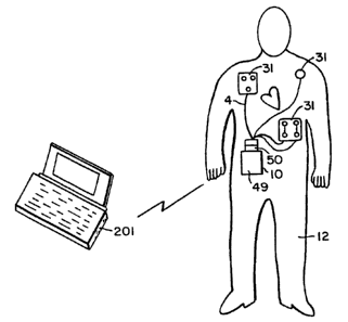
representative embodiment of the invention is shown in Figures 1 and 2.
As shown in these figures, defibrillation system 1 is comprised of base
station 2, electrode harness 4, personal computer 6, patient simulator 7,
central repository 9, and wearable defibrillator 10. A brief overview of the
operation of each of these components is provided below, followed by
detailed descriptions thereof.
Defibrillator 10 is capable of interfacing either to base station
2, as shown in Figure 1, or to electrode harness 4, as shown in Figure 2.
To this end, both electrode harness 4 and base station 2 include physical
-12-
t
CA 02283339 1999-09-03
WO 98/39061 PCT/US98/04585
connector identifiers at their respective interfaces to defibrillator 10. By
reading these connector identifiers, defibrillator 10 is able to determine
both the type of interfaced device (i.e., a base station or electrode harness)
and the identity of a particular interfaced device (i.e., one electrode
harness
as opposed to another), and then to react accordingly
Electrode harness 4 includes one or more sensing electrodes
31 which interface to patient 12, and which are used both to monitor the
patient and to transmit defibrillation energy to the patient. In this regard,
although defibrillator IO may be utilized with non-segmented electrodes
having a low surface area or with traditional defibrillation electrodes,
sensing electrodes 31 comprise segmented electrodes since these require the
most description. Defibrillation energy, which can comprise an electric
signal having a bi-phasic waveform, a mono-phasic waveform, or a
truncated exponential waveform, is generated by defibrillator 10 in the
event that predetermined conditions have been detected in the patient.
These predetermined conditions include whether the patient has suffered a
cardiac arrhythmia, whether the patient is conscious, as well as other
conditions, such as patient impedance, that are monitored by sensing
electrodes 31.
Defibrillator 10 is also capable of providing pacing impulses
and tactile stimulation signals to the patient via electrode harness 4. The
tactile stimulation signals alert the patient to abnormal cardiac activity in
the patient, whereas the pacing impulses stimulate contractions of the
patient's heart. While electrode harness 4 is being worn by the patient,
data may be transmitted directly from defibrillator 10 to personal computer
201 via non-contact interface 16.
When defibrillator 10 is interfaced to base station 2, as
shown in Figure 1, base station 2 is able to perform diagnostics on the
defibrillator, to reprogram the defibrillator, and to retrieve data stored in
the defibrillator. Such data can include an operational history of the
defibrillator, information concerning the patient's cardiac activity, and the
like. All or some of this retrieved data may be transmitted, via personal
computer interface 14, to personal computer 6 for display and/or
-13-
i i
CA 02283339 1999-09-03
WO 98/39061 PCT/US98/04585
processing.
Data retrieved by base station 2 from defibrillator 10 may be
transmitted to central repository 9 via external data link 17. Central
repository 9 preferably stores this data, together with patient and
S defibrillation information corresponding to a plurality of other patients,
all
of whom use the same type of defibrillator. A personal computer 19 is in
communication with central repository 9. This personal computer may be
used to analyze the patient and defibrillation information received from
defibrillator 10 in view of corresponding information from the plurality of
other patients, and, if desired, to provide the results of this analysis back
to
base station 2.
As shown in Figure 1, defibrillator 10 also includes a link to
patient simulator 7. Patient simulator 7 comprises test equipment which
simulates bodily functions and characteristics of a patient, including cardiac
activity and thoracic impedance. During testing, defibrillator 10 monitors
patient simulator 7 in much the same way that defibrillator 10 monitors a
patient and, in a case that predetermined conditions have been detected in
patient simulator 7, transmits defibrillation energy to patient simulator 7.
To aid in the testing process, patient simulator 7 also simulates patient
responses to the defibrillation energy provided by defibrillator 10 and
provides response information back to defibrillator 10. This response
information may be transmitted to, and analyzed by, base station 2, and
then provided to any one or more of central repository 9, computer 6, or
defibrillator 10.
Electrode Harness
Figure 3 shows a close-up view of electrode harness 4.
Electrode harness 4 is preferably disposable and, in preferred embodiments
of the invention, can be worn for approximately 2 to 7 days or longer for a
cumulative period of 1 week to 12 months. To this end, electrode harness
may include a means for defibrillator 10 to determine how long electrode
harness 4 has been connected thereto. For example, in one embodiment of
the invention, electrode harness 4 includes an identification resistor {not
-14-
T
CA 02283339 1999-09-03
WO 98/39061 PCT/US98/04585
shown) as its physical connector identifier. Defibrillator 10 measures the
resistance across this resistor and then starts a countdown, after which
defibrillator 10 notifies the patient that it is time to change the electrode
harness. In this regard, each electrode harness may include a different,
unique resistance associated therewith. Defibrillator 10 may measure this
resistance by passing a current therethrough and, in this manner, determine
the identity of an interfaced electrode harness.
As shown in Figure 3, electrode harness 4 includes power
supply 20, connector 21, non-electrically conductive padding 22 and 24,
electrical leads (or "lines") 26, 27, 29 and 30, and sensing electrodes 31.
Sensing electrodes 31 comprise the defibrillator's interface to the patient.
Specifically, sensing electrodes 31 attach to the patient so as to monitor the
patient, transmit tactile stimulation energy, and to transmit defibrillation
energy to the patient under appropriate circumstances. Each electrode may
comprise a single layer of conductive material. In preferred embodiments
of the invention, however, each electrode is multi-layered as shown, for
example, in the cross-sectional view of electrode 31a in Figure 4. In the
example shown in Figure 4, electrode 31a includes three layers, namely top
cover assembly 32, conductor/wire assembly 33, and skin interface 32
Skin interface 32 contacts directly with the patient's skin and
comprises a layer of material, such as a hydrogel, that is capable of
transmitting defibrillation energy to the patient without causing substantial
irritation or harm to the skin. For larger patients, or hypoallergenic
patients, conductive screens or meshes can be used in addition to or in
place of hydrogel. These screens or meshes may be used in combination
with a cream, such as a hydrating cream or a skin healing cream. Such
creams also may be applied to the patient's skin before attaching the
electrodes thereto.
Skin interface 32 contacts to conductor/wire assembly 34,
which can be either substantially coextensive with, or smaller than, skin
interface 32. Conductor/wire assembly 34 includes conductive layer 34a,
wire connection 34b, wire 34c, and sealing layer 34d. Conductive layer
34a preferably comprises a silver/silver chloride polymer base ink silk-
-15-
CA 02283339 1999-09-03
WO 98/39061 PCT/US98/04585
screened onto a layer of Tyvek (used as an insulator and as a carrier)
which is die-cut and folded. A wire with a welded washer is then attached
to conductive layer 34a by means of a washer (tin plated nickel) and eyelet
(a hollow rivet that is crimped in order to hold the assembly together).
Insulating tape is then wrapped around this connection in order to reduce
corrosion.
As an alternative to the silver/silver chloride formulation,
conductor 34a may comprise conductive metal such as tin, silver, gold,
copper, salts or oxides of these conductive metals, carbon, a substrate
which has been coated with a conductive compound (e.g.,
polytetrafluoroethylene), an ink silkscreened on a carrier, metallized cloth,
solid metal or carbon grid, foil, plate, etc. Conductor 34 preferably has a
thickness which is sufficient to transmit at least ten successive
defibrillation
energy singals having peak amplitudes of 23 amperes for a duration of
10.75 msec each.
Top cover assembly 33 includes foam insulating layer 33a
and wearable adhesive layer 37. Adhesive 37, which can comprise an
adhesive material fixed to a backing, such as tape or the like, is disposed
adjacent to conductor 34 and/or skin interface 32 and is used to attach
electrode 31a to the patient's skin. In preferred embodiments of the
invention, adhesive 37 may also be temperature sensitive, meaning that
adhesion thereof increases or decreases in response to temperature
variations. Adhesive 37 is preferably non-conductive.
Adhesive 37 should also be adapted for long-term wear. To
this end, an adhesive having a high moisture vapor transmission rate
("MVTR") of approximately 300 to 1500 g/mzlday is suitable for use with
the invention. By virtue of this feature of the invention, the adhesive is
made breathable, meaning that it permits air to be transmitted therethrough
to the patient's skin. This increases the amount of time an electrode may
be worn without causing substantial harm to the patient's skin. Adhesive
37 should also be sufficient to adhere to the patient's body in the face of
normal movements or muscle contractions and in the face of normal water
exposure such as might occur during bathing or sweating. However,
-16-
r ~
CA 02283339 1999-09-03
WO 98/39061 PCT/US98/04585
adhesive 37 should not be so strong as to cause substantial discomfort
during removal of an electrode. To this end, adhesive 37 preferably has a
peel strength of 500 g/cm or less.
Also shown in Figure 4 are applicator 35 and release liner
assembly 36. Applicator 35 includes two layers - a bottom layer having
cut-outs 35a and a top layer having an adhesive 35b. Cut-outs 35a limit
the amount of adhesive that contacts the top side of the electrode. Release
liner assembly 36 includes cut-out 36a on upper layer 36b {closest to the
hydrogel) which causes only a portion of urethane bottom layer 36c to
come into contact with upper layer 36b. This configuration of release liner
assembly 36, particularly cut-out 36a, allows bottom layer 36c to be
removed first from an electrode, followed by upper-layer 36b, without
causing any separation of the electrode from the applicator assembly.
Moreover, cut-outs 35a on applicator 35b facilitate removal of applicator
35b from top cover assembly 35 without causing harm to the electrode.
An alternative electrode configuration to that shown in
Figure 4 is shown in Figure 18. Figures 19 to 24 show additional
alternative electrode configurations. Figure 19 shows a finger-patterned
electrode 200 comprising a conductive adhesive polymer layer 220, a
carbon sheet 240, and a medical adhesive carrier 260, covering and
extending beyond the edges of the polymer layer 220 and the carbon sheet
240. In other embodiments, a metal sheet or a metal fabric may replace
carbon sheet 220.
Figure 20A and Figure 20B show a rectangular electrode 300
comprising metal foil sheet 320, pressure pad backing 340 and medical
adhesive carrier 360, covering and extending beyond the edges of the metal
foil sheet 320 and the pressure pad backing 340. The front surface of
metal foil sheet 320 comes in contact with patient's skin 380 and pressure
pad backing 340 contacts the back surface of metal foil sheet 320 and keeps
metal foil sheet 320 in close contact with skin 380. In this regard, a
pressure pad is a unit which can be deformed by pressure applied in the
direction perpendicular to the skin. By maintaining a thickness that is less
than a free dimension, pressure in the pad is assured. Medical adhesive
-17-
i i
CA 02283339 1999-09-03
WO 98/39061 PCT/IJS98/04585
carrier 360 contacts pressure pad backing 340. In another embodiment, a
metal fabric replaces metal foil sheet 320.
Figures 21A and 21B show rectangular electrode 400
comprising metal foil sheet 420, pressure pad backing 440, stiffener 460,
and medical adhesive carrier 480. The front surface of metal foil 420 is in
contact with skin 490; the front surface of pressure pad 440 is in contact
with the back surface of metal foil 420; the front surface of stiffener 460 is
in contact with the back surface of pressure pad 440; and medical adhesive
carrier 480 is in contact with stiffener 460 and covers and extends beyond
the edges of all other layers (420, 440, and 460). Stiffener 460 comprises
a material which will resist bending, and stiffener 460 is used to transmit
force into the electrode area. In a preferred embodiment, stiffener 460
comprises a thin plastic material with an area that is slightly larger than
areas of metal foil sheet 420 and pressure pad backing 440. In another
embodiment, a metal fabric replaces metal foil sheet 420.
Figure 22 shows electrode 500 with alternating strips of
active areas 520 and space 540 for breathing. Each active area 520
comprises a metal foil sheet and a pressure pad backing. The strips of
active areas 520 are in contact with stiffener 560, and stiffener 560 is in
contact with medical adhesive carrier 580. In another embodiment, a metal
fabric replaces the metal foil in each active area 520. In yet other
embodiments, each active area comprises a metal foil sheet or a metal
fabric, a pressure pad backing and a stiffener. The back surfaces of the
active areas are in contact with a medical adhesive carrier.
Figure 23 show electrode 600 with multiple small square-
shaped active areas 620. As in the alternative strip configuration, active
areas 620 are spaced for breathing. Each active area 620 comprises a
metal foil and a pressure pad backing. The back surfaces of active areas
620 are in contact with the front surface of stiffener 640, and medical
adhesive carrier 660 is in contact with the back surface of stiffener 640. In
another embodiment, a metal fabric replaces the metal foil sheet. In yet
other embodiments, each active area comprises a metal foil sheet or a metal
fabric, a pressure pad backing and a stiffener. Back surfaces of the active
-18-
r
CA 02283339 1999-09-03
WO 98/39061 PCT/CTS98/04585
areas are in contact with a medical adhesive carrier.
Figure 24 shows an embodiment of the electrode in which
the adhesive surface is covered with a pull-tab covering to allow the patient
to place the electrode on the skin and then pull the tab to expose the
adhesive surface and maintain the electrode in place.
A conductive portion of each sensing electrode, which in this
case are segmented electrodes, preferably has a surface area that is roughly
8 to 10 cm2; although other dimensions may be used. The present
invention, however, takes advantage of "spreading resistance" in the
patient's bodily tissue so as to permit this reduction in the surface area of
the conductive portion each electrode. Spreading resistance is a property of
human tissue which causes defibrillation energy (or any other electric signal
for that matter) applied to the patient's skin to spread outward over the skin
and downward and outward through the patient's tissue. In the context of
i5 the present invention, once current from the defibrillation energy is
applied
from an electrode to the patient's skin, the current diffuses beyond the
electrode and continues to diffuse as the current moves into the underlying
tissue. As a result of this diffusion, the density of the current decreases
with increasing distance from the perimeter of the electrode. The present
invention compensates for this by placing sensing electrodes 31 in a
geometric pattern such that the interaction between diffusing current from
each sensing electrode results in an accumulation of spreading current in
areas between sensing electrodes 31. The result is that an effective area is
created in which current densities in the path between groups of sensing
electrodes (e.g., sensing electrodes 31a, 31b and 31c shown in Figure 3)
are similar to that of a large electrode having a perimeter equal to an outer
perimeter of all of the sensing electrodes in the group. A similar effect
may be achieved through random placement of the electrodes on the
patient.
Thus, by spatially arranging the sensing electrodes to take
advantage of human tissue's spreading resistance, the present invention is
able to create a "virtual" conductive surface using relatively small
electrodes. The virtual conductive surface can be significantly larger than a
-19-
i
CA 02283339 1999-09-03
WO 98/39061 PCT/US98/04585
combined conducive area of the individual sensing electrodes. This also
contributes to a lower impedance for a combined surface area of the
sensing electrodes than would be the case for a continuous electrode having
a similar surface area.
Each of sensing electrodes 31 may be shaped so that the
conductive portion thereof has a perimeter which is greater than a
circumference corresponding to a radius of the electrode. That is, since
charge tends to migrate to the perimeter of an electrode, the present
invention attempts to maximize the perimeter of each electrode, particularly
conductive surfaces thereof, thereby increasing the amplitude of the
defibrillation energy that the electrode can handle without causing
substantial burns to the patient. Examples of electrodes with such a
perimeter include star-shaped electrodes, square electrodes, swirled-shaped
electrodes, etc. It is, however, noted that conventional circular electrodes
may be used in conjunction with the present invention as well
To increase operational efficiency of sensing electrodes 31,
sensing electrodes 31 should be placed within a "thoracic window" on the
patient's body. A thoracic window is defined as an area of the patient's
body which is suitable for placing electrodes so as to optimize delivery of
defibrillation energy to the patient's heart, and is described in an article
by
Geddes et al. the American Heart Journal, volume 94, page 67 (1977), the
contents of which are hereby incorporated by reference into the subject
application as if set forth herein in full. In this regard, there are two
currently defined thoracic windows on a patient. These comprise the
anterior-posterior thoracic window and the apex-sternum thoracic window.
In the apex-sternum thoracic window, electrodes are typically placed
underneath the patient's left rib cage and over the patients right shoulder
area. In the anterior-posterior thoracic window, electrodes are typically
placed on a patient's lower left back and left front. Preferably, the sensing
electrodes are placeable over the thoracic window either randomly or in a
geometric pattern which is sufficient to cover a large enough area of the
patient's myocardium to cause adequate defibrillation upon application of
defibrillation energy.
-20-
r
CA 02283339 1999-09-03
WO 98/39061 PCT/US98/04585
Electrodes 31 can be attached or placed in contact with the
skin by various methods. Proper defibrillation requires that the electrodes
be in close contact with the patient's skin, in addition to being placed in an
appropriate location within the patient's thoracic window area. Preferably,
the electrode are attached to the patient's skin using an adhesive thereon, as
described in more detail below. However, other attachment means are
possible. For example, a thoracic wrap made out of cotton or spandex can
be used to assure proper placement of the electrodes and good contact
between the electrodes an the skin.
In order to ensure proper current accumulation in areas
between the sensing electrodes, each sensing electrode in a group (e.g., the
group of electrodes 31a, 31b and 31c) should be placed within a
predetermined distance of other sensing electrodes in the group. In
preferred embodiments of the invention, each sensing electrode in each
group of electrodes is separated from other sensing electrodes in that same
group by between 0.5 and 3 times an effective diameter of each electrode,
where the effective diameter corresponds to the farthest distance between
two points on the electrode. To ensure proper separation among the
sensing electrodes, electrode harness 4 includes non-electrically conductive
pads 22 and 24 (i.e., the passive areas), on which groups of sensing
electrodes can be mounted in predetermined geometric configurations. For
example, as shown in Figure 3, sensing electrodes 31a, 31b and 31c (i.e.,
the active areas) are mounted on pad 22 in a triangular configuration, while
sensing electrodes 31d, 31e, 31f and 31g are mounted on pad 24 in a
rectangular configuration. Although Figure 3 shows only two geometric
arrangements for sensing electrodes 31, the invention is not limited to
these. Rather, any geometric arrangement may be utilized, including, but
not limited to, a checkerboard pattern, swirl patterns, interlocking patterns,
star patterns, crescent patterns, E-shaped patterns, F-shaped patterns, L-
shaped patterns, X-shaped patterns, H-shaped patterns, O-shaped patterns,
C-shaped patterns, etc. Of course, the electrodes may be arranged in a
random manner as well.
Preferably, pads 22 and 24 are flexible so as to facilitate
-21-
i i
CA 02283339 1999-09-03
WO 98/39061 PCT/iJS98/04585
placement of sensing electrodes 31 on contours of the patient's body. It is
noted, however, that pads 22 and 24 need not be flexible. Rather, an
adhesive tape can be used in place of pads 22 and 24 or, alternatively, in
addition to pads 22 and 24. As still another alternative, pads 22 and 24
can be used selectively in electrode harness 4, meaning that pads can be
used to mount some of sensing electrodes 31 and not others. In fact, this is
the case in the representative embodiment of the invention depicted in
Figure 3. That is, in the embodiment shown in Figure 3, segmented
electrodes is not mounted on a pad, but rather "floats" , meaning that it can
be mounted anywhere on a patient's body, constrained, of course, by the
length of its electrical lead 30. As described below, electrode 31h does not
provide the defibrillation energy to the patient, but rather is used only to
monitor the patient's ECG. However, to provide greater flexibility in
electrode placement, in alternative embodiments of the invention, all
electrodes may float in the same manner as sensing electrode 31h. In the
case that all or some electrodes float, the invention may include an
applicator tray, such as tray 39 shown in Figure 5, having cups 40 which
arrange the electrodes in geometric pattens so as to ensure accurate
placement within the patient's thoracic window. That is, the applicator tray
ensures that the electrodes will be spatially arranged in the manner
described above so as to take advantage of human tissue's spreading effect.
As noted above, electrodes 31 may include a hydrogel or
other conductive material on a surface thereof which comes into contact
with the patient's skin, i.e., on the skin interface of the electrode. The
hydrogel is electrically conductive, thereby permitting transmission of the
defibrillation energy to the patient, but has a relatively low ion
concentration that is low enough so as to not to cause substantial skin
irritation. In preferred embodiments, the conductivity of the hydrogel is
variable based, e.g., on temperature, etc. In addition, the hydrogel
preferably has a relatively high MVTR, thereby making the hydrogel
breathable. As was the case above with respect to adhesive 37, this
reduces skin irritation caused by wearing the electrodes, and thus increases
the amount of time that the electrodes can be worn.
-22-
CA 02283339 1999-09-03
WO 98/39061 PCT/US98/04585
Hydrogels or other conductive materials used with
conventional ECG electrodes may be used in the present invention, since
the deleterious effects of such materials will be countered by the present
invention for reasons described both above and below. In addition,
S conductive materials which meet the above qualifications include
electrolytes, such as sodium chloride (NaCI), potassium chloride (KCl), or
lithium chloride (LiCI). Currently preferred hydrogel materials include
hydrophilic polymers, such as karaya gum, gum acacia, locust bean gum,
polysaccharide gum, modified polysaccharide, or polyacrylamide. A
hydrating agent, such as water or polyhydric alcohol (e.g., glycerine,
propylene glycol, triethylene glycol, glycerol, etc.) may also be included in
the conductive material. In these cases, water is typically present at a
concentration from about 1 % to 60% by weight. whereas polyhydric
alcohol is typically present at a concentration from about 10 % to 50 % by
weight.
As noted above, the electrode interface to the skin may
include, instead of or in addition to a hydrogel, a mesh, screen, or other
porous material. These elements are conductive and, due to their porous
nature, allow air to pass therethrough to the patient's skin. As was the
case with the hydrogel described above, this feature of the invention
provides for prolonged wearability of the electrodes.
The hydrogel on each sensing electrode may also include a
therapeutic or prophylactic agent which reduces skin irritation caused by
the electrode, and/or which promotes healing of wounds or skin irritation
that may be caused by the sensing electrodes. Such an agent may be
applied directly to each electrode, or capsules which release the agent in
response to the defibrillation energy may be applied to the electrode. A
therapeutic or prophylactic agent may also be included on each of pads 22
and 24 in order to promote skin health. Agents which render the patient's
skin porous, such as keratolytic agents (e.g., salicylic acid) or rubefacient
(e.g., methyl salicylate) may be included on each electrode or pad so as to
facilitate transmission of the therapeutic or prophylactic agent into the skin
and/or to permit use of low water content hydrogels.
-23-
l
CA 02283339 1999-09-03
WO 98139061 PCT/US98I04585
Examples of therapeutic or prophylactic agents that may be
used with the present invention include moisturizers, emollients,
bactericides, mold inhibitors, stabilizers or buffers to maintain a neutral PH
and to reduce corrosion and skin sensitivity, gelation inhibitors (e.g.,
Mg(OAc)~), healing agents, hormonal agents (e.g., hydrocortisone
(steroids)), protective agents, etc. Examples of acceptable bactericides and
mold inhibitors include antibactierals, antiseptics, antifungals, boric acid,
bacitracin, acriflavine, formaldehyde, gentian violet, mercuric sulfide,
mercurochrome, neomycin, and iodine. Examples of acceptable stabilizers
include oligo or polybasic organic acids and their salts (including chelating
agents), polyethers, tartaric acid, citric acid, and n-alkyl sulfonate, where
n
is from 8 to 16 carbon atoms. Examples of acceptable healing agents
include allantoin, peruvian balsam, vitamin A, and vitamin B. Examples of
acceptable protective agents include benzoin, charcoal, talc, zinc oxide, and
aloe vera. These therapeutic agents may be used both prior to use or after
use to promote healing. The amount of therapeutic or prophylactic material
used corresponds to an amount effective to reduce irritation, or to promote
recovery of irritated skin. The therapeutic or prophylactic agent may be in
any form useful to achieve the intended purpose, including liquid solutions,
creams, gels, solids, granules, powders or any other form, including
microcapsules. As noted above, the therapeutic or prophylactic agent may
be included as part of the electrode, e.g., incorporated in the conductive
areas of the electrode, or incorporated in a passive area of the electrode
array. Alternatively, the electrode array may comprise three areas:
electrode areas, passive areas and areas containing the therapeutic or
prophylactic material. In addition, the therapeutic or prophylactic material
may be applied to the skin prior to attaching the electrodes (pre-treatment),
or after the electrodes have been removed (post-treatment). In another
embodiment, skin irritation may be reduced by including in the electrode
array means for monitoring, controlling and/or correcting the skin
environment in contact with the electrode array. For example, it is
possible to monitor the electrode-to-skin PH, and adjust the PH
accordingly. Along these lines, the electrodes may comprise a multi-
-24-
r r ,
CA 02283339 1999-09-03
WO 98/39061 PCT/U898/04585
layered matrix for controlling ion flow between the skin and the electrode.
In alternative embodiments of the invention, rather than
using the sensing electrode configuration described above, i.e., segmented
electrodes, non-segmented electrodes having conductive portions of less
than 60 cm2 and, in some cases, even to less than 30 cm' may be utilized.
In this regard, traditionally, it was necessary for conductive portions of
defibrillation electrodes to have a surface area of 60 cmz to 80 cmz in order
to deliver a sufficient defibrillation energy to the patient. The present
invention, however, takes advantage of the "spreading resistance" effect
described above so as to permit reduction in the surface area of each
electrode. Of course, the features described herein with respect to sensing
electrodes 31 may also be used in conjunction with the non-segmented
electrodes described herein. These features include, but are not limited to,
using hydrogels having low ion concentrations, therapeutic and prophylactic
IS agents, and/or high MVTRs, effecting electrode movement relative to the
patient so as to reduce the deleterious effects of electrode-to-skin contact,
etc., utilizing an adhesive designed for long-term wear, etc. As well, the
following monitoring and energy-transmitting functions described with
respect to segmented electrodes may also be used in conjunction with the
non-segmented electrodes described herein.
In still other embodiments of the invention, traditional non-
segmented defibrillation electrodes, i.e., electrodes having a surface area of
60 cmz to 80 cm2, may be used in conjunction with all aspects of the
invention described herein, particularly those aspects of the invention that
provide for long term (i.e., greater than two days) wearability of the
electrodes. In this regard, these aspects include, but are not limited to,
using the electrodes in conjunction with hydrogels having low ion
concentrations, therapeutic and prophylactic agents, and/or high MVTRs,
effecting electrode movement relative to the patient so as to reduce the
deleterious effects of electrode-to-skin contact, etc. , utilizing an adhesive
designed for long-term wear, etc. As well, the following monitoring and
energy-transmitting functions described with respect to sensing electrodes
31 may also be used in conjunction with the traditional non-segmented
-25-
I I
CA 02283339 1999-09-03
WO 98/39U61 PCT/US98/04585
electrodes described herein.
It is noted that defibrillator 10 may also be used with any
type of commercially-available subcutaneous electrode as well. It is still
further noted that the invention may include combinations of two or more
of the foregoing types of electrodes.
Referring back to Figure 3, in electrode harness 4, each
group of electrodes and/or single electrode has at least one electrical lead
mechanically connected between its lead interface and power supply 20.
The leads can be standard cables comprising electrically conductive wires
sheathed in a flexible, protective material, e.g., a flexible plastic
material.
The wires used in the leads preferably are able to carry repeated
defibrillation impulses of at least 20 A for a 20 millisecond duration, and
preferably have adequate insulation qualities satisfying a high potential test
for about 1750 V. In addition, the cables preferably are flexible, durable,
soft and comfortable while having sturdy insulation.
An exploded view of power supply 20 is shown in Figure
17. As shown in Figure 17, power supply 20 includes base 180, top 181
and, sandwiched therebetween, batteries 184. Power supply 20 is also
comprised of connector 21, which includes the physical connector identifier
for electrode harness 4, and which interfaces power supply 20 and
electrical leads 26, 27, 29 and 30 to defibrillator 10. To this end,
connector 21 includes socket 186 which receives the electrical leads via
holes 185. Socket 186 fits within top 181 such that pin 188 on top 181
contacts with hole 190 on socket 186. Socket 186 is shielded so as to
protect signals being transmitted therethrough from batteries 184. This
shielding is tied to the "floating" ground in the battery and also to shields
from the leads to the electrodes.
In preferred embodiments of the invention, each electrical
lead is non-removably connected to connector 21 on power supply 20,
meaning that each lead is hard-wired to connector 21 or is otherwise
connected to connector 21 in a way in which removal of the electrical leads
from connector 21 will damage either one or both of these components. It
is noted, however, that alternative embodiments of the invention are
-26-
f I
CA 02283339 1999-09-03
WO 98/39061 PCT/US98/04585
possible, in which each lead is removably connected to connector 21
Power supply 20 comprises the primary power cell for
wearable defibrillator 10. To this end, batteries 184 comprise non-
rechargeable lithium batteries (e.g., DL123A, size 2/3 A) which are
capable of providing 2 to 7 days of normal operation of defibrillator 10,
including delivering at least six defibrillation energy (i.e., shocks) having
peak currents of 23 A to the patient. In this regard, the working voltage of
power supply 20 is preferably 3.3 to 6.6 V, with the maximum output
being 6.6 V. In preferred embodiments, power supply 20 is made water-
resistant by sealing power supply 20 within a silicone membrane (not
shown) or the like. In this regard, power supply 20 may be made water-
resistant by a number of other means as well. For example, it is possible
to use a variety of other insulating materials in place of silicone. By
sealing power supply 20 in this manner, it is possible to reduce the risk of
unintended electric shock during activities, such as showering or the like.
Each electrical lead on electrode harness 4 has a length that
is sufficient for each corresponding electrode or group of electrodes to be
placed on the patient at a predetermined distance away from others of the
electrodes or groups of electrodes. As noted above, by spatially arranging
defibrillator (as opposed to solely ECG) electrodes in this manner, the
invention ensures proper accumulation of current in areas between the
sensing electrodes. The present invention provides an additional advantage
in this regard in that groups of electrodes arranged in geometric patterns on
the patient are movable relative to the patient in a manner which ensures
that the geometric patterns are substantially retained, but are at different
orientations relative to the patient. For example, it is possible to rotate
pad
22 containing sensing electrodes 31a, 31b and 31c on a patient such that
pad 22 is still within the patient's thoracic window (e.g., either the apex-
sternum thoracic window, the anterior-posterior thoracic window, or some
combination thereof), but such that sensing electrodes 31a, 31b and 31c do
not contact the patient's skin at the same location. Random rotation or
movement may also be used to accomplish the same result. For example,
in a case that electrodes 31 all float, random motion may be the best way
-27-
l l
CA 02283339 1999-09-03
WO 98/39061 PCT/US98/04585
in which to achieve the desired result. Moreover, in accordance with the
invention, and particularly with non-segmented electrodes, it is possible
merely to shift each electrode from a first position to a second position,
such that portions of the first and second positions overlap.
By providing for the foregoing rotation and/or movement,
together with the therapeutic and prophylactic agents described above, the
present invention reduces skin damage which may occur due to prolonged
use of wearable defibrillator 10. To achieve optimum reduction in skin
damage, each sensing electrodes 31, group of sensing electrodes, or
individual non-sensing electrodes should be rotated in the above manner
roughly once every I2 hours to 7 days.
In operation, sensing electrodes 31 are capable of
transmitting electrical signals from defibrillator 10 to a patient. These
electrical signals include, but are not necessarily limited to, defibrillation
energy, tactile stimulation signals, pacing signals, and AC and DC signals
used to measure a patient's skin and thoracic impedance. The same
information used to measure the patient's thoracic impedance may also be
used to determine the patient's respiration and pulse rates. The
defibrillation energy preferably has a bi-phasic waveform with two phases.
These phases may have substantially equal durations or, alternatively, may
have different durations, e.g., the first phase may be roughly 6 msec and
the second phase may be roughly 4 msec. An example of a bi-phasic
waveform having substantially equal durations is shown in Figure 6
(described below).
As noted above, the waveform of the defibrillation energy
may also be mono-phasic, i.e., the waveform may comprise just the first
one of the two phases shown in Figure 6, or may comprise a truncated
exponential waveform. Monophasic waveforms typically require greater
current, generally on the order of 15 % to 20 % greater, in order to achieve
substantially the same effect as hi-phasic waveforms. Regarding the pacing
impulses, these preferably comprise waveforms having a low peak current
of roughly 150 mA.
Sensing electrodes 31 are also capable of transmitting patient
-28-
CA 02283339 1999-09-03
WO 98/39061 PCT/US98/04585
information from the patient to defibrillator 10. This patient information
includes, but is not limited to, information relating to the patient's skin
and
thoracic impedance, artifact noise in the patient's body (caused, e.g., by
cardiopulmonary resuscitation, agonal breathing, seizures, patient handling,
and ambulatory or vehicular motion), sensory stimulation signals from the
patient, and the patient's ECG, including any cardiovascular signals
evidencing abnormal heart activity.
In this regard, electrode harness 4 preferably includes two
independent differential channels, namely "ECG 1 " and "ECG 2" shown in
Figure 3, for measuring the patient's ECG. In this regard, groups of
electrodes 31a, 31b and 31c and 31d, 31e, 31f and 31g comprising ECG 1
both provide the defibrillation energy to the patient and monitor the
patient's ECG, whereas electrode 31h comprising ECG 2 is used solely for
monitoring the patient's ECG. ECG 1 serves as the primary monitoring
channel for ECG analysis, whereas ECG 2 serves as a backup for use in a
case that ECG 1 is not operating or is not operating properly (e.g., if ECG
1 is relatively noisy). Alternatively, ECG 2 can be used as a means of
verifying the validity of an ECG obtained via ECG 1. That is, by
comparing ECGs obtained from ECG 1 an ECG 2, it is possible to confirm
the validity of the patient's ECG. It is also possible to confirm whether the
electrodes are properly attached to the patient based on this comparison.
As still another alternative, it is possible to "average" ECGs from ECG 1
and ECG 2, in order to obtain an averaged ECG for the patient, or to
increase ECG resolution by using inputs from both ECG 1 and ECG 2.
The use to which defibrillator 10 puts the ECG signal, as well as the other
signals obtained via an electrode harness 4, is provided in detail below.
For example, defibrillator 10 provides several ways to
measure the patient's thoracic impedance based on information received
from sensing electrodes 31. Specifically, the patient's impedance is
determined directly by applying an AC or DC signal through an electrode,
sampling data obtained from the electrode in response to the AC or DC
signal, and analyzing the sampled data. More specifically, in preferred
embodiments of the invention, this method of measuring the patient's
-29-
l l
CA 02283339 1999-09-03
WO 98/39061 PCT/US98/04585
thoracic impedance entails sampling such data from an electrode within 5
seconds of defibrillator power-on and then every 10 seconds for a 160 msec
period at a sample rate of 4 msec or to continuously sample the impedance
signal. Filtering is then performed on the sampled data so as to reduce the
bandwidth of the sampled data to less than 10 Hz. The sampled data is
then analyzed so as to provide a noise magnitude estimate for the signal
and a frequency content of the signal. Subsequent processing is then
performed to obtain a measurement of the patient's thoracic impedance by
averaging all of the data samples in each 160 msec period. This averaging
and filtering is performed in order to reduce the effects of artifact noise
associated with CPR, muscle tremors, and agonal breathing. Of course,
this processing occurs in defibrillator 10, and not in electrode harness 4.
In addition to the foregoing method, the patient's thoracic
impedance is also calculated by defibrillator 10 each time the defibrillation
energy is transmitted to the patient. Specifically, the patient's thoracic
impedance may be determined before, during and after transmission of
defibrillation energy, based on patient information received from the
electrodes in response to the defibrillation energy.
Wearable Defibrillator
A close-up view of defibrillator 10 is shown in Figure 7A.
As shown in Figure 7A, defibrillator 10 includes housing 40, light emitting
diode (hereinafter "LED") 41, visual indicator 42, auditory indicator 44,
annunciator 46, user interface 47, communications link 49, and connector
50. Figure 7B is an exploded view showing the mechanical construction of
the features shown in Figure 7A. A description of these features of the
invention is provided below.
Housing 40 is preferably small enough to make the
defibrillator portable, and thus wearable, yet large enough to house the
circuitry included within defibrillator 10. In this regard, housing 40 can
comprise a belt, or the like, which a patient can wear around his or her
waist, chest, etc. However, in preferred embodiments of the invention,
housing 40 comprises a rectangular casing that is roughly 6 inches in
-30-
CA 02283339 1999-09-03
WO 98/39061 PCT/US98/04585
length, by 4 inches in width, by 2 inches in depth, or preferably 5.2 inches
by 3.2 inches by 1.5 inches. As shown in Figure 7B, housing 40 is
comprised of base 40a and top 40b. The preferred weight of defibrillator
is approximately 1.5 pounds or preferably 1.0 pound or lighter. In this
S regard, the invention is able to achieve its small size by using high-energy
- capacitors (e.g., 500 uF, 400 ~F, etc.) to store and deliver defibrillation
energy, as described in more detail below.
Connector 50 and communications link 49 comprise external
interfaces on defibrillator 10. Connector 50 is disposed on housing 40 so
10 as to permit connection of defibrillator 10 to mating connector 21 on power
supply 20 {see Figure 2), and to permit connection of defibrillator 10 to
corresponding mating connector 51 on base station 2 (see Figure 1). Ta
this end, connector SO preferably includes two high-speed data pins (not
shown), which permits base station 2 to interface to defibrillator 10 at a
same point that electrode harness 4 interfaces to defibrillator 10. By virtue
of this arrangement, information can be transmitted between defibrillator 10
and either base station 2 or a patient. Communications link 49 comprises a
non-contact interface to personal computer 201 (see Figure 2), over which
information may be transmitted between defibrillator 10 and personal
computer 201 (see Figure 2) while defibrillator 10 is being worn by the
patient or, if desired, at other times as well. Examples of such a non-
contact interface include an infrared (hereinafter "IR") link or a radio
frequency (hereinafter "RF") link.
Visual indicator 42 comprises a liquid crystal display
(hereinafter "LCD") or the like (in preferred embodiments, a Standish
162SLC 2x16 dot matrix display), having two lines for displaying
information, including text and errors, to a patient or to a clinician
operating defibrillator 10. This information can include, but is not limited
to, information concerning the patient's heart activity, such as the patient's
ECG or the like; messages upon detection of abnormal activity in the
patient's heart; statements indicating that the patient has been wearing
electrode harness 4 for greater than a recommended period of time;
-31-
i
CA 02283339 1999-09-03
WO 98/39061 PCT/LTS98/04585
instructions for the use of defibrillator 10; prompts for using defibrillator
I0; errors that have occurred in defibrillator 10; messages transmitted to
defibrillator 10 from an external location via base station 2, personal
computer 6, or personal computer 201; an indication that one or more of
sensing electrodes 31 have become detached from the patient; and an
indication that defibrillator 10 cannot differentiate between artifact noise
in
a patient's body and a cardiac arrhythmia. In preferred embodiments of the
invention, the information is displayed for about 15 seconds.
In addition to the LCD, one or more visual indicators, such
as light emitting diode (hereinafter "LED") 41, may be provided on
defibrillator 10 to indicate different operational states thereof. For
example, in preferred embodiments of the invention, LED 41 blinks when
defibrillator 10 is operating normally, is off when defibrillator 10 is
without power (including when power supply 20 has failed), and is
continuously illuminated during power-up error detection diagnostics
performed by defibrillator 10.
Auditory indicator 44 comprises a speaker (in preferred
embodiments, an MG Electronics MCS298 speaker) or the like, which
provides verbal messages corresponding to information displayed on visual
indicator 42. These messages may be output in a variety of different
languages on both visual indicator 42 and auditory indicator 44. In
preferred embodiments of the invention, auditory indicator 41 echos visual
display 42, and has a volume that is adjustable at least up to 60 or 70
decibels. Auditory indicator 44 may also be configured to provide
additional sounds, such as tones, buzzing, beeping, etc. to indicate error
conditions within defibrillator 10. Examples of such error conditions
include, but are not limited to, a low or drained power supply, improper
attachment of sensing electrodes 31 to the patient, detachment of sensing
electrodes 31 from the patient, and inoperability of defibrillator 10. In
addition, another auditory indicator, such as annunciator 46, may also be
included on defibrillator 10. Annunciator 46 is preferably separate from
auditory indicator 42, and produces a buzzing or other unique sound to
indicate error conditions within defibrillator 10, particularly a low or
-32-
t
CA 02283339 1999-09-03
WO 98/39061 PCT/LJS98/04585
drained power supply.
User interface 47 comprises a response button, whereby a
patient or clinician can provide an input to wearable defibrillator 10 simply
by pressing the button. As shown in Figure 7B, the response button is
comprised silicone 47a, button 47b, paddle 47c and foil 47d. The response
button cancels any upcoming defibrillation operation, meaning that, when
pressed, the response button terminates a current defibrillation and, in some
embodiments of the invention, disarms defibrillator 10. In preferred
embodiments of the invention, the response button is relatively large,
making it easily accessible to the patient, particularly through clothing and
the like. In addition, in preferred embodiments of the invention,
defibrillator 10 confirms when the response button has been pressed by
issuing both audio and visual messages. In operation, the response button
is pushed in response to, e.g., a "PLEASE RESPOND" verbal message so
as to confirm patient consciousness or lack thereof. In preferred
embodiments of the invention, defibrillator 10 also includes a cardiac event
recording button which may or may not be separate from the response
button, and which is pushed when the patient wants to record occurrence of
a cardiac event. It is noted that these two functions are combined during a
potential rescue situation; i.e., the patient will want to respond to the
defibrillator's response requests at the same time, possibly recording the
occurrence of a cardiac event. Combining these functions into a single
button helps train the patient to habitually push the same button whenever
there are requests from the defibrillator or cardiac events.
Figure 8 is a system functional block diagram of defibrillator
10. Briefly, the hardware and software functional blocks shown in Figure
8 monitor and analyze patient information (e.g., the patient's ECG)
received from electrode harness 4 in order to determine if a cardiac rhythm
is life threatening and requires defibrillation, and also permits
communication to base station 2, personal computer 6, and personal
computer 210. In addition, these blocks within defibrillator 10 determine
the patient's thoracic impedance based on information received from the
patient via electrode harness 4, and adjust the defibrillation energy
-33-
CA 02283339 1999-09-03
WO 98/39061 PCT/US98/04585
accordingly. Alternatively, a predetermined thoracic impedance value may
be stored in data logging memory block 57. That predetermined thoracic
impedance value may then be retrieved and used to adjust the defibrillation
signal.
As shown in Figure 8, defibrillator 10 comprises auditory
indicator 44, visual indicator 42, LED 41, user interface 47, and
annunciator 46. Descriptions of these features of the invention were
provided above and, therefore, will not be repeated here. It is worth
noting, however, that annunciator 46 receives power from backup battery
54, which is rechargeable, and which is independent of the primary source
of power (i.e., power supply 20) for defibrillator 10. Backup battery 54
provides power to annunciator 46 in the event that power supply 20 fails,
thereby giving annunciator 46 the ability to warn the patient in such an
event.
Defibrillator 10 also includes patient measurements block 56,
data logging memory block 57, accessory communications block 59, real
time clock (hereinafter "RTC") 60, RTC/RAM backup battery 61, which
may or may not be separate from backup battery 54, and signal generator
62. In the representative embodiment of the invention described herein,
signal generator 62 comprises processing block 64, defibrillation control
block 66, discharge/protection switches 67, capacitors 69, charger 70 and
charge control block 71. In this regard, although the present invention
includes the forgoing blocks in signal generator 62, any combination of
hardware and/or software which effects the same function as these features
can be employed in the practice of the present invention.
RTC 66 maintains the current date and time, receiving power
from rechargeable RTC/RAM backup battery 61 when defibrillator 10 is
powered-off. In preferred embodiments of the invention, RTC 60 is a
Dallas 1306 serial RTC, which is able to maintain the correct time and date
for up to 30 days without connection to power supply 20. RTC/RAM
backup battery 61 also provides power to a memory (e.g., a RAM (not
shown)). Accessory communications block 59 performs any filtering and
protocol conversion necessary to enable transmission of data between
-34-
r i ,
CA 02283339 1999-09-03
WO 98/39061 PCT/US98/04585
defibrillator 10 and base station 2, personal computer 6 andlor personal
computer 210. Patient measurements block 56 comprises analog signal
conditioning hardware which filters and digitizes ECG signals, thoracic
impedance measurements, and electrode-to-skin impedance measurements
received via electrode harness 4, and which transmits the resulting data to
processing block 64. Patient measurements block 56 also receives
information from an accelerometer, described below, relating to the
patient's motion, and provides this information to processing block 64.
Data logging memory block 57 stores both the operational
history of defibrillator 10 and information relating to the patient. More
specifically, data logging memory block 57 stores abnormal heart activity
of the patient; the patient's ECG before, during and after application of
defibrillation energy; an indication that the patient has been trained for use
with defibrillator 10; analyzed ECG conditions; ECG markings, including
defibrillation synch, external pace pulse, high slew rate, and saturation;
patient thoracic and electrode-to-skin impedance measurements over time;
voice, tone, and buzzer prompts; displayed messages; information
concerning patient interaction with the defibrillator 10 (e.g., if and when
the patient has pressed the response button); transmitted defibrillation
waveform measurements, including current and voltage versus time;
execution time measurements of defibrillator 10 for use in determining if
defibrillator 10 operated as expected; detected operational errors of
defibrillator 10 (including a type of error, persistence of the error, whether
defibrillator 10 was in the operational mode when the error occurred,
whether defibrillation had begun when the error occurred, and whether a
cardiac arrhythmia had been detected when the error occurred); calibration
data for defibrillator 10; the serial number of defibrillator 10; a harness
identification ID of an electrode harness interfaced to defibrillator 10; cold
and warm start information for defibrillator 10; artifact noise in the
patient;
data from an accelerometer relating to motion of the patient; documentation
regarding the defibrillator; instructions on how to use the defibrillator; and
patient parameters. These patient parameters include, but are not limited
to, the patient's ID number which is a unique assigned identifier for each
-35-
i
CA 02283339 1999-09-03
WO 98/39061 PCT/CTS98/04585
patient (range of 1 to 9,999; default=0); the patient's name; the language
used for voice and corresponding text messages; a minimum audio level to
which a patient responds; minimum tactile stimulation signal to which a
patient responds; maximum tactile stimulation signal for a patient; pacing
S bradycardia rate - heartbeat rate below which bradycardia will be declared
and pace rescue initiated; pace current level - current level needed to
ensure pace rescue; a ventricular tachycardia rate at which defibrillation
energy is to be delivered to the patient (range of 150 to 180 beats per
minute (hereinafter "bpm")); the patient's thoracic impedance range,
meaning, the impedance range during which defibrillation energy may be
transmitted to the patient (range of 15 to 200 ohms); a time and a date at
which the defibrillator was issued to the patient; a name of a clinic at an
external location (e.g., central repository 9, a hospital, a doctor's office,
etc.); a name of a clinician at the external location; and an electrode-to-
skin
impedance range which is used to determine whether the electrodes are not
attached, or are improperly attached, to the patient. Also stored with the
patient parameters is a checksum which is used to determine their validity.
In summary, data logging memory block essentially stores
any information provided to, or transmitted from, defibrillator 10 over a
predetermined span of time, such as two days. This data is preferably
stored in a log format and includes time data specifying a time at which
each event occurred. In addition, embedded in the data are validation and
synchronization mechanisms for use in detecting areas of corrupted or
missing data.
In preferred embodiments of the invention, data logging
memory block 57 has a capacity (e.g., 24 megabytes or 48 megabytes)
which is sufficient to record the foregoing information over a 48 hour
period of continuous use of defibrillator 10. In embodiments of the
invention where only a portion of the foregoing information is stored in
data logging memory block 57, there may be a corresponding decrease in
the size of data logging memory block 57 or a corresponding increase in
the available period of use for the device. On the other hand, to achieve a
reduction in the required amount of memory space without sacrificing data
-36-
r . ,
CA 02283339 1999-09-03
WO 98/390b1 PCT/US98/04585
stored therein, it is possible to compress the data, preferably at a 4-to-1
compression ratio, and then to store the compressed data in data logging
memory block 57. It is noted that the invention is not limited to
compressing the data at a 4-to-1 ratio, and that any compression ratio may
be used. Any of a number of well-known compression algorithms,
particularly those relating to biological signal compression, may be
employed to effect the necessary compression. Compression techniques
that result in lossless compression, however, are preferred. In particularly
preferred embodiments of the invention, data logging memory block 57 is
removable, and can be transferred to the base station or to an external
location at which point data stored therein may be downloaded.
Signal generator 62 generates, based on patient impedance
data provided from patient measurements block 56, defibrillation energy
which preferably has a bi-phasic waveform may have two phases with
substantially identical durations, different durations, or a first phase with
a
longer duration than a second phase, and having relatively low peak
amplitudes over a patient impedance range. A mono-phasic waveform or a
truncated exponential waveform having similar characteristics may also be
used.
Figure 6 shows a representative example of defibrillation
energy generated by signal generator 62. As shown in Figure 6, waveform
72 has two phases 74 and 76, each of which has substantially the same
durations 77 and 79 (i. e. , t~,ax - -- 5 .4 seconds) and a peak amplitude of
less
than 23 amperes (i.e., ImaX= --20A). Specifically, the bi-phasic waveform
has a 5.37 msec (+/- 5%o) initial "positive" phase, followed by a 100 to
300 ,usec zero-potential plateau (i.e., tsW;,~,,_ --180 sec), followed by
another 5.37 msec (+/- 5%) "negative" phase. In preferred embodiments
of the invention, the amplitude at the end of the pulse is 60 to 80% of its
initial value. Of course, the example shown in Figure 6 merely
representative, and waveforms having different values can also be
generated by the present invention. In fact, in preferred embodiments of
the invention, the duration of each phase of a bi-phasic waveform may be
-37-
i i
CA 02283339 1999-09-03
WO 98/39061 PCT/US98104585
between 4.5 and 6.4 msec.
The waveform corresponding to the defibrillation energy
preferably also has a substantially consistent tilt, as shown in Figure 6. In
preferred embodiments, the tilt of the defibrillation energy is between 53 %
and 79% . The present invention provides a way in which to alter this tilt
using capacitors 69. In this regard, capacitors 69 store energy which is
discharged to the patient via discharge/protection switches 67 in the form of
the foregoing defibrillation energy. This energy is discharged as a
waveform having the foregoing characteristics. The tilt of the waveform
(i.e., the rate of the waveform's exponential decay) is determined by the
"RC" time constant z of the circuit used to generate the waveform. In this
regard, that circuit's time constant z may be altered by switching
capacitors 69, as described below, so as to alter the overall, combined
capacitance value of capacitors 69. Thus, by switching capacitors 69 in
this manner, it is possible to vary the tilt of the waveform and the achieve
a low-current waveform.
To ensure safe operation, the energy for defibrillation energy
is discharged by capacitors 69 only in the case that a life-threatening
cardiac arrhythmia is detected in the patient (as determined by comparing
the patient's ECG to pre-stored patient parameters), the patient is deemed
to be unconscious (as determined, e.g., by the patient's response, or lack
thereof, to messages provided by auditory indicator 44, visual indicator 42,
and annunciator 46), and the patient's thoracic impedance is within a
predetermined range. In preferred embodiments of the invention, this
range is between 15 ohms and 200 ohms. The process of determining
whether and when to transmit defibrillation energy to a patient is described
in more detail below. One (or more) of capacitors 69 is also used to
provide the above-noted tactile stimulation signal to the patient. The tactile
stimulation signal may be used to alert the patient of the occurrence of a
cardiac event, and may vary based on the patient and based on which time
it is administered to the patient in response to a message from the
defibrillator. That same capacitor may also be used to deliver a pacing
-38-
r r
CA 02283339 1999-09-03
WO 98/39061 PCT/US98/04585
signal to the patient's heart in order to stimulate normal contractions
thereof.
The invention preferably includes 5 capacitors having
capacitances of 500 ~cF {350 V) each; although capacitors having other
capacitances may be used as well. The capacitors are switchable into the
"221 " configuration shown in Figure 9, in which there are two sets of
parallel capacitors in series with a single capacitor; the "2111"
configuration shown in Figure 10, in which there is one set of parallel
capacitors in series with three single capacitors; and the " 11111 "
configuration shown in Figure 11, in which there are five single capacitors
in series. In general, the higher the impedance of the patient, the lower the
peak current that is required for the defibrillation energy (a lower tilt is
also required at higher impedances). Consequently, the higher the
impedance of the patient, the lower the capacitance that is required to
generate the defibrillation energy. For example, for a patient whose
impedance is relatively high (e.g., 120 ohms), the 11111 capacitor
configuration may be used to generate defibrillation energy having a
maximum peak current of 15 A.
In this regard, the lower the impedance of the patient, the
higher the capacitance that is required to generate the defibrillation energy.
Thus, for patients whose impedance is relatively low (e.g., 40 ohms), the
221 capacitor configuration may be used to generate defibrillation energy
having a maximum peak current of 21 A.
-39-
l l
CA 02283339 1999-09-03
WO 98/39061 PCT/US98/04585
Table 1, shown below, provides values for I",~x_,., h~~R+, Ie,~n,
tmax+, tsW~«~,~ and t~,ax- (see Figure 6) for defibrillation energy for
patients
having impedances of 40, 60, 80 and 120 ohms. These values are merely
representative of a particular case, and other values, of course, may be
substituted therefor. In this regard, as noted above, the duration of each
phase is roughly between 4.5 - 6.5 msec, and the peak current is roughly
between 12 and 25A.
Table 1
40 ohms 60 ohms 80 ohms 120 ohms
ImaX~ 21A 20A 20A 15A
Ia~~~+15A 15A 15A 11A
Ir,~~~--7.OA -6. OA -S .OA -S . 5A
tmax+ 5.4ms 5.4ms 5.4ms 5.4ms
tsW~n 0.2ms 0.2ms 0.2ms 0.2ms
tmax- 5.4ms 5.4ms 5.4ms 5.4ms
In addition to using capacitors 69 to control the waveform of
the defibrillation energy in the foregoing manner, the invention may also
include a resistor, placed in series with the capacitors. Due to the
resistor's effect on the circuit's time constant, the resistor has the effect
of
"smoothing", i.e., decreasing the tilt of, the waveform of the defibrillation
energy. In this regard, generally speaking, an increase in the resistance of
the resistor decreases the tilt of the waveform. As an alternative to the
foregoing configuration, the invention may include a single capacitor and
one or more switchable resistors to achieve the effect of varying the
circuit's time constant and thereby varying the waveform of the
defibrillation energy.
As noted above, discharge/protection switches 67 control
delivery of the defibrillation energy from capacitors 69 to the patient.
Discharge/protection switches 67 are controlled by defibrillation control
-40-
..... T
CA 02283339 1999-09-03
WO 98/39061 PCT/US98/04585
block 66 which, in turn, is controlled by processing block 64. Specifically,
when defibrillator 10 is not in use, or is not required to provide an output
signal, processing block 64 commands defibrillation control block 66 to
open discharge/protection switches 67, thereby providing protection from
unwanted electric signals. On the other hand, when processing block 64
determines that defibrillation energy is to be transmitted to the patient,
processing block 64 immediately commands defibrillation block control
block 66 to close discharge/protection switches 67, thereby providing the
energy to the patient. In this regard, as noted above, one of capacitors 69
IO is also able to transmit tactile stimulation and pacing signals to the
patient.
In a case that a tactile stimulation or a pacing signal, as opposed to the
defibrillation energy, is to be transmitted, processing block 64 issues a
command to defibrillation control block 66 which, in response, may switch
one of discharge protection switches 67 so as to output a signal from only
one of capacitors 69. As shown in Figure 8, defibrillator control block 66
also monitors discharge/protection switches 67 and provides the results
thereof to processing block 64.
Charger 70 controls charging of capacitors 69 from power
supply 20, the input of which is labeled 20a in Figure 8. Specifically,
charger 70 comprises hardware which transfers energy from power supply
20 to capacitors 69. Charger control block 71 controls charger 70 in
response to commands received from processing block 64 so that capacitors
69 charge to a level commanded by processing block 64. In this regard,
processing block 64 may issue commands to charge capacitors 69 to one of
a plurality of different levels depending on a determined type of
arrhythmia, e.g., ventricular fibrillation versus ventricular tachycardia.
Thus, if a ventricular fibrillation is detected, then a signal having a higher
amplitude is output, whereas if a ventricular tachycardia is detected, then a
''cardio" or lower amplitude defibrillation signal is output. Processing
block may also issue commands to charge processing block 69 based on a
type of signal to be transmitted, e.g., defibrillation energy, a pacing
impulse, or a tactile stimulation signal.
Processing block 64 can comprise a microprocessor,
-41-
i
CA 02283339 1999-09-03
WO 98/39061 PCT/US98/04585
controller, or the like, as described below, which includes an internal
program memory (not shown in Figure 8). This memory is used to store
software modules comprised of process steps that are executable by
processing block 64. Specifically, process steps in these modules are
executable to control operation of defibrillator 10 based on input received
from a patient or, alternatively, from base station 2 (see Figure 1). These
software modules comprise input/output (hereinafter "I/O") module 80,
including communications submodule 81 therein, data recording module 82,
diagnostics module 84, defibrillation control module 86, ECG analysis
module 87, user interface protocols 89, and master control module 90.
I/O module 80 preferably comprises a BIOS module which
controls the transfer of data between software modules running within
processing block 64 and hardware components within defibrillator 10. I/O
module 80 also controls communications between processing block 64 and
auditory indicator 44, visual indicator 42, LED 41, and user interface 47.
To this end, the invention includes user interface protocols 89, between
master control module 90 and I/O module 80. User interface protocols 89
comprise command sequences for controlling transmission of various
prompts, such as tones, verbal messages, or the like, to the user via
auditory indicator 44, visual indicator 42, and LED 41 in response to
detected events, such as a cardiac arrhythmias or the like. Similarly, user
interface protocols 89 include command sequences for controlling receipt of
signals input by the user via user interface 47 (i.e., the response button).
Communications submodule 81 controls communications between
processing block 64 and an interfaced device, such as electrode harness 4
or base station 2, via accessory communications block 59. Processing
block 64 also executes a low-level, run time executive (hereinafter "RTE")
module (not shown), which supports communication between the various
software tasks running in processing block 64.
ECG analysis module 87 comprises process steps which
monitor the patient for a predetermined condition based on information
provided through electrodes 31. ECG analysis module analyzes the
patient's ECG and impedance data provided from sensing electrodes 31 (via
-42-
r i. ,
CA 02283339 1999-09-03
WO 98/39061 PCT/US98J04585
patient measurements circuit S6), together with other information provided
from the patient, including artifact noise and patient motion, in order to
determine whether, when, and what types of arrhythmias are present in the
patient's ECG.
S More specifically, ECG analysis module 87 performs any
necessary signal processing on the patient's ECG and thoracic impedance
data in order to remove any extraneous data such as may be present due to
noise in the patient's body, including artifact noise or noise caused by
patient motion. Assuming that ECG analysis module 87 is able to remove
extraneous noise from the patient's ECG and impedance data, ECG analysis
module 87 compares the patient's ECG and thoracic impedance data to the
patient parameters stored in data logging memory block S7 (e.g., the
patient's thoracic impedance range and ventricular fibrillation and
ventricular tachycardia rates at which defibrillation, pacing or cardio
1S (described below) signals should be administered) in order to determine
whether an arrhythmia has occurred. In the case that ECG analysis module
87 determines that an arrhythmia has occurred, ECG analysis module 87
analyzes the patient's ECG and the patient parameters in order to determine
the type of arrhythmia, i.e., whether the arrhythmia comprises ventricular
fibrillation, ventricular tachycardia, asystole, bradycardia, or
indioventricular rhythms, and whether defibrillation energy should be
transmitted to the patient in response to the arrhythmia. Specifically, ECG
analysis module 87 determines if the patient's rhythm is normal, meaning
that the patient has a normal heartbeat, in which case no intervention is
2S required; bradycardia, meaning that the measured heartbeat is less than S
bpm; ventricular fibrillation which comprises uncoordinated rapid
contractions of the heart which replace normal synchronous pumping
action, specifically in the heart's lower chambers; ventricular tachycardia
which comprise a threatening heart condition associated with a very rapid
heart rate ( > 1 SO bpm) but minimum pumping action; marginal ventricular
tachycardia (between 120 and 1S0 bpm - referred to as "walking VTs");
noise; and non-determinate signals, which are described below. The
invention may use one or more well-known analysis algorithms to make
-43-
CA 02283339 1999-09-03
WO 98/39061 PCT/US98/04585
these determinations. However, in preferred embodiments of the invention,
ECG analysis module 87 performs the algorithm shown in Figure 16.
The algorithm shown in Figure 16 essentially includes three
stages, ECG input and prefiltering stage 150, arrhythmia risk assessment
stage 151, and therapy decision algorithm stage 152. ECG input and
prefiltering stage 150 includes preconditioning filter step 154, during which
baseline stabilizer processing, noise filtering, bandpass filtering, and auto-
gain control are performed on the patient's input ECG data. In preferred
embodiments, at least some of these functions are performed in hardware in
patient measurements block 56 (e.g., ECG filter 119 and ECG amplifier 92
described below with respect to Figure 13). Following preconditioning
filter step 154, the ECG data is then transmitted to both rhythm analysis
step 156 and fault detection step 157.
Rhythm analysis step 156 detects peaks in the patient's ECG,
a beat morphology of the patient's heart, and a rhythm interval of the
patient's heart. Fault detection step 157 determines a noise level estimate
for noise in the patient's ECG, the electrode quality, meaning connection of
electrodes 31 to the patient, and the patient's ECG signal amplitude.
Results from steps 156 and 157 are provided to preliminary decision matrix
158 which makes a preliminary determination, based on the information
provided from steps 156 and 157 and based on stored patient parameters,
whether the patient's ECG comprises a benign rhythm, a complex rhythm,
or a rhythm that constitutes a risk of arrhythmia. This preliminary
determination is then provided to periodic arrhythmia review step 160.
Periodic arrhythmia review step 160 samples the input ECG data for fixed
time intervals (e.g., 10 to 20 seconds), or at variable time intervals for
arrhythmias which are preliminarily determined to be complex. This data,
along with the preliminary determination made in preliminary decision
matrix 158, is then passed along to arrhythmia risk assessment stage 151,
specifically to rhythm analysis step 161.
Rhythm analysis step 161 performs segment morphology
analysis on data received from periodic arrhythmia review step 160, and
also performs a temporal analysis of the results of the segment morphology
-44-
r i
CA 02283339 1999-09-03
WO 98!39061 PCT/US98/04585
analysis. Processing then proceeds to decision matrix 162 which either
confirms or refutes the preliminary decision made in preliminary decision
matrix 158 based on the processed data and stored patient parameters.
Specifically, decision matrix 162 either confirms or refutes that the
patient's ECG comprises a benign rhythm, an arrhythmia, or a complex
rhythm. Processing then proceeds to therapy decision algorithm stage 152,
specifically to rhythm analysis step 164. Rhythm analysis step 164
processes the patient's ECG data by performing thereon, a Fourier
analysis, a power spectral analysis, spectral analysis extraction, and a
multi-variate temporal analysis.
The processed ECG data output from rhythm analysis step
164 is then provided to noise analysis step 166. Noise analysis step 166
performs adaptive noise discrimination on the processed ECG data, and
then performs noise identification and classification so as to characterize
noise in the data. For example, the noise may comprise artifact nose, noise
from an internal pacemaker, etc. Thereafter, processing proceeds to
decision matrix 167. Decision matrix 167 either confirms that the patients
ECG is a benign rhythm, in which case decision matrix 167 merely permits
continued monitoring of the patient's ECG, or declares that there is an
arrhythmia. The results of decision matrix 167 are then output to therapy
decision step 169, which determines whether to provide defibrillation
energy to the patient based on the results of decision matrix 167, together
with patient physiologic measurements provided by step 170. These
physiologic measurements include consciousness detection, hemodynamic
assessment, and stimulus response tests.
By virtue of the foregoing, ECG analysis module 87 is able
to differentiate "treatable rhythms", meaning ECG and physiologic analysis
results which warrant application of therapy, from "non-treatable rhythms" ,
meaning ECG and physiologic analysis results which do not warrant
application of therapy. Non-treatable rhythms include normal sinus
rhythms, supraventricular tachycardia, atria! fibrillation (with or without
bundle branch block), atria! flutter, second and third degree heart block,
ventricular ectopy, premature ventricular contractions, and pacing (see
-45-
CA 02283339 1999-09-03
WO 98/39061 PCT/US98/04585
below). ECG analysis module 87 is also capable of recognizing
spontaneous organized cardiac rhythms that frequently follow defibrillation,
and are associated with the presence of pulse and blood pressure. These
rhythms are also classified as non-treatable. Treatable rhythms include
ventricular fibrillation (coarse), and high rate ventricular tachycardia that
are hemodynamically compromised and result in patient unconsciousness.
For ventricular fibrillation, the peak-to-peak amplitude should be greater
than 150 ~cV for the rhythm to be considered treatable, and for a
ventricular tachycardia the patient's rate must exceed the corresponding
patient parameter, namely the patient parameter corresponding to the
ventricular tachycardia rate at which defibrillation energy (or a cardio
signal (i.e., a low-energy defibrillation signal)) is to be delivered to the
patient.
When identifying treatable versus non-treatable rhythms, the
i5 foregoing process errs on the side of caution, meaning that it is more
likely
that the ECG analysis module 87 will identify a non-treatable rhythm as
treatable, than identify a treatable rhythm as non-treatable. This feature is
built into the system as a safety measure, so that the likelihood of
misidentifying a life-threatening arrhythmia is reduced.
ECG analysis module 87 is also able to identify other
rhythms. These include non-determinative rhythms and pace rhythms.
Pace rhythms correspond to heart rates that are sustained below 30 bpm
and which result in patient unconsciousness. These rhythms are treated
with a pacing signal, as opposed to a full defibrillation/cardio signal. Non-
determinate rhythms comprise rhythms which require additional analysis to
make a definitive decision as to whether defibrillation is required in
response thereto. Such rhythms may be the result of extraneous artifact
noise in the patient's body caused, e.g., by muscle contractions resulting
from movement. In the event that ECG analysis module 87 is unable to
differentiate between extraneous noise and the patient's ECG, ECG analysis
module 87 notifies master control module 90 which, in turn, outputs a
message to visual indicator 42 and/or auditory indicator 44.
-46-
r ,
CA 02283339 1999-09-03
WO 98/39061 PCT/US98/04585
In addition to the foregoing, ECG analysis module 87 is able
to determine, based on electrode-to-skin impedance data received from
electrodes 31 and the electrode-to-skin impedance range stored in data
logging memory block 57, whether and which electrode in electrode
harness 4 has become detached from the patient. To this end, patient
measurements block 56 preferably monitors separate, identifiable terminals
which permits ECG analysis module 87 to identify inputs from particular
electrodes in electrode harness 4. In the case that one or more of these
electrodes has become detached from the patient, ECG analysis module 87
passes such information along to master control module 90. ECG analysis
module 87 is also able to determine whether electrodes are attached to the
patient's skin based on thoracic impedance measurements. That is, if the
patient's thoracic impedance is determined to he above a predetermined
value, such as 200 ohms, ECG module ascertains that the electrodes are no
longer attached to the patient.
ECG analysis module 87 also determines the patient's heart
rate and "R-wave synchronization trigger" based on received ECG
information. In this regard, R-waves are present in the patient's ECG,
both intrinsically and, in some cases, due to an internal or external
pacemaker. Transmission of defibrillation signal (in this case a cardio
signal) to the patient must be synchronized with an R-wave in order to
avoid triggering ventricular fibrillation during a vulnerable period of the
patient's ventricles. This vulnerable period occurs during repolarization of
the ventricles, and usually begins 30 to 40 msec before the apex of a T-
wave in the patient's ECG, and ends near the apex of the T-wave. If
ventricular ischemia is present, the vulnerable period starts approximately
at the same time, but may persist for as long as 120 msec after the end of
the T-wave. In all cases, however, the onset of the vulnerable period
follows the peak of the R-wave by an amount of time that depends on the
patient's heart rate and on the patient's ECG. The onset ranges from
approximately 220 msec at a heart rate of 60 bpm to 120 msec and to as
low as 100 msec at a heart rate of 150 bpm. The R-wave synchronization
trigger determined by ECG analysis module 87 is used by master control
-47-
CA 02283339 1999-09-03
WO 98/39061 PCT/US98/04585
module 90, described below, to synchronize the defibrillation energy so
that the defibrillation energy is not applied during these vulnerable periods.
Specifically, defibrillation energy is synchronized so that it is delivered
within 60 msec of the patient's R-wave peak, if one is present. If no R-
wave is detected within a 500 msec monitoring window, then the
defibrillation energy is transmitted at the end of this window. In
determining the R-wave synchronization trigger, ECG analysis module 87
disregards stand-alone pacemaker pulses, meaning that only the heart's
reaction to these pulses is factored into determination of the R-wave
synchronization trigger, and not the pacemaker pulses themselves.
Master control module 90 integrates patient information from
ECG analysis module 87, patient responsiveness from user interface 47,
and outputs to auditory indicator 44, visual indicator 42, and LED 4I.
Figure 12 is a flow diagram which provides an overview of this aspect of
master control module 90's operation, including some operational aspects
of ECG analysis module 87. In step 51201 of the flow diagram, master
control module 90 receives analysis results from ECG analysis module 87,
which indicate whether the patient has suffered an arrhythmia, the type of
arrhythmia that the patient has suffered, and whether the arrhythmia is life
threatening. Master control module 90 also receives information
concerning the patient's thoracic impedance. As described below, this
information is used to determine the amplitude of the defibrillation energy
to be transmitted by defibrillator 10.
After step S 1201 receives the ECG analysis results and
impedance information, processing proceeds to step S1202, which
determines whether the analysis results indicate that the patient has suffered
a life-threatening arrhythmia. In a case that step 51202 determines that the
patient has not suffered a life-threatening arrhythmia, processing proceeds
to step S1203. Step 51203 issues commands to output a message, such as
"SEE A DOCTOR", to the patient, in the form of audio and visual signals
via audio indicator 44 and visual indicator 42, respectively. Thereafter,
processing returns to step S 1201.
On the other hand, in the case that step 51202 determines
-48-
CA 02283339 1999-09-03
WO 98/39061 PCT/US98/04585
that the patient has suffered a life-threatening arrhythmia, processing
proceeds to step S 1204. Step 51204 determines whether other pre-
conditions have been met before defibrillation energy is transmitted to the
patient. Specifically, step 51204 determines whether the patient's thoracic
S impedance is within a predetermined range, preferably between 15 and 200
ohms, and whether the patient's ECG has been confirmed by, e.g., both
ECG 1 and ECG 2 above taking into account artifact noise in the patient's
body. In a case that step 51204 determines that these pre-conditions have
been met, processing proceeds to step 51205; otherwise processing
proceeds to steps S 1203 and back to step S 1201.
Step S1205 determines whether the patient is conscious.
More specifically, in step 51205, master control module 90 outputs a
message (e. g. , a query) such as "ARE YOU THERE" . This message is
output both visually and audibly. After the message is output, master
control module 90 waits a predetermined period of time for a response
signal. This response signal may be input by the patient via user interface
47, specifically, by pressing the response button. In a case that master
control module 90 detects the response signal within the predetermined
period of time, master control module 90 ascertains that the patient is
conscious. Since defibrillator 10 does not administer defibrillation energy
to conscious patients, in this case, master control module 90 will not cause
defibrillation energy to be transmitted to the patient. Instead, processing
proceeds to step 51203, in which master control module simply issues
instructions to the patient, which can vary depending upon the severity of
the arrhythmia.
Thus, by pressing the response button, transmission of
defibrillation energy to the patient is averted. In this regard, transmission
of the defibrillation energy can also be averted upon detection of certain
errors in the defibrillator, disconnection of electrode harness 4 from
defibrillator 10, and removal of sensing electrodes 31 from the patient.
In step 51205, in a case that master control module 90 does
not detect a response signal within the predetermined period of time,
master control module 90 issues another audio or visual message (e.g., a
-49-
i i
CA 02283339 1999-09-03
WO 98/39061 PCT/LTS98/04585
louder message). After this second message has been transmitted to the
patient, master control module 90 again waits for a response from the
patient. In a case that the patient does not respond to this second message
within a predetermined period of time, master control module 90 issues a
third message to the patient. This message can be a still louder message or
can include a tactile stimulation signal which is transmitted via an electrode
on the patient. If the patient does not yet respond, master control module
90 issues a fourth and final message together with a tactile stimulation
signal. In this regard, it is noted that the format of these four messages
can vary. For example, verbal messages need not be louder, the tactile
stimulation signal can increase in intensity with each message or may be
applied at each message, etc. Moreover, there need not be four messages.
Rather, there can be more or less messages, as desired.
In the event that the patient responds to none of the
foregoing messages, master control module 90 ascertains that the patient is
unconscious. In this case, processing proceeds to step 51206, in which
case master control module 90 immediately thereafter issues a command to
defibrillation control module 86 instructing that the defibrillation energy be
transmitted to the patient. That is, in preferred embodiments of the
invention, as soon as the patient is determined to be unconscious, master
control module causes defibrillation energy to be transmitted to the patient
immediately, without waiting for further input from the patient, a clinician,
or other party. Included with the command to transmit the defibrillation
energy is data defining the amplitude and duration of phases in the
defibrillation energy. Master control module 90 determines this
information based on the measured thoracic impedance of the patient (see,
e.g., Table 1 above).
In preferred embodiments of the invention, the defibrillator
trains patients with periodical tests in use of the response button via a
response test training protocol. Clinicians introducing the defibrillator to
new patients are able to initiate execution fo the protocol from an attached
clinician station (not shown). The same protocols that are be used to
periodically test (and train} patients whenever the electrodes are replaced.
-50-
CA 02283339 1999-09-03
WO 98/39061 PCT/IJS98/04585
The protocol is based on first two alert levels (described below) and an
imminent response protocol (i.e., when the patient must response to a
message from the defibrillator) so that the patient learns and becomes
familiar with the first part of the imminent response protocol. The patient
learns what the voice message prompt sounds like at Level 1, and learn
what the Level 2 combined voice and tactile simulation signal sounds and
feels like. The repetition of the Level 2 prompts is to ensure that the
patient was given more than enough time to response. The response/test
training protocol serves to develop a habitual reaction in the patient to the
push response button in response to voice and tactile stimulation signal
prompts during a potential rescue situation; regularly expose the patient to
the "PLEASE RESPOND" voice menage prompt at Level l, Level 2 with
tactile stimulation signal if they do not respond to the first prompt;
indirectly show the patient that before electrical therapy will be applied to
them, they will be exposed to a series of voice and tactile stimulation
signals to confirm that the patient is unconscious each time the test is
performed, establish that the defibrillator is generating the voice and
tactile
stimulation prompts and that the patient can hear/feel them, can respond,
and the defibrillator is registering the response; each time, record how fast
the patient is responding to the prompts; and each time, determine that
patient interface is working and, otherwise, declare the defibrillator
unusable.
In some embodiments of the invention, in a case that a
patient is determined to be unconscious, master control module 90 may also
cause a message, such as "STAND BACK", to be output prior to
defibrillation so as to advise bystanders of an upcoming defibrillation. In
this regard, other messages may be provided to bystanders as well. For
example, it is possible to output a message such as "CALL FOR HELP" in
the event that a patient wearing the defibrillator is unconscious. Such a
message may even be output after defibrillation energy is administered to
the patient. In this regard, any number of different types of messages may
be included in the invention, which may be output at any time during the
defibrillation process.
-51-
i i
CA 02283339 1999-09-03
WO 98/39061 PCT/US98/04585
In addition to applying defibrillation energy to the patient,
master control module 90 also determines when it is time to provide a
pacing signal to the patient, and applies that signal accordingly. This
process is similar to the above process for applying defibrillation energy.
That is, in a case that master control module 90 determines that a patient's
heart rate is below 30 bpm and the patient is unconscious, a pacing signal
may be applied via the electrode harness 4.
Defibrillation control module 86 controls defibrillation
control block 66 and charge control block 71 in accordance with the
command received from master control module 90 so that defibrillation
energy which is appropriate for the patient can be generated and transmitted
to the patient.
Regarding the remaining software modules executing within
processing block 64, diagnostics module 84 performs diagnostics on
1 S defibrillator 10 relating to the operation and safety thereof prior to
transmitting defibrillation energy to the patient. These diagnostics include
diagnostics that are performed at power-on of defibrillator 10 in order to
determine if there are operational defects therein. In a case that diagnostics
module 84 detects operational defects as a result of these diagnostics, this
information is stored in data logging memory block 57 and is transmitted
back to master control module 90, which alerts the patient. Such
information also may be transmitted to base station 2 or to personal
computer 6.
Data recording module 82 controls transmission of data
between defibrillator 10, base station 2 and computers 6 and 210. This
data can include as noted above, abnormal heart activity of the patient; the
patient's ECG before, during and after application of defibrillation energy;
analyzed ECG conditions; ECG markings, including defibrillation synch,
external pace pulse, high slew rate, and saturation; patient thoracic and
electrode-to-skin impedance measurements over time; voice, tone, and
buzzer prompts; displayed messages; information concerning patient
interaction with the defibrillator 10; transmitted defibrillation waveform
measurements, including current and voltage versus time: execution time
-52-
r ,
CA 02283339 1999-09-03
WO 98/39061 PCT/US98/04585
measurements of defibrillator 10 for use in determining if defibrillator 10
operated as expected; detected operational errors of defibrillator 10;
calibration data for defibrillator 10; the serial number of defibrillator 10;
a
harness identification ID of an electrode harness interfaced to defibrillator
10; cold and warm start information for defibrillator 10; artifact noise in
the patient; data from an accelerometer relating to motion of the patient;
and patient parameters. Data recording module 82 also controls storage of
the foregoing data in data logging memory block 57.
Figure 13 is a block diagram showing representative
hardware used to implement the functions of defibrillator 10 described
above with respect to the Figure 8. The hardware components shown in
Figure 13 include processor board 91, which is comprised of ECG
amplifier 92, flash memories 94, program ROM 96, static RAM
(hereinafter "SRAM") 97, backup battery 99, voice chip 100, LCD circuit
i01, processor field programmable gate array (hereinafter "FPGA") 102,
voice driver 103, output circuit 104, reset/watchdog circuit 106, power
supply monitor 107, microprocessor 109, control FPGA 110, address and
data bus 111, crystal oscillator 112, and RTC 113. Also included within
defibrillator 10 are capacitors 69, transformer 114, power supply 20, input
circuitry 116, output circuitry 117, ECG filter 119, and accelerometer 120.
Generally speaking, processor 109, processor FPGA 102 and
control FPGA 110 perform the functions of processing block 64; flash
memories 94 perform the functions of data logging memory block 57;
control FPGA 110 performs the functions of defibrillation control block 66,
charge control block 71, and charger 70; processor FPGA 102, input
circuitry 116, ECG filters 119, accelerometer 120, and ECG amplifiers 92
perform the functions of patient measurements block 56; and control FPGA
110, together with a serial interface (not shown) and a non contact interface
(not shown), perform the functions of accessory communications block 59.
Remaining hardware components of Figure 13 that perform the functions
shown in Figure 8 are self-evident
The hardware components shown in Figure 13 are all tied to
an internal, "floating" ground during operation of defibrillator 10. This
-53-
CA 02283339 1999-09-03
WO 98/39061 PCT/ITS98/04585
means that there are no connections to an external ground when
defibrillator 10 is interfaced to a patient (although there may be external
ground connections when defibrillator 10 is interfaced to base station 2).
The use of floating grounds during operation of defibrillator 10 is important
from the patient's perspective, since it reduces the chances of unwanted
electric shock to the patient. This floating ground may be on power supply
20, which was described above. A detailed description of power supply 20
is therefore omitted here for the sake of brevity. Suffice it to say that
power supply 20 provides power to all components on defibrillator 10,
including processor board 91, input circuitry 116, and capacitors 69.
Accelerometer 120 measures a patient's motion and provides
this information to processor 109 via control FPGA 110. Processor 109
analyzes the information received from accelerometer 120, stores
information relating to patient motion, and uses this information in its
IS calculations of artifact noise noted above.
Input circuitry 116 receives signals from each of sensing
electrodes 31, negative and positive inputs from power supply 20, and a
connector ID from an interfaced device. Input circuitry 116 also controls
output of defibrillation, tactile stimulation and pacing signals to a patient.
To this end, included within input circuitry 116 are a plurality of switches,
one corresponding to each input/output. These switches open and close in
response to instructions from processor board 91 so as to ensure that
signals, such as the defibrillation energy, are not inadvertently transmitted
to a patient. Input circuitry 116 also includes shielding and the like on its
input/output signal lines so as to reduce the chances of damage to
defibrillation circuitry during application of defibrillation energy to the
patient.
Signals received from input circuitry 116 are transmitted to
ECG filter 119. ECG filter 119 comprises plurality of bandpass filters
used to filter signals received from electrode harness 4. These filtered
signals are transmitted to ECG amplifier 92 on processor board 91. ECG
amplifier 92 includes amplifying circuitry for amplifying the filtered signals
received from ECG filter 119, and an analog-to-digital converter for
-54-
1
CA 02283339 1999-09-03
WO 98/39061 PCT/US98/04585
converting the amplified signals from analog form into digital form. These
digital signals are then transmitted to processor 109 via processor FPGA
102.
Processor 109 receives clocking signals from crystal
S oscillator 112, which preferably provides signals up to 40 MHz. Processor
109 can comprise a microprocessor, microcontroller, or the like, and is
used to execute the software modules described above so as to control
- operation of defibrillator 10. Examples of microprocessors which have
been identified as suitable for use with the present invention include the
Intel 196 processor family, Intel 386EX (386EXTB), TMS320F206,
Motorola 68332, the TI "2x3" family DSP Processor, Amtel 8051 or
equivalent, Hitachi 8/S00 series, Mitsubishi M37700, and Motorola
68HC16, to name a few. Processor 109 also controls the application of
power to other components in defibrillator 10, particularly those on
1S processor board 91, and is able to cause these components to be powered-
up and powered-down for predetermined time intervals. This feature of the
invention reduces the amount of power consumed by defibrillator 10.
As another power saving feature, processor 109 is capable of
operating in different modes, during which processor 109 consumes
different amounts of power. Specifically, processor 109 is operable in a
normal mode, during which processor 109 samples data from electrodes 31
and controls defibrillator 10 in the manner described herein. Processor 109
is also operable in a low-power mode. In some embodiments of the
invention, processor 109 may be turned off completely. In preferred
2S embodiments, however, most processing in processor 109 ceases, but some
elementary routines remain running. During the low-power mode, all
internal registers in processor 109 retain their data, thereby making it
possible for processor 109 to resume normal operation upon re-entering the
normal mode. Processor 109 enters the low-power mode periodically,
e.g., at intervals of 1 to 2 ms, for predetermined periods of time, e.g., 20
ms. Alternatively, processor 109 can operate in the low-power and normal
modes at equal intervals, such as every 4 ms.
In preferred embodiments of the invention, the length of time
-SS-
CA 02283339 1999-09-03
WO 98/39061 PCT/US98/04585
that processor 109 operates in the low-power mode is variable based on
information received from the patient or, alternatively, based on
information received from central repository 9. For example, if processor
109 determines based, e.g., on the patient's ECG and previous history, that
S the patient is at a relatively low risk for a cardiac arrhythmia, processor
109 can lengthen the period of the low-power mode. Likewise, if
processor 109 determines, based on similar information, that the patient is
at a high risk for an arrhythmia, processor 109 can shorten, or even
eliminate, the low-power mode.
Another feature of the low power mode is that the amount of
power consumed therein may be varied. For example, in a case that
processor 109 determines that benign rhythms have occurred for a
relatively long time, processor 109 may enter a "deep" low power mode,
in which processor 109 is off, or in which only the most elementary of
routines remain running. On the other hand, in a case that processor 109
determines that a treatable rhythm occurred recently, processor 109 may
enter a "light" low power mode, in which less power is consumed than
when the processor operates in the normal mode, but in which more than
just elementary routines remain running in the processor.
To enter the low-power mode, in preferred embodiments of
the invention, processor 109 simply executes an "IDLE" instruction, during
which most internal processing in processor 109 is disabled. In response to
this IDLE instruction, processor 109 stops its internal clock and may
execute only low-level routines so as to perform minimal tasks, such as
determining when it is time to re-enter the normal mode. To this end, in
the low-power mode, processor 109, via control FPGA 110 (described
below), monitors signals received from RTC 113 and, based on these
signals, determines when it is time to re-enter the normal mode. In a case
that defibrillator 10 includes a button or the like (not shown) on its user
interface for placing processor 109 in the normal mode manually, processor
109 also monitors such a button during the low-power mode.
Flash memories 94 comprise removable EPROMs or the
like. Program ROM 96 stores the software modules described above with
-56-
~,
CA 02283339 1999-09-03
WO 98/39061 PCT/US98/04585
respect to Figure 8 which are executed by processor 109. Static RAM 97
comprises a memory out of which those software modules may be
executed. As described above, SRAM 97 is backed-up by backup battery
99. In this regard, backup battery 99 contains a rechargeable lithium coin
cell battery which is sufficient to back up both RTC 113 and SRAM 97.
Battery backup 99 may also be used to supply power to power annunciator
46. Alternatively, a second backup battery (not shown) may be used for
this purpose.
Reset/watchdog circuit 106 monitors processor 109 and
control FPGA 110 in order to determine if either processor 109 or control
FPGA has lost program control. For example, in a preferred embodiment
of the invention, control FPGA 110 outputs a square wave signal called
WATCHDOG OUT in a case that control FPGA 110 and processor 109
are communicating properly. Reset/watchdog circuit 106 monitors this
signal for variations therein. In a case that this signal is interrupted, or
has
an unexpected waveform, reset/watchdog circuit 106 ascertains that there
has been a system malfunction. As another example, processor 109 is
programmed to generate a signal called CONTROL PLD FAULT in a
case that control FPGA 110 has failed. Reset/watchdog circuit 106
monitors for this signal as well in order to ascertain if there has been a
failure in control FPGA 110.
In the event that either processor 109 or control FPGA 110
has failed, a system reset will be attempted by reset/watchdog circuit 106.
If the problem persists, reset/watchdog circuit 106 will instruct output
circuit 109 to output an alarm, e.g., a "tone" or a "buzzing" via
annunciator 46. In this regard, in the event of a system error which causes
a reset to be asserted, an alarm will not be sounded immediately. That is,
if the system successfully recovers after one reset, no alarm will be
sounded. This design allows the system to rebound from a temporary fault
without alerting the user unnecessarily. If, however, a persistent fault
condition exists which reasserts itself for at least 200 msec per second, the
alarm is sounded for 5 to 10 seconds. If the system subsequently recovers,
the alarm will cease to sound at the conclusion of this 5 to 10 second
-57-
i i
CA 02283339 1999-09-03
WO 98/39061 PCT/US98/04585
period; otherwise, the alarm will continue as long as the system reset signal
is asserted for 200 msec or more per second, stopping only when the
backup battery for annunciator 46 has been drained.
The system will also reset itself if power output by power
supply 20 drops below a predetermined level or goes above a
predetermined level. To determine if power supply 20 has gone below or
above these predetermined levels, power supply monitor 107 monitors
power supply 20. In preferred embodiments of the invention, power
supply monitor comprises plural comparators with associated circuitry for
making these determinations. In a case that power supply 20 is low, or is
outputting greater than a predetermined amount of power, this information
is transmitted to reset/watchdog circuit 106. In response, reset/watchdog
circuit 106 causes annunciator 46 to output an alarm via output circuit 104.
In a case that power supply 20 has failed, processor 109 shuts down
defibrillator 10 just after resetlwatchdog circuit 106 has caused annunciator
46 to output the alarm.
Processor board 91 also includes control FPGA 110 and
processor FPGA 102, which comprise glue logic for controlling inputs to,
and outputs from, processor board 91. Processor FPGA 102 contains
memory page registers, glue logic, and processor internal clock
stopping/starting circuitry. This clock stopping/starting circuitry stops an
internal clock of processor 109 for predetermined periods of time, such as
4 msec, during processor 109's low-power modes. Processor FPGA 102
also controls outputs to LED 41, auditory indicator 44 and visual indicator
42 (see Figure 8). In this regard, interfaced to processor FPGA 102 is
voice chip 100, e.g., an ISD33000 series Chip Voice Record/Playback
device which can store up to 60 seconds of prerecorded voice messages.
Voice driver 103 is also required to drive auditory indicator 44. An
example of such a driver is a TI TPA4861D (SOIL-8). Defibrillator 10
also includes LCD controller/driver 101 which, in preferred embodiments
of the invention, is an OKI MSM6555B or MSM6665 chip.
Control FPGA 110 contains a defibrillator state machine and
various registers for controlling operations of defibrillator 10. Among
-58-
r i r
CA 02283339 1999-09-03
WO 98/39061 PCT/US98/04585
these registers are capacitor configuration registers (not shown). By
writing into these registers, capacitors 69 can be programmed to enable
different capacitor configurations, i.e. , the 221, 2111 or 11111
configurations shown in Figures 9 to 11. More specifically, control FPGA
110 includes CAP SW1, CAP-SW2, CHRGELCB and CHARGE registers.
Writing a " 1 " into the CHARGE register enables capacitor charging,
whereas writing a "0" into the CHARGE register disables charging.
Writing a "1" into the CHRGELCB register switches all capacitors into a
parallel configuration for charging, whereas writing a "0" into the
CHRGELCB register charges only one of capacitors 60 and allows that one
of capacitors 69 to be placed into a series configuration for transmitting a
tactile stimulation or pacing signal. Writing a " 1 " into the CAP SW 1
register switches two of capacitors 69 into a series configuration, whereas
writing a "0" into CAP'SW 1 register switches the two of capacitors 69
into a parallel configuration. Writing a "1" into the CAP-SW2 register
switches two others of capacitors 69 into a series configuration, whereas
writing a "0" into CAP SW2 register switches the two others of capacitors
69 into a parallel configuration. Thus, values in CAP SW 1 and CAP SW2
control the configuration of capacitors 69 during a defibrillation.
Control FPGA 110 comprises a plurality of other registers
as well, including a defibrillator control register. Bits are written to the
defibrillator control register to set defibrillator 10 to provide either
defibrillation energy, a tactile stimulation signal, or a pacing signal.
Control FPGA 110 also has a number of other functions, including
monitoring the charge in capacitors 69 and adjusting the charge based on
signals (e.g., patient impedance) received from an interfaced device such as
electrode harness 4, monitoring inputs from user interface 47, e.g., the
response button, and providing output via a serial interface (not shown) to
base station 2 and via a non-contact interface (not shown) to personal
computer 6.
As noted above, outputs from control FPGA 110, namely
CAPSW1 and CAPSW2, control switching of capacitors 69 from the 11111
configuration into the 2111 and 221 configurations. Figure 14 shows a
-59-
CA 02283339 1999-09-03
WO 98/39061 PCT/ITS98/04585
detailed circuit diagram of capacitors 69 (and of transformer 114). In
preferred embodiments of the invention, the circuitry shown in Figure 14
fits on a circuit board having a surface area of roughly 2 inches2 or less.
As shown in Figure 14, capacitors 69a, 69b, 69c, 69d and 69e are charged
by power supply 20 via transformer 114. CAPSW I controls switching of
capacitors 69b and 69c via transistor switch 122, whereas CAPSW2
controls switching of capacitors 69d and 69e via transistor switch 124. As
noted above, this switching controls the overall, combined capacitance of
69a, 69b, 69c, 69d and 69e, which affects the amplitude and the tilt of a
waveform output therefrom. Additional signal conditioning and output
circuitry is also shown in Figure 14 but, since this particular circuitry is
not essential to the invention, a detailed description thereof has been
omitted for the sake of brevity.
It is worth noting, however, that Figure 14 also shows
circuitry I26 which is used to output tactile stimulation and pacing signals
from capacitor 69a. As shown in Figure 14, PACEP and TENSP, which
are output by control FPGA 110, control application of the pacing and
tactile stimulation and signals, respectively, from capacitor 69a. That is,
charge from capacitor 69a, namely CAP1 + 127, is applied to circuitry 126
and processed for output as the pacing or tactile stimulation signal. Figure
I4 also shows the CHARGELCB signal, which was described above, and
ISNSCB and IDSNSCB signals. The ISNSCB signal comprises a current
sense signal which is used to determine the charge of capacitors 69,
whereas the IDSNSCB signal is used to determine the defibrillation energy
current. This information is passed back to processor board 91, which
processes this information and responds in the manner described above.
For example, in the event that processor 109 determines that capacitors 69
are charged excessively for a particular patient such that over-current or
even over-time defibrillation could occur, defibrillator 10 may be shut
down or temporarily disabled so that capacitors 69 can be discharged
without harm to the patient.
Returning to Figure 13, output circuitry 117 includes signal
conditioning circuitry as well as control circuitry which ensures that
-60-
CA 02283339 1999-09-03
WO 98/39061 PCT/US98/04585
defibrillation energy will not be output inadvertently. In this regard, the
invention also includes other safety features which limit transmission of the
defibrillation control signal. For example, prior to delivery of the
defibrillation energy, processor 109 monitors and demonstrates that state
control clocks controlling the defibrillation energy are operational, and also
tests the hardware components in order to detect any single point failures
therein. These tests include testing switches in input circuitry 116, through
which the defibrillation energy is transmitted. These switches are tested,
one at a time, to demonstrate that each switch is capable of holding off a
full magnitude of the defibrillation energy. At each step of this switch test,
a voltage across each transistor (i.e., switch) is monitored to record a test
voltage and to record a transistor gate drive time constant in addition to
combined opto isolator and transistor turn-off times. These voltages are
then used to measure patient voltage during application of the defibrillation
IS energy. Upper transistor switches, through which the defibrillation energy
is transmitted, are tested first, followed by lower transistor switches. Each
transistor switch is monitored and tested to demonstrate that the
defibrillation energy would be terminated independently of hardware
control timing.
In addition, as noted above, two pre-conditions must be met
before defibrillation energy is transmitted the patient, namely, (l) the
patient has experienced a treatable rhythm and (ii) the patient is
unconscious. When these two conditions occur, processor 109 arms the
defibrillation controller, e.g., control FPGA 110. Specifically, processor
109 provides the following sequence of control signals to initiate arming of
the defibrillation controller. First, processor 109 tests its own internal
safety signal ("I'D SAFE") to demonstrate its ability to override any
hardware defibrillation control signals. Processor 109 then confirms that
defibrillation energy can be detected properly by activating test signals and
reading current feedback signals based on these test signals. Master control
module 90, executing within processor 109, sets a "defibrillator arm
request status" bit in memory for use and checking by an executive control
module (not shown). This executive control module monitors operation of
-61-
l l
CA 02283339 1999-09-03
WO 98/39061 PCT/US98/04585
the software and updates a watchdog timer in processor 109 (which outputs
the WATCHDOG OUT signal noted above) when the software is
confirmed to function properly. The executive control module then sets an
arm request signal in the defibrillation controller and calls a watchdog
update subroutine which transitions the signal output from the watchdog
timer. This causes a watchdog timer update and transitions the "armed
request" status to the "armed" status in the defibrillation controller. The
PD_SAFE signal is then put in the active state to allow the activation of
hardware control signals for defibrillation therapy.
Next, processor 109 provides a final synchronized trigger
signal to the defibrillation controller for delivery of the defibrillation
energy. This synchronized trigger signal will be accepted only after the
"armed" status has been established. The defibrillation controller will clear
the armed status if the synchronization trigger signal is not provided within
a 500 msec time period, thereby providing a limited acceptable period for
defibrillation therapy. In a case that the defibrillation energy is
transmitted
to the patient, following transmission thereof, processor 109 determines if
the defibrillation energy was transmitted properly. In this regard,
defibrillation energy dosage errors of over-current are protected by the
reset/watchdog circuit described above, but, if such an event does
inadvertently occur, a fault condition is retained in a hardware fault
register. Similarly, under-dosages of the defibrillation energy are also
detected and stored. In addition to being stored, these and other
defibrillator operational errors may be transmitted to base station 2.
Diagnostics module 84 also performs a plurality of
diagnostics on defibrillator 10 to test defibrillator 10's hardware. These
diagnostics include cold start diagnostics, which are executed when
defibrillator 10 powers-up normally, warm start diagnostics which are
executed when defibrillator 10 experiences transient reset or power loss,
runtime diagnostics which are continuing, periodic tests performed in the
background of normal operating conditions, and specific conditions
diagnostics which are tests that are performed prior to, or when certain
operations are performed, such as transmission of defibrillation energy.
-62-
r ,
CA 02283339 1999-09-03
WO 98/39061 PCT/US98/04585
Cold start diagnostics include RAM test, ROM test, D/A and
A/D converter tests, FPGA tests, RTC and ECG sampling rate tests, LCD
tests, voice circuitry tests, backup battery voltage tests, primary power
supply voltage tests, internal voltage tests, old electrode harness test
(i.e.,
whether the electrode harness has been changed after transmission of
defibrillation energy), safety tests so as to verify that safety controls are
operational, watchdog timer tests, shutdown tests, and patient parameters
validation. Warm start diagnostics include operational state data tests
(protected RAM validation) and patient parameters validation. Runtime
diagnostics include watchdog timer active tests so as to confirm that a clock
signal is active, software clock to real time clock comparison tests,
software execution times checks, system voltage tests so as to test if the
system is within voltage specifications, AID runtime reference voltage
tests, backup battery voltage tests, primary power supply voltage tests,
internal voltage tests, old electrode harness test, operational state data
integrity test, operational temperature tests, lead off tests, stuck keys
test,
safety tests including electrode harness time limit tests, defibrillation
capacitor voltage tests, defibrillation output circuitry tests, output voltage
tests, patient parameter validation, and impedance measurements. Specific
conditions diagnostics include tests which are performed prior to
transmission of defibrillation energy to the patient. These tests include
cross checking processor tests whereby control FPGA 110 checks processor
109 for correct performance of a command sequence, and processor 109
checks control FPGA 110 for correct progression of states during set-up
and transmission of defibrillation energy. Other tests include a watchdog
timer test which resets the system in a case that processor 109 fails to
respond periodically, electrodes-off tests in which processor 109 confirms
that electrodes are attached to the patient, operational therapy state data
integrity tests, pre-therapy dosage tests whereby energy to be transmitted as
defibrillation energy is compared with two impedance measurements prior
to transmission, stuck ECG relay contacts tests (prior to defibrillation), H-
bridge therapy tests, and delivered defibrillation current limiting tests.
-63-
CA 02283339 1999-09-03
WO 98/39061 PCT/US98104585
Base Station
In brief, a base station for use with the present invention
includes a defibrillator interface, over which information is exchanged with
the defibrillator, and an external interface over which information is
exchanged with an external entity, such as central repository 9, a doctor's
office, a hospital, etc. Also included in the base station is a controller
which receives patient information and defibrillation information from the
defibrillator, transmits the patient information and defibrillation
information
to the external entity, receives defibrillator programming information from
the external entity, programs the defibrillator in accordance with the
defibrillator programming information, performs diagnostics on the
defibrillator, and transmits results of the diagnostics to at least one of the
defibrillator and the external entity. Communication between the
defibrillator and the base station may be via an RF, IR, or direct electrical
connection. In addition, communication/testing may be effected by direct
contact between the sensing electrodes and the base station.
A block diagram of base station 2 is shown in Figure 15. As
shown, base station 2 includes, RAM 130, program memory 131, user
interface 132, non-volatile memory 133, defibrillator interface 134,
external interface 136, controller 137, address/data bus 139, and personal
computer interface 140. Base station 2 may receive power from an
external source, such as a wall outlet, or from a battery (not shown). Each
of the features of base station 2 shown in Figure 15 is described in more
detail below.
User interface 132 can comprise a keyboard, buttons,
switches, or the like, which provide a user with a way to control base
station 2 directly. Defibrillator interface 134 corresponds to connector 51
shown in Figure 1 and comprises an interface to defibrillator 10, over
which patient information (e.g., ECG information) and defibrillator
information (e.g., errors in operation of defibrillator 10) is received from
the defibrillator, and over which external information (i.e., information
received from an external source, such as new patient parameters) is
transmitted to the defibrillator. Defibrillator interface 134 includes base
-64-
r r ,
CA 02283339 1999-09-03
WO 98/39061 PCTIUS98/04585
station 2's physical connector ID. Defibrillator interface 134 is preferably
a serial interface and, as described above, mates to the same connector on
defibrillator 10 that is used to interface defibrillator 10 to electrode
harness
4.
Personal computer 6, which is interfaced to base station 2
via personal computer interface 140, may be used to indicate operational
characteristics of base station 2, such as when base station 2 is uploading
data, whether that upload was successful, a display of all uploaded data,
etc.
External interface comprises a link to an external location,
such as central repository 9 (see Figure 1) or a personal computer, over
which patient and defibrillation information is transmitted to the external
source, and over which the external information is received from the
external source. External interface can comprise a modem link, a network
connection, or the like, over which data may be transmitted to and from
base station 2. At this point, it is noted that all information stored in data
logging memory block 57 above can be transmitted over defibrillator
interface 134 and external interface 136 of base station 2.
Controller 137 comprises a microprocessor or the like, which
is capable of executing stored program instructions so as to control
operation of base station 2. Any type of processor may be employed, such
as those described above with respect to defibrillator 10. Program
instructions that can be executed by controller 137 are stored in program
memory 131. Program memory 131 preferably comprises an EPROM, or
the like, which can be reprogrammed with newly-received information or
routines by controller 137. In preferred embodiments of the invention,
program memory 131 stores control module 140, diagnostics module 142,
and patient parameters 144, among other data and software modules.
Patient parameters 144 correspond to the patient parameters
described above and, as noted, can be reprogrammed based on information,
such as instructions, provided from an external source. Control module
140 is executed so as to control transfer of information between
defibrillator 10, base station 2, and central repository 9. Diagnostics
-65-
l
CA 02283339 1999-09-03
WO 98139061 PCT/LTS98/04585
module 142 comprises a module which performs various safety diagnostics
on defibrillator 10 when defibrillator 10 is interfaced to base station 2. By
way of example, control module 140 may be executed to retrieve
information relating to operational errors of defibrillator 10 from data
logging memory block 57 of defibrillator 10. Diagnostics module 142 may
then use this information to target-test components and/or software on
defibrillator 10 that may be responsible for these errors. Alternatively,
diagnostics module 142 may perform a complete safety diagnostic check on
all aspects of defibrillator 10 each time defibrillator 10 is mated to base
station 2.
In this regard, base station 2 is capable of performing
diagnostics comprising an audio test to confirm that messages and tones
output by defibrillator 10 are clearly audible; measurement tests to confirm
that all measurements in defibrillator 10 needed to perform a defibrillation
procedure are as expected; ECG analysis tests so as to confirm that
defibrillator 10 is able to detect and differentiate various cardiac
arrhythmias; defibrillation waveform tests so as to confirm that defibrillator
10 can generate and output a waveform appropriate for a patient having a
particular impedance; patient leakage current tests so as to confirm that
leakage current in defibrillator 10 is not above an acceptable level; over-
dosage defibrillation tests so as to confirm that defibrillator 10 will
automatically terminate an over-current defibrillation (e.g., I",~x >_ 30 A)
and an over-time defibrillation (e.g., t",ax ? 20 msec); under-dosage
defibrillation tests so as to determine if defibrillator is providing an under-
current defibrillation (e. g. , Imax is less than a required current for a
patient's impedance measurement by more than 20%) or an under-time
defibrillation (e.g., t",aX is less than a required duration for a patient's
impedance measurement by more than 20%); power consumption tests so to
confirm that defibrillator 10 meets with the power requirements set forth in
Table 2 below; and button switch tests to confirm that each button on user
interface 47 is operating properly.
-66-
CA 02283339 1999-09-03
WO 98/39061 PCT/US98/04585
Table 2
Defibrillator O eratin Conditions Current Constraint
Low-Power Mode < 2 mA
Patient Monitoring Only < 20 mA
Detection of Ventricular Fibrillation < 400 mA
When any of the following operations < 900 mA
are active
i) Flash read/write
0 ii) LCD
iii) Voice message/Tone
iv) Accessory Communications
Charging of Capacitors < 3.0 A
Diagnostics module 142 may also be executed to check
proper transmission of patient parameters or other programming
information from base station 2 to defibrillator 10. More specifically, in
operation, base station 2 receives new patient parameters over external
interface 136 from central repository 9, and transmits these patient
parameters over defibrillator interface 134 to data logging memory block
57 in defibrillator 10. Diagnostics module 142 may then be executed to
issue a request for the patient parameters stored in data logging memory
block 57 over defibrillator interface 134, to receive the patient parameters
therefrom over defibrillator interface 134, and to compare the patient
parameters, including a checksum, to the same patient parameters, which
are stored in program memory 131, so as to verify valid receipt of the
patient parameters by defibrillator 10.
In preferred embodiments of the invention, the results of the
foregoing diagnostics may be transmitted back to defibrillator 10 in order
to warn the user of the defects via, e.g., an LCD or a speaker. In
addition, the results of such diagnostics may also be transmitted to an
external location via external interface 136 for analysis or the like.
Base station 2 also includes RAM 130, out of which
controller 137 executes program instructions stored in program memory
-67-
CA 02283339 1999-09-03
WO 98/39061 PCT/US98/04585
131, and non-volatile memory 133, which stores information received from
defibrillator 10 and from an external source. Non-volatile memory 133 can
comprise an NVRAM, battery backed-up RAM, EPROM, or the like, and
has a storage capacity which is the same as or greater than that of data
S logging memory block 57 on defibrillator 10. This is preferable, since
non-volatile memory 133 should be capable of storing any information
downloaded to base station 2 from defibrillator 10, including all or part of
the information described above that is stored in data logging memory
block 57, i.e., abnormal heart activity of the patient, the patient's ECG
before, during and after application of defibrillation energy, etc. In this
regard, upon connection of defibrillator 10 to defibrillator interface 134, in
preferred embodiments of the invention, controller 137 requests
defibrillator 10 to upload data stored in data logging memory block 57 and,
if the data has been uploaded successfully, controller 137 requests
defibrillator 10 to reset all recorded data in data logging memory block 57,
and also to clear patient parameters stored therein to their default settings.
As noted above, it is possible to reprogram defibrillator 10
and/or base station 2 with information received from the external location.
In fact, it is even possible to use information received from defibrillator 10
to affect such reprogramming. More specifically, information relating to
patients using the same type of defibrillator, i.e., defibrillator 10's type,
is
stored in central repository 9. This information can be analyzed in order to
test algorithms used in defibrillator 10. One such algorithm that may. be
tested is an algorithm used by ECG analysis module 87 above to detect
irregular heart activity. Once this algorithm is tested based on analysis
results from plural defibrillators, it is possible to pinpoint errors in the
algorithm, and to correct these errors. Thereafter, the corrected algorithm
can be transmitted back to base station 2, as well as to a plurality of other
base stations. In this regard, central repository may identify base station 2
as corresponding to defibrillator 10 based on defibrillator 10's ID number.
Base station 2 can then reprogram defibrillator 10 using the corrected
algorithm. As a result the invention provides a means by which to improve
its performance based on information collected thereby.
-68-
r y
CA 02283339 1999-09-03
WO 98/39061 PCT/US98/04585
The present invention has been described with respect to
particular illustrative embodiments. It is to be understood that the
invention is not limited to the above-described embodiments and
modifications thereto, and that various changes and modifications may be
S made by those of ordinary skill in the art without departing from the spirit
and scope of the appended claims.
-69-