Note: Descriptions are shown in the official language in which they were submitted.
CA 02283430 1999-09-07
WO 98/41266 - 1 - PCT/CA97/00186
TTTLE OF INVENTION
Elimination of vapour anaesthetics from patients after surgical
procedures
FIELD OF INVENTION
r 5 The purpose of this invention is to provide a simple breathing
circuit that can, for example, be added to a standard circle anaesthetic
circuit known to persons skilled in the art to hasten recovery of patients
administered vapour anaesthetics prior to an operation.
This invention also relates to the use of the breathing circuit in
hastening the recovery of patients who have been administered vapour
anaesthetics prior to a surgical operation.
This invention also relates to methods of treatment of patients to
hasten their recovery from administration of the vapour anaesthetics to
them prior to surgical procedures.
BACKGROUND OF THE INVENTION
Ph~siolow
Venous blood returns to the heart from the muscles and organs
depleted of oxygen (~) and full of carbon dioxide (~). Blood from
various parts of the body is mixed in the heart (mixed venous blood) and
pumped to the lungs. In the lungs the blood vessels break up into a net of
small vessels surrounding tiny lung sacs (alveoli). The net of vessels
surrounding the alveoli provides a large surface area for the exchange of
gases by diffusion along their concentration gradients. A concentration
gradient exists between the partial pressure of C02 (PC02) in the mixed
venous blood (PvCO~) and the alveolar PC02. The C02 diffuses into the
alveoli from the mixed venous blood from the beginning of inspiration
until an equilibrium is reached between the PvC02 and the alveolar PC02
at some time during the breath. When the subject exhales, the end of his
exhalation is considered to have come from the alveoli and reflect the
equilibrium concentration between the capillaries and the alveoli; the
PC02 in this gas is called end-tidal PCO~ (PE'rCO~ ).
When the blood passes the alveoli and is pumped by the heart to
the arteries it is known as the arterial PCO~ (Pa ). The arterial blood has
a PC02 equal to the PC02 at equilibrium between the capillaries and
alveoli. With each breath some C02 is eliminated and fresh air
containing little C02 {assumed to be O) is inhaled and dilutes the residual
CA 02283430 1999-09-07
WO 98/41266 PCT/CA97/00186
-2
alveolar PC02, establishing a new gradient for C02 to diffuse out of the
mixed venous blood into the alveoli. The rate of breathing, or ventilation
(V), usually expressed in L/min, is exactly that required to eliminate the
C02 brought to the lungs and maintain an equilibrium PC02 (and PaC02 )
of approximately 40 mmHg (in normal humans). V1/hen one produces
more C02 ( e.g. as a result of fever or exercise), more C02 is produced and
carried to the lungs. One then has to breathe harder (h~nerventilate) to
wash out the extra C02 from the alveoli, and thus maintain the same
equilibrium PaC02. But if the C02 production stays normal, and one
hyperventilates, then the PaC02 falls.
It is important to note that not all V contributes to blowing off C02 .
Some V goes to the air passages (trachea and bronchi) and alveoli with
little blood perfusing them, and thus doesn't contribute to blowing off
C02. That portion of V that goes to well perfused alveoli and participates
in gas exchange is called the alveolar ventilation (VA).
There are a number of circumstances in therapeutic medicine and
research where we want the subject to breathe harder but not change his
PaC02 (see Table 1).
..... .. r . . .,. .. . . ..... ....,m.,. . ....... . .... .
i
CA 02283430 1999-09-07
WO 98/41266 PCT/CA97/00186
-3
Table 1
Type of Investigation Reference Method of Source of C02
adjustment
Respiratory muscle fatigue 5 M R
12 M E
7 M R
Respiratory muscle training 2 M R
3 M R
Increased V during 6 M R
anaesthesia
Carotid chemoreceptor 8 M E
function 1 M E
Effect of hypoxia on 10 M E
sympathetic response 4 M E
Control of respiration 9 A E
Tracheobronchialtone 11 M E
Table 1:
Title: Summary of previous studies attempting to maintain
constant PETC02 during hyperpnea
Legend: Method of adjustment of inspired PC02 : M = manual; A =
automated. Source of C02: R = rebreathing; E = external.
1. Angell-James, J.E., Clarke, J.A., de Burgh Daly, M. and Taton, A.,
Carotid chemoreceptor function and structure in the atherosclerotic
rabbit: respiratory and cardiovascular responses to hyperoxia and
hypercapnia. Cardiovascular Research 23{6): 541-53, 1989.
2. Belman, M.J. and C. Mittman. Ventilatory muscle training
improves exercise capacity in chronic obstructive pulmonary
disease patients. Am. Rev. Respir. Dis. 121:273-280, 1980.
CA 02283430 1999-09-07
WO 98/41266 PCT/CA97/00186
-4
3. Bradley, M.E. and Leith, D.E. Ventilatory muscle training and the
oxygen cost of sustained hyperpnea. J. Appl. Physiol. 45(6}:885-
892,1978.
4. Busija, D.W., Orr, J.A., Rankin, J.G.H., Liang, H.K. and Wagerle,
L.C., Cerebral blood flow during normocapnic hyperoxia in the
unanaesthetized pony. J. Apple. Physiol. 48(1):10-15, 1980.
5. Jonsson, L.O. Predictable PaC02 with two different flow settings
using the Mapleson D. system. Acta Anaesthesiol Scand. 34:237-240,
1990.
6. McKerrow, C.B., and Otis, A.B. Oxygen cost of hyperventilation. J.
Apple. Physiol. 9:375-79, 1956.
7. Bobbins, P.A., Swanson, G.D. and Howson, M.G. A prediction-
correction scheme for forcing alveolar gases along certain time
courses. J. Apple. Physiol. 52(5):1353-1357, 1982.
8. Smith, D.M., Mercer, R.R. and Eldridge, F.L., Servo control of end-
tidal C02 in paralyzed animals. J. Apple. Physiol. 45(1):133-136,
1978.
9. Somers, V.K., Mark, A.L., Zavaia, D.C. and Abboud, F.M. Influence
of ventilation of hypocapnia on sympathetic nerve responses to
hypoxia in normal humans. J. Appl. Physiol. 67(5):2095-2100, 1989.
10. Sorkness, R. and Vidruk, E. Reflex effects of isocapnic changes in
ventilation of tracheal tone in awake dogs. Respir. PhysioI. 69:161-
172,1987.
11. Tenney, S.M. and Reese, R.E. The ability to sustain great breathing
efforts. Respir. Physiol. 5:287-201, 1968.
12. Wahba, R.W.M. and Tessler, M.J. Misleading end-tidal COZ
tensions. Can. J. Anaesth. 43(8):862-6, 1996.
This requires compensating for excess ventilation by inhaling COZ
either from exhaled gas or some external source. The amount of C02
required to be inhaled needs to be adjusted manually or by an automated
servo-controlled mechanism, depending on how fine the control of PaCOz
is required. The input signal is the PETC02 . Stability of PaC02 depends on
the variability of C02 production and ventilation on the one hand, and
the ability of a system to compensate for this variability on the other.
The termination of the anaesthetic effects of intravenously
administered drugs depends on metabolism and redistribution. The
CA 02283430 1999-09-07
WO 98/41266 PCT/CA97/00186
- 5
recovery time from anaesthesia is therefore determined by the drug's
pharmacology and cannot be accelerated.
This is not so for inhaled anaesthetic vapours. The uptake and
elimination of anaesthetic vapours is predominantly through the lungs.
The partial pressure of an anaesthetic vapour in the blood going to the
brain is dependent upon the equilibration of vapour between the blood
and the lungs. The concentration of vapour in the lungs in turn is
dependent on the concentration of vapour in the inhaled gas, the rate of
breathing, and the rate of transfer of gas between the lung and the blood.
The newer anaesthetic agents desflurane and sevoflurane have very low
blood solubility. Therefore the amount of drug transferred between the
lungs and the blood is small and can, for discussion purposes, be ignored.
Thus, for a patient waking up from a vapour anaesthetic, the greater the
rate of breathing, the more vapour is eliminated from the lungs.
However, in anaesthetized patients breathing spontaneously, ventilation
is often depressed as a result of combined effects of residual intravenously
administered anaesthetic drugs, pain relieving drugs (i.e. narcotics), the
effects of surgery, as well as the respiratory depressant effect of the
residual
anaesthetic vapour itself
Practically, there has been limited scope for intervention to hasten
the process of eliminating vapour from the lung and thus hastening the
rate of emergence from the effects of vapour anaesthesia.
Provosals in Prior Art
1. Artificial ventilation
Manually or mechanically hyperventilating patients at the end of
surgery is generally ineffective in shortening the time of recovery
from anaesthesia.
a) High ventilation using the circle anaesthetic circuit results in
rebreathing of exhaled gases. These gases contain anaesthetic
vapour as well as C02 . The C02 is eliminated by the C02
absorber in the circuit, but the exhaled anaesthetic vapour is
returned to the patient.
b) The attempts at hyperventilation will result in a decrease in
arterial PC02. The low arterial PC02 removes the stimulus to
breathe, which in turn delays elimination of vapour (and
may also prevent adequate oxygenation of the blood).
CA 02283430 1999-09-07
WO 98/41266 - 6 - PCT/CA97I00186
This is seldom practised.
2. Flushing the circuit:
High fresh gas flows in the circuit are inefficient in washing out the
vapour from the circuits. The circle anaesthetic circuits have
volumes of approximately 8 L (not counting the patient's lung
volume of approximately 2.5 L). At the maximum fresh gas flows
on the oxygen flow meter of 10 L/min, it would take about 4
minutes to wash out the anaesthetic vapour from the circuit alone!
3. Stimulate breathing
In the past, some anaesthetists tried to stimulate the patient's
breathing by adding C02 to the breathing circuit. The rationale was
to increase the C02 concentration in the circuit, stimulate the
patient to breathe harder until he managed to ventilate off the C02
and some of the vapour as well. This has largely been abandoned
and has been labeled a wasteful and dangerous practice.
a) It is wasteful for the reasons enumerated in 1a and 1b (vide
supra}. As well, the practice is wasteful in that extra C02
absorbing crystals are consumed.
b) The technique may put a patient at risk if the patient cannot
respond to the extra C02 by increasing their ventilation.
They will absorb it and develop a high blood C02
concentration which can be detrimental. The high C02 in
the patient also causes them a good deal of distress on
waking up as it makes them feel like they are not getting
enough air to breathe.
4. Increase ventilation, keeping PC02 constant
To increase the ventilation without lowering PC02 requires adding
C02 to the circuit. This can be supplied from an external source or
from the subject's exhaled gas. All the presently described systems
depend on a servo-controlled system, or feedback loop to regulate
the amount of C02 supplied to the patient. These devices are
complex, cumbersome and expensive. No such device has been
reported used for hastening the elimination of anaesthetic vapour
during recovery from anaesthesia.
With respect to 4 above, there are considerable limitations of servo-
controlled methods, both manual or automatic. These may be discussed
_w _... ~ .. .. ..
CA 02283430 1999-09-07
WO 98/41266 - ,~ - PCTICA97100186
as follows:
1. Input signal
Whereas the parameter that we want to keep constant is the arterial
PC02, feedback systems use the C02 concentration in the expired
gas, the so called end tidal PC02 (PETC02) as the input signal and
endpoint. The PETC02 can be very different from the arterial PC02
in many circumstances. Furthermore, changes in PETC02 may not
correlate with those in arterial PCO2. This will result in PETC02
being an inappropriate input for the control of arterial PC02. For
example, a smaller than usual breath decreases PETC02 (tending to
increase arterial PCOZ), causing a servo-controller to respond with
an inappropriate increase in inspired C02.
2. Gain
If, in an attempt to obtain fine control, the gain in a servo-control
system is set too high, the response becomes unstable and may
result in oscillation of the control variable. Conversely, if the gain
is set too low, compensation lags. Over-damping of the signal
results in a the response never reaching the target. To address these
problems, servo-controllers require complex algorithms and
expensive equipment.
3. Inherent limitation
Servo-control systems work on the principle of detecting, and
subsequently attempting to correct for, changes in PETC02. Even
under ideal conditions, no such system can predict the size of an
impending VT in a spontaneously breathing subject and thus
deliver the appropriate C02 load.
As is apparent, people have tried to hasten the recovery of patients
who have been anaesthetized and have made substantial efforts in this
regard. However, they have been, for the most part, as seen above,
unsuccessful. The reason for the attempts is that the benefits of faster
return to consciousness, the less the need for recovery care and the less
risk of nausea and post-operative respiratory complications. Thus the
health care system will save substantial dollars. In this regard, the cost to
the health care system of operating room and recovery area time is
approximately $5.00 (Canadian Dollars) and $2.00 (Canadian Dollars) ~r_
minute respectively. The total number of anaesthetics given in North
CA 02283430 1999-09-07
WO 98/41266 PCT/CA97/00186
_ g _
America is approximately 35,000,000/year (3.5 million and about
30 million in the United States), a conservative estimate with as high as
about 50,000,000/year. The North American estimate does not include
Mexico or countries in Central America. A modest average decrease in
recovery time in the operating time and in the recovery room of 5
minutes each can potentially result in billions of dollars savings per year
worldwide. In North America alone, the expectation of saving 5 minutes
In eacn or the operanng room ana recovery area can amount to
$1,000,000,000 in savings.
It is therefore an object of this invention to provide an improved
breathing circuit or circuit components that can be added to a standard
circle anaesthetic circuit to be used to hasten recovery of patients who
have been administered vapour anaesthetics.
It is a further object of the invention to provide methods of
treatment using the said circuit and the use of the said circuit during the
administration of vapour anaesthetics to hasten recovery of the said
patients.
Further and other objects of the invention will be realized by those
skilled in the art reading the following summary of the invention and
detailed description of embodiments thereof.
SUMMARY OF THE INVENTION
According to one aspect of the invention, there is provided a new
breathing circuit and components thereof that can, for example, be added
to a standard circle anaesthetic circuit for hastening the recovery of
patients administered vapour anaesthetics.
In accordance with the invention, the said circuit and components
thereof, when combined with the general anaesthetic circle circuit cause
the administration of carbon dioxide gas to the patient to maintain the
same PC02 in the patient independent of the rate of ventilation (so long as
the said rate of ventilation is greater than a control rate of ventilation)
but
permit the rate of anaesthetic vapour elimination from the lungs of the
patient to vary directly as the total ventilation by the patient, whether the
patient is breathing normally or is hyperventilating. Thus, the vapour
anaesthetic is eliminated from the lungs. However, the carbon dioxide is
not eliminated from the lungs at a rate greater than the resting rate of the
patient or a predetermined control rate. (The predetermined rate of
.__~.~.w..__.~ w... , , .
CA 02283430 1999-09-07
WO 98/41266 PCT/CA97/00186
-9
elimination of C02 may be set based on the rate of administration of the
fresh gas into the circuit as discussed below.)
Thus, according to another aspect of the invention, the simple
breathing circuit comprises components which together form the simple
circuit and comprise (a) an exit port from which the gases exit from the
circuit to the patient, (b) a non-rebreathing valve which constitutes a one-
way valve permitting gases to be delivered to the exit port to be delivered
to the patient but which non-breathing valve when the patient breathes
into the exit port does not permit the gases to pass the non-rebreathing
valve into the portion of the circuit from which the gases are delivered
but passes them to ambient or elsewhere, (c) a source of gas (which may be
oxygen or air or other gases but does not contain C02 (air contains
physiologically insignificant amounts of C02) in communication with the
non-breathing valve to be delivered through the valve to the patient, (d) a
fresh gas reservoir in communication with the source of fresh gas flow for
receiving excess gas not breathed by the patient from the source of gas and
for storing same and when the patient breathes and withdraws amounts
of gas from the source of gas flow also enables the patient to receive gas
from the fresh gas reservoir in which the gases have been stored, (e) a
reserve gas supply containing C02 and other gases (usually oxygen)
wherein the partial pressure of the C02 is approximately equal to the
partial pressure of the C02 in the patient's mixed venous blood, for being
delivered to the non-rebreathing valve as required by the patient to make
up that amount of gas required by the patient when breathing that is not
fulfilled from the gases delivered from the source of gas_ flow and fresh gas
reservoir, the said source of gas and fresh gas reservoir and reserve gas
supply being disposed on the side of the valve remote from the exit port.
Preferably a pressure relief valve is in communication with the
fresh gas reservoir, in the event that the fresh gas reservoir overfills with
gas so that the fresh gas reservoir does not break, rupture or become
damaged in any way.
The reserve gas supply preferably includes a demand valve
regulator so that where the additional gas is required, the demand valve
regulator opens the communication of the reserve gas supply to the non-
rebreathing valve for delivery of the gas to the non-breathing valve and
where not required the demand valve regulator is closed and only fresh
CA 02283430 1999-09-07
WO 98/41266 PCT/CA97/00186
- I0
gas flows from the source of fresh gas and from the fresh gas reservoir to
the non-rebreathing valve. The source of fresh gas is set to supply fresh
gas (non-C02-containing gas) at a rate equal to the desired alveolar
ventilation for the elimination of C02.
The basic concept underlying my approach is that when breathing
increases, the rate of flow of fresh gas (inspired PC02 = 0} from the fresh
gas flow contributing to elimination of C02 is kept constant. The
remainder of the gas inhaled by the subject (from the reserve gas supply}
has a PC02 equal to that of mixed venous blood, does not contribute to a
C02 concentration gradient between mixed venous blood and alveolar gas,
and thus does not contribute to elimination of C02. If there is access to
mixed venous blood (such as if a catheter is present in the pulmonary
artery, the mixed venous PC02 can be measured directly. If there is no
possibility of measuring, then an estimation can be made from PETCO2.
PETCO2 is determined by measuring the PC02 of expired using a
capnograph usually present or easily available in an operating facility by
persons skilled in the art.
In effect, the device passively, precisely and continuously matches
the amount of C02 breathed in by the patient to the amount of total
breathing, thereby preventing any perturbation of the arterial PC02. This
is opposed to servo-controllers which are always attempting to
compensate for changes. Persons skilled in the art, however, may
automate the circuit by using a servo-controller or computer to monitor
and deliver the amounts from the reserve gas supply.
According to another aspect of the invention, the new simple
breathing circuit is used to treat a patient to enable the patient to recover
more quickly from, and to hasten the recovery of the patient after, vapour
anaesthetic administration.
According to another aspect of the invention, the use of the said
circuit is made in the manufacture of a device to hasten the recovery of
patients from administration of vapour anaesthetics.
According to another aspect of the invention, the use of the said
circuit is made to hasten the recovery of patients from vapour
anaesthetics administration.
According to another aspect of the invention, a method of
treatment of an animal (for example, a person) is provided (such as to
CA 02283430 1999-09-07
WO 98/41266 PCT/CA97/00186
- 11
enable such animal to recover from vapour anaesthetics administration),
the method comprising delivering to a patient gases which do not contain
C02 at a specified rate, and gases containing C02 to maintain the same
PCO2 in the animal independent of the rate of ventilation, at the rate of
ventilation of the animal which exceeds the rate of administration of the
gases which do not contain C02.
Therefore, when the rate of ventilation of the animal exceeds the
rate of delivery to the animal of non-C02-containing gases inhaled by the
animal, the C02-containing gases inhaled by the animal maintain the
PC02 in the animal constant.
Thus, with respect to the use of the invention to eliminate
anaesthetic vapour from the lungs, the total ventilation of combined
gases which includes the C02-containing, and non-C02 containing, gases
act to eliminate vapour from the Iungs.
25 This circuit and methods of treatment can also be used for any
circumstance where one wants to dissociate the minute ventilation from
elimination of carbon dioxide such as respiratory muscle training,
investigation of the role of pulmonary stretch receptors, tracheobronchial
tone, expand the lung to prevent atelectasis, and control of respiration
and other uses as would be understood by those skilled in the art.
The circuit and methods of treatment may also be used by deep sea
divers and astronauts to eliminate nitrogen from the body. It can also be
used to treat carbon monoxide poisoning under normal baric or hyper
baric conditions. The fresh gas will contain 100% oxygen, and the reserve
gas will contain approximately 6% C02 and approximately 94% oxygen.
Neither the fresh gas nor the reserve gas supply will, in this case, contain
nitrogen.
BRIEF DESCRIPTION OF THE DRAWINGS
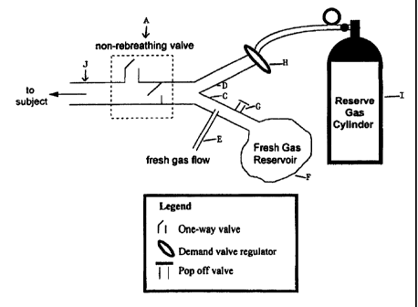
Figure 1 illustrates schematically the nature of the simple breathing
circuit and components which enable the patient to recover more quickly
from vapour anaesthetics administration. The said device shown enables
the PC02 to remain constant despite increase in minute ventilation which
thereby permits faster elimination of the vapour anaesthetic.
Figure 2 illustrates schematically portions of a standard circle
anaesthetic circuit generally known to persons skilled in the art.
Figure 3 illustrates schematically the simple breathing circuit in one
CA 02283430 1999-09-07
WO 98/41266 PCT/CA97100186
- 12
embodiment added to the portions of the circle anaesthetic circuit shown
schematically in Figure 2, illustrating modifications of the circuit shown
schematically in Figure 1 for use with the generally known circuit shown
in Figure 2. (It would be clear to persons skilled in the art that depending
upon the circuit used as the circle anaesthetic circuit, different
modifications on the basic circuit shown in Figure 1 will be made.}
Figure 4A illustrates the structure shown in Figure 3, now
combined with the general structure shown in Figure 2. (Figure 3 shows
the modifications made specifically to the structure of Figure 1 to combine
it with the structure in Figure 2 which is now shown in Figure 4A.)
Figures 4B and 4C illustrate schematically close up portions of one
portion of the structure shown in Figure 4A in different positions.
Figure 5 graphs the VT (Tidal Volume} and PETC02.
Figure 6 graphs traces of airway PC02 and VT.
Figures 7A, 7B and 8A and 8B graphically depict changes in PaC02
and PETC02.
DETAILED DESCRIPTION OF EMBODIMENTS
The circuit (Figure 1) consists of a non-rebreathing valve (A)
connected distally to two ports (C and D). The first port is connected in
parallel to a source of fresh gas (E) (which does not contain C02) and a
fresh gas reservoir (F). A one-way pressure relief valve (G) prevents
overfilling of the reservoir (F} by venting excess fresh gas. The second port
(D} is connected via a one-way valve (H), to a source of gas (containing
C02) whose PC02 is equal approximately to that of the mixed venous PC02
. We call this the "reserve gas" (I). Non-rebreathing valve A is connected
to exit port J (from which the patient breathes).
Functional analysis of circuit maintaining constant PC02 with
hyperventilation
When the minute ventilation "V" is less tYtan or equal to the fresh
gas flow "FGF" from (E), the subject inhales only fresh gas (non-C02-
containing gas). When V exceeds FGF, the reservoir (F) containing fresh
non-C02-containing gas empties first and the balance of inhaled gas is
drawn from the reserve gas (I) which contains C02. The reserve gas is
considered not to participate in COz exchange ensuring that the actual
ventilation provided is limited by FGF. If the rate of FGF is 5L/minute
and the patient breathes at 5L/minute or less, then the patient will inhale
..
CA 02283430 1999-09-07
WO 98/41266 - 13 - PCT/CA97100186
only non-C02-containing gas that comes from fresh gas flow sources (E
and F). If minute ventilation exceeds FGF, the difference between minute
ventilation and fresh gas flow is made up from gas from reserve gas (I)
which contains C02 at a concentration that does not provide a gradient for
elimination of C02 in the patient.
Ayylication of circuit to anaesthesia circle circuit
The schematic of the standard anaesthetic circle circuit, spontaneous
ventilation (Figure 2)
When the patient exhales, the inspiratory valve (1) closes, the
expiratory valve (2) opens and gas flows through the corrugated tubing
making up the expiratory limb of the circuit (3) into the rebreathing bag
{4). When the rebreathing bag is full, the airway pressure-limiting (APL)
valve (5) opens and the balance of expired gas exits through the APL valve
into a gas scavenger (not shown). When the patient inhales, the negative
pressure in the circuit closes the expiratory valve (2), opens the inspiratory
valve (1), and directs gas to flow through the corrugated tube making up
the inspiratory limb of the circuit (6). Inspiration draws all of the gas from
the fresh gas hose (7) and makes up the balance of the volume of the
breath by drawing gas from the rebreathing bag (4). The gas from the
rebreathing bag contains expired gas with C02 in it. This C02 is extracted
as the gas passes through the C02 absorber (8) and thus is delivered to the
patient (P) without C02, (but still containing exhaled anaesthetic vapour,
if any).
Modification of the circuit (Figure 3) to allow hyperventilation of patients
under anaesthesia
The modified circuit consists of
1. a circuit which acts functionally like a standard self inflating bag
(such as made by Laerdal ) consisting of
a) a non rebreathing valve, such as valve #560200 made by
Laerdal, that functions during spontaneous breathing as well
as manually assisted breathing (9);
b) an expired gas manifold, such as the Expiratory Deviator
#850500, to collect expired gas (10) and direct it to a gas
scavenger system (not shown) or to the expiratory limb of the
anaesthetic circuit {figure 4);
c) a self inflating bag (11) whose entrance is guarded by a one
CA 02283430 1999-09-07
WO 98/41266 _ 14 _ PCT/CA97/00186
way valve directing gas into the self inflating bag (12).
2. a source of fresh gas, (i.e. not containing vapour) e.g. oxygen or
oxygen plus nitrous oxide (13) with a flow meter (22).
3. a manifold (24} with 4 ports:
a) a port (15} for input of fresh gas (13);
b) a port (16) for a fresh gas reservoir bag (17);
c) a port to which is attached a one way inflow valve that opens
when the pressure inside the manifold is 5cm H20 less than
atmospheric pressure, such as Livingston Health Care
Services catalog part #9005, (18) (assuring that all of the fresh
gas is utilized before opening);
d} a bag of gas (19) whose PC02 is equal approximately to that of
the mixed venous PCOZ connected to inflow valve
(18)(Alternatively, the valve and gas reservoir bag can be
replaced by a demand regulator, such as Lifetronix
MX91120012, similar to that used in SCUBA diving, and a
cylinder of compressed gas);
e) a port to which is attached a one way outflow valve (20), such
as Livingston Health Care Services catalog part #9005, that
allows release of gas from the manifold to atmosphere when
the pressure in the manifold is greater than 5cm H20.
Method of operation in an anaesthetic circuit (Figure 4A)
The distal end of the nonrebreathing valve (Laerdal type) (9), is
attached to the patient.
The proximal port of the nonrebreathing valve is attached to a 3
way respiratory valve (21) which can direct inspiratory gas either from the
circle anaesthetic circuit (Figure 4B) or from the new circuit (Figure 4C).
The expiratory manifold (10) of the self inflating bag's non rebreathing
valve is attached to the expiratory limb of the anaesthetic circuit (3).
Regardless of the source of inspired gas, exhalation is directed into the
expiratory limb of the anaesthetic circuit.
To maximize the elimination of anaesthetic vapour from the
patient's lungs, the 3-way respiratory stopcock is turned such that patient
inspiration is from the new circuit (Figure 4C). Thus inspired gas from
the very first breath after turning the 3-way valve onward contains no
vapour, providing the maximum gradient for anaesthetic vapour
_m..__.~w., w. . . T , .
CA 02283430 1999-09-07
WO 98!41266 - 15 - PCT/CA97/00186
elimination.
An increased breathing rate will further enhance the elimination of
vapour from the lung. If breathing spontaneously, the patient can be
stimulated to increase his minute ventilation by lowering the FGF (22)
thereby allowing the PC02 to rise. Using this approach the PC02 will rise
and plateau independent of the rate of breathing, resulting in a constant
breathing stimulus. All of the ventilation is effective in eliminating
vapour.
If the patient is undergoing controlled ventilation, he can also be
hyperventilated with the self-inflating bag (11). In either case, the
patient's PC02 will be determined by the FGF (22). As long as the FGF
remains constant, the PC02 will remain constant independent of the
minute ventilation.
To illustrate the effectiveness of the circuit we performed a number
of tests with respect to humans and dogs. The humans were breathing
spontaneously and the dogs were mechanically ventilated.
Human subjects
After obtaining institutional ethics board approval and informed
consent, four healthy subjects aged 19 - 25 y breathed through the circuit by
means of a mouth piece while wearing nose clips. During normal
breathing, the FGF was set equal to V by adjusting the FGF such that the
bag containing fresh gas just emptied at the end of each inhalation.
Subjects were then instructed to breathe maximally ("breathe as hard as
you can") for 3 min. Flows were recorded by means of a Pitot tube (Voltek
Enterprises, Willowdale Canada) and the signal integrated to obtain
volume. C02 was sampled continuously at the mouthpiece (Medical Gas
Analyzer LB-2, Sensormedics Corp., Anaheim California). Analog signals
were digitized at 60 samples~s 1 and recorded using data acquisition
software (WINDAQ/200, DATAQ instruments, Inc. Akron Ohio).
Studies in dogs
Following institutional ethics board approval, 6 mongrel dogs of
either sex weighing 20-25kg were anaesthetized with methohexital (5-7
mg~kg-1 for induction followed by 150-300 mg~kg l~miri 1) and intubated.
Adequacy of anaesthetic depth was deduced from the eye lash reflex, lack
of spontaneous movements, and stable heart rate and blood pressure. A
catheter was placed in the femoral artery for monitoring blood pressure
CA 02283430 1999-09-07
WO 98141266 - 16 - PCTICA97/00186
and periodic sampling of blood for gas analysis., The dogs were ventilated
with a conventional mechanical piston ventilator (Harvard Apparatus
model 618, South Natick, MA). For each dog, an inflation volume (VT} of
400 ml and a frequency (f) of 10 miri 1 (duty cycle, 0.5) were used. All dogs
were ventilated to just below their apneic thresholds (by increasing VT
about 50 mL) so that they made no respiratory efforts. Tidal C02 was
sampled continuously (Ametek, Thermox Instruments Division,
Pittsburgh, PA) at the proximal end of the endotracheal tube. Flow was
measured with a pneumotachograph (Vertek series 47303A, Hewlett-
Packard) and the signal integrated to obtain volume. Analog signals were
digitized at 17 samples~s 1 and recorded using the same data acquisition
software as that used in studies on human subjects.
Because of differences in initial PaC02s among dogs (reflecting
individual sensitivities to C02, differences in anaesthetic levels, or
differences in VT/body weight ratio), the C02 concentration in the reserve
gas was arbitrarily adjusted for each dog to 1.5 ~ 0.5% above its FetC02 to
approximate the mixed venous PC02 (PvC02) (see Table II}. To allow
greater flexibility in setting the concentration of C02 in the reserve gas,
the
circuit was modified by replacing the demand valve with a one-way PEEP
(positive end expiratory pressure) valve and the cylinder with a bag
containing premixed gas. This circuit is functionally identical to that used
in studies on humans. The circuit was connected to the intake port of the
ventilator. Under control conditions, FGF was adjusted so that the fresh
gas reservoir just emptied during each ventilator cycle; this end point was
confirmed by a slight rise in FICOz above zero. After a steady-state had
been reached (difference < 1.5 mm Hg in two successive PaC02's taken 5
minutes apart), VT was increased at 5 minute intervals from 400 to 600 to
900 to 1200 mL. In a second trial at a fixed VT (approximately 400 mL} and
fixed FGF, f was increased at 5 minute intervals from 10 to 14 to 18 to 22
min-1. A blood sample for the determination of blood gases was drawn
from the femoral artery at the beginning and end of each 5 min interval.
All data are expressed as means ~ standard deviation. We tested for
significant differences using one- or two-way ANOVA with post hoc
analysis where appropriate. A p value Iess than 0.05 was considered
significant.
_,.~... ~ , ,
CA 02283430 1999-09-07
WO 98/41266 _ l,~ _ PCT/CA97100186
Results:
Human subjects
Figure 5 presents the VT and PETCOz of subject 1 during 3 min of
maximal ventilatory effort. Results for all subjects are summarized in
Table III; data represent average values for 10 breaths at 0 (the onset of
hyperventilation), 2.5 and 3 min. PETCOZ did not change significantly
from control values throughout the course of hyperventilation (p = 0.08,
ANOVA). There was considerable variability in V and breathing patterns
between subjects but individual subjects tended to sustain a particular
breathing pattern throughout the run.
Dogs
Figure 6 presents traces of airway PC02 and VT for dog #5 during
changes in f or VT. Figures 7 and 8 show the changes in PaC02 and the
PETC02 in all dogs during changes in f or VT. Increases in f did not
significantly affect mean PaC02 or PETCOz (p=0.28 and p=0.11, respectively;
ANOVA). Increases in VT decreased mean PaC02 from control only at VT
of 1200 mL (p=0.01); in contrast, changes in VT did not affect mean PETCOz
(p=0.25). The mean absolute change in PaC02 between control and the
highest ventilation was 2.2 ~ 1.8 mmHg (range 0.4 to 4.8) for f and 3.4 ~ 2.3
mmHg (range 0.4 to 5.6) for VT.
COMMENTARY
The system minimized decreases in PETCOz over a wide range of
ventilation (56 to 131 L miri') and breathing patterns, in hyperventilating
human subjects and in mechanically hyperventilated dogs (4 to 12 L
min'). The variability in PaCOz in the hyperventilated dogs, although
small, may have been due to a) imprecise matching of reserve gas PCOZ to
the dog's PvCO2s; b) prolonged duration of the maneuver in dogs (> 15
min versus 3 min for human subjects) and c) the extent of
hyperventilation (see below). In addition, the different levels of
ventilation may have induced changes in systemic and pulmonary blood
flow (ventilation-perfusion matching, physiological and anatomical dead
space), thereby affecting PaC02 and PvC02. Despite these sources of
variability, the range over which PaC02 varied in my studies in dogs was
similar to those reported in studies utilizing more complex equipment
(see Table 1).
Conventional servo-controlled techniques designed to prevent
CA 02283430 1999-09-07
changes in PCO~ with hyperpnea are less affected by changes in CO
production than the circuit; however, they have other limitations. The
assumption that detected changes in PETCOz are due to a change in PaC02
is not always warranted (14). Small changes in ventilatory pattern can
'uncouple' PETCOz from PaCO~, resulting in PETCO~ being an inappropriate
input for the control of PaCOz. For example, a smaller VT decreases VA
(which tends to increase PaCOZ) but will also decrease PETCO~, causing a
servo-controller to respond with an inappropriate increase in inspired CO,
Even under ideal conditions, a servo-controlled system attempting to
correct for changes in PETCOZ cannot predict the size of an impending VT
in a spontaneously breathing subject and thus deliver the appropriate CO~
load. If in an attempt to obtain fine control the gain in a servo-control
system is set too high, the response becomes unstable and may result in
oscillation of the control variable (11). Conversely, if the gain is set too
low, compensation lags (9). Over-damping of the signal results in a the
response never reaching the target. To address these problems, servo-
controllers require complex algorithms (16) and expensive equipment.
When CO~ production is constant, the circuit has the theoretical
advantage over servo-controlled systems in that it provides passive
compensation for changes in V. This minimizes changes in VA, pre
empting the need for subsequent compensation. Maintenance of a nearly
constant VA occurs even during irregular breathing, including brief
periods when V is less than the FGF. Under this circumstance, excess FGF
is stored in the fresh gas reservoir and subsequently contributes to VA
when ventilation exceeds FGF.
When COZ production increases during hyperventilation, as would
occur with increased work of breathing or exercise, my method requires
modification. To compensate, additional VA can be provided either by
increasing FGF or by lowering the PCOZ of the reserve gas below the
PvCOZ, as expressed in the following equation:
VA = FGF + (V-FGF) (PvC02 - reserve gas PCOz)/PvC02
Because spontaneously breathing subjects had such variable V
during hyperventilation, compensating for the COZ production by
modifying FGF would have required constant adjustment. We therefore
chose to decrease the PCOZ of the reserve gas to establish a concentration
~A~~NDE.D SI-kE-~T
:y
i
CA 02283430 1999-09-07
WO 98/412bb PCT/CA97/00186
- 19
gradient between the PCOZ of the reserve gas and the PvC02; when this
gradient is constant, Vr~ is a function of V. We found that, over the wide
range of V exhibited by the subjects, a concentration of 5.5% C02 in the
reserve gas (instead of 6.5% which corresponds to a PvC02 of 46 mrnHg)
provided the optimal gradient to compensate for increases in C02
production resulting from increased work of breathing.
I therefore have described a simple circuit that disassociates VA
from V. It passively minimizes increases in Vr~ that would normally
accompany hyperventilation when COZ production is constant. It can be
modified to compensate for increases in COZ production. The circuit may
form the basis for a simple and inexpensive alternative to servo-
controlled systems for research and may have therapeutic applications.
TABLE II
Dog # Weight Initial FETCOZ Bag FC02
(kg) (%) (%)
1 22 5.3 7.0
2 20 4.6 6.6
3 20 7.1 9.0
4 24 7.3 9.0
5 25 5.5 6.9
6 20 6.0 7.2
CA 02283430 1999-09-07
WO 98/41266 _ 20 - PCT/CA97/0018G
TABLE III
End Tidal PC(~2(mmHg) Frequency (ruin-1)
Time
Subject Control0 1.5 3 Subject # 0 1.5 3
#
1 40.3 33.6 34.935.6 1 57 50 47
2 36.6 30.9 28.128.0 2 89 87 88
3 42.0 42.5 43.242.7 3 31 30 30
4 41.0 34.5 38.838.8 4 149 130 127
Tidal Volume Minute Ve ntilation min-I)
(L) (L
Time Time
Subject 0 1.5 3 Subject # 0 1.5 3
#
1 2.30 2.49 2.58 1 131 124 118
2 0.85 0.72 0.63 2 75 63 56
3 2.60 2.64 2.26 3 80 78 68
4 0.78 0.62 0.60 4 117 80 76
While the foregoing provides a detailed description of a preferred
embodiment of the invention, it is to be understood that this description
is illustrative only of the principles of the invention and not limitative.
Furthermore, as many changes can be made to the invention without
departing from the scope of the invention, it is intended that all material
contained herein be interpreted as illustrative of the invention and not in
a limiting sense.
_, _ ... _ ...