Note: Descriptions are shown in the official language in which they were submitted.
CA 02283708 1999-09-15
WO 98/41154 PCT/LIS98/05661
BIODEGRADABLE TISSUE RETRACTOR
Field of the Invention
The invention relates generally to surgical devices, and more particularly to
materials and
methods for retraction and stabilization of tissues, using biodegradable
patches adhered to tissue
in a releasable manner.
Background of the Invention
During surgery, it is often necessary to retract tissue, especially internal
tissue, to operate
~o upon it or upon adjacent tissue. This is conventionally accomplished using
forceps, or other
mechanical gripping devices adapted for the purpose. Retraction or
manipulation may also be
accomplished by implanting a suture into the tissue, but there is a
significant risk of tissue
tearing even with strong, muscular tissue.
U.S. Patent No. 4,621,619 {Sharpe) describes a plastic disposable, hand
applied medical
~s retractor for retracting flesh at the edges of an incision or aperture in
the human or animal body.
The retractor includes a hook for impaling flesh and a pad that has an
adhesive surface that can
be adhered to skin to anchor the retractor to keep flesh in a retracted
position.
U.S. Patent No. 4,899,762 (Muller) describes a combination surgical drape,
dressing, and
closure structure and method of use before, during, and after a surgical
procedure. The dressing
2o portion can be adhered to a patient over a surgical field and, after an
incision is made, the
incision can be retracted with retractors, forceps, or clamps secured to the
dressing portion.
U.S. Patent No. 5,026,389 (Thieler) describes a surgical method and apparatus
for
opening and closing a surgical wound. An elastic member is adhered across a
patient's skin at a
treatment site. An incision is made by cutting through the elastic member and
through the
2s patient's skin to permit a surgical procedure to be conducted. The incision
is closed by
reapproximating the patient's skin at the treatment site and by bringing the
cut edges of the
elastic member together and adhering a relatively inelastic sealing member
over the elastic
member to maintain the cut edges while the wound heals.
International Patent Publication No. WO 96/29370, published September 26,
1996,
3o describes a barrier or drug delivery system that is adherent to a tissue
surface to which it is
applied. Tissue can be stained with a photoinitiator, then a polymer solution
or gel having added
thereto a defined amount of the same or a different photoinitiator can be
applied to the tissue.
CA 02283708 1999-09-15
WO 98/41154 - 2 - PCT/US98/05661
Exposure of light causes polymerization at the surface, providing adherence
and forming a gel.
The resulting polymerizable barrier materials are useful for sealing tissue
surfaces and junctions
against leaks of fluids. Tissue surfaces also can be adhered to each other.
The adhesive qualities
of the material are demonstrated in an example in which pieces of abdominal
wall were excised
from a euthanized rat and were clamped to a glass slide with binder clips.
Polymeric material
was adhered to the abdominal wall tissue. The polymer was urged away from the
tissue and
fractured, with portions remaining adherent to the tissue.
While the above and other disclosures describe, in some cases, useful tissue-
adherent
devices, there exists a need for retraction of soft internal tissues such as
liver and spleen.
Accordingly, it is an object of the present invention to provide a device for
retraction of tissue,
internally of a patient, in connection with a surgical procedure.
Summary of the Invention
A novel technique to accomplish retraction of tissue is described herein. The
present
invention provides a variety of methods, kits, systems, devices, and new uses,
all concerning
retraction of tissue using an agent that adheres to the tissue. All of the
devices, kits, and systems
can be used in conjunction with improved uses and methods of the invention,
and vice versa.
In one aspect, the invention provides a method that involves applying a tissue-
affixing
article to a surface of tissue internally of a patient. The tissue-affixing
article is adhered to the
2o surface of the tissue, and the tissue is retracted by exerting a
manipulative force on the tissue
affixing article. In one embodiment the method is to affix an adhesive to
tissue, internally of a
patient, and to retract the tissue via the adhesive, that is, to physically
manipulate the tissue by
applying force directly or, preferably, indirectly, to the adhesive. This can
be accomplished by
affixing a patch of a material having su~cient tensile strength to the tissue,
using a suitable
adhesive. In one embodiment, an unadhered portion of the patch may then be
easily grasped and
moved or stabilized by suitable retraction means, such as a retracting
instrument, or a suture that
is passed through the unadhered portion of the patch, or which is permanently
pre-attached to the
patch. Alternatively, the patch may be completely adhered to the tissue, and a
suture may then
be passed through the adhered patch to provide tension without necessarily
penetrating the tissue,
or a retracting instrument may be applied to the adhesive or the patch. In
each case, the patch
can distribute the applied force over a larger surface area of the tissue than
that which would be
contacted directly by the retracting means, thus reducing potential tissue
trauma, as might occur
CA 02283708 1999-09-15
WO 98/41154 - 3 - PCT/US98105661
if a suture were passed directly through tissue and retracted. Moreover, the
ability to achieve and
maintain retraction of a tissue in a confined space using a means of
retraction which minimizes
obstruction of access ports is particularly important in minimally-invasive
surgery, such as
laparoscopy or other endoscopic surgery. This concept is particularly
applicable to organs such
as cardiac tissue, lung tissue, and GI (gastrointestinal) tissue in minimally
invasive surgical
procedures.
The above and other methods of the invention can be carried out in minimally
invasive
procedures, in which a tissue-affixing article that can be a patch, or a
suture/patch combination
can be passed through a small diameter needle access port and adhered to the
target tissue. The
1 o suture then passes out of the body through the needle access port, and
does not obstruct larger
endoscopic ports through which the surgery may be performed. Alternatively, a
retracting suture
can be passed outwardly through tissue to the exterior of the body with a
needle, allowing
retraction without adding an access port for that purpose.
In another embodiment the invention involves securing an area of tissue
internally of a
patient with an adhesive and applying a force to the tissue via the adhesive
thereby retracting the
tissue in connection with a surgical procedure. Force can be applied to the
tissue via the
adhesive by grasping the adhesive directly, (via, for example, a tab formed
from the adhesive
material) or by applying a force to the adhesive indirectly by applying a
force to an article
adhered to or secured within the adhesive. For example, a patch can be adhered
to the adhesive,
2o a mesh can be formed within the adhesive when the adhesive is a fluid that
is solidified by light
or the like, etc.
In another embodiment a method is provided that involves percutaneously
inserting a
tissue-affixing article into a patient, adhering the affixing article to a
tissue surface internally of
the patient, and retracting the tissue surface via the affixing article.
Another aspect of the invention involves tissue-retracting devices and
arrangements. In
one embodiment, a tissue-affixing article is provided in combination with an
adhesive and a
suture fastenable to the article.
According to another embodiment the invention provides a percutaneously
deliverable
assembly including a distal portion constructed and arranged to be inserted
into a patient
3o percutaneously, and a proximal portion constructed and arranged to remain
outside of a patient
during a surgical procedure, the distal portion constructed and arranged to
contain a
tissue-affixing article.
. ~,
.. .
CA 02283708 1999-09-15
-4-
The invention also provides a kit that includes a tissue-a~fi~inl; article for
application to
and retraction of tissue internally of a patient, and ~.n adhesive for bonding
the tissue-affixing
article to the tissue.
Also provided is a device for the percutaneous application of a. tissue-
a~'~xing article to
tissue. The device includes an obturator with a notch for holding the :issue-
affixing s~rtiele, and a
needle or cax~nula for percutaneous access of the obturator.
In another aspect the invention provides the use of kits, devices, and systems
as desdribed
above in surgical procedures. In one embodiment the devices, kits, or systems
can be used Sri
minimally invasive surgery. In another embodiment they can be used in open,
internal surgery.
I 0 Other advantages, novel features, and objects of the invention will become
apparent from
the following detailed description of the invention when considered vi
conjunction with the
accompanying drawings, which are schematic and which are not intended to be
drawn to scale.
In the figures, each identical or nearly identical component that is
iIlu.strated in va.~ions figures is
represented by a single numeral. For purposes of clarity, not overt' component
is labeled in every
figure.
Brief Description of_ h l7rawings_
Figt;rc 1 i.Ilustrates schematically a tissue-affi:cing article connected to a
suture, the
tissue-affixing article adhesively secured to a tissue surface of a heart,;
and
Figure 2 illustrates, schematically, a percutaneously-iasertabl~: portal
including a distal,
insertable portion having a port in which is mounted a tissue-axing article.
Figure 2a is a cross-sectional view taken along line 2~2a of Fig. 2.
Figure 2b is a cross-sectional view taken along line 2b-2b of 1?ig, z.
Figure 2c is a cross-sectional view taken along line 2~-2c of Fig. 2.
Detailed Description of the Inventis~n
The Retractor and Associated Devices
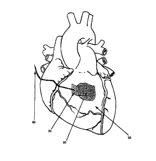
Figure I is a schematic illustration of a tissue-affixing article (20) which
has been
adhered to a surface of tissue (10} external of a heart by a biologically-
compatible adhesive, and
to which retractive force is being applied to m unadhered tab (30) ofthe patch
via a suture (40).
Figure 2 shawl an etnbadiment of a percuraneously-passable application
assembly for
percutaneously inserting a called tissue-axing article (154) with a lire-
attached suture (100)
AMENDED SHEET
..._ , . _ . ... . ... ...,~... . . - ... . ..., _- .. .. . . _ . . . . ~ _..
",u -~o.rw~rmo.~. n m
CA 02283708 1999-09-15
- ~/ 1 -
with an optional retainer (110). The rolled-up tissue-affixing article is
placed in a notch in an
obturator (I40}, as shown in cross-section 2b-2b, with the suture running in a
groove in the
obturator as shown in cross section Z:i-2a. The cbturator is enclosed vi a
needle (130), which
may
I
I'
,.
AMENDED SHEET
k«. ~t.u\:I~.I':~-vfi l:.\<Ill~..v «_ , r:.m m-:u~ . ...r.r ~ ." r i_" -.r..r
r_ r.,.:r «:~ .:..~:r:r~twv:~.rr i~~
- CA 02283708 1999-09-15 -
-5-
be capped at the extracorporeal (p:oxitr~al) end with a removable lock:-out
I?0 to prevent motion
of the obturator during insertion of the needle. The needle defines one:
embodiment of a "portal"
as that term is used herein, the obturator and needle together defining one
embodiment of a
percutaneously-passable assembly 1 b0 of the invention. After insertion of th
a needle through the
skin and into the body cavity where retraction is to occur, the o'aturatar is
advanced and the
tissue-af#ixing article is removed from the notch in the obturator. ThE:
obturator and optionally
the needle may then be removed, leaving only the suture passing through the
wall of the badly
cavity. Tine tissue-aft-axing article is toeri achered to the target tissue
':o allow retraction thereof
In use, the device illustrated in Figu:e 2 can be used to apply the tissue-
affi.~ing article to
the tissue surface, for example via tamping or pressing the tissue-affixing
srticle onto the
surface, or another device can be used. For example, the portal can ve
inserted percutaneously
(via a puncture created by the needle, through a trocar cannuia, or thc~ like)
and another
instrument, opiionally inserted through another incision, can be used to
manipulate the tissue
affixing article and apply the tissue-affixing article to tissue. The device
illusuated in Figure 2
can include (not shown) an adhesive-dispensing putt, and an emitter of
electromagnetic radiation
such that, in the embodiment described in which a phcto-activated a,ihesive a
tried to secure the
tissue, the device in Figure 2 ca.-a, be used to deploy the tissue-affixing
article, to apply adhesive
to a tissue surface, and to photo-adhere the tissue to the tissue-affixvag
article. The tissue-
affixing article can be applied entirely via the device of Figure 2 in that
embodiment. In other
2C embodiments the tissue-;3fftxing article will be de~loyzd by the device of
figure 2, another
device will be used to apFly a photoadhesive to the tissue surface and to
apply the tissue-affixing
article to the photuadhesive and photo-adhere the a>issue to th.e tissue-
affixing article.
In a snore complex arrangement, not illustrated, the device u~hiGh inserts the
tissue-
affi.cing article percutaneously can also be used to deploy and adhere the
tissue-affixing article.
For example, the tissue-afF~xing article could be pretreated with dried
adhesive, as described
below, and could be loosely attached to a set of expandable prongs, for
example of the type used
for ret:ievinp small objects in mechanical repairs. Thezt after percirtancous
insertion, the tissae-
affi~cing article is expanded by the prangs, applied to the tissue and allowed
to hydrate in bodily
fluids or supplementally added fluids to activate the adhesive. Theca the
adhesive is cured, either
by external means, such as light or a sprayed chemical, or by self a;,tivation
from the waterlin the
bodily and supplementally added fluids. After appropriate curing time, which
can be determined
in advance, the retractor is ready for use.
AMENDED SHEET
CA 02283708 1999-09-15
WO 98/41154 - 6 - PCT/US98/05661
Materials for the Tissue-affixing article
The invention comprises, in one embodiment, at least two material components,
a
medically acceptable tissue-aff xing article and a tissue-compatible adhesive
for the tissue-
affixing article. These may be combined for convenience of application.
Further optional
elements include retraction elements, such as a pre-attached suture or a tab
for grasping or for
peeling. When the retraction elements include a tab, the tab may be a separate
element that is
attachably connected to the tissue-affixing article, or the tab may be formed
from an area of the
tissue-affixing article itself that is not adhered to the tissue surface.
Additional optional elements
include biologically active materials. In another embodiment the invention
comprises a
tissue-compatible adhesive, and an adhesive-manipulating object such as a
forceps or the like for
physically manipulating the adhesive directly, preferably percutaneously,
thereby retracting the
tissue.
The tissue-affixing article material and/or adhesive, after application to
tissue and curing
(if required) of the adhesive, must possess sufficient mechanical strength and
tenacity to
withstand the force required to retract the tissue when such force is applied
to tissue-affixing
article material and/or adhesive at a single point, such as via a suture or a
forceps. The tissue-
affixing article and/or adhesive must distribute the force over a sufficient
area of the tissue
surface to allow retraction without damage. As used herein, "force" is meant
to define the
application of a physical force, as opposed to a driving force in chemical
equilibria, osmotic
pressure, or the like. A "force", in the context of the invention, if left
unopposed, causes physical
movement of the tissue.
The tissue-axing article can be a patch made of VicrylTM degradable mesh or of
MersileneTM non-degradable mesh which is believed to have sufficient strength
for the purposes
of the invention. The patch material may be the adhesive material, if the
adhesive has sufficient
tensile strength once cured.
Alternatively, the tissue-affixing article can be a portion of adhesive
itself,
The tissue-axing article, or patch, may be formed of any of a variety of
materials, in
any suitable form, which are suitable for surgical use. Physical forms
suitable for application
include, but are not limited to, filaments, fibrils, meshes, fabrics, felts,
sponges, membranes,
imperforate films and wafers, and pre-formed adhesives or adhesive precursors.
The physical
form is most typically preformed before application to the tissue surface, but
may also form
'spvntaneou~Iy, for example as a phase-separation-created membrane or
coacervate, after
RECTIFIED SHEET (RULE 91 )
.. .... ~ ,
CA 02283708 1999-09-15
WO 98/41154 - ~ - PCT/US98/05661
application. The materials used to make the tissue-affixing article or patch
can include any
material with adequate biocompatibility and sufficient tensile strength.
Materials known to be
suitable for medical use are preferred. Suitable materials include, but are
not limited to,
polyolefin meshes, such as MersiieneTM mesh described below; poly(fluorinated
alkylene)
membranes and meshes, such as Gore-TexTM perfluoropolymers; medical-grade
materials in
general, including polyurethanes, polyolefins, polycarbonates, silicones,
polyesters, polyacryiates
and polyamides; natural fibers of cotton, silk, alginate and the like; and
inorganic materials such
as glass, ceramic and metal. The tissue-affixing article or patch can be an
elastomer as described
in U.S. patent no. 5,026,389, incorporated herein by reference.
to Degradable materials are preferred, especially when the materials are to be
left behind
after the surgical procedure. Many suitable degradable materials are
commercially available,
such as VicrylTM mesh; these are commonly made of the same homopolymers and
copolymers
used to make absorbable sutures. Monomers for such polymers include but are
not limited to
lactide, glycolide, caprolactone, 1,3-dioxan-2-one and other aliphatic
carbonates,
1,4-dioxan-2-one, anhydrides, and orthocarbonates. Polymers of absorbable
natural materials
such as proteins and polysaccharides are also useful, if formed so as to have
suitable tensile
properties. Such materials include catgut and other collagenous materials, and
degradable
saccharides such as hyaluronic acid, alginate or partially oxidized cellulose.
Degradable materials will be preferred for many purposes. The exact
degradation time is
not, in most cases, an important variable as long as it is sufficiently long
to allow completion of
the operation. When used solely for retraction, the tissue-affixing article or
patch material is
preferably biodegradable, so that it may be left in place after the operation.
Non-degradable
materials are preferred if the tissue-affixing article or patch is intended to
also serve an ancillary
function, such as reinforcement of the tissue location. Degradable mesh
materials are
particularly preferred.
The Adhesive Component
The adhesive may be any medically acceptable adhesive which provides a
sufficiently
strong bond between the tissue-affixing article or patch and the tissue to
withstand the forces
involved in retraction of the tissue. Examples of suitable adhesives include
those disclosed in
3o U.S. patent no. 5,026,389, referenced above, or as described in U.S. patent
no. 4,621,619,
incorporated herein by reference. The adhesive should be strong enough and
bond sufficiently
well so that, when a 1 cmZ tissue-affixing article or patch is adhered, via
the adhesive, to an organ
CA 02283708 1999-09-15
WO 98/41154 - g - PCT/US98/05661
weighing about 200 g, (such as, depending on species, a heart, liver, or
spleen,) the organ can be
easily moved, or prevented from moving (stabilized), for example rotated,
elevated, and the like
via force applied to the organ via the adhesive. The adhesive bond typically
will have a strength
of at least 1000 g in a 90 degree peel test, preferably at least 1500 g, more
preferably still at least
2000 g in this test. As used herein, all physical manipulations of tissue of
this nature (rotation,
elevation, translation, transposition, and the like, or prevention thereof)
will be called
"retraction", which is to be understood to apply to any method of physical
manipulation or
physical stabilization of interior organs, structures, and tissues of the body
entailing movement or
prevention of movement thereof.
1 o Some preferred adhesives will be formed in situ by reaction of active
monomers.
Frequently, these reactive monomers will be based on macromolecules, both for
mechanical
stability and to minimize toxicity. These larger monomers are also known as
"macromers".
However, in the discussion herein, the terms "macromer" and "monomer" are not
distinguished
from each other unless specifically so stated.
One set of suitable adhesives are disclosed in detail in International Patent
Publication
No. WO 96/29370 and in co-pending, commonly-owned U.S. patent application
serial nos.
08/478,104 and 08/710,689, which are hereby incorporated by reference. These
documents
disclose adhesive gel materials which adhere strongly to tissue, yet are
biodegradable. The
synthesis of such polymers is described in U.S. Patent No. 5,410,016,
incorporated herein by
2o reference, in which application of biodegradable macromers to tissue,
followed by
photopolymerization to form a gel, is described; while methods for forming
gels on tissue
surfaces are described in 5,573,934, incorporated herein by reference. These
methods and
materials, described in more detail below, are currently preferred for the
invention, but other
bioadhesive materials are known in the art may be suitable. Such materials
include
cyanoacrylates, poly(meth)acrylates, polyurethanes, and protein-containing
glues incorporating
collagen and fibrin. The adhesive material may be of any sort which has
appropriate tissue
adherence and low toxicity, and may be a solid or a gel after being cured with
gels being
preferred.
The adhesive is preferably biodegradable, i.e., degradable in the body to
metabolizable or
3o excreted components, and is preferably biocompatible, i.e., minimally
stimulatory of
inflammation or other tissue reaction.
.r...... r. .. .....
CA 02283708 1999-09-15
WO 98/41154 - 9 - PCT/US98/05661
In addition to the photopolymerizable materials described in 5,410,016,
systems for
forming adhesives on surfaces may comprise other polymers known in the art,
including the
polymers described in U.S. Patent No. 4,938,763 to Dunn, et al., U.S. Patent
Nos. 5,100,992 and
4,826,945 to Cohn et al, U.S. Patent Nos. 4,741,872 and 5,160,745 to De Luca
et al, US
5,527,864 to Suggs et al., and U.S. Patent No. 4,511,478 to Nowinski et al.,
all incorporated
herein by reference. These materials, which either are able to covalently
crosslink by
free-radical-initiated polymerization, or can be made so by known chemical
modifications such
as those described in U.S. Patent Number 5,410,016, are preferred materials of
the invention. In
addition, materials which cross-link by other mechanisms, or which comprise
low-molecular
1 o weight reactive monomers, are also potentially suitable for the invention
if they are
biocompatible and non-toxic.
For some applications, particularly where retraction of heavy internal
structures is not a
critical function, a preformed adhesive layer on a tissue-affixing article or
patch may be suitable.
However, classical pressure-sensitive adhesives can have poor adherence when
surfaces are
~ 5 mucoid or bloody. For critical applications, of which cardiac surgery is
one example, loss of
adherence of a retractor can be dangerous, and highly adherent adhesives are
preferred. Such
highly adherent adhesives preferably include crosslinkable groups which are
capable of forming
covalent bonds with other compounds while in the presence of an aqueous
solution. These
crosslinkable groups permit crosslinking of the monomers to form a gel or
solid, either after, or
2o independently from thermally dependent gelation or preciptation of the
monomer. Chemically or
ionicaliy crosslinkable groups known in the art may be provided in the
monomers. The
crosslinkable groups in one preferred embodiment are polymerized using
photoinitiation to
generate free radicals , preferably with visible or long wavelength
ultraviolet radiation. The
preferred crosslinkable groups are unsaturated groups including vinyl groups,
allyl groups,
25 cinnamates, acrylates, diacrylates, oligoacrylates, methacrylates,
dimethacrylates,
oligo(meth)acrylates, or other biologically acceptable polymerizable groups.
Other polymerization chemistries which may be used include, for example,
reaction of
amines or alcohols with isocyanate or isothiocyanate, or of amines or thiols
with aldehydes,
epoxides, oxiranes, or cyclic imines; where either the amine or thiol, or the
other reactant, or
3o both, may be covalently attached to a monomer. Mixtures of covalent
polymerization systems
are also contemplated. Sulfonic acid or carboxylic acid groups may be used.
CA 02283708 1999-09-15
WO 98/41154 - 10 - PCT/US98105661
Preferably, especially if the adhesive is to form a hydrogel, at least a
portion of the
reactive monomers will be crosslinkers, i.e., will have more than one reactive
group, to permit
formation of an adherent layer and/or a coherent hydrogel by ensuring the
crosslinking of the
polymerized monomers. Up to 100% of the monomers may have more than one
reactive group.
Typically, in a synthesis, the percentage will be of the order of 50 to 95%,
for example, 60 to
80%. The percentage may be reduced by addition of co-monomers containing only
one active
group. A lower limit for crosslinker concentration will depend on the
properties of the particular
monomer and the total monomer concentration, but typically will be at least
about 3% of the total
molar concentration of reactive groups. More preferably, the crosslinker
concentration will be at
least 10%, with higher concentrations, such as 30% to 90%, being optimal for
maximum
retardation of diffusion of many drugs. Optionally, at least part of the
crosslinking function may
be provided by a low-molecular weight crosslinker. When the drug to be
delivered is a
macromolecule, higher ranges of polyvalent monomers (i.e., having more than
one reactive
group) are preferred. If the gel is to be biodegradable, as is preferred in
most applications, then
the crosslinking reactive groups should be separated from each other by
biodegradable links.
Any linkage known to be biodegradable under in vivo conditions may be
suitable, such as a
degradable polymer block. The use of ethylenically unsaturated groups,
crosslinked by free
radical polymerization with chemical and/or photoactive initiators, is
preferred as the
crosslinkable group. The monomer may also include an ionically charged moiety
covalently
2o attached to a monomer, which optionally permits gelation or ionic
crosslinking of the monomer.
Methods of Application
In a preferred embodiment, a tissue-affixing article or patch is adhered to
the tissue by a
process called "priming", which is described in greater detail in
International Patent Publication
No. WO 96/29370 and in co-pending U.S. patent application serial nos.
08/478,104 and
081710,689.
As described therein, one or more initiators are applied to a surface to form
an absorbed
layer. "Absorbed" is used herein to encompass both "absorbed" and "adsorbed".
A solution of
polymerizable molecules, referred to herein as "monomers", is then applied.
There are several
embodiments of this application method.
3o In its simplest embodiment, one or more initiators or components of an
initiation system
are applied directly to the surface, and the unabsorbed excess is optionally
removed by washing
or blotting. The initiator solution may further contain one or more
polymerizable monomers, and
.._ r t ..
CA 02283708 1999-09-15
WO 98/41154 PCT/US98/05661
-11-
other useful formulating ingredients, including accelerators, co-initiators,
sensitizers, and
co-monomers. Then a liquid, containing polymerizable monomers in combination
with one or
more initiators or components of an initiation system (which may be the same
as or different
from that absorbed in the first step) is applied. The system, if not self
polymerizing, is then
stimulated to polymerize, for example by exposure to an appropriate wavelength
of light.
The priming and monomer-application steps can also be combined. For example,
if
excess initiator is not removed before monomer addition, then subsequent
application of
monomer will result in mixing of initiator into the monomer layer. Similarly,
if the monomer
layer contains an initiator with a high affinity for the surface, then it is
possible to apply a
monomer layer containing initiator, and wait an appropriate time to allow
preferential absorption
of the initiator to the surface to achieve the same effect.
All of these methods may collectively be described as application of the
monomer in an
"initiator-incorporating manner", encompassing any means of application and
mixing which
results in both an absorbed layer of initiator, and a layer of monomer
incorporating an initiator,
t s being present on a surface to be coated.
The initiators may be chemical, photochemical, or a combination thereof. With
non-photochemical systems, a reductant component and an oxidant component may
be present in
the two parts of the solution, i.e., in the priming layer and the coating
layer.
Alternatively, a two-step process can be used to form polymers, especially
bioabsorbable
2o hydrogels, on tissue. In the first step, the tissue is treated with an
initiator or a part of an initiator
system for the polymerization of olefinic (e.g. acrylic) or other functional
monomers, optionally
with monomer in the priming solution. This provides an activated tissue
surface. In the second
step, monomers) and, if appropriate, the remainder of an initiator system, are
together placed in
contact with the activated tissue, resulting in polymerization on the tissue.
An example of such a
2s system is the combination of a peroxygen compound in one part, and a
reactive ion, such as a
transition metal, in another.
This process of spontaneous polymerization does not require the use of a
separate energy
source. Moreover, since the process of polymerization is initiated when part
one contacts part
two, there are no "pot life" issues due to initiation of polymerization. If
desired, part one or part
3o two can contain dyes or other means for visualizing the hydrogel coating.
In application of a photoiniated process to adhere a patch to tissue, the
tissue, and
preferably the patch as well, are stained (primed) with a photoinitiator,
optionally also containing
CA 02283708 1999-09-15
WO 98/41154 - 12 - PCT/US98/05661
a polymerizable material, and then a polymer solution having added thereto a
defined amount of
the same or a different photoinitator is applied to the tissue and to the
patch. The patch is applied
to the tissue region either before or after application of the primer and the
polymer solution. On
exposure to light, the resulting system polymerizes to form a gel throughout
the applied volume
which has excellent adherence both to the tissue and to the patch. As used
herein, "photoadhere"
is meant to define adhering an article to a tissue surface using
electromagnetic radiation, as
described for example in the discussion above.
Thus, a tissue-affixing article or patch may be applied onto the adhesive, or
can be a mesh
or other arrangement that can be contained within a fluid pre-adhesive which
is hardened and
1 o adhered to tissue and encases the tissue-affixing article or patch. A tab,
suture, or other
component affixed to the tissue-affixing article or patch may be allowed to
extend outside of the
hardened adhesive to be grasped by the operator, optionally via an instrument,
for manipulation
of tissue via the adhesive.
In any adhesive system, especially in one involving more than one component,
one or
more of the components of the adhesive system may be applied to the tissue-aff
xing article or
patch before its application to tissue. For example, some or all of the
components of an adhesive
system could be dried or lyophilized onto the tissue-affixing article or
patch. On application to
tissue, fluid would be absorbed, reconstituting the adhesive. The adhesive
could then polymerize
or otherwise adhere to the tissue, via an external stimulus such as light or
via endogenous
2o chemical reactions.
In a further embodiment, the degree of adhesion of the tissue-affixing article
or patch to
the tissue, which is provided by the adhesive, can be tailored, for example to
resist the stress of
retraction, but to allow removal by peeling from an edge, or by other
atraumatic removal
procedures. If edge-peeling for removal is contemplated, the retraction point
(suture, tab, or point
of application of forceps to the adhesive surface) is preferably near the
middle of the tissue-
affixing article or patch or adhesive area; and retraction force should be
applied substantially
normal to the plane of the tissue-affixing article or patch.
In a further embodiment, an anchoring patch could be provided within the body
cavity,
allowing the retracted organ or tissue to be secured, via an attached patch,
to the anchoring patch,
3o for example via a suture, staple or clip. Alternatively, the surfaces of a
retracting patch and an
anchoring patch could interact, as in VelcroTM loop/hook closures.
Biologically Active Agents
,.
CA 02283708 1999-09-15
WO 98/41154 PCT/US98/05661
-13
In any embodiment, the applied tissue-affixing article or patch, the adhesive,
or both can
contain biologically active ingredients such as drugs for local or systemic
controlled release. The
active materials can provide therapy for existing conditions, or for the
effects of the surgery
itself, or as ancillaries to a medical treatment (for example, antibiotics) or
as the primary
objective of a treatment (for example, a gene to be locally delivered). A
variety of biologically
active materials may be included, including passively-functioning materials
such as hyaluronic
acid, as well as active agents such as growth hormones. All of the common
chemical classes of
such agents are included: proteins (including enzymes, growth factors,
hormones and
antibodies), peptides, organic synthetic molecules, inorganic compounds,
natural extracts,
nucleic acids, lipids and steroids, carbohydrates, glycoproteins, and
combinations thereof.
Further examples include analgesics (for example, LidocaineTM), anti-
irritants, anti-inflammatory
agents (both steroidal and nonsteroidal), regulators of wound healing, growth
factors and
antagonists, and hemostatic agents.
Examples for use in particular applications include anti-thrombotic agents
(e.g.,
prostacyclin and salicylates), thrombolytic agents (e.g. streptokinase,
urokinase, tissue
plasminogen activator (TPA) and anisoylated plasminogen-streptokinase
activator complex
(APSAC)), vasodilating agents (e.g.. nitrates, calcium channel blocking
drugs), anti-proliferative
agents (e.g. colchicine and alkylating agents, intercalating agents), growth
modulating factors
(such as interleukins, transformation growth factor 8 and congeners of
platelet derived growth
factor, monoclonal antibodies directed against growth factors), and other
agents which may
modulate local or systemic physiological function, or the healing response to
organ injury post
intervention.
The adhesive material may also incorporate cells as therapeutic agents, such
as cells
producing drugs (including growth factors and inhibitors), or progenitor
cells, for example
progenitors of the same cell type as the involved organ, to accelerate healing
processes.
Sites of Retraction
The invention is applicable to retraction of any organ or internal tissue. In
addition, the
techniques and devices of the invention can also be used for the retraction of
external tissue, such
as skin. The retraction method is especially critical with "soft" organs, for
which, to the
knowledge of the inventors, there are no truly satisfactory non-damaging
methods of retraction at
present. These include, without limitation, liver, spleen, pancreas, gall
bladder, and kidney;
RECTIFIED SHEET (RULE 91)
ISA/EP
CA 02283708 1999-09-15
WO 98141154 - 14 - PCT/US98/05661
components of the gastrointestinal tract, including stomach and intestine;
components of the
genitourinary tract, including bladder, uterus, ovary, fallopian tubes, and
testicles;
compression-sensitive organs such as nerve bundles, spinal cord and brain; the
lungs; and the eye
and internal components thereof.
The invention is also useful for retraction or displacement of more muscular
tissues,
where conventional retraction can leave bruises. As shown in the example
below, the
compositions and methods of the invention can be applied to the beating heart.
Other muscular
tissues which could be manipulated include the tongue, the uterus, and major
blood vessels
including the aorta and vena cava. The invention could also be used for
positioning a tissue wall,
to such as a chest wall or the abdominal wall, during laparoscopic and similar
procedures. While
such structures can he manipulated with current devices, the required devices
can be bulky and
obstructive compared to the invention.
The size of the applied tissue-affixing article or patch will be scaled to the
size of the
target organ or area. For example, a larger adhered area will be preferred for
a liver compared to
~ 5 a spleen. Alternatively, the adhesive strength of the adhesive bond to the
tissue can be varied by
manipulation of the adhesive or of the application technique. For small
components, for example
in the eye, inner ear or ovary, the required area of adhesive will be small,
and it may be sufficient
to adhere a knotted or looped suture as the retracting component, or to apply
force to the
adhesive directly without reinforcement. There is no sharp lower limit to the
size of the applied
2o adhesive area which might be useful in microsurgery. On the other hand, for
retraction of the
abdominal wall, for example in minimally-invasive surgery, a relatively large
area of the skin
could be adhered with reinforced adhesive, to spread the retraction force
widely over the
relatively friable epidermis.
25 E am a
The nature of the invention is clearly demonstrated by an operational example.
The
technique was used to manipulate the beating heart of a live dog. A primer
solution buffered to
physiological pH and containing 50 ppm Eosin Y, 0.10 M neutral triethanolamine
{TEA} buffer,
and 10% of a polymerizable macromer (which contained a polyethylene glycol
backbone (3500
3o MVO with about 5 lactate residues attached, and endcapped with acrylic acid
esters) was brushed
onto a tissue-affixing article, specifically a 2 cm by 4 cm area of a 2 cm x 6
cm polyester
(MersileneTM) mesh patch, and also onto the epicardial surface of the LV apex
of a beating dog
__. a , .
CA 02283708 1999-09-15
WO 98141154 - 15 - PCTIUS98/OSb61
heart that was exposed via thoracotomy. A layer of sealant prepolymer (20%
35,000 MW PEG
with trimethylene carbonate linkages and acrylate end caps) also containing
Eosin Y and TEA
buffer were applied over the primer on the heart. The mesh was placed onto the
coated tissue,
and a layer of sealant was brushed onto the mesh/tissue surface. Two 20-sec
pulses of visible
light (approx. 500 mW) were applied to the laminate to crosslink it, thereby
forming a layer of
hydrogel on the surface of the tissue in which the mesh was embedded. The gel,
as expected,
was strongly adherent to both the tissue surface and to the mesh.
The free portion of the mesh (2x2 cm) was then gripped with a conventional
mechanical
retractor, and used to lift the beating heart out of the opened pericardium,
pointing the apex
t o upward. The retractor was then released, and a suture was passed through
the gel/mesh
composite (but not the tissue) and likewise used to retract the heart. The
heart continued to beat
normally throughout the application and retraction.
The adhesive gel used to adhere the mesh to the heart surface was a material
designed as
a tissue sealant and disclosed in more detail in International Patent
Publication No. WO
96/29370 and in co-pending U.S. patent application serial nos. 08/478,104 and
08/710,689. The
prepolymers were synthesized according to U.S. Patent No. 5,410,016.
The ability to manipulate a beating heart during surgery without trauma to the
heart
muscle is important in virtually all cardiac surgery, especially in minimally-
invasive surgery, and
in particular in cardiac bypass operations. Arteries requiring replacement may
be located on the
2o posterior ("back") side of the heart, requiring sustained retraction for
access to these areas.
What is claimed is: