Note: Descriptions are shown in the official language in which they were submitted.
CA 02284283 2005-11-03
74583-35
1
NOVEL POROUS COMPOSITE AND ITS USE IN IMPLANTS
This invention concerns a porous composite..
The invention is also concerned with an implant comprising
the composite.
GENERAL DEFINITIONS
The definitions below are to be understood herein as
follows:
"Biomaterial" means non-living material, which is intended
to be used in the body of a human or an animal. A
biomaterial can be 1) inert, 2) bioactive, or 3) capable of
bioresorption (solubilizable).
"Inert" means nonreactivity of the respective biomaterial
with a tissue.
"A bioactive material" reacts in the physiological
conditions within the body so that the outermost layer of a
block manufactured from said material is converted to form
.a chemical bond with the surrounding host tissue.
An "osteoconductive" material means a material which
facilitates the growth of newly forming bone along its
surface but without giving rise to newly forming bone when
introduced, for example, in muscle.
An "osteoinductive" material is generally a so called
growth factor isolated from the interstitial matter of bone
tissue or made synthetically, which induces the formation
of newly forming bone for example in muscle.
An "implant" is any manufactured device of an artificial
material, such as an artificial joint or a part of it, a
CA 02284283 2005-11-03
74583-35
2
screw, a fixation plate or a corresponding orthopedical or
odontological device, which is to be introduced into a
tissue.
"Host tissue" or " tissue" means bone tissue or soft tissue
into which for example an implant has been surgically
introduced.
"Micromotion" means microscopic motion (generally below 500
m) within the interfacial region of a surgical implant and
the host tissue caused by a dynamic load.
BACKGROUND OF THE INVENTION AND PRIOR ART
Biomaterials and the biological anchoring thereof
Implants for both medical and odontological purposes have
already been manufactured from various materials for a long
time. Various metals, alloys, plastics, ceramic materials,
glass ceramic materials and the newest or biologically
active glasses are distinguished from each other not only
by their durability but also by the properties of the
interfacial layer between the implant and the tissue. Inert
materials, such as metals and plastics, do not react with a
tissue. In this case there always exists an interfacial
layer between the implant and the tissue because the
implant and the tissue form two distinct systems.. Bioactive
materials such as hydroxyapatite, glass ceramics and
bioactive glasses react chemically with the tissue and
produce a relatively strong chemical bond in the interface
between the implant and the tissue, especially for the
bioactive glasses. The implant and the tissue are thus
anchored to each other. The rate of healing of the tissue
CA 02284283 1999-09-20
WO 98/47465 PCT/F198/00331
3
and the potential chemical fixation to the implant is
dependent on the activity of the implant material towards
. the tissue.
In designing the outermost layer of the implant it has to
be considered that implants intended for functional
activity are subjected to motion under a load immediately
after the surgical operation. This compromises the healing
and impairs the final result. In addition, the load is not
communicated to the flexible bone by the structure of a
non-elastic implant but the interfacial region in question
is disturbed and the integration is blocked. Problems are
often generated also by the lack of bone or the
unacceptable quality thereof. If for example a dental
implant is surgically placed into an insufficient or
qualitatively unacceptable bone, the stability in the early
phase is not attained and the surgical operation fails, if
any bone is not generated beforehand. Under the functional
conditioris mentioned above, the undisturbed healing is not
achieved with the currently used implants.
Specific clinical problems
1. Mechanical micromotions between the implant and the host
tissue prevents the fast integration (osseal joining)
within 6-12 weeks, in which case the device is left without
a permanent firm anchorage to the surrounding tissue. The
lack of this anchorage is kriown to lead to clinical
detachment in an early phase (within 1-2 years) or even a
number of years later and to the need of a repeat surgery
(1), (2)=
2. One approach is to have the surface of the implant made
porous for example by means of a few millimeters deep
three-dimensional surface structure constructed from
microscopic titanium spheres or from titanium tape. Newly
forming bone is expected to grow from the host tissue into
this surface structure. Such a porous biologically inactive
CA 02284283 1999-09-20
WO 98/47465 PCT/F198/00331
4
surface structure gives rise to a microscopic locking
structure towards the ingrowing newly forming bone but the
mechanical properties of this attachment do not allow a
sufficient adaptation under the control imposed by the load
conditions. The optimal anchoring structure between the
implant and the host tissue is in a state of a continuous
readaptation to make the strength of the structure to
correspond to the load conditions.
3. It has been shown (3) that the attachment of a metallic
bone implant (such as an artifi-cial joint) to the host bone
can be facilitated by a bioactive coating. The material
used most often is synthetic hydroxyapatite. It has been
demonstrated that hydroxyapatite 1) facilitates the
mechanical attachment of an implant to the host bone after
it has been attached firmly by means of a surgical
operation, 2) diminishes the interference in the
integration of the implant to the host bone caused by the
micromotion, and 3) diminishes the retardation of the
integration of the implant caused by local lack of bone and
by the lack of contact to the bone implant. Hydroxyapatite
is caused to attach to the surface of the implant by using
a spraying technique, in which case the coating material is
applied to the surface mostly only from the spraying
direction. In the biomechanical and biological sense, the
most optimal implant surface forms a three-dimensional
structure, wherein the interstitial space of the structure
forms a growth space to accommodate the ingrowing bone
tissue. In such a case, healing leads to the formation of a
connective locking structure. The growth of a newly formed
tissue is facilitated, if the porous structure is entirely
made of a bioactive material. In such a case the bioactive
coating material forms a three-dimensional osteoconductive
surface for the growth of newly forming bone. In
exceptionally difficult conditions, where the growth of
host bone is particularly poor for example because of low
quality or small amount of the bone, the growth of the
newly forming bone can optionally be improved by combining
CA 02284283 2005-11-03
74583-35
an osteoinductive component, which directly promotes the
generation of bone, to a bioactive coating material.
Although a bioactive coating can improve the integration of
the implant to the host bone, it must nevertheless be noted
5 that this technique is associated with many problems. The
combination of two materials which differ by their
properties (elasticity, thermal expansion), is a technically
demanding task. The coating of a metallic implant with a
bioactive ceramic material can lead to the early breakdown
of the coating, its fast corrosion, or slow detachment
(delamination). This has shown to be the most common
complication in efforts to use bioceramic materials,
including hydroxyapatite, as a smooth coating material of
metallic implants (4), (5), (6).
The optimal approach would be a construction which makes use
of the advantages of a bioactive coating material to ensure
early ossification but in which the possibility has been
taken into account that the permanent integration can be
secured by using other constructional approaches concerning
the surface.
One problem with implants provided with bioactive coatings
is also in that the bioactive surface, which is rather
fragile, is damaged rather easily in the chasing of the
implant into the bone.
SUMMARY OF THE INVENTION
The invention provides a new composite, which when combined
into the implant secures both rapid ossification and
permanent integration of the implant.
The invention also provides an implant, which allows the
micromotion of the implant and the surrounding tissue (bone)
CA 02284283 2005-11-03
74583-35
6
and nevertheless secures rapid growth leading to the
integration of the implant and the bone.
The invention also provides an implant which can be chased
into the bone without a risk of damaging the bioactive
structural component, which promotes the growth of a newly
forming bone.
Further the invention provides an implant wherein the
fracturability and the risk of detachment of the bioactive
structural component are smaller than those of the known
implants.
Thus, according to one aspect, the invention concerns a
porous composite, which is characterized in that it
comprises:
- particles A manufactured from a bioactive
material, and
- particles B, which are manufactured from a
material which is non-bioactive or weakly bioactive and
which is sintratable to the said bioactive material,
and that the particles A and particles B have been sintered
together to form a porous composite.
According to a further aspect, the invention concerns an
implant which is composed of a core and a bioactive
structural component which extends to the surface of the
implant. The implant is characterised in that into the body
has been made a recess or a through-passing hole which
comprises the above mentioned composite according to the
invention, said composite forming the surface layer of the
implant at the recess or at the through-passing hole.
CA 02284283 2005-11-03
74583-35
6a
In one embodiment, the invention provides a porous
composite, which is intended to be filled into a recess or a
through-passing hole of an implant, and which comprises:
particles A, which are prepared from a bioactive material;
and particles B, which are prepared from a non-bioactive
material or from a bioactive material the bioactivity of
which is lower than that of the bioactive material of
particles A, wherein the non-bioactive material can be
sintered together with the bioactive material, wherein the
particles A and the particles B are sintered together into a
porous composite, and wherein the particles A are
homogeneous in size and the particles B are homogeneous in
size.
In a further embodiment, the invention provides a porous
composite, which is intended to be filled into a recess or a
through-passing hole of an implant, and which comprises:
particles A, which are prepared from a bioactive material
which will react in the physiological conditions within the
body so that an outermost layer of a block of said bioactive
material forms a chemical bond with surrounding host tissue;
and particles B, which are prepared from a non-bioactive
material or from a weakly bioactive material which under
physiological conditions does not dissolve within the first
few months, wherein the particles A and the particles B are
partially melted together to form a porous composite having
a three dimensional structure in which individual particles
are connected to at least one adjacent particle but retain a
substantially spherical individual shape, wherein the
particles A are homogeneous in size and the particles B are
homogeneous in size, and wherein the particles A and the
particles B are the same size compared to one another, and
have a diameter of at least 250 microns.
CA 02284283 2005-11-03
74583-35
6b
In a still further embodiment, the invention provides an
implant comprising a core having a recess or a through-
passing hole and a bioactive structural component contained
within said recess or through-passing hole and which extends
to the surface of the implant, wherein said bioactive
structural component comprises a layer of a porous composite
of the invention.
BRIEF DESCRIPTION OF THE DRAWINGS
Figures 1A-1C show schematically tissue reactions of the
CA 02284283 1999-09-20
WO 98/47465 PCT/FI98/00331
7
composite according to this invention as a function of
time,
Figures 2A-2B show schematically the behaviour of the
continuous and the discontinuous coating in the bending of
the implant framework,
Figures 3A-3C show as cross sections the recesses made into
the body of the implant,
Figure 4 shows a hip prosthese, which has three recesses
for the composite of the invention,
Figure 5 show as a cross section a recess made into the
body of the implant, said recess being filled with a
composite according to the invention, wherein the composite
is comprised of distinct layers,
Figures 6A-6F show the use of the composite according to
this invention in joining and bone screws,
Figure 7 shows a light micrograph of glass spheres
manufactured by using a torch spraying technique,
Figures 8A-8C show X-ray diffractograms of finely-ground
glass-based cones,
Figure 9 shows a scanning electron micrograph showing
bioactive glass spheres sintered together,
Figure 10 shows implant cones used in tests in vivo, and
Figures 11A-11C show push-out or detachment curves of the
cones implanted into the bones in tests in vivo.
CA 02284283 1999-09-20
WO 98/47465 PCT/F198/00331
8
PREFERRED EMBODIMENTS AND DETAILED DESCRIPTION OF THE
INVENTION
In the definition of this invention, the bioactive material
means a material which under the physiological conditions
dissolves at least partly within a few months, most
preferably within a few weeks, preferably in about six
weeks. For example, a bioactive material can be a bioactive
glass, a bioactive ceramic material or a bioactive glass
ceramic material.
In the definition of this invention the term "non-bioactive
or weakly bioactive material" i.e. the material from which
the particles B have been prepared, means a material which
under physiological conditions does not dissolve within the
first few months. For example, this material can be a non-
bioactive or weakly bioactive glass; a ceramic material, a
glass ceramic material or hydroxyapatite. Thus, this
material can be any physiologically acceptable material,
the bioactivity of which is clearly lower than the material
of the particles A and which additionally allows the
particles A and particles B to be sintered together to form
a porous composite. Particularly preferably, the non-
bioactive or weakly bioactive material (the material of the
particles B) begins to dissolve before the bioactive
material (the material of particles A) has dissolved
completely. In this case the superimposed formation of a
chemical and mechanical bond between the tissue and the
implant with respect to each other is best secured.
Preferably, the particles A and the particles B are
essentially homogenous in size and approximately of the
same size relative to each other.
Preferably, the diameter of the particles A and the
particles B is in the range of 100-500 m.
According to a preferred embodiment the particles are
CA 02284283 1999-09-20
WO 98/47465 PCT/F198/00331
9
spherical, for example spheres manufactured by a torch
spraying process wherein the raw material is glass. In such
a case the particles A are made of bioactive glass and
particles B of glass without or almost without bioactivity.
The problem with many traditional bioactive glasses is that
they have poor workability because they easily crystallize.
Such bioactive glasses cannot be manufactured into spheres.
The international patent application WO 96/21628 (7)
describes new types of bioactive glasses, the working
region of which is suited for the manufacture of glass and
which thus allow the production of spheres. Typically,
these glasses have the following composition:
Si02 53-60 % by weight
Na20 0-34 % by weight
K20 1-20 % by weight
Mg0 0-5 % by weight
CaO 5-25 % by weight
B203 0-4 % by weight
P205 0,5-6 % by weight
provided that
Na20 + K20 = 16-35 % by weight
K20 + Mg0 = 5-20 % by weight
MgO + CaO = 10-25 % by weight
The above glasses are particularly suitable for use in this
invention as the bioactive glass, i.e. as starting material
for the particles A.
Preferably, the ratio of the amounts of particles A and B
in the composite is adjusted so that the amount of
particles A is 1/5 to approximately 1/1 of the total amount
of the composite. A particularly suitable mixing ratio is
CA 02284283 1999-09-20
WO 98/47465 PCT/FI98/00331
one where the amount of particles A is about 1/3 of the
total amount of the composite.
Of course, the composite of this invention can comprise
particles of several bioactive materials and/or several
5 non-bioactive materials or weakly bioactive materials.
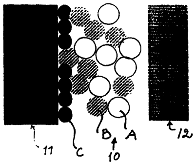
Figures lA-1C show a tissue reaction of the composite
according to this invention or a growing locking structure
as a function of time. Figure 1A represents a situation
immediately following the surgical placement of the
10 implant. Immediately next to the surface of the core 11 of
the implant are positioned spheres C, which are composed,
for example, of the same material as the core 11. A
composite layer 10, which is composed of bioactive spheres
A and spheres B, which are composed of non-bioactive or
very weakly bioactive material, is left between the core 11
and the tissue (bone) 12. The spheres A and B are sintered
together into a porous composite 10. Figure 1B, which shows
the situation after about 6-12 weeks, shows that newly
formed bone 12a has grown into -the pores formed by the
spheres A and B. Said newly formed bone 12a forms together
with the composite 10 a microscopic locking structure
between the bone 12 and the core 11. Figure 1C, which
represents the situation months or years after placing the
implant, shows a microscopic locking structure, wherein
newly formed bone 12a and spheres B are found. The
bioactive spheres A have been completely dissolved.
The series of Figures 1A-1C illustrates the formation of a
chemical bond and a mechanical bond. Table 1 summarizes the
amount of various bonds present.
Table 1 The types of bonds prevailing in Figures lA-1C
1A 1B 1C
Chemical bond
Particle A --> bone - +++ -
Particle B --> bone - ++
Mechanical bond - ++ +++
CA 02284283 1999-09-20
WO 98/47465 PCT/F198/00331
11
Figures 2A and 2B show a continuous coating 10 (Figure 2A)
and a non-continuous coating 10 (Figure 2B) of the core 11,
respectively. The bending of the core 11 in the case of
Figure 2A in the direction of the arrows results in a large
ratio between the elongation of the coating 10 and the
original length. Therefore, there exists the possibility
that the above mentioned problems might be encountered. In
contrast, when the core of Figure 2B bends, the ratio
between the elongation of the coating 10 and the original
length is small. Thus the bioactive structural component
functioning as a non-continuous structure retains its
position much better.
The implant according to this invention utilizes the
principle of non-continuous coating. Into the core 11 of
the implant are formed one or more recesses 13 (Figures 3-
5) or a through-passing hole, and the composite according
to this invention is applied into such recesses or holes.
Thus, the composite will not cover the surface of the core
as a continuous coating. Instead, the composite layer forms
a layer 10 extending to the surface only at the recess or
recesses 13 (or a hole or holes across the structure).
Figure 4 shows a hip prostheses having three circular
recesses 13 containing the composite according to the
invention. Figures 3A-3C show examples of some profiles of
the recesses. In Figure 3A, the edges 13a and 13b of the
recesss are perpendicular to the surface of the core 13, in
Figure 3B the recess is widening outwardly, and Figure 3C
shows an outwardly closing or locking recess structure. The
profile of the recess of Figure 3C is particularly good
because it secures the holding of the composite therein.
Figure 5 shows an implant according to this invention
wherein the composite layer 10 is composed of several
sublayers 10a...lOn. The advantage with this structure is
that the various sublayers can have a distinct mixing ratio
between the particles A and B. The mixing ratios are
preferably chosen so as to cause an increasing content of
CA 02284283 1999-09-20
WO 98/47465 PCT/F198/00331
12
particles A in the composite from the innermost sublayer
l0a towards the sublayer lOn in contact with the tissue 12.
Particularly preferred is the composite layer 10 forming a
gradient with respect to bioactivity.
Particularly preferred is an implant wherein the amount of
the particles A in the sublayer l0a of the composite facing
the interior of the core is 1/10 of the amount of the
sublayer in question, and wherein the sublayer lOn to come
in contact with the tissue is composed exclusively or
almost exclusively of particles A.
In the approach of Figure 5, if desired, inert particles
preferably made of the material of the core can be sintered
into the surface of the recess before the formation or the
application of the composite into the recess.
According to one embodiment, the implant of this invention
can be prepared so that the composite within a recess or in
a through-passing hole is formed by applying the particles
A and B into the recess, for example, as a mixture with an
organic binding material. Sintering is then performed
wherein the organic binding material is burned. If the
composite layer is composed of several sublayers, the
particles A and B required for each sublayer, respectively,
are applied separately and sintered.
According to another embodiment the composite can be shaped
into a block of the desired form and size capable of
attaching to the recess or the through-passing hole in the
inplant core. Such a composite block can be composed of
several sublayers, in which case the different sublayers
have a different mixing ratio of particles A and B so that
the content of particles A increases from the sublayer
facing inwardly into the implant core of the composite
towards the sublayer of the composite in contact with the
tissue.
CA 02284283 1999-09-20
WO 98/47465 PCT/F198/00331
13
By a proper selection of a narrow fraction and a suitable
particle size and shape, the void space between the
particles can be controlled so as to allow newly forming
bone with its blood vessels to penetrate into the
structure. When the ossification proceeds, the spheres
prepared from, for example, a bioactive glass, are
gradually resorbed. This generates more space for the bone,
whereby the structure of the bone is strengthened.
Therefore, the amount of the biomaterial is diminished as
function of time. The diminution can be controlled by a
proper selection of bioactive particles which are variable
in their bioactivity and in their size and shape as well as
by changing the mixing ratios of the various materials. In
order to increase the durability of the final fixation of
the bone, it is possible to use an inactive; porous
structure made of the implant material in the bottom of the
recesses. An essential feature of this surface, sintered
for example from spheres, is its three-dimensionality. A
conductive and inductive osseal contact is formed quickly.
A coating made only by using bioactive glass (enantelling)
would result only in the generation of a two-dimensional
reaction surface and the healing would be more difficult.
By virtue of the bioactive material in the recesses an
active healing reaction takes place already within a few
weeks leading to a mature stage within a few months. This
represents a noticeable improvement to the current
situation, in which most of the failures are due to the
fact that the fixation of the implant does not occur within
the first six weeks.
The composite within the recesses of the implant is
intended to function as a conductive, and in some
applications inductive, surface for the rapid growth of
newly forming bone and the chemical binding of the host
tissue. The functions of recesses made into the implant
core (or into the through-passing holes) can be summarized
as follows:
CA 02284283 2005-11-03
74583-35
14
The recess creates for the bone tissue a healing process
which is protected mechanically (from the mechanical
micromotion). The static and dynamic load towards the
implant and the consequent micromotion is thus not directly
directed to the interface between the implant and the host
tissue. This mechanically protected interface between the
implant and the host tissue provides optimal conditions for
the ossification and for the formation of a chemical
bonding, in other words, undisturbed conditions are created
for a fast integration of the device into the host bone.
The recess also protects the surface material mechanically
during the surgical placement of the implant. The implant
can be affixed tightly to a pre-formed site (press-fit
fixation) without causing a direct abrasive force to the
bioactive material in the recess. The requirements upon the
mechanical structural properties of the material can thus be
less demanding.
The recess also diminishes the size of the uniform structure
of the bioactive material. Especially for the enamellized
material, the mechanical integrity is improved with the
reduction in the size of the attachment region. Similarly,
the coating of the whole circumference of the device is
avoided, which contributes to the improvement of the
mechanical integration durability of the bioactive material.
Thus, the susceptibility of fracturing and the risk of loss
of the bioactive structure is diminished.
The noncontinuous bioactive material placed into the recess
partly counteracts the different elastic properties of the
implant core and the bioactive material. The different
elasticities of the materials can cause problems, for
example, for keeping the bioactive structural component
CA 02284283 2005-11-03
74583-35
14a
attached in the implant under different conditions of
dynamical load.
CA 02284283 2005-11-03
74583-35
The recess also creates a macroscopic surface structure for
the locking of the newly forming bone, which surface
structure in itself strengthens the mechanical bonding of
the implant due to the ingrowth of newly forming bone. The
5 inclined locking structure (Figure 3C) provides a
macroscopic locking structure between the host tissue and
the device.
A porous surface structure which is unreactive (non-
bioactive) with the tissue can be formed on the bottom of
10 the recess. This surface structure fulfils the function of
creating, when needed, a microscopic three-dimensional
mechanical locking joint between the device and the bone
tissue, based on the growth of the newly forming bone. The
object of this bottom structure is to secure a permanent
15 mechanical bone junction between the implant and the host
tissue in those cases where the bioactive coating structure
has completely eroded. A second object of the bottom
structure is to cover those bearer regions where the use of
a bioactive component is unwanted but which are needed to
secure the circular fixation of the device in the wanted
direction.
An example of other applications is a tightening joining
screw for various orthopedical operations to the bone as
shown in Figure 6A. The approach of Figure 6 is suited
particularly for osteoporotic bones. In th'is application
the joining screws 14 are fixed into the bone with
separate, for example cone-shaped devices 15 having
recesses 13, which in themselves comprise material causing
bioactive ossification. The bioactive agent (or bioactive
agents) are attached to the surface of the device according
to the method described above. The reference numbers 12 and
12' denote bone and the number 12" denotes marrow. Figure
6B shows a cross section of the conical device 15 along the
line A-A of Figure 6A. Figure 6C shows a plating operation
of a fracture 16 of an osteoporotic hollow bone 12 and 121,
wherein the metallic plate has been given the number 17.
CA 02284283 1999-09-20
WO 98/47465 PCT/F198/00331
16
Figure 6D shows the fixation of a fracture 16 of the
navicular of the wrist by using the tightening joining
screw described above.
An another application is, as illustrated in Figur 6E, an
ordinary bone screw 18, which has recesses 13 made for the
bioactive material 10. Figure 6F shows a cross section of a
bone screw along the line B-B of Figure 6E.
EXAMPLES
Example 1
Preparation of the glasses
For the experiments described below,-two types of glasses
were prepared of which a was bioactive and b was very
weakly bioactive. The glasses were prepared by mixing a
paste from PA (pro analys) grade raw materials. The raw
materials were Na2CO3, KZC03, MgO, CaCO3r CaHPO4*Hz0, H3BO3 and
fired Si02. The composition of the prepared glasses is given
in Table 2.
Table 2
The composition of the prepared glasses (weight-%)
Glass Na20 K20 Mg0 CaO P205 B203 Si02
a 6 12 5 20 4 - 53
b 25,5 - - 11 2,5 1,3 59,7
After weighing and mixing, the paste was melted in a
platinum crucible at a temperature of 1360 C with a melting
time of 3 hours. The glass melt was casted in a graphite
mould into blocks which were cooled at 520 C for 30 minutes
and subsequently in the oven, which was left to cool after
switching the power off. The finished glasses were crushed
and melted again in order to homogenize the glass mass. The
CORRECTED
CA 02284283 1999-09-20
WO 98/47465 PCT/F198/00331
17
glasses, which had been re-casted and cooled, were crushed
and sieved into the 250-297 m fraction, whereafter the
sieved crush was treated with a magnet to remove the small
iron particles detached during the crushing operation.
Example 2
Preparation of glass spheres
Using a torch spraying technique, the small glass particles
were heated for a short time to a sufficient degree to have
them melted and become rounded by virtue of the surface
tension. After a quick cooling, the glass spheres were
collected into a receptacle.
The torch spraying device used in the experiments comprised
of a container for the crushed glass, a feeding tube, a
common input head for the gases and crushed glass, and a
nozzle. A mixture of acetylene and oxygen was used for
heating. The nozzle was Castodyn 8000 nozzle nr. 30, which
is intended for ceramic spraying. This nozzle gives a
sufficient heat to round even the largest particles. The
crushed glass flowed into the nozzle from the container
above the device by its own weight. After a suitable mixing
ratio has been found, the different quantity of heat
required for the melting of different fractions can be
controlled by adjusting the flow rate of the gases. Smaller
particles melt faster than the larger ones and thus
necessitate passing through the flame at a greater
velocity, that is a greater flow rate of the gases. A
suitable gas flow for the fraction 250-297 m was 4 dm3/min
for acetylene and 6 dm3/min for oxygen. A funnel made of
stainless steel with a glass container below was used to
collect the glass spheres.
In order to assure a good quality of the glass spheres,
sieving (o 250-297 m), magnet treatment and light
microscope checking were performed immediately after the
CA 02284283 1999-09-20
WO 98/47465 PCT/F198/00331
18
preparation. After ultrasonic washing in ethanol, the
spheres were stored in ethanol in a closed vessel.
Figure 7 shows a light micrograph of glass spheres (m 250-
297 m) manufactured by torch spraying technique. The glass
speres had been prepared of bioactive glass (glass a, Table
2).
Example 3
Preparation of glass-based cones
The implants used in the experiments described below were
prepared by sintering glass spheres prepared according to
the previous example into porous devices having the shape
of a truncated cone. For the preparation of the glass
cones, the glass spheres prepared from the glasses a and b
of Table 2 were used. Two types of glass cones were
prepared, type I and type II. The first type (I) of glass
cones was prepared by sintering glass spheres which were
torch sprayed from the glass a of Table 2. The second type
(II) of glass cones was prepared by sintering a mixture of
glass spheres of which 1/3 were glass spheres prepared from
the glass a of Table 2 and 2/3 glass spheres prepared from
the glass b of Table 2.
Figures 8A and 8B show an X-ray diffractometric analysis of
a randomly chosen crushed cone. Figures 8A and 8B are X-ray
diffractograms of the cone type I and the cone type II,
respectively. It can be seen from these Figures that the
glass has retained its amorphic structure after the heating
processes associated with the preparation of the cones.
Figure 8C shows an X-ray diffractogram of a control cone,
wherein the observed peaks demonstrate the occurrence of
crystallization in the glass structure. The control cone
was prepared from glass spheres, for which a conventional
bioactive glass, in other words glass without potassiuin or
magnesium oxide, was used as a raw material.
CA 02284283 1999-09-20
WO 98/47465 PCT/F198/00331
19
For the sintering a rectangular mold (50 x 30 x 20 mm) was
prepared from graphite, into which ten 14 mm deep holes
were made using a cone-shaped 4 mm bit. The holes were
filled with the prepared glass microspheres, and the mold
with the spheres was heated in a preheated Naber L 49 oven.
Both of the cone types I and II were prepared at the
sintering temperature of 760 C. The sintering time for the
cone type I was 5 min 15 s and the sintering time for the
cone type II was 3 min 40 s.
The cones which were overshrinked (overmelting) during the
heating were discarded and the accepted cones were checked
for the thickness of the necks between the spheres by using
a light microscope. The length of the cones was 14 mm and
the 8= 2,9 mm and 3,9 mm. The finished cones were washed
in ethanol by using an ultrasonic treatment and stored in
ethanol in a closed vessel.
Figure 9 represents a scanning electron micrograph, which
shows bioactive glass spheres of type I sintered together m
= 250 - 297 m.
Example 4
Preparation of titanium-based cones
For comparison, a titanium-based cone type was prepared by
sintering titanium microspheres. Microspheres prepared from
medical grade titanium by atomizing in a protective argon
gas were purchased from Comp Tech, Tampere. The spheres
were sieved to a fraction 250-297 m and washed
ultrasonically in ethanol. Because titanium reacts very
easily with oxygen at higher temperatures, the sintering of
titanium must be done in a vacuum oven. For the sintering,
molds resembling the ones used in Example 3 were prepared
by drilling holes with a 4 mm cone-shaped bit into a
graphite block. The blocks were filled with titanium
microspheres and the sintering was performed in a vacuum
CA 02284283 1999-09-20
WO 98/47465 PCT/F198/00331
oven at a temperature of 1500 C and with a sintering time
of 2 h 30 min. A successful result was checked after the
sintering by using a light microscope.
Figure 10 represents cones used as implants in this study.
5 The cone shown on the right represents the glass cone type
I and the cone shown in the middle the glass cone type II
from Example 3. The titanium-based cone described above is
shown on the left. The spheres have a m= 250-297 m.
TEST RESULTS
10 The durability of the sintering necks observed in Figure 9
is influenced essentially by not only the behaviour of the
glass in the tissue but also by the successfulness of the
sintering. The sintering result, i.e. the mechanical
strength of the matrix, is compromised by the sintering of
15 more than one type of glass together. This is due to the
fact that different glasses have different coefficients of
thermal expansion; during the cooling, microfractures
develop in the structure of the matrix. In order to clarify
the differences in the mechanical strength of the different
20 matrices, a mechanical compression test was performed on
the cones made of glass spheres.
1) Compression strength of the cones
For the compression test, blocks with dimensions
corresponding to those of the types I and II of the cones
made of glass spheres, respectively, were prepared by
sawing off the excessive material from the both ends, in
which case the cone block to be tested was 4 mm in length,
o= 3,3 and 3,4 mm, respectively. The compression strength
of the titanium cones was not measured, because the
strength of the sintered titaniuni cone would have exceeded
the maximum load of the measuring device.
The measuring device was composed of an Alwerton
CA 02284283 1999-09-20
WO 98/47465 PCT/F198/00331
21
compression device and a recorder. In the device, a
downward-moving probe proceeding at a constant velocity
compresses a block on a solid platform. The velocity can be
controlled and the probe measures the load upon the block.
The device is connected to a recorder and this is arranged
to record the maximum load before the disintegration of the
block.
The compression strength of the cones made of glass spheres
is shown in the Table 3.
Table 3
Glass cone Number of Compression strength
type tests (MPa)
I 8 17,5 3,9
II 7 5,0 1,0
2) Push-out test of the cones
Into the femur of rabbits (n= 8) were implanted cones,
which represented the glass cone types I and II described
in Example 3, and the titanium-based cone type described in
Example 4. A similar series of three cones were implanted
into both femurs, one for histomorphological determinations
and the other for biomechanical determinations. The total
number of implants was 3 x 16 = 48 cones. After a six week
follow-up period, the rabbits were sacrificed, the femurs
removed and the force (the push out force) needed for the
detachment of the cone from the bone was determined.
The biomechanical push-out test was performed on the same
device as the compression strength test described above.
For the test, the ends of the femur were cut and the bone
was split longitudinally. The excess part of the implant
inside of the bone was removed and the exterior of the bone
was carefully cleaned. The bone was then placed against a
solid support. The support had a central hole with
dimensions suitable for fitting the other end of the
CA 02284283 1999-09-20
WO 98/47465 PCT/F198/00331
22
detaching cone. The device was switched to register the
maximal load, the compression rate was 0,5 mm/min. In
addition, the device was connected to a recorder with a
paper velocity of 30 mm/min.
A summary of the results of the push-out test is shown in
Table 4.
Table 4 The push-out force needed for the implanted cones
after a six week tissue reaction
Cone type Number of tests Push-out force
(N)
Glass cone type I 8 216,2 20,6
Glass cone type 8 293,3 43,8
II
Titanium-based 8 230,6 15,4
Given that the contact surface to the bone was the same for
all of the cones (the depth of hard bone = 1 mm = the
height of the cone) the push-out strength was calculated by
dividing the push-out force by the contact surface of the
cone. The push-out strength is shown in Table 5.
Table 5 The push-out strength of the implanted cones after
a 6 week tissue reaction
Cone type Number of tests Push-out
strength (MPa)
Glass cone type 8 20,8 2,0
I
Glass cone type 8 28,3 4,2
II
Titanium-based 8 22,2 1,5
cone
Figure 11 shows the push-out curves for the different
cones, wherein the push-out force is expressed as a
function of dislocation (11A = glass cone type I, 11B =
glass cone type II, and 11C = titanium-based). The slopes
of these curves can be used to calculate the so called
CA 02284283 1999-09-20
WO 98/47465 PCT/F198/00331
23
push-out stiffness, which is a ratio:
push-out force
---------------
dislocation
and which describes the stiffness of the implant core
during the push out test. The push-out stiffnesses for the
different cones is given in Table 6. The stiffness of the
matrix is directly proportional to the slope of the curve.
Table 6 The push-out stiffness of the implanted cones after
a 6 week tissue reaction
Cone type Number of Push-out stiffness
tests (N/mm)
Glass cone type I 8 301,6 150,6
Glass cone type II 8 214,5 99,4
Titanium-based cone 8 277,4 149,2
DISCUSSION
Testing of the cones made of glass spheres
The matrix sintered from two different types of spheres
allows the combinatory sintering between the spheres of
three different types: a-a, a-b, and b-b. Because the
glasses differ by their coefficients of thermal expansion,
the necks a-b are weak or partially broken (tensioned)
after cooling. Only the necks between the two similar
glasses are strong and these are mainly responsible for the
mechanical strength of the matrix.
The test results of the compression strengths of the cones
made of glass spheres (Table 3) demonstrate that the matrix
sintered from microspheres prepared from two different
glasses is notably weaker than a matrix of spheres prepared
from a single glass. It can be supposed that all the necks
in a matrix of spheres prepared from a single glass are
intact after cooling. The strength of the matrix sintered
CA 02284283 1999-09-20
WO 98/47465 PCT/F198/00331
24
from a mixture of glass spheres (glass cone type II) is
improved if the ratio between the a/b spheres is
diminished, or the fraction of the spheres prepared from
the bioactive glass (a) is reduced. In this case the
mixture of the spheres is made more homogenous and the
number of necks is increased. In this study the ratio 1/3
was used.
The behaviour of the implanted cones in vivo
1) Cones sintered from the bioactive glass spheres (glass
cone type I)
In connection with the implanting of the cones it was
possible to observe immediately the fast penetration of
bone marrow fluid into the cone matrix. The matrix was, due
to the capillary force, filled completely with tissue fluid
and blood, so that there was a plentiful amount of reaction
surface between the glass and the tissue/tissue fluid.
The bioactive glass reacts with all of its surface, in
which case all the necks are solubilized with time. This
leads gradually to the weakening of the matrix with the
onset of breaking of the necks.
In the push-out test, which was made after a six-week
tissue reaction, it is observed that the cones sintered
from bioactive glass are attached to the bone rather
strongly. In spite of the essentially weaker core, the
push-out strength (20,8 2,0 MPa) is approximately of the
same order of magnitude as the push-out strength (16-23
MPa) measured for a cone molded from a bioactive glass in
previous studies (8). This can be explained by the ingrowth
of newly forming bone into the matrix. Concurrently with
the growth of bone into the cone, the bioactive glass is
solubilized and the matrix as a whole is weakened. At the
maximal load of the push-out value the considerably
wealcened necks break near the outer edge of the cone and
CA 02284283 1999-09-20
WO 98/47465 PCT/F198/00331
the cone is displaced as the newly ingrown bone yields at
the edges of the cone. The abrupt rupture of the bond
between the implant and the bone is also illustrated by the
so called push-out stiffness of the cone sintered from
5 bioactive glass spheres (301,6 150,6 N/mm), which is of
the same order of magmitude as the cone sintered from
titanium spheres (277,4 149,2 N/mm).
2) Cones sintered from a mixture of glass spheres (glass
cone type II)
10 The tissue reaction of this cone type begins vigorously
only on the surface of the bioactive spheres. In contrast,
spheres prepared from very weakly bioactive glass (b) react
or dissolve very slowly. Newly forming bone has the
opportunity to grow into the pores as induced by the
15 bioactive component. However, mainly the bioactive
component dissolves from the matrix with time and the
spheres prepared from glass b remain as a support for the
core.
The push-out test shows clearly the push-out strength ( 28s3
20 4,2 MPa) as compared to the corresponding figure for the
cone type I or the titanium-based cone (approximately 22
MPa). Similarly, the push-out stiffness (214,5 99,4 MPa),
which illustrates the stiffness of the core, demonstrates
that the core is more flexible by virtue of the remaining
25 matrix composed of the remaining intact glass type b which
provides support for the core. The stiffness of the cone
sintered from the mixture of spheres is clearly smaller
than the corresponding figure for the cones sintered from
the bioactive glass spheres or the titanium spheres. The
structure of the core is markedly more heterogenous than in
the cones sintered from only one type of glass spheres. The
newly formed bone grown into the pores gets support in the
push-out process from the remaining matrix composed of the
glass type b, in which case the bonding between the bone
and the cone becomes iiiarkedly more durable and more
CA 02284283 1999-09-20
WO 98/47465 PCT/F198/00331
26
flexible than the bonding between the cones sintered from
only bioactive glass spheres or titanium spheres and the
bone. The large standard deviation in the push-out strength
and in the push-out stiffness is explained by the different
compositions of the individual cones prepared from the
mixture of spheres (which again results from the fact that
the mixture of spheres from which the various cones were
sintered, was not completely homogenous). Thus the
durability of the matrix is variable.
3) Cones sintered from titanium microspheres
Titanium is an inert material used widely in surgical
implants. The bonding between the implant and the tissue is
good but there is no bonding at the interface. In studies
performed previously (8) it has been shown that the push-
out strength of a smooth titanium cone (approximately 2
MPa) is considerably inferior when compared with the
strength of a corresponding cone molded from a bioactive
glass (16-23 MPa). In this study the push-out strength of a
cone sintered from titanium microspheres (22,2 1,5 MPa)
was about ten-fold as compared to the corresponding
strength of the smooth titanium cone measured in the above-
mentioned reference and of the same order of magnitude as
the push-out strength of a cone sintered from bioactive
glass spheres. The increased strength of a porous titaniurn
cone results from not only the coarseness of the interface
but apparently also from the implant-supporting influence
of the newly formed bone grown into the implant matrix. At
the time of detachment the bone strings at the interface
are broken and the cone is detached. The push-out stiffness
(277,4 149,2 N/mm) is of the same order of magnitude than
the push-out stiffness of the cones sintered from bioactive
glass spheres and markedly larger than that of the cones
(214,5 99,4 N/mm) prepared from the mixture of the
spheres. The matrix sintered from titanium spheres can not
even be supposed to be flexible.
CA 02284283 1999-09-20
WO 98/47465 PCT/F198/00331
27
The composite according to this invention can be used to
facilitate the bonding of any orthopedical (medical or
veterinary) or odontological implant.
The above mentioned embodiments of this invention represent
merely examples of the application of the idea of this
invention. It is evident to the one skilled in the art that
the various embodiments of this invention can be varied
within the scope of the following claims.
CA 02284283 1999-09-20
WO 98/47465 PCT/F198/00331
28
References
1. Freeman MAR, Plante-Bordeneuve P: "Early migration and
late aseptic failure of proximal femoral prostheses". J
Bone Joint Surg 76-B:432-438, 1994.
2. Karrholm J et al., "Does early micromotion of femoral
stem prostheses matter?" 4-7-year stereoradiographic
follow-up of 84 cemented prostheses. J Bone Joint Surg
76-B: 912-917, 1994.
3. Jaffle WL, Scott DF: "Total hip arthroplasty with
hydroxyapatite-coated prostheses". J Bone Joint Surg
78-A:1918-1934, 1996.
4. Ducheyne P, Cuckler J: "Bioactive prosthetic coatings".
Clin Orthop 276:102-114, 1992.
5. Ido K et al., "Cementless total hip replacement.
Bioactive glass ceramic coating studies in dogs". Acta
Orthop Scand 64:607-612, 1993.
6. Pajamaki J: "Bioactive glass and glass-ceramic
interfacial reactions to bone". Acta Univ Tamperensis vol
406, 1994.
7. Brink et al., WO 96/21628.
8. O.H. Andersson et al., "Evaluation of the acceptance of
glass in bone", J. Mater. Sci.: Mater. in Medicine 3(1992)
145-150.