Note: Descriptions are shown in the official language in which they were submitted.
CA 02285056 1999-09-30
WO 98/43702 PCT/IB98/00487
METHOD FOR INTRODUCING PHARMACEUTICAL DRUGS AND
NUCLEIC ACIDS INTO SKELETAL MUSCLE
FIELD OF THE INVENTION
The present invention is related to a method for making skeletal muscle
semipermeable
to pharmaceutical dings and nucleic acids. More specifically, skeletal muscle
is made
semipermeable by electrically stimulating the muscle at low field strengths
following
pharmaceutical drugs and nucleic acids injection.
2. TECHNICAL BACK RO
Scientists are continually discovering genes which are responsible for many
human
diseases, such as genes responsible for some forms of breast cancer, colon
cancer, muscular
dystrophy and cystic f brosis. In addition, scientists are continually
discovering genes that code
for bacterial and viral antigens (e.g., viral capsid proteins). Despite these
new discoveries, a
1 S major obstacle facing the medical profession is how to safely deliver
effective quantities of these
agents to patients to treat disease or for genetic immunization.
Currently, most pharmaceutical agents are taken orally or intravenously. Oral
and
intravenous drug and gene delivery methods, however, have several
shortcomings. First, a
large percent of orally or intravenously delivered drugs are degraded by the
body before
arnving at the target organ or cells. Acids and enzymes in the stomach and
intestine, for
example, can break down many pharmaceutical drugs. Similarly, genes would be
rapidly
destroyed by proteins found in the blood and liver which break down DNA.
Additionally,
intravenously delivered drugs and genes are often sequestered by the liver or
immune system
before arriving at the diseased organ or cells. Second, oral and intravenous
drug and gene
delivery is non-specific. That is, the drug or gene is delivered to both
target and non-target
cells.
Skeletal muscle is a promising candidate for drug delivery, gene therapy and
genetic
immunization. First, skeletal muscle constitutes over 50% of a human's body
mass, most of
which is easily accessible compared to other tissues and organs of the body.
Second, there are
numerous inherited and acquired disorders, such as Duchenne muscular dystrophy
(DMD},
diabetes mellitus, hyperlipidaemia and cardiovascular disease which are good
candidate
disorders for drug and gene delivery into the muscle. Third, muscle is an
ideal site for genetic
immunization because it is easily accessible and proteins made in the muscle
are secreted, thus
eliciting an immune response. Finally, skeletal muscle cells are non-dividing.
Therefore,
CONFIRMATION COPY
CA 02285056 1999-09-30
WO 98/43702 PCT/IB98/00487
skeletal muscle cells are capable of expressing a protein coded by a gene for
a longer time
period than would be expected of other cell types that are continually
dividing. Because the
protein is expressed for a longer time, fewer treatments would be necessary.
Currently, however, there is no non-viral method for effectively delivering
S pharmaceutical drugs and DNA into skeletal muscle in vivo. There are several
methods known
in the art for transferring pharmaceutical drugs and DNA into skeletal muscle,
such as
intramuscular injection of DNA. The clinical applicability of direct muscle
injection, however,
is limited mainly because of low transfection efficiency, typically less than
1 % transfection
efficiency. It has been demonstrated that the efficacy of transfection can be
improved if DNA
injections are done in regenerating muscle. Injection is induced three days
before DNA
injection with the drug Bivucain. While injection in regenerating muscles
induced by Bivucain
show higher efficiency, the method has limited applicability in humans because
of the severe
damage caused to the muscle.
From the foregoing, it will be appreciated that it would be an advancement in
the art to
provide a non-viral method of delivering pharmaceutical drugs and DNA only to
diseased
organs and cells. It would also be an advancement in the art to provide an
electroporation
method of delivering pharmaceutical drugs and DNA directly into skeletal
muscle. It would be
yet another advancement in the art if the electroporation method could deliver
therapeutically
effective quantities of pharmaceutical drugs and DNA into the skeletal muscle
at multiple sites
simultaneously. It would be a further advancement if the method permitted the
delivery
efficiencies to be regulated.
Such a method is disclosed herein.
3. BRIEF SUMMARY OF THE INVENTION
The present invention provides a method for delivering or transfecting
pharmaceutical
drugs and DNA into skeletal muscle. Without being bound by theory, the method
is thought to
be similar to electroporation. Electroporation works on the principle that
cells act as an
electrical capacitor generally unable to pass current. Subjecting cells to a
high-voltage electric
field, therefore, creates transient permeable structures or micropores in the
cell membrane.
These pores are large enough to allow pharmaceutical drugs, DNA and other
polar compounds
to gain access to the interior of the cell. With time, the pores in the cell
membrane close and
the cell once again becomes impermeable.
-2-
CA 02285056 1999-09-30
WO 98/43702 PCT/IB98100487
Conventional eiectroporation, however, employs high field strengths from 0.4
to several
kV/cm. In contrast to conventional electroporation, the field strength used in
the present
invention ranges from about 25 V/cm to 250 V/cm. These lower field strengths
are thought to
cause less muscle damage without sacrificing, and indeed increasing,
transfection efFciencies.
Furthermore, using the method of the present invention, transfection
efficiencies can be tightly
regulated by altering such parameters as frequency, pulse duration and pulse
number.
The increase in DNA transfection efficiency is observed only if the muscle is
electrically
stimulated immediately, or shortly aRer the DNA injection. Thus, the
semipermeable quality of
the tissue induced by the stimulation is reversible. Moreover, it is dependent
on current
through the muscle; activity induced through the nerve does not affect
transfection effciency.
Once transfected, the muscle cells are able to express the proteins coded by
the nucleic
acid. Therefore, the transfection method of the present invention can be used,
for example, to
transfect expression vectors for genetic immunization (i.e., DNA vaccines). In
one
embodiment, rabbits were transfected with a plasmid containing the cDNA for
rat agrin. The
transfected muscles produced and secreted agrin protein. Nineteen days post-
transfection,
rabbit serum contained significant antibodies against rat agrin.
In a second embodiment, mice and rats were transfected using the method of the
present
invention with one or more of three different eukaryotic expression vectors
containing the
coding sequences for DH-CNTF, an agonistic variant of human ciliary
neurotrophic factor,
AADH-CNTF, an antagonistic variant of human ciliary neurotrophic factor and
sec-DHCNTF,
a secreted form of DH-CNTF. The muscles were either not electrically
stimulated or stimulated
immediately after DNA injection. Blood was collected at various time points
and the antibody
titers determined. In both rats and mice, electrical stimulation immediately
after DNA injection
led to approximately 5 to IO-fold higher antibody titers than simple DNA
injection.
The transfection method ofthe present invention can also be used to
systemically deliver
proteins to treat diseases. In one preferred embodiment, a DNA plasmid
harboring the
erythropoietin (EPO) gene was injected into skeletal muscle and stimulated
according to the
method of the present invention. Controls were either not stimulated or
transfected with a
control vector not harboring the EPO gene. After 14 days, only the mice
transfected with EPO
according to the method of the present invention displayed an increased
hematocrit indicating
the transfected muscles were able to produce and secrete into the blood stream
substantial
amounts of EPO.
-3-
CA 02285056 2004-03-24
Non-nucleic acids may also be transfected by the method of the present
invention. In
one embodiment, r~odamin conjugated dextran was injected into the muscle
followed by
electrical stimulation. Three to five days later the muscles were frozen in
liquid nitrogen and
sectioned on a cryostat. Fluorescence was observed in cells injected and
stimulated,
indicating the rhodamin conjugated dextran was able to enter and remain in the
muscle cells.
The invention provides an apparatus suitable for delivering a molecule to a
skeletal
muscle of a mammal in vivo, said apparatus comprising: a means suitable for
injecting a
molecule into an injection site in the skeletal muscle of a mammal; electrodes
suitable for
positioning near the injection site such that current travelling through the
electrodes passes
through the injection site; and a means for electrically stimulating the
muscle with an
electrical current: wherein, in use an electrical current having a field
strength between
25 V/cm and 200 V/cm is provided.
These and other objects and advantages of the present invention will become
apparent
upon reference to the accompanying drawings and graphs and upon reading the
following
I S detailed description and appended claims.
4. SUMMARY OF THE DRAWINGS
A more particular description of the invention briefly described above will be
rendered by reference to the appended drawings and graphs. These drawings and
graphs only
provide information concerning typical embodiments of the invention and are
not therefore to
be considered limiting of its scope.
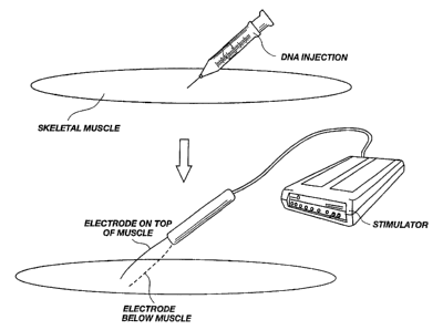
Figure 1 - graphically illustrates the method of delivering pharmaceutical
drugs and
DNA into skeletal muscle of the present invention.
Figure 2 - is a graphical illustration of an electrical stimulation delivered
according to
2~ the method of the present invention.
Figure 3 - illustrates whole amounts of muscles which have been injected with
50 ~1
of RSV-Lac Z Plasmid DNA solution at a concentration of 1 ,uglul. Muscles in
3a and 3b
were taken out 15 days after DNA injection. Muscles in 3c and 3d were taken
out 7 days
after DNA injection. All muscles are pairs from the same rat.
Figure 4 - pictures a whole muscle and a 1 mm slice of a transfected muscle.
Dark
stain indicates o-nitrophenyl-b-D-galactopyranoside (ONPG) that has been
catalyzed by
-4-
CA 02285056 2003-09-03
(3-galactosidase in the muscle to yield a dark precipitate. Arrows illustrate
muscle fibers that
were successfully transfected using the method of the present invention.
Figure 5 - includes mean number of transfected fibers from each group of
skeletal
muscles shown in Figure 3.
Figure 6 - is a bar graph illustrating mean transfected fibers of muscles from
several
different experiments and several different batches of DNA grouped together.
In columns
marked SOL S and EDL S the muscles (16 in each group) have been stimulated
directly after
the injection of DNA. In columns marked SOL NS and EDL NS the muscles (10 in
each
-4a-
CA 02285056 1999-09-30
WO 98/43702 PCT/IB98/00487
group) have been stimulated by the nerve, not stimulated at all or stimulated
directly 10 minutes
before the DNA injection.
Figure 7 - is a graph illustrating the number of skeletal muscle fibers
transfected versus
- the log of the stimulation frequency. The duration of the stimulation train
was kept constant at
1 second.
Figure 8 - is a photograph of transfected muscles from which data in Figure 7
were
generated.
Figure 9 - illustrates the results achieved when whole mounts of muscles were
transfected according to the method of the present invention using two
different electrodes.
Figure 10 - is a graph illustrating the number of skeletal muscle fibers
transfected with
increasing frequency compared to increasing pulse number.
Figure 11 - is a graph illustration of the number of skeletal muscle fibers
transfected
versus the number of pulses at constant frequency.
Figure 12 - is a graph illustrating mean luciferace activity versus the number
of pulses.
Figure 13 - is a graph illustrating the voltage dependency of the stimulation
method of
the present invention. Figure 13a illustrates the luciferase activity of
muscle stimulated with
varying volts. Figure 13b illustrates the mean luciferace activity of muscles
stimulated with an
amplitude above 13 volts and below 5 volts.
Figure 14 - is a graph illustrating the effect of pulse duration on the
transfection
efficiency.
Figure 15 - is a bar graph illustrating a comparison of transfection
efl<iciencies for
varying pulse durations and pulse numbers.
Figure 16 - is a bar graph illustrating the effect of DNA concentration on
transfection
efficiency.
Figure 17 - is a photograph of transfected muscles illustrating damage caused
by
stimulation and regeneration of the muscle after a short period of time.
Figure 17a illustrates an
injected muscle that was not stimulated. Figure 17b illustrates muscle damage
following muscle
stimulation. Figure 17c illustrates muscle stimulated under harsher
stimulation conditions.
Figure 17d illustrates that muscles stimulated under the conditions of muscles
in 17c are
completely regenerated and repaired after 14 days. Figure 17e illustrates
muscles transfected
with green fluorescent protein (GFP). Figure 17f illustrates that transfected
fibers can bee seen
in the vicinity of the damaged area.
-5-
CA 02285056 1999-09-30
WO 98/43702 PCT/IB98100487
Figure I 8 - is a photograph of cells stained with anti-agrin polyclonal
antibodies derived
from a rabbit genetically immunized with an expression vector coding for rat
agrin using the
stimulation technique of the present invention.
Figure 19 - are graphs illustrating improved genetic immunization of mice and
rats using
the stimulation technique of the present invention versus naked DNA injection.
Figure 20 - is a photograph of muscles transfected with rhodamine-conjugated
dextran
and green fluorescent protein. Top: rhodamin fluorescence from rhodamine
conjugated
dextran. Middle: The same section as above but with filters revealing GFP
fluorescence.
Bottom: hematoxilin and eosin staining of a neighboring section.
5. DETAILED DESCRIPTION OF THE INVENTION
The present invention is directed to a novel method for increasing the
permeability of
skeletal muscle tissue, thus allowing pharmaceutical drugs and nucleic acids
to enter or
transfect the cells. The method of the present invention passes a
predetermined amount of
I S electrical current through the skeletal muscle tissue. Unlike previously
described
electroporation methods, however, the parameters of the method of the present
invention are
unique, particularly with respect to the low field strength used and the
amount of damage that
occurs. Other parameters such as the number of trains, frequency, pulse number
and pulse
duration can be varied in order to regulate the amount of pharmaceutical drug
or nucleic acid
delivered.
As illustrated in Figure 1, generally, skeletal muscle is exposed and a
predetermined
amount of a molecule is injected into the muscle. In one embodiment the DNA is
dissolved in
0.9% sodium chloride (NaCI). The exact solvent, however, is not critical to
the invention. For
example, it is well known in the art that other solvents such as sucrose are
capable of increasing
DNA uptake in skeletal muscle. Other substances may also be co-transfected
with the
molecule of interest for a variety of beneficial reasons. For example, P 188
{Lee, et al. PNAS.,
4524-8, 10, 89 (1992)), which is known to seal electropermeabilized membranes,
may
beneficially affect transfection efficiencies by increasing the survival rate
of transfected fibers.
With continued reference to Figure 1, electrodes are placed on the muscle,
about I-4 mm
apart, near the area where the molecule was injected. The exact position or
design of the
electrodes is not critical so long as current is permitted to pass through the
muscle fibers
perpendicular to their direction in the area of the injected molecule.
-6-
CA 02285056 1999-09-30
WO 98/43702 PCT/IB98100487
Once the electrodes are in position, the muscle is electroporated or
stimulated. As
illustrated in Figure 2, the stimulation is delivered as a square bipolar
pulse having a
predetermined amplitude and duration. In order to optimize the transfection
efficiencies, these
parameters have been widely varied and transfection efficiencies compared. For
example, the
voltages have ranged from approximately 0 to 50 volts; the pulse durations
have ranged from 5
ps to 5 ms; the number of pulses have ranged from a single pulse to 30,000
pulses; and the
pulse frequency within trains have ranged from 0.5 Hz to 1000 Hz.
The conclusion from these results is that so long as the field strength is
above about 50
V/cm, the other parameters may be varied depending on the experimental
conditions desired.
While no upper limit was detected, effective transfection effciencies were
observed with much
higher field strengths. The field strength of the stimulation can be
calculated using the formula:
E=V/(2r In(D/r)),
which gives the electric field between wires if D »r. In the formula, V=
voltage = 10 V, D =
distance between wire centers = 0.1-0.4 cm, r = diameter of electrode = 0.06
cm. See
Hofmann, G. A. Cells in electric fields. In E. Neumann, A. E. Sowers, & C. A.
Jordan (Eds.),
Electroporation and electrofusion in cell biology (pp. 389-407). Plenum
Publishing Corporation
(1989). At 10 volts, the field strength is between 163 V/cm - 43 V/cm (from
0.1 to 0.4 cm
between electrodes, respectively). Because D is not much greater than r, it
may be more
appropriate to use the formula for electric fields between large parallel
plates:
E=VID
This gives a similar field strength of between 100 V/cm - 25 V/cm (from 0.1-
0.4 cm between
electrodes, respectively). It will be appreciated that the field strength, as
well as other
parameters, are affected by the tissue being transfected, and thus optimal
conditions may vary.
Using the parameters given in the present invention, however, optimal
parameters can be easily
obtained by one skilled in the art.
As illustrated in Figures 3 and 5-8, the method of the present invention
dramatically
increases the e~ciency of drug and DNA delivery into skeletal muscle. In one
embodiment, rat
soleus or EDL muscles were injected with DNA plasmid containing the (3-
galactosidase gene
(lac Z). The (3-galactosidase gene yields a protein capable of converting a
colorless substrate
into a blue substrate that can be visually analyzed or measured
spectrophotometrically. Figure
3 depicts representative soleus and EDL muscles that have been transfected
with ~3-
galactosidase gene using various stimulation parameters.
-7-
CA 02285056 1999-09-30
WO 98/43702 PCT/IB98100487
Figure 3a illustrates the improved DNA delivery efficiency of soleus and EDL
muscles that
have been transfected according to the method of the present invention. Soleus
and EDL
muscles {n=3) were first denervated by transecting the sciatic nerve. This was
done to eliminate
any influence of nerve-induced activity that arguably could contribute to the
increased
S transfection efficiency observed. Three days post-denervation, the muscles
were injected with
the Vii- galactosidase gene as described above. After the DNA injection, the
muscles were either
untreated or, immediately after the DNA injection, the muscles were stimulated
according to
the method of the present invention.
Fifteen days after DNA injection the soieus and EDL muscles were analyzed. As
illustrated
in Figure 3a, muscle cells that were stimulated immediately after DNA
injection {bottom panels)
contain more blue product indicating that more ~i-galactosidase gene was
introduced into the
muscle cells. The transfection efficiency was quantitated by counting the
muscle fibers in a I
mm cross section of the muscle that contained blue product as illustrated in
Figure 4. As
illustrated by the bar graph in Figure Sa, soleus muscle transfected using the
method of the
present invention showed a 47-fold increase over muscles that were not
stimulated. Similarly,
EDL muscle transfected using the method of the present invention showed a 12-
fold increase
over muscles that were not stimulated.
To determine whether nerve activity affected the transfection efficiency, the
method of the
present invention was performed on innervated (sciatic nerve not transected)
and denervated
(sciatic nerve transected) soleus and EDL muscles as described above. As
illustrated in Figure
3b, fifteen days after DNA injection both innervated and denervated muscles
produced a
generous quantity of blue product indicating high efficiency transfer of the
(3-galactosidase
gene. As illustrated in Figure Sb, quantitation of transfected muscle fibers
confirms high
efficiency transfection of both innervated and denervated muscles.
To rule out the possibility that the increased transfection efficiency
observed was due to
muscle activity, direct stimulation of the sciatic nerve was compared to
stimulation of the
muscle (n=5). If the increased transfection effciency was due to muscle
activity, the
transfection e~ciency in muscles stimulated via the nerve should yield similar
efficiencies as
direct muscle stimulation. As illustrated in Figure 3c, direct nerve
stimulation did not
significantly increase transfection efficiencies compared to direct muscle
stimulation. As
illustrated in Figure Sc, in both soleus and EDL muscles a 10-fold increase in
transfection
e~ciency was observed with direct muscle stimulation.
_g_
CA 02285056 1999-09-30
WO 98/43702 PCT/IB98/00487
As illustrated in Figure 3d, the increased efficiency is transient, consistent
with
electroporation. Muscles stimulated directly after DNA injection display
significantly more blue
dye than muscles that were stimulated prior to DNA injection. In fact, muscles
that were
stimulated directly after DNA injection displayed transfection efficiencies
between 10- and 25-
fold greater than muscles that were stimulated 10 minutes prior to DNA
injection (Figure Sd).
Figure 6 summarizes the results of the present invention. Muscles from several
different
experiments and several different batches of DNA are grouped together. In
columns marked
SOL S and EDL S the muscles ( 16 in each group) have been stimulated directly
after the
injection of DNA. In columns marked SOL NS and EDL NS the muscles { 10 in each
group)
have been stimulated by the nerve, not stimulated at all, or stimulated
directly 10 minutes before
the DNA injection.
The electrical stimulator used for the experiments was manufactured by FHC
(Brunswick,
ME 04011 ). Both Pulsar 6bp and the Pulsar 6bp-a/s stimulators have been used.
The Pulsar
6bp-a/s delivers a maximal voltage is 150 V and a maximal current of 50 mA.
The maximal
voltage that can be delivered requires a resistance between the electrodes of
greater than 3000
ohms. The stimulators have been operated at constant voltage mode. Because of
the low
resistance in the muscle, the voltages have been lower as discussed in the
Examples below. In
all experiments the current has been maintained at SOmA.
It will be appreciated by one skilled in the art that numerous other electrode
configurations
can be employed. For example, Figure 9 illustrates the results obtained using
two different
electrodes configuration. The electrode shown in (A) was placed perpendicular
to the muscle
fibers. It consisted of a silver wire with diameter (d) of 0.6 mm, (C) (this
is the electrode which
was used in all experiments except in (B)). One electrode was placed on each
side of the
muscle. A short segment in the middle third of the muscle is positive for the
Lac Z staining {A),
indicating localized expression. In (B) a I .5 cm electrode made from an
insulated silver wire
was used (d=0.3 mm). Insulation was removed from short segments (0.5 - 1.0 mm)
along the
wire at 2 mm intervals (D). The electrode was penetrated into the muscle in
parallel with the
muscle fibers. One of the two wires of the electrode was penetrated into the
muscle parallel
with the muscle fibers. The second wire was placed on the muscle surface, also
parallel with
the fibers. Both types of electrodes (Figures 9c and 9d) gave a similar number
of transfected
fibers (approximately 250). Using the longer electrode in parallel with the
muscle fibers,
however, gave a more wide spread staining, indicating a transfection along a
longer segment of
the fibers and/or increased transfection.
-9-
CA 02285056 1999-09-30
WO 98/43702 PCT/IB98/00487
Muscles were stained for Lac Z in whole mounts by methods well known in the
art. After
staining, the pictures were taken with the bluest side of the muscle up.
Thereafter the muscle
was cut in three pieces as seen in Figure 2. The number of blue fibers in
about I mm thick slice
from the middle of the muscle were counted (fibers transfected distally or
proximally from the
S slice are therefore not counted). In order to count the transfected fibers,
the slices were
dissected into smaller bundles so single fibers could be distinguished under a
dissection
microscope.
In four (4) muscles the pSV40-luc construct was used. It was injected into the
soleus
muscle, 3 days after the muscles were removed and luciferase activity was
measured using the
Promega Luciferase Assay System (Daviset et al., 1993). Uninfected EDL from
the same rats
were used as control.
It will be appreciated that any nucleic acid can be used with the method of
the present
invention, for example, plasmid DNA, linear DNA, antisense DNA and RNA. In one
preferred
embodiment, the nucleic acid is a DNA expression vector of the type well known
in the art.
Generally, an expression vector contains a promoter operably linked to a DNA
molecule that
codes for the protein of interest followed by a termination signal such as a
poiyadenylation
signal. Other elements required for bacterial growth and proper mammalian
processing may be
included, such as the (3-lactamase coding region, an fl origin and ColEl-
derived plasmid
replication origin. Similar constructs containing a DNA coding region of
interest can be
constructed by one skilled in the art.
As illustrated in the examples below, molecules other than nucleic acids can
be delivered to
the muscle using the technique of the present invention. In one embodiment,
rhodamin
conjugated dextran injected into the muscles and stimulated according to the
method of the
present invention was able to enter muscle cells. In addition, nucleic acid
and proteins can be
simultaneously introduced into an electroporated muscle. In one embodiment,
the large
T-antigen nuclear localization signal was mixed with a plasmid containing the
DNA coding
region for Lac Z. The large T-antigen nuclear localization signal is a protein
that binds DNA
and facilitates its transport into the nucleus of a cell. In other systems,
large T-antigen nuclear
localization signal has been shown to increase transfection efficiency. Using
the method of the
present invention, large T-antigen nuclear localization signal also increased
the transfection
efficiency of Lac Z indicating that the protein was able to bind the DNA arid
enter the muscle
cell.
-10-
CA 02285056 1999-09-30
WO 98/43702 PCT/IB98100487
6. EXAMPLE
The following examples are given to illustrate various embodiments which have
been made
of the present invention. It is to be understood that the following examples
are not
comprehensive or exhaustive of the many types of embodiments which can be
prepared in
S accordance with the present invention.
Example 1 - Stimulated Versus Unsimulated Muscles:
Transfection efficiencies were determined by injecting skeletal muscles with
the pSV40-luc
reporter construct into the soleus muscle. Three days after injection, the
muscles were removed
and luciferase activity was measured using the Promega Luciferase Assay System
(Madison,
WI) according to manufacturer's protocols. Unstimulated EDL muscles from the
same rats
were used as control. The data are shown below in Table 1.
TABLE 1
STIMULATED VERSUS UNSTIMUALTED MUSCLES
Stimulated Unstimulated
Muscle (Relative luciferase-(Relative luciferase-Percent
activity) activity) Increase
Soleus animal I 34.40 1.950 1664%
Soleus animal II 21.50 0.250 8500%
EDL animal I 0.045
~~ EDL animal II 0 046
Example 2 - Transfection Efficiency Versus Frequency:
Rats were injected with 50 pl of 1 mg/pl of a plasmid carrying lac Z gene.
Immediately
following injection, electrodes were placed between 2-3 mm apart and the
muscle was
stimulated with the following stimulation parameters: voltage = 30 volts;
pulse duration = 0.2
ms (total 0.4 ms, bipolar); trains = 30, 1 second on t second offfor 1 minute.
Transfected
fibers were counted from a 1 mm slice from middle of muscle. The number of
transfected fibers
is shown below in Table 2 and illustrated in Figure 7. These data also
illustrate that the method
of the present invention transfects more than just surface muscle f bers;
muscle fibers several
cell layers deep are also transfected.
CA 02285056 2002-09-20
____ TABLE
2 ~L'C'~' 3
..~-.j.~,~
~
TRANSFECTION
EFFICIENCY
VERSUS
FREQUENCY
~~a ~
,"_.~
_ -
Mean
Frequency Percent
'
(
Transfected
Increase with
Stimulation
Fibers)
p 22 _
1 83 277%
10 I53 595%
100 2'15 877%
1000 315 1332%
Example 3 - Transfection Efficiency Versus Pulses:
Soleus muscles of Wistar rats (200-270 grams) were injected with 50 pg of RSV
luciferase
DNA plasmid in 501r1 0.9% NaCI. Shortly after injection, the n'uscles were
electrically
stimulated using the following parameters: 1000 Hz, between 0 - I 000 bipolar
pulses of 200ps
duration in each train were applied to the muscle 30 times over a period of 1
minute. Muscles
were removed 3 days after transfection and frozen in liquid nitrogen. Cryostat
sections were
TM TM TM
taken from the of the muscles and stained with I-iematoxolin, Eosin and Safran
(see Example 9).
The remaining pieces were homogenized as described in Example 4 below. As
illustrated in
Figure 10-12, transfection efficiency increased with the number of pulses
delivered to the
muscle.
Example 4 - Determining the Effect of Voltage on Transfection Efficiency:
EDL and soleus muscles of Wistar rats (245-263 grams) were injected with 251tg
of RSV
driven luciferace plasmid DNA in 501r1 0.9% NaCI. Shortly after injection, the
injected muscles
were electrically stimulated using the following parameters: 100 Hz, 100
bipolar pulses in each
train of 200 ps duration, voltage varied from between 0 to 47.5. Muscles were
removed 4 days
post injection and stimulation, homogenized in Promega (Madison, Wl)
iuciferace assay buffer
and luminescence was measured according to manufacturer's protocols. Macintosh
computer
and a LabWiev acquisition program were used to capture the first voltage
pulses. Recordings
were done in parallel with the stimulation electrodes. The voltage
measurements were done
manually on prints as the average of the maximal voltage of 10 pulses
approximately 100 ms
after onset of stimulation.
As illustrated in Figure 13a, there was a pronounced increase in transfection
efficiency with
increased voltage. As illustrated in Figure 13b, under the conditions of this
experiment,
-12-
CA 02285056 1999-09-30
WO 98/43702 PCT/IB98/00487
muscles stimulated with 13 volts or higher showed 40-fold greater luciferace
activity compared
to muscles stimulated with S volts or less.
Example 5 - Determining Optimal Pulse Duration:
Soieus muscles of Wistar rats (200-270 grams) were injected with 50 ~tg of DNA
plasmid
containing the p-galactosidase gene in SOpI 0.9% NaCI. Shortly after
injection, the muscles
_were electrically stimulated using the following parameters: 100 Hz, 25
volts, 100 bipolar
pulses in each train having pulse durations ranging from S-200 ps. The number
of transfected
fibers were counted in a 1 mm thick section from the middle of the muscle
under a dissection
microscope. A second set of rats were injected with 2Sltg of RSV-driven
luciferace plasmid
DNA in SO~tI 0.9% NaCI and electrically stimulated with the same parameters as
above except
that the pulse durations were varied from SO-2000 lls. As illustrated in Table
3 below and
Figure 14, under these stimulation parameters, the optimal pulse duration
ranged from about 50
ps to about 200 ps. This method can be used to optimize the pulse duration of
other
stimulation parameters.
TABLE
3
TRANSFECTION EFFICIE NCY ERSUS PULSE
V DURATION
Pulse Transfected Pulse Luciferase-
Duration Fibers Duration activity
(ps) (Mean ) (us) (Mean)
0 ' 0 52.7
S S1 SO 631
20 107 200 S36
50 228 500 348
2S 200 272 2000 194
Example 6 - Current versus number of pulses:
Soleus muscles of six Wistar rats (178-193 grams) were injected with 50 ug of
DNA
plasmid containing the [3-galactosidase gene in SOpI 0.9% NaCI. Shortly after
injection, the
muscles were electrically stimulated as described above except that the pulse
duration was
varied. The following electroporation parameters were compared: ( 1 ) I 00
pulses of 50 ps
duration versus 1 pulse of 5000 ps; and (2) 10 trains of 100 pulses of 50 ps
versus 10 pulses of
5000 ps. Muscles were removed 14 days later and sectioned on a cryostat. Cross
sections
were stained as previously described. The number of transfected fibers were
counted. As
3S illustrated in Figure 15, longer pulse durations result in higher
transfection efficiency.
-13-
CA 02285056 1999-09-30
WO 98143702 PCT/IB98/00487
Example 7 - DNA Concentration:
EDL muscles of six Wistar rats (178-193 grams) were injected with either 1
ug/pl or Spglpl
of DNA plasmid containing the (3-galactosidase gene in 501 0.9% NaCI. Shortly
after
injection, the muscles were electrically stimulated with 30 trains of 100
pulses of 200 ps
duration or not stimulated at all. Muscles were removed 14 days later and
sectioned on a
cryostat. Cross sections were stained as previously described and transfected
fibers were
counted. As illustrated in Figure 16, greater transfection efficiencies were
obtained with higher
DNA concentrates.
I O Example 8 - Large T Antigen Nuclear Localization Signal:
Wistar rat muscles were injected with DNA plasmid containing the ~3-
galactosidase gene
containing a 100:1 molar excess of large T-antigen nuclear localization
signal. This has been
shown in other transfection studies to improve the transfection. (See, P.
Collas et al. Trans~enic
Res., 6: 451-8 (1996)). The muscle were stimulated with 10 trains of 100
pulses of 50 ps
duration. The muscles containing the large T-antigen nuclear localization
signal had the highest
number of transfected fibers. Specifically, the muscle co-transfected with
large T-antigen
nuclear localization signal had 100 and 38 transfected fibers versus 7.3 and
4.7 for the muscles
transfected only with DNA, respectively. These data illustrate that
transfection efficiencies can
be aided by mixing the DNA with non-nucleic acid molecules. In addition, this
data illustrates
that non-nucleic acid molecules can also be delivered to the muscle using the
electroporation
techniques of the present invention. No improvement was seen in cells that
were not stimulated
following injection.
Example 9 - Muscle Damage Resulting from Stimulation:
Muscles from Example 3 that were sectioned and stained to assess the muscle
damage from
electroporation. As illustrated in Figure 17a, some damage can occur with
injection alone,
although the majority of unstimulated muscles were undamaged. In muscles
stimulated with
300 pulses, more damage was observed (Figure 17b). As illustrated in Figure
17c, muscle
stimulated with 30 trains of 1000 pulses displayed greater damage, indicating
that damage is
proportional to the extent of stimulation. Figure 17d illustrates that muscles
stimulated under
the conditions of muscles in 17c are completely regenerated and repaired after
14 days.
In another muscle which got the highest amount of stimulation (30 trains of
1000 pulses),
plasmid DNA encoding green fluorescent protein (GFP), was also included.
Figure 17e
-14-
,.
CA 02285056 1999-09-30
WO 98/43702 PCTlIB98/00487
illustrates muscles transfected with GFP. Transfected fibers can bee seen in
the vicinity of the
damaged area (Figure l7fJ. Transfected regenerating fibers were never observed
in cross
sections 3 days after electroporation.
Exampie 10 - Genetic Immunization of Rabbits:
A female rabbit (4.5 kg) was injected into the right femuraiis rectus with 2
milliliters of 1 ~tg
/pl of DNA plasimd containing the rat neural agrin cDNA driven by the CMV
promotor (Cohen
et al. MCN, 9, 237-53 (1997)). The first milliliter was injected equally in
ten places superficial
in the muscle followed by 10 trains of 1000 pulses delivered at a frequency of
1000 Hz. The
second milliliter was placed further down in the muscle. To test the rabbit
serum, rat muscles
and COS cells were transfected with the same construct. Muscles were taken out
5 days after
transfection and the COS cells were stained 4 days after transfection.
Bleeds were collected at days 0, 19, 50, 81 and 106 and diluted 1:100 and
1:1000. After 19
days the bleed contained enough antibody in the serum to give a weak staining
of transfected
I 5 fibers when diluted 1:10. As a positive control the monoclonal antibody
(mAb) AG-86 was
used. See Hoch et al. EMBOJ, 12 (13): 2814-21(1994). Preimmune serum did not
show any
staining of transfected fibers. Later bleeds all had agrin antibodies in the
serum. Bleed collected
at day 50 or later contained sufficient antibodies to stained sections at a
dilution of 1:1000.
Figure 18a illustrates the agrin transfected COS cells stained with antiserum
from
immunized rabbit (diluted 1:100) and fluorescein conjugated secondary
antibody. COS cells
were stained first fixing the cells in I.5% paraformaldehyde for 10 minutes,
followed by a 30
minute wash with phosphate buffered saline (PBS). The cells were then blocked
with 0.2%
bovine serum albumin, triton X-100, 0.1% in PBS O.1M, for 4 minutes. Serum
diluted in same
solution was added to the cells and allowed to incubate for 20 minutes. Cells
were wash for 4
minutes in PBS and incubated with the secondary antibody (Cappel, 55646) for
10 minutes
followed by a PBS wash. Mouse primary mAb Agr-86 was included in the same
antibody
mixture and rhodamin conjugated secondary antibody (Sigma T-5393, St. Louis.
MO) was used
at a dilution of 1:100. Figure 18b illustrates the same cells stained with mAb
Ag-86/rhodamin
conjugate. These data illustrate the potential of the technique of the present
invention for
genetic immunization or DNA vaccine technology.
Example 11 - Genetic Immunization of Mice:
-15-
CA 02285056 1999-09-30
WO 98/43702 PCTIIB98/0048?
Groups of two-month old male Sprague Dawley rats were inoculated bilaterally
in the EDL
and soleus muscles with a total of 200 micrograms (4 x 50 microliters of a 1
mg/ml solution of
DNA in saline) of three different eukaryotic expression vectors containing the
cytomegalovirus
immediate early promoter (CMV} and the coding sequences for the following
proteins:
DH-CNTF, an agonistic variant of human ciliary neurotrophic factor (Saggio et
al. EMBO J.
14, 3045-3054, 1995); AADH-CNTF, an antagonistic variant of human ciliary
neurotrophic
factor (Di Marco et al. Proc. Natl. Acad. Sci. USA 93, 9247-9252, 1996); sec-
DHCNTF, a
secreted form of DH-CNTF. The muscles were either not electrically stimulated
or stimulated
immediately after DNA injection using 30 trains of 100 or 1000 square bipolar
pulses (duration
200 microseconds; amplitude setting 150 V, effective voltage --25 V) each,
delivered at a
frequency of 1000 Hz with a two second interval between successive trains.
Groups of two-month old male CD 1 mice were inoculated bilaterally in the
quadriceps
muscles with 100 micrograms (2 x 50 microliters of a 1 mg/ml solution of DNA
in saline) of
sec-DHCNTF plasmid, with or without electrical stimulation of the muscle
immediately after
DNA injection. Stimulation conditions were 10 trains of 1000 square bipolar
pulses (amplitude
setting 150 V) delivered at a frequency of 1000 Hz with a two second interval
between
successive trains.
Blood was collected from the retroorbital sinus at selected time points and
serum was
prepared and stored at -20° C. The presence of anti-CNTF antibodies in
rat and mouse sera was
determined by ELISA. Microtiter plates coated with recombinant human CNTF were
incubated
with serial dilutions of sera, followed by alkaline phosphatase-conjugated
antibody against rat
or mouse IgG (Pierce). The plates were then incubated in the presence of
p-nitrophenyl-phosphate and the absorbance at 405 nm was determined using a
microplate
reader. Antibody titers were defined as the dilution of serum producing an
absorbance reading
equal to 50% of that obtained with a saturating concentration of anti-CNTF
antiserum.
The results are shown in Figure 19. Titers could not be averaged with
precision, due to the
fact that some animals did not develop detectable amounts of antibody. Data
are therefore
presented for individual animals, with a value of 1:100 representing a low or
undetectable
antibody titer (reciprocal titer 3/4 100). The results were similar for all
plasmids used, as well as
for rats and mice, as depicted in Figure 19. Similar results were also
obtained in both rats and
mice with another plasmid encoding an unrelated viral protein (data not
shown). In both rats
and mice, electrical stimulation immediately after DNA injection led to
approximately 5 to
10-fold higher antibody titers than simple DNA injection. This was true for
stimulation with
_ 16_
CA 02285056 1999-09-30
WO 98/43702
PCT/IB98/00487
both high and low numbers of pulses. These results demonstrate that the
electroporation
method increases the effciency of DNA-mediated immunization.
Example 12 - Secreted Proteins with Systemic Biological Activity:
Fifty micrograms (50 microliter of a 1 mg/ml solution in 0.9% NaCI) of a
eukaryotic
expression plasmid (CMV-EPO) containing the cDNA of mouse erythropoietin under
the
_ control of the cytomegalovirus immediate early promoter was injected in the
left quadriceps
muscle of three-month old 129xBa1b/C female mice. In five mice (group 1 ), the
muscles were
electrically stimulated immediately after DNA injection using 10 trains of
1000 square bipolar
I O pulses of 200 microseconds duration, with an interval of 2 seconds between
successive trains.
The frequency of the trains was 1000 Hz and the amplitude set at 150 V
(effective voltage ~25
V}. In another group of 5 mice (group 2) the muscles were not stimulated after
DNA injection.
As a control, a group of 4 mice (group 3) was injected with a plasmid (CMV-
GFP) containing
the coding sequence for green fluorescence protein under the control of the
CMV promoter,
I S followed by electrical stimulation at the same conditions as group 1.
Group 4 consisted of 5
mice injected only with saline solution without electrical stimulation.
Blood was collected from the retroorbital sinus at selected time points and
hematocrit was
measured by centrifugation in capillary tubes. Serum samples were analyzed for
the presence of
EPO using a commercial ELISA kit (R&D Systems). The results are shown in Table
4. In all
20 groups of mice, except those that were injected with the EPO construct and
electrically
stimulated immediately thereafter, circulating EPO levels were below the limit
of detection of
the ELISA kit (<I S mU/ml). In contrast, mice injected with the EPO construct
and electrically
stimulated had significantly elevated serum EPO levels 5 days after injection
(average of
approximately 50 mU/mi). The serum concentration of EPO remained elevated for
up to 28
25 days following DNA injection (latest time point examined; data not shown).
These levels of
EPO produced an increase in hematocrits, which rose from 46.2% prior to
injection to 70.0%
and 76.7% at 14 and 28 days after DNA injection, respectively. These values
were significantly
different from those obtained with both control groups (groups 3 and 4) and
from those of mice
injected with the EPO expression vector without electrical stimulation of the
muscle (group 2).
30 Indeed, the latter had hematocrit levels not significantly different from
those of the control
groups (see Table 4}. These results demonstrate that the electroporation
method is superior to
simple DNA injection both in terms of the expression levels of a secreted
protein and in
producing a biological effect mediated by the secreted protein.
-17-
CA 02285056 1999-09-30
WO 98/43702 PCT/IB98/00487
TABLE 4
EPO Serum
Concentrations
and
Activity
Day Day Day
2 ~ 14
Mouse mEPO mEPO mEPO
No.
HCT HCT% HCT%
/
(mUlml) (mU/ml) (mU/ml)
7 45 ND ND 55.7 71 72.4
8 48 ND ND 54.6 68 5.3
9 47 ND ND 59 75.5 48.7
Group 1 CMV-EPO 10 44 ND ND 62.2 69.5 62.9
Stimulated 11 47 ND ND 7.9 66 22.4
Avg. 46.2 ND ND 47.9 70.Oa'"48.3
Stand
. 1 3.6
6
Dev. .
12 45 ND ND ND 50 <15
13 45 ND ND ND 50 < 15
14 ND ND ND ND 48 <15
Group 2 CMV-EPO 15 46 ND ND ND 49.5 <15
No stimulation 16 44 ND ND ND 52 <15
Avg. 45 ND ND ND 49.9 <15
Stand.
8
0
Dev. .
2 ND ND ND <15 43.5 <15
3 ND ND ND <15 48 <15
5 ND ND ND < l5 46 < I S
Group 3 CMV-GFP
Stimulated 6 ND ND ND < 15 46 < 15
Avg. ND ND ND < 15 45.9 < 15
Stand.
1.8
Dev.
17 45 ND ND < 15 45.5 ND
18 45 ND ND < I 49 ND
~
19 43 ND ND < 1 48 ND
~
20 45 ND ND < 15 51.5 ND
Group 4 CMV-EPO 21 50 ND ND < 15 47 ND
Avg. 45.6 ND ND < 15 48.2 ND
Stand.
2.6 2.3
Dev.
ND= not determined. a p < 0.0001 vs. group 2; ° p < 0.0001 vs. group 3;
' p < 0.0001 vs.
group 4 (Fisher's protected least significant difference).
Example 13 - Delivery on Non-nucleic Acid Molecules:
Muscles were injected with 50 ~tl of a mixture of GPF plasmid DNA 1 ~g/pl and
2 ~g/pl
rhodamin-conjugated dextran (10 kD from Molecular Probes). Three to S days
later the
- I 8-
CA 02285056 1999-09-30
WO 98143702 PCT/IB98/00487
muscles (n=b) were frozen in liquid nitrogen and sectioned on a cryostat. As
illustrated in
Figure 20, stimulated muscles (bottom) were transfected with rhodamin-
conjugated dextran
(top) and GFP (middle). As further illustrated, the same muscle fibers were
transfected with
both GFP and rhodamin-conjugated dextran. These data indicate that non-nucleic
acid
molecules can be delivered to muscle cells using the technique of the present
invention.
Figure 2
Whole muscle and a 1 mm thick slice cut out from the middel of it.
Number of transfected fibers were counted after it was split into
smaller bundels and single fibers could be seen through a direction
microscop. In some areas of the muscle rmst fibers were transfected
(black arrows). These areas were close to were the electrodes were
situated during stimulation.
Figure 9
'Itao different electrodes have been used in order to improve transfection
efficiency. The injection pirocedure and stinnilation pattern (100 Hz)
was the same as previously described. The electrode sYwwn in (A) was
placed perpendicular to the muscle fibers. It consisted of a silver
wire with diameter (d) of 0.6 mm, (C) (This is the electrode which
was used in all experiments except in (B)). One electirode was placed
on each side of the muscle. A short segment in the m-fiddle third of the
muscle is positive for LacZ staining (A), irx3i.cating localised
expression. In (B) a 1.5 cm electrode made from an insulated silver
wire was used (d=0.3 mm). Insulation was removed from short segments
(0.5-1.0 mm) along the wire at 2 mm intervals (D). The electrode was
penetrated into the muscle in parallell with the muscle fibers. A
second electrode was placed on the surface of the muscle. Positive blue
staining was observed in approximately 250 fibers which were localised
to the middle third of the muscle. In (B) the fibers shc7wed widepread
staining. indicating transfection along a longer segment og the fiber
and/or increased transgene expression.
-19
SUBSTITUTE SHEET (RULE 26)