Note: Descriptions are shown in the official language in which they were submitted.
CA 02285233 1999-09-30
WO 98/43557 PCT/US98/06333
PROSTHETIC HEART VALVE LEAFLET SELECTION METHOD AND
APPARATUS
. Field of the Invention
The present invention relates to methods for selecting leaflets for use in
a prosthetic heart valve, and, more particularly, to methods and apparatuses
for
selecting individual
pericardial leaflets for a multi-leaflet heart valve prothesis.
Background of the Invention
Prosthetic heart valves are used to replace damaged or diseased heart
valves. In vertebrate animals, the heart is a hollow muscular organ having
four
pumping chambers: the left and right atria and the left and right ventricles,
each
provided with its own one-way valve. The natural heart valves are identified
as
the aortic, mitral (or bicuspid), tricuspid, and pulmonary valves. Prosthetic
heart
valves can be used to replace any of these natural valves. The two primary
types
of prosthetic heart valves known in the art are mechanical valves and bio-
prosthetic valves. Mechanical valves include rigid leaflets and a pivoting
mechanism, and bio-prosthetic valves utilize flexible tissue leaflets,
typically
mounted to a manufactured support frame. The present invention provides
methods for selecting leaflets in bio-prosthetic valves.
Bio-prosthetic valves may be formed from an intact, multi-leaflet porcine
(pig) heart valve, or by shaping a plurality of individual leaflets out of
bovine
pericardial tissue and combining the leaflets to form the valve. The
pericardium
is a sac around theheart of vertebrate animals, and bovine (cow) pericardium
is
commonly used to make individual leaflets for prosthetic heart valves. The
bovine pericardium is first harvested from the animal and then chemically
fixed
to crosslink collagen and elastin molecules in the tissue and increase the
tissue
CA 02285233 1999-09-30
WO 98/43557 PCT/US98/06333
2
durability, before being cut into leaflets. Various physical characteristics
of the
tissue may be examined before or after fixation.
One drawback faced by a patient having an implanted bio-prosthetic heart
valve is the potential for calcification of the leaflets if the valve remains
in place
for an extended period of time (more than ten years). Calcification tends to
make the leaflets less flexible. A significant amount of research has been
accomplished in mitigating calcification of bovine pericardial leaflets to
lengthen
the useable life of the heart valve. Calcification may reduce the performance
of
the heart valve, and thus, the highest quality materials and design in the
heart
valve is required to forestall a failure of the valve from excessive calcium
deposits.
Despite the drawbacks of artificial heart valve material, over twenty years
of clinical experience surrounding implanted artificial heart valves has
produced
a proven track record of success. Research in extending the useful life of the
bio-
prosthetic valves continues, however. One aspect of designing heart valves
which
is very important in improving their performance is the selection of the
pericardial
tissue used in the leaflets. In all heart valves, the natural action of the
flexible
heart valve leaflets, which seal against each other, or co-apt, is desirable.
The
difficulty in simulating the leaflet movement of an actual heart valve
(especially
a mitral valve) in a prosthetic valve is that the leaflets used are
"inanimate."
There are no muscular attachments to the leaflets as in the natural valve, and
the
prosthetic leaflets must co-apt to function properly solely in response to the
fluid
pressures within the heart chambers. Indeed, natural coaptation of the
leaflets
in bio-prosthetic valves comprising a plurality of individual leaflets sewn
together
is particularly difficult, even when compared to inanimate but intact valves,
such
as harvested porcine valves.
Much of this research involves the mechanical properties of fresh or fixed
bovine pericardium. A good discussion of the various physical properties of
fixed
bovine pericardium is given in Simionescu, et al, Mapping of Glutaraldehyde-
Treated Bovine Pericardium and Tissue Selection For Bio-prosthetic Heart
CA 02285233 1999-09-30
-WO 98/43557 PCT/US98/06333
3
Valves, Journal of Bio-Medical Materials Research, Vol. 27, 1993. Simionescu,
et al, recognized the sometimes striking variations in physical properties of
the
pericardial tissue, even in the same pericardial sac. Their research mapped
out
areas in individual pericardial sacs and tested those areas for fiber
orientation,
suture holding power, and thickness. In another paper by Sacks, Bi-axial
Mechanical Behavior of Fixed Bovine Pericardium, Fifth World Biomaterials
Congress, May-June 1996, the collagen fiber architecture within bovine
pericardial
tissue was examined and various specimens were tested in a bi-axial tester.
The
results indicated that by presorting for uniform collagen fiber architecture,
more
uniform bio-pericardial specimens could be obtained for better controlled use
in
bioprosthetic applications. Finally, in another study, Zioupos, et al,
Anisotropic
Elasticity and Strength of Glutaraldehyde Fixed Bovine Pericardium For Use In
Pericardial Bioprosthetic Valves, Journal of Biomedical Materials Research,
Vol.
28, 1994, various tests were performed on fixed bovine pericardial tissue to
determine the stress/strain behavior along various axes. The results suggest
that
leaflets can be made from fixed bovine pericardium possessing pronounced
anisotropy in strength and stiffness along two orthogonal directions. In the
leaflets circumferential direction, which bears most of the stress during
function,
the stiffer pericardium is desired, while in the radial direction, more
flexible tissue
is desired. Leaflets are thus cut from bulk tissue whose properties have
generally
been examined, and the leaflets categorized accordingly. D e s p i t e t h e
extensive research into bulk tissue characteristics there remains a need for a
more
reliable method of selecting leaflets to insure maximum functional
compatibility
with the other leaflets in the dynamic operating environment of a prosthetic
heart
valve.
Summary of the Invention
The present invention provides methods and apparatuses for selecting
leaflets for use in producing multi-leaflet prosthetic heart valves. The
selection
of leaflets to be combined in a heart valve is based on grouping a plurality
of
CA 02285233 1999-09-30
-WO 98/43557 PCT/US98/06333
4
leaflets by strain response to an applied load which is designed to simulate
physiological pressures within the heart. A stress load sufficient to stress
the
leaflets within a high modulus region of their stress/strain characteristic is
applied
to each leaflet, and leaflets within a predetermined observed deflection range
of
each other are grouped together. In an exemplary embodiment, glutaraldehyde-
fixed leaflets are stressed within a generally linear, high modulus region of
the
bulk tissue stress/strain curve, and the deflection measured for grouping the
leaflets. In one embodiment, the strain response is observed relative to a
deflection of bovine pericardium leaflets resulting from applying a load
thereto,
and two or three leaflets from a group of leaflets having deflections within
.030
inches of each other are combined to form a prosthetic heart valve
One aspect of the present invention is a method of selecting leaflets for an
implantable heart valve, including providing a collection of similarly sized
leaflets,
applying a load to each leaflet, observing the resulting strain response, and
sorting
the leaflets based on their respective strain responses. The collection may be
natural tissue leaflets which are chemically fixed prior to testing. The
natural
tissue leaflets may be made of bovine pericardium. In one embodiment, the load
applied is sufficient to create an average stress in at least some of the
leaflets of
between 300 and 600 kPa. The load is preferably applied for a predetermined
number of times prior to observing the strain response. Another aspect of the
invention is a bioprosthetic heart valve manufactured with leaflets selected
by the
aforementioned method, wherein the number of leaflets selected may be three.
The present invention also provides a method of testing a leaflet for use
in an implantable heart valve, including mounting the leaflet in a frame so
that
portions which are to be sutured in the valve are held stationary. A load is
applied to the leaflet in a location adapted to simulate a point at which an
average load is applied in the valve, and the resulting strain in the leaflet
is
sensed. The natural tissue leaflet typically defines a cusp and a coapting
edge
generally opposite the cusp, and the step of mounting may comprise holding
CA 02285233 2003-04-02
stationary at least the cusp of the leaflet. The leaflet may be positioned in
a
framing assembly having a recess for receiving at least the edges of the cusps
of
the leaflet, and a cavity circuinscribed by the recess. Moreover, the load may
be
applied by a mechanical deflector to an upper surface of the leaflet over the
5 cavity. Preferably, the framing assembly includes an upper rnember and a
lower
member, the lower member having the recess and the upper member shaped to
mate over the recess. 'I'he method further includes piercing the leaflet edges
with
needles extending between and supported from movement by the upper and lower
members.
The present invention provides an apparatus for testing heart valve leaflets
having a leaflet framing assembly including a holder with a recess for
receiving a
leaflet to be tested and a frame which cooperates with the holder to hold
stationary the cusps of the leaflet. '1'he apparatus includes a base having
indexing
structure for locating the framing assembly thereon, and a deflection assembly
indexed with respect to the base and having a deflector mounted for movement
above the framing assembly to contact the leaflet. The recess may be cusp-
shaped, and the holder includes a cavity substantially surrounded by the
recess
over which the leaflet is suspended. The apparatus may further include
structure
adapted to hold stationary discrete points of the leaflet around the cavity.
To
secure discrete points of' the leaflet around the cavity, the frame preferably
includes a plurality of needles having their pointed ends downward, and the
recess
includes receptor holes for the needles, wherein the cusp of the leaflet is
secured
against movement at the discrete points defined by the needles.
According to an aspect of the present invention, there is provided a multi-
leaflet bioprosthetic heart valve comprising a plurality of leaflets, wherein
each of
the leaflets has a measured deflection response within 0.030 inches of the
other
leaflets upon application of a loaci sufficient to stress each of the leaflets
between
300 and 600 kPa.
According to anotlier aspect of the present invention, there is provided a
:30 multi-leaflet bioprosthetic heart valve comprising a. plurality of bovine
pericardium leaflets, wherein each of the leaflets has a measured deflection
response of
CA 02285233 2003-04-02
5a
between 0.17 and 0.34 inches upon application of a load sufficient to stress
each
of the leaflets between 300 and 600 kPa.
According to another aspect of the present invention, there is provided a
method of selecting leaflets fr-r an implantable heart valve, comprising the
steps
of:
providing a collection of similarly sized leaflets;
applying a load to each leaflet;
observing the strain response in each leaflet from applying the load; and
sorting the leaflets based on their respective strain responses.
According to another aspect of the present invention, there is provided a
method of testing a leaflet for use in an implantable heart valve, comprising
the
steps of:
mounting the leaflet in a framing assembly so that portions which are to
be sutured in the valve are held stationary, wherein the leaflet defines a
cusp edge
and a coated edge generally opposite the cusp edge, and the framing assembly
includes an upper member and a lower member, the lower member having a
recess for receiving at least the cusp edge of the leaflet, the upper member
being
shaped to mate over the recess, and the framing assembly defining a cavity
circumscribed by the recess, the step of mounting including positioning the
leaflet
in the recess and piercing the leaflet cusp edge with needles extending
between
and supported from movement by the upper and lower members, to hold at least
the cusp edge of the leaflet stationary;
applying a load to the leaflet in a location adapted to simulate a point at
which an average load is applied in the valve; and
25. sensing the resulting strain in the leaflet.
According to another aspect of the present invention, there is provided a
method of testing a leaflet for use in an implantable heart valve, wherein the
leaflet defines a cusp edge and a coating edge generally opposite the cusp
edge,
comprising the steps of
positioning the leaflet in a frame assembly including an upper member
CA 02285233 2003-04-02
5b
and a lower member, the lower niember having a recess for receiving and
securing the cusp edge of the leaflet the user member being shaped to mate
over
the recess, and the framing assembly further defining a cavity circumscribed
by
the recess;
supporting the leaflet by piercing the leaflet cusp edge with needles
extending between and support.ed from movement by the upper and lower
members to hold at least the cusp edge of the leaflet stationary such that a
mid-
portion remains unsupported;
applying a load to the mid-portion of the leaflet; and
sensing the resulting strain at the mid-portion of the leaflet.
According to another aspect of the present invention, there is provided an
apparatus for testing heart valve leaflets, each leaflet including an arcuate
cusp
edge and a free edge, the apparatus, comprising:
a leaflet framing assembly including a holder with a recess for receiving a
leaflet to be tested and a franie which cooperates with the holder to
substantially
hold stationary the cusps of the leaflet;
a base having indexing structure for locating the framing assembly
thereon; and
a deflection assembly indexed with respect to the base and having a
deflector mounted for movement above the framing assembly to contact the
leaflet.
According to another aspect of the present invention, there is provided an
apparatus for testing heart valve leaflets, each leaflet including an arcuate
cusp
edge and a free edge, the apparatus comprising:
a leaflet framing assembly including a holder for receiving a leaflet to be
tested and a frame which cooperates with the holder to hold stationary those
portions of the leaflet that are positionally stable in the to-be constructed
heart
valve, the holder including a cavity substantially over which the leaflet is
suspended;and
a deflection assembly having a deflector mounted for movement above
the framing assembly to contact the leaflet and deflect the leaflet into the
cavity.
CA 02285233 2003-04-02
5c
According to a further aspect of the present invention, there is provided an
apparatus for testing heart valve leaflets, each leaflet including an arcuate
cusp
edge and a free edge, the apparatus comprising:
a leaflet framing assembly including a holder with a cusp-shaped recess
for receiving a leaflet to be tested and structure adapted to positionally
hold
stationary the leaflet cusp edge around the recess, the holder including a
cavity
substantially surrounded by the recess over which the leaflet is suspended;
and
a deflector positioned above the framing assembly and adapted to apply a
force and deflect the leaflet into the cavity.
Brief Description of the Drawings
Figure 1 is a front perspective view of an exemplary leaflet tester
illustrating the principles of the present invention;
Figure 2 is a rear perspective view of the leaflet tester 1;
of Figure
CA 02285233 1999-09-30
-WO 98/43557 PCT/US98/06333
6
Figure 3 is a perspective view of a leaflet mounting frame for use in the
leaflet tester;
Figure 4 is an exploded perspective view of the leaflet mounting frame and
a needle calibration gauge for use therewith;
Figure 5 is a perspective view of an exemplary leaflet holder for use in the
leaflet tester;
Figure 6 is an exploded perspective view of the leaflet mounting frame
over the leaflet holder, with a leaflet held therein;
Figure 6a is a top elevational view of the leaflet holder, with leaflet
therein, taken along line 6a-6a of Figure 6;
Figure 7 is an assembled perspective view of a leaflet framing assembly
comprising the leaflet mounting frame, and leaflet holder;
Figure 8a is an elevational view of a deflector in contact with a leaflet to
be tested and mounted within the framing assembly prior to a deflection test;
Figure 8b is a front elevational view of the framing assembly with a leaflet
support removed and the deflector deflecting a leaflet;
Figure 9 is a graph showing tissue deflection values for a plurality of 29
mm CEP mitral valve leaflets;
Figure l0a is a graph showing a distribution of deflection values for a
number of leaflets which have been previously grouped and categorized by droop
characteristic, Category A;
Figure lOb is a graph showing a distribution of deflection values for a
number of leaflets which have been previously grouped and categorized by droop
characteristic, Category B;
Figure lOc is a graph showing a distribution of deflection values for a
number of leaflets which have been previously grouped and categorized by droop
characteristic, Category C.; and
Figure 11 is a graph illustrating a typical stress-strain curve for
pericardial
tissue.
. .. . _ .._.. . ... . . .. . _. _..... . ...r . . ,r.._. ._. ,. .. . . .. ..
.. ,. . .. ,. . _ _...,.. ...... .. ...._..
CA 02285233 1999-09-30
-WO 98/43557 PCT/US98/06333
7
Description of the Exemplary Embodiments
The present invention involves testing individual leaflets for use in
producing heart valves which, in its broadest sense, provides methods and
apparatuses for obtaining and grouping the deflection response of individual
leaflets in order to better sort or group them for later selection and
combining
with other leaflets to form a valve. Unlike prior art bulk tissue testing, the
present invention characterizes individual leaflet response under loads
similar to
those the leaflets are subjected to under normal physiological conditions
within
the heart. Not only are the loads higher than previously used in tissue
testing, but
the leaflets are subjected to repeated loadings, which conditions the leaflet
tissue,
prior to observing a deflection. Although the bulk mechanical properties of
tissue
in general have been studied, prior art non-destructive tests of individual
tissues
already cut to leaflet shape have not been developed or utilized to group
tissue
leaflets for assembly into prosthetic valves.
An exemplary deflection testing apparatus, disclosed within the teachings
of the present invention, closely simulates dynamic pressure on the individual
leaflets with a mechanical deflector having a smooth, generally spherical tip
on
the end for repeatedly contacting a framed and supported leaflet at a pre-
determined contact location. The leaflet is framed and secured around its
periphery at a number of discrete points designed to simulate the lines of
suturing
that would retain the leaflet within an actual prosthetic heart valve. The
invention should not be limited to the specific apparatus shown, however, and
is
intended to cover any equivalent apparatuses or methods which take individual
leaflets and subject them to loading while measuring their deflection
response.
For example, an alternative apparatus contemplated as being within the scope
of
the present invention may apply a pressure to the leaflet, as opposed to a
discrete
or diffuse mechanical load.
CA 02285233 1999-09-30
-WO 98/43557 PCTIUS98/06333
8
Exemplary Deflection Tester Apparatus
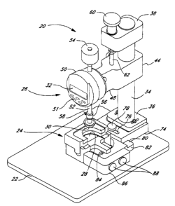
One particular embodiment of a leaflet deflection tester 20 for loading
individual leaflets is shown in Figures 1 and 2. Referring to Figure 1, the
leaflet
deflection tester 20 comprises a flat base 22 supporting a leaflet framing
assembly
24 and a deflector assembly 26 thereabove. A leaflet 28 is shown mounted
within
the framing assembly 24 and a deflector 30 is positioned to apply a load to
the
leaflet to result in a deflection which can be read from display 32. For
purposes
of discussion, the display 2 faces in a forward longitudinal direction, and
lateral
left and right directions are defined from the perspective of looking at the
display.
The deflector assembly 26 comprises a support post 34 vertically oriented
with respect to the base 22 and attached thereto with a post holder 36.
Referring
to Figure 2, at the top of the post 34, a cap 38 is vertically adjustable via
a set
screw 40 engaging a vertical groove 42 in one side of the post. An indicator
carriage 44 is also vertically adjustable along the post and may be secured at
various locations using a pair of carriage locking screws 46 which also engage
the
groove 42. A carriage arm 48 extends longitudinally forward from the post 34
and
terminates in a position indicator 50 mounted thereto. Referring to Figure 1,
the
position indicator preferably includes electronic circuitry and a digital
readout 32,
but may be of a variety of configurations, and the particular embodiment
illustrated herein should not be construed as limiting. Control buttons 51
including a zero reset function are provided on the face of the indicator 50.
The position indicator 50 is generally centrally located above the leaflet
framing assembly 24 and includes an indicator shaft 52, vertically passing
therethrough and engaging position-sensing equipment within the indicator.
That
is, various known mechanical or electro-mechanical devices for sensing the
displacement of a shaft within a housing are contemplated for this purpose and
will not be described further herein. A mass 54 attaches to an upper end of
the
indicator 52 above the position indicator 50. At the lower end of the shaft
52, a
. i. . . . . . . . . . .
CA 02285233 1999-09-30
- WO 98/43557 PCTIUS98/06333
9
collar 56 is fastened thereon via a locking screw 58. The collar continues
downward and terminates in the aforementioned deflector 30.
The deflector assembly 26 further includes a means for vertically adjusting
the position between the post cap 38 and the indicator carriage 44. A vertical
adjustment knob 60 is mounted for rotation above a vertical axis through the
post
cap 38. The adjustment knob 60 engages a connecting rod 62 which extends
between the post cap 38 and the indicator carriage 44. In one embodiment, the
vertical adjustment knob 60 rotates a threaded nut within the post cap 38
which
engages male threads on an upper end of the connecting rod 60 to cause its
vertical displacement. The connecting rod 62 is preferably firmly connected to
the indicator carriage 44 and thus turning the vertical knob 60 vertically
displaces
the indicator carriage 44. The use of the vertical adjustment knob 60 in
calibrating and operating the tester 20 will be described below.
With reference still to Figure 1, and, more particularly, to the rear
perspective view in Figure 2, the post holder 36 is formed as a monolithic T-
shaped block, having a pair of overhanging edges through which longitudinally
oriented adjustment slots 68 are provided. The slots 68 are provided on either
lateral side of the support post 34 and receive locking bolts 70 which extend
downward into engagement with a step 72 formed in a longitudinal adjustment
bracket 74. The longitudinal adjustment bracket 74 can thus be adjusted
longitudinally with respect to the post holder 36 and secured with the bolt
70.
On a front end of the longitudinal adjustment bracket 74, an overhanging
portion includes a lateral adjustment slot 76 receiving a locking screw 78.
Referring to Figure 1, the locking screw 78 continues through the overhanging
portion of the adjustment bracket 74 into contact with a step 80 formed in a
lateral adjustment bracket 82 which is generally L-shaped, having a forwardly
extending arm portion 84. An L-shaped clamp 86 is adjustable longitudinally
with
respect to the arm portion 84 and is fastened thereto with a pair of clamping
screws 88. The combination of the adjustment brackets 74 and 82, and L-shaped
CA 02285233 1999-09-30
- WO 98/43557 PCT/US98/06333
clamp 86, index and secure the leaflet framing assembly 24 with respect to the
support post 34 and, in turn, the position indicator 50.
An upper framing assembly member or leaflet mounting frame 94,
illustrated in Figures 3 and 4, comprises a generally rectangular shaped base
96,
5 having an upper stepped recess 98 open to a front side of the rectangle. An
undercut 100 is formed in the recess 98 to receive a plate-shaped needle clamp
102 therein. The needle clamp 102 includes a semicircular cutout 104 in an
edge
facing toward the open edge of the recess 98. The cutout 104 conforms to a
semicircular cutout 106 formed in the base 96. It should be noted that
although
10 the cutouts 104, 106 are described as generally semicircular, the
particular shape
of the leaflet 28 may be somewhat oval in shape, which may correspondingly
alter
the shape of the cutouts.
Both the base 96 and the needle clamp 102 include a plurality of
registered, vertical through holes 108, arranged equidistantly around the
semicircular cutouts 104 and 106. In a preferred embodiment, there are seven
such through holes 108, arrayed at specific circumferential angles around the
cutouts 104 and 106. The through holes 108 receive leaflet framing needles 110
which are vertically retained therein through the use of a needle clamp screw
112
threaded through a rear wall of the frame body 96 and into contact with the
needle clamp 102.
The frame base 96 further includes a plurality of positioning tabs 114
depending downward therefrom. In the illustrated embodiment, there are three
such tabs 114, two on left and right sides, respectively, of the frame base 96
and
one on the rear side. With reference to Figure 4, the tabs are utilized to
orient
a needle gauge or calibration member 116 under the cutouts 104 and 106. More
particularly, the needle gauge 116 comprises a generally rectangular base 118
and
a recessed pocket 120. The base 118 is guided between the two side tabs 114
and
abuts against the rear tab of the mounting frame 94. In this orientation, the
pocket 120 is positioned directly below all of the through holes 108 so that
the
CA 02285233 1999-09-30
-WO 98/43557 PCT/US98/06333
11
needles 110 depend downward below the lower surface of the frame base 96, as
seen at 122, only as far as the pocket. The needles 110 are inserted through
the
holes 108 into contact with the pocket 120, and then the needle clamp screw
112
is tightened to push the needle clamp 102 i4 a direction out of the recess 98
and
create a compression against the needles 110. That is, the shear force exerted
on
the needles 110 by the through holes 108 in the frame base 96 and needle clamp
102 maintains the needles in the vertical position as calibrated by the needle
gauge 116. Once the needles are calibrated to depend downward the same
distance, the frame 94 is ready for use in the framing assembly 24.
Figure 5 illustrates a lower framing assembly member or leaflet holder 126
comprising a block-shaped body 128 having a flat lower surface adapted to rest
on the base 22 (Figure 1) and a flat upper platform 130. The body 128 is
generally rectangular in shape and includes a rectangular base locator 132
projecting from a front side and shorter in height than the body 128. The
outer
edges of the body 128, other than the edge from which the base locator 132
extends, include positioning channels 134 opening to the platform 130. The
positioning channels 134 receive the positioning tabs 114, previously
described for
the leaflet mounting frame 94, as best seen in Figure 6, to locate the
mounting
frame with respect to the leaflet holder 126. The lower surface of the leaflet
mounting frame base 96 is flat and is juxtaposed with the flat platform 130.
In
the center of the body 128, and opening toward the base locator 132, a cavity
136
is formed having a generally semicylindrical shape. A stepped leaflet edge
recess
138 is formed in the platform 130 surrounding the cavity 136 and is sized and
shaped to receive a leaflet, such as the leaflet 28 as shown in Figures 1 and
2.
Referring to Figure 5, a paddle-shaped leaflet support 140 has a generally
semicircular end which fits closely within the cavity 136, with a handle 142
extending outward from the cavity 136 and resting on a top surface of the base
locator 132. The leaflet support 140 has a height which is identical to the
height
from the top surface of the base locator 132 to the elevation of the leaflet
edge
CA 02285233 1999-09-30
'WO 98/43557 PCT/US98/06333
12
recess 138 so that the upper surface of the leaflet support 140 is in the same
plane as the edge recess 138. The edge recess 138 further includes a plurality
of needle receptor holes 144 sized and positioned in an array identical to the
array in which the through holes 108 and associated needles 110 are positioned
around the leaflet mounting frame 94. This arrangement allows the needles 110
to extend through the peripheral edge of the leaflet 28 into the receptor
holes
144, thus holding stationary portions of the leaflet at the edge recess 138.
With reference to Figure 6a, the leaflet typically includes a straight
coapting edge 148 having opposed tab ends 150, and a generally semicircular
cusp
152 therebetween and opposite the coapting edge. The tab ends 150 include
angled sides 153 transitioning to the coapting edge 148. The edge recess 138
is
sized and shaped to receive the cusp 152 and tabs 150 with the coapting edge
148
oriented parallel with but spaced from a front edge of the holder 126.
Figure 6a also illustrates a point 154 at which an axis through the center
of the deflector 30 intersects the leaflet 28. This point 154 will be referred
to
herein as the point of contact between the deflector 30 (Figure 1) and leaflet
28,
but in the exemplary embodiment the deflector is a relatively large diameter
smooth hemisphere, and contacts the leaflet over a circular area to better
simulate a distributed load and to help avoid stress risers. The point 154 is
determined from a model of the stress distribution in the leaflet based on
assumed forces applied to the leaflet in a human heart valve. The forces
applied
to the leaflet in a human heart valve originate from fluid pressures upstream
and
downstream of the valve, and the stress distribution is found from the
leaflets'
shape and boundary conditions (i.e., geometry of the lines of sutures
attaching the
leaflets in the valve). The point 154 is thus an idealized concentrated load
point
(or concentrated area) equivalent to the actual distributed pressure load.
The leaflet is symmetrical about an axis perpendicular to and bisecting the
coapting edge 148, and is typically continuously sutured in an actual valve
along
the cusp 152, and thus the point 154 is desirably on that axis. The dimension
"A"
r 11
CA 02285233 1999-09-30
'W0 98/43557 PCT/US98/06333
13
is the distance from the point 154 to the coapting edge 148 determined from
the
aforementioned stress distribution model. The dimension "A" will vary
depending
on the size and geometry of the leaflet, its thickness and bulk material
properties,
and the assumed stress distribution. It will be noted, however, that the point
154
is spaced from the coapting edge 148, which prevents undue tensile stresses
between the deflector 30 and the points closest to the coapting edge at which
the
leaflet is held stationary in the framing assembly 24 (i.e., needles 110, as
will be
described below). The particular apparatus and methods disclosed, and the
concentrated loading, requires that the point 154 be spaced from the coapting
edge 148 to best distribute the tensile stresses between the deflector 30 and
the
stationary points at the leaflet periphery. Of course, those of skill in the
art will
recognize that a more accurate test setup with actual suturing around the cusp
152
and a pressure loading over the surface of the leaflet could be substituted
within
the scope and teaching of the present invention, and the presently illustrated
test
setup is an approximation driven by practical manufacturing considerations.
The interaction of the leaflet mounting frame 94 with the leaflet holder
126 will be explained with reference to Figure 6 and 7. As mentioned, the
positioning tabs 114 fit within the positioning channels 134 to orient the
mounting
frame 94 with respect to the leaflet holder 126. The registration between the
tabs
114 and channels 134 insures that the needles 110 in the through holes 108 in
both the base 96 and needle clamp 102 of the mounting frame 94 align with the
needle receptor holes 144 in the leaflet holder 126. The assembly arrow 146
illustrates the movement of the mounting frame 94 when coupling to the leaflet
holder 126. In an anticipated alternative embodiment, the mounting frame 94
will
be hingedly or otherwise pivotally coupled to the holder 126, with the final
relative movement being vertical to avoid skewing the needles within the
receptor
holes 144.
The leaflet 28 is pre-positioned so that its outer edges conform to the
shape of the leaflet edge recess 138, and the middle portion is supported by
the
CA 02285233 1999-09-30
WO 98/43557 PCT/US98/06333
14
leaflet support 140. The needles 110 extending down below the leaflet mounting
frame 94 thus pierce and pass through the tissue of the leaflet 28 and extend
into
the receptor holes 144. In a heart valve, the cusps 152 of each leaflet are
supported by a wireform, and the coapting edges 148 remain free to cooperate
with the coapting edges of the other leaflets. The framing assembly 24 thus
closely simulates the static points of attachment so that the stress
distribution, and
accompanying deflection response, in the leaflet 28 is as near to the actual
distribution as possible. The needles 110 hold stationary peripheral portions
of
the leaflet 28 to approximate an actual line of sutures peripherally securing
the
leaflet within a heart valve. Furthermore, the lower surface of the mounting
frame base 96 rests on the upper surface of the platform 130. In this regard,
it
is important to note that the leaflet 28 is preferably not compressed, or only
lightly compressed, by the weight of the mounting frame 94 because it is
positioned within the recess 138. The final assembled leaflet framing assembly
24 is illustrated in Figure 7.
Figures 8a and 8b illustrate two positions of the deflector 30 during a
leaflet deflection test. The leaflet 28 is mounted in the framing assembly 24
with
the leaflet support 140 supporting the leaflet 28 from underneath in a plane
at the
same elevation of the leaflet edge recess 138, and thus the leaflet 28 does
not
bend or sag in its mid-portion. The deflector 30 is lowered into a position
just
contacting the top of the leaflet 28, as shown in Figure 8a, prior to a
deflection
test. Subsequently, the leaflet support 140 is removed from underneath the
leaflet
28, and the indicator shaft 52 is allowed to drop as in Figure 8b, thus
causing the
deflector 30 to displace the leaflet 28 until an equilibrium is reached. The
equilibrium depends on the framing geometry, the size of the mass 54 (Figure
1),
and the stress/strain characteristics of the leaflet 28. The total deflection
of the
leaflet at the point of contact with the deflector 30 is illustrated in Figure
8b by
the dimension "d". As described below in the Exemplary Test Assembly section,
an approximate measurement of the true deflection "d" is made by disregarding
T
CA 02285233 1999-09-30
WO 98/43557 PCTIUS98/06333
the relaxed thickness of the leaflet being tested for simplicity of
calibration of the
apparatus and method.
Preferably, the deflector 30 is a relatively large diameter smooth
hemisphere so that the load imposed on the upper surface of leaflet 28 is
5 somewhat distributed. The deflector 30 is made of a biocompatible material,
such
as a plastic, and preferably a thermoplastic. Other variations of deflector 30
are
envisioned and the present invention should not be construed to be limited to
the
illustrated embodiment. For example, a more uniformly distributed load such as
a pressure load may be imposed upon the leaflet 28 and the subsequent
deflection
10 measured. In all cases, the aim is to closely simulate the conditions
experienced
by the leaflet 28 in an actual heart valve. Indeed, it would be desirable to
load
test leaflets after being installed on a heart valve wireform and support
ring.
However, even if such a test could be accurately configured, it would be
difficult
to test individual leaflets within a three leaflet prosthetic valve, for
example.
15 Furthermore, once the valve has been constructed, many of the benefits of
the
leaflet selection process are rendered moot. That is, the primary
consideration
is finding similar leaflets to combine within a single heart valve. A
secondary
consideration, which is not insignificant, is being able to select a leaflet
prior to
construction of the valve to reduce manufacturing time and expense.
Construction of a heart valve involves many intricate steps of sewing leaflets
together and to the wireform and surrounding fabric covering. The work must be
done by highly skilled technicians and thus testing of individual leaflets
within
fully constructed valves is prohibitively expensive, although not outside of
the
scope of the present invention.
The present invention thus seeks to provide a selection method for
individual leaflets prior to construction of a heart valve which most
accurately
predicts the ultimate mechanical response of each leaflet within the
constructed
valve and ensures optimum performance in coaptation with the other leaflets.
To
that end, the presently illustrated test apparatus 20 is believed to closely
simulate
CA 02285233 1999-09-30
- WO 98/43557 PCT/US98/06333
16
the forces and stresses imposed on the leaflet during use, in a relatively
easy to
set up and operate environment. Because of the modular nature of the test
apparatus 20, repeatability of tests for various sizes of leaflets is
enhanced. That
is, the leaflet holder 126 is sized for a particular diameter of leaflet, and
a
number of leaflet holders having different leaflet edge recesses 138 may be
provided for different sized leaflets. The external dimensions of the leaflet
holder
126 remain the same so that it may be indexed within the aforementioned
brackets on the platform 22 (Figure 1) and the same deflection assembly 26 is
utilized. Concurrently, the leaflet mounting frame 94 may be provided in a
variety of sizes to cooperate with different sized holders. An additional
advantage
is the relatively small size and portable nature of the tester 20. The
platform 22
may be set up on assembly lines, laboratory tables, and even desktops.
Exe=larXTest Assemblv
The steps in preparing the exemplary tester apparatus 20 for use will now
be described with respect to the drawings. First of all, the equipment is
cleaned
to remove any particulate matter and dirt adhered thereto. The test equipment
is then sterilized through a process including a bio-burden reduction process
(BREP) well known in the art.
With reference to Figures 1 and 2, the post cap 38 is first secured on the
post 34 by tightening the set screw 40. The locking screws 46 are loosened to
allow the carriage 44 to freely move vertically on the post 34. Additionally,
the
locking bolts 70, locking screws 78, and clamping screws 88 are loosened.
Prior
to installing the leaflet framing assembly 24, the framing assembly 24 must be
indexed under the deflector assembly 26. To accomplish this, an indexing tip
(which is not shown) is fastened to the lower end of the indicator shaft 52 in
place
of the collar 56 and deflector 30. The indexing tip on the lower end of the
indicator shaft 52 fits through an indexing hole (not shown here) within the
cavity
136 formed in the leaflet holder 126. The indexing hole allows the indexing
tip
CA 02285233 1999-09-30
-WO 98/43557 PCT/US98/06333
17
to contact the platform base 22. Once the indexing tip has contacted the base
22,
the carriage loclcing screws 46 are tightened to locate the carriage 44. As
the
indexing hole is sized just large enough to receive the indexing tip, the
leaflet
holder 126 is located in its proper position with respect to the position
indicator
50. That is, the vertical axis of the indicator shaft 52 is positioned at the
precise
location with respect to the leaflet holder 126 so that the deflector 30, when
eventually installed, will contact the leaflet 28 in the proper position.
The longitudinal adjustment bracket 74 and lateral adjustment bracket 82
are then manipulated to contact the framing assembly 24 under the deflector
assembly 26. The longitudinal adjustment bracket 74 and lateral extending
portion of the lateral adjustment bracket 82 are displaced to contact the
associated sides of the leaflet holder 126, and the arm portion 84 contacts
the end
of the base locator 132. The locking bolts and locking screw 78 are tightened.
The carriage locking screws 46 are then loosened and the vertical adjustment
knob 60 manipulated to raise the carriage 44 upward. The indexing tip is
removed.
The correct size deflector 30 is chosen depending on the size of the leaflet
28 to be tested. The collar 56 of the deflector 30 is attached to the lower
end of
the indicator shaft 52 via the locking screw 58. Next, the proper size mass 54
is
selected for the leaflet 28 to be tested. In this regard, a single mass for a
particular size of leaflet 28 is preferred, although different masses may be
used
on the same leaflet for a variety of deflection results. The mass 54 must be
selected so as not to over stress the leaflet 28 being tested. Thus, for
example,
stress loading for a glutaraldehyde-fixed pericardial tissue leaflet within a
mitral
valve is up to 1,000 kPa. For this application, therefore, the mass 54 should
be
chosen so that the stress imparted to the leaflet 28 is no greater than 1,000
kPa.
At this point, the position indicator 50 is calibrated. With the leaflet
support 140 in position, the position indicator 50 is reset so that the
display 32
reads zero, using one of the control buttons 51. The carriage locking screws
46
CA 02285233 1999-09-30
WO 98/43557 PCT/US98/06333
18
are then loosened and the entire position indicator 50 is lowered using the
vertical adjustment knob 60 on the top of the post cap 38. The carriage 44,
along
with the position indicator 50, is lowered until the deflector 30 contacts the
upper
surface of the leaflet support 140. The vertical adjustment knob 60 is further
turned to lower the position indicator 50 while the indicator shaft 52 remains
stationary until the display 32 reads a deflection of between approximately
0.390"
and 0.410". Then the carriage locking screws 46 are tightened to lock the
position
indicator 50 in place.
The display 32 is then again set to a zero reading, using one of the control
buttons 51. This sequence ensures that the deflector 30 can drop a sufficient
distance below the level of the leaflet support 140 to ensure the leaflet
under test
is properly stressed (i.e., not understressed). That is, a proper deflection
reading
is desirably obtained within a nearly linear, high modulus region of the
particular
leaflet stress/strain curve, prior to reaching the yield stress, as described
below
with reference to Figure 11. In general, the optimum stress level is first
approximated, and the mass and total allowable deflection selected accordingly
from that approximation to result in stress in a linear region of the tissue
stress/strain curve.
It should be noted that the leaflet deflection is measured from a zero
datum of the top of the leaflet support 140, and the thickness of the
particular
leaflet is disregarded. The leaflet thickness is relatively small in
comparison to
the deflection, and the ultimate test results are used to compare leaflets, so
the
slight inaccuracy from not taking the leaflet thickness into account applies
to all
of the leaflets, and is thus rendered even less important. Thus, the dimension
"d"
indicated in Figure 8b is the true deflection, while the deflection actually
measured is off by the relaxed thickness of the leaflet being tested, and is a
close
approximation of the true deflection.
The next step in the test preparation process is to secure the leaflet 28
within the framing assembly 24. First, the leaflet mounting frame 94 is
assembled
._.._ . ...._. . __._........._. _.._ w.......,..w _ . ..__ . .. .T... . .1,.
,. ... . _.. .. . . .._. ... .. ._. . . ...
CA 02285233 1999-09-30
- WO 98/43557 PCT/US98/06333
19
by inserting the needle clamp 102 in the base 96. As mentioned previously, the
appropriately sized base 96 and needle clamp 102 are chosen for the particular
leaflet 28 being tested. The needles 110 are inserted into the through holes
108,
until their tips just extend beyond the lower surface of the base 96 as seen
in
Figure 4. Of course, throughout this operation, the needle clamp screw 112 is
loose to remove any shear force between the needle clamp 102 and the base 96.
As shown in Figure 4, the leaflet mounting frame 94 is then positioned
over the needle gauge 116 on a flat surface and the needles 110 allowed to
drop
until their lower tips contact the upper surface of the pocket 20. At this
stage, the
needle clamp screw 112 is tightened to apply a shear between the needle clamp
102 and the base 96, which holds the needles 110 in their calibrated
elevation.
The needles 110 are individually pulled to insure that they are tightly held
in the
proper position and if any of the needles move, the needle clamp screw 112 is
recalibrated and tightened further. Before placement of the leaflet 28 within
the
leaflet holder 126, the leaflet mounting frame 94 is first positioned over and
mated with the leaflet holder to insure that the needles 110 register with and
extend freely into the receptor holes 144. The mounting frame 94 is then
removed from the leaflet holder 126.
The leaflet support 140 is then installed in the cavity 136 of the leaflet
holder 126, and the leaflet 28 to be tested positioned on the leaflet support
so
that its peripheral edges conform to the appropriately sized leaflet edge
recess
138. The mounting frame 94 is then brought vertically over the leaflet holder
126
and displaced downward so that the needles 110 pass through the tissue of the
leaflet 28 and into the receptor holes 144. In its assembled state, as shown
in
Figure 7, the lower surface of the base 96 rests on the upper surface of the
platform 130, with the positioning tabs 114 oriented in the positioning
channels
134. In this arrangement, therefore, the peripheral edges of the leaflet are
not
pinched or otherwise compressed between the framing assembly halves. This
CA 02285233 1999-09-30
- WO 98/43557 PCT/US98/06333
helps reduce damage to the leaflet which may be assembled in a prosthetic
valve
and implanted for use in a patient.
The leaflet framing assembly 24 with leaflet 28 mounted therein is then
placed back into its previously indexed position under the deflector assembly
26.
5 The L-shaped clamp 86 is brought into contact with the side of the base
locator
132, and the clamping screws 88 tightened to secure the framing assembly 24 on
the base 22.
At this point, the deflector 30 is elevated manually with the position
indicator 50 remaining stationary. The deflector 30 is then placed gently on
the
10 top of the leaflet 28 by manually lowering the shaft 52. The leaflet
support 140
is then removed carefully from underneath the leaflet 28 which is allowed to
deflect under the weight of the mass 54. The deflector 30 is elevated away
from
contact with the leaflet 28, and the test is repeated several times to insure
correct
readout. Preferably the leaflet 28 is deflected five times, and the readouts
of the
15 fourth and fifth deflections are then recorded.
Upon removal of the leaflet mounting frame 94, the leaflet 28 should stay
with the frame by virtue of the needles 110 piercing the leaflet tissue. If
all seven
of the needle tips are visible through the leaflet tissue, then the leaflet 28
is
removed from the mounting frame 94 by loosening the needle clamp screw 112
20 and pulling the needles 110 out from above. The leaflet 28, if useable, is
then
placed in its particular deflection grouping and stored for later combination
with
similar leaflets to produce a heart valve.
If any of the needles 110 are not visible through the tissue, then the
mounting frame 94 is reinstalled onto the leaflet holder 126. After removing
the
mounting frame 94 once again, the needle tips should be visible through the
leaflet 28. When all the needle tips are visible through the leaflet 28, the
mounting frame 94 is replaced on the leaflet holder 126 and one or more
deflection tests are repeated. The data from the second set of deflection
tests
are then used to select and classify the leaflet for later grouping with other
CA 02285233 1999-09-30
-WO 98/43557 PCTIUS98/06333
21
leaflets. After this second test, the leaflet 28 is removed from the mounting
frame 94 and placed in its particular deflection grouping.
Exem lary Tissue Selection Methodolog,y
Studies in the prior art have demonstrated there can be a significant
variation in the stress/strain curves from specimen to specimen of pericardial
tissue. Tests have also demonstrated that typical stress loading of
glutaraldehyde-
fixed pericardial tissue results in varying strains for different tissue
samples, even
from the same pericardium sac. Moreover, leaflets may experience localized
stresses within a mitral valve of up to 1,000 kPa, with a typical high average
range
of between 500 and 600 kPa. Previous studies have shown that the average
stress/strain curve of leaflet tissue material non-linearly increases until a
particular stress is reached after which the curve is approximately linear
(the
tissue stretches significantly more at low loads). In general, tissue is
significantly
stiffer in the high stress region, and is more flexible at low stresses.
Figure 11 illustrates a typical stress-strain curve for pericardial tissue. It
will be understood that the curve is exemplary for a particular tissue fixed
in a
particular way. Other tissues may respond differently, but the trends shown
are
generally seen in fixed bovine pericardium tissue. The curve shows a low
elastic
modulus of the tissue at low stresses under about 300 kPa, and an increasing
modulus at higher stresses. The curve is generally linear above about 300 to
600
kPa. For purposes of discussion, a high modulus region (HMR) of the curve is
shown in Figure 11 within which the stress/strain curve is generally linear.
The
HMR is an approximation of an average high stress range within a particular
fixed bovine pericardium leaflet in an implanted heart valve. This approximate
information can be combined with knowledge of the operating conditions and
valve size to design an appropriate deflection test method. That is, the bulk
tissue stress/strain curve along with the valve size and assumed loading and
boundary conditions can be combined to predict a stress distribution in the
leaflet.
CA 02285233 1999-09-30
-WO 98/43557 PCTIUS98/06333
22
The size of the mass 54 in the illustrated exemplary test apparatus 20 (Figure
1),
for example, is then selected to stress the leaflet into the HMR of the
tissue.
Understressing the leaflet during the test may not obtain optimum results, and
over-stressing the leaflet may damage it. Thus, for example, a preferred
stress
level applied to glutaraldehyde-fixed pericardial tissue leaflets for a 29 mm
CEP
valve in the tester apparatus 20 has been found to be in the HMR of between
300
and 600 kPa.
The present tissue deflection test addresses the observed variation in
resulting strain in tissue leaflets when the applied load is similar to
pressure
loading under physiological conditions. As mentioned previously, localized
stresses on a leaflet in use may reach 1,000 kPa. Testing of leaflets within
the
tester 20 is preferably accomplished using a significantly lower stress level,
while
still sufficiently deflecting the leaflet in the linear stress/strain region
for useful
results. Empirical testing or finite element stress analysis on specific
leaflet
material is desirably used to predict the probable stress-strain relationship
of
individual leaflets. This preliminary testing or analysis is then used to
design the
proper deflection test method, as described herein.
The particular testing stress level, however, is also affected by the type of
test configuration. The needles 110 secure the edges of the leaflet 28 in a
uniform circumferential array which simulates the sutures which attach the
leaflet
cusp within a heart valve prosthesis. In particular, the cusp 152 of the
leaflet 28
is held stationary at discrete points defined by the needles 110, while the
coapting
edge 148 remains free. The number of needles 110 should be sufficient to
simulate this edge connection of the leaflet in use, but not be too numerous
as
the needles pierce through the tissue of the leaflet. Therefore, between at
least
five and nine, and preferably at least seven needles 110 as shown are adequate
for a uniform framing configuration of the leaflet without creating an
inordinate
number of holes therein. As the load is applied by the deflector 30 the stress
distribution within the leaflet 28 will not be completely uniform because the
_ _ _ . _..._..__. . .. .. _. ~ , .
CA 02285233 1999-09-30
WO 98/43557 PCT/US98/06333
23
leaflet is only held at discrete locations. Therefore the load applied must be
carefully gauged, so as not to create undue levels of stress concentration in
and
around the points at which the needles pierce through the leaflet tissue. The
stated range of between 300 and 600 kPa for glutaraldehyde-fixed pericardial
tissue leaflets has been determined to be suitable when using seven needles as
shown. Of course, other arrangements for framing the leaflet are possible,
such
as using more than seven needles, and the stress range may be appropriately
modified. Further, the stress range is not determined solely with reference to
the
leaflet holding arrangement. Those of skill in the art will recognize the
exemplary
test apparatus is an attempt to simulate true stresses imposed on the leaflet,
with
certain tradeoffs, including simplifying the test apparatus and minimizing the
number of needles used.
The mass 54 for a 29 mm CEP leaflet is chosen to be approximately 100
g to set up stress levels of between 300 and 600 kPa in the leaflet. The mass
54
for other size valve leaflets are scaled from the 100 gram load used for the
29 mm
valve leaflet, as seen in Table I.
Table I
Valve Leaflet Size Deflection Load (g)
mm 74
27 mm 87
29 mm 100
31 mm 112
Additional testing may be performed to insure that the appropriate mass
54 selected for various valve size leaflets imparts a stress in a generally
linear
region of the tissue stress/strain curve. One example of such testing is to
use an
Instron tensile test tester in place of the position indicator 50. The Instron
CA 02285233 1999-09-30
- WO 98/43557 PCT/US98/06333
24
Tensile tester can be used to vary the load on the leaflet 28 and a series of
stress/strain curves can be generated for each leaflet size. Based on these
test
results, the minimum load on the tissue leaflets for all sizes to ensure that
the
stress/strain response is in the linear regime is approximately 60 grams.
Other means of categorizing leaflets may be used in conjunction with the
presently described deflection test. For example, selection of individual
leaflets
to be grouped with other leaflets in a heart valve has been accomplished by
the
assignee of the present invention using a so-called "droop" test of the
leaflets.
That is, the leaflets are cantilevered over the end of a rod, or other
structure, and
the droop of the leaflet for different lengths of extension is observed. The
droop
test can thus be generally termed an intrinsic loading test, wherein there is
no
applied load and the leaflet deflects solely under its own weight. The droop
test
is used to categorize leaflets, so that leaflets with similar droop
characteristics can
be put together for assembly into a heart valve in an attempt to improve
leaflet
cooperation and coaptation. The droop test in combination with the presently
described deflection test is particularly useful in grouping individual
leaflets with
similar characteristics for assembly into a multi-leaflet valve.
Results for loading of leaflets for use in various size valves is given in
Figure 9. After the leaflets were deflected five times in succession to
account for
conditioning or change in the Young's modulus, a final deflection comprising
the
last observed deflection or an average of the last two observed deflections
were
recorded. Figure 9 shows the distribution of deflection values from the tissue
deflection test. The deflection values for the leaflets measured ranged from
0.19
to 0.36 with the majority grouped between 0.23 and 0.30.
To illustrate the effectiveness of the methods and apparatuses of the
present invention relative to conventional tissue categorizing techniques, the
droop test and tissue deflection test of the present invention were applied to
169
leaflets. The leaflets tested for deflection response in Figure 9 were then
categorized by droop value. Figures 10a, 10b, and lOc show the population of
. ...._. ...... ._,....y.. . i. , . .... .. .. . .... ..
CA 02285233 1999-09-30
-WO 98/43557 PCTIUS98/06333
leaflets categorized by letters A, B and C based on droop characteristic. In
general, the leaflets from Group A had the lowest deflection values with Group
B somewhere between Group A and Group C. There is significant overlap of
deflection values between categories A, B, and C.
5 In an exemplary embodiment, the deflection test described herein is first
used to categorize a number of similarly shaped leaflets into subgroups, such
as
is shown in Figure 9. Subsequently, a droop test is performed on a subgroup of
leaflets within a predetermined deflection range, and only leaflets within an
acceptable droop range from that subgroup are combined into a prosthetic heart
10 valve. Alternatively, the droop test may be performed first to obtain a
number
of subgroups, one or more of which is then droop tested to arrive at a
selected
group of leaflets suitable for combining together in a prosthetic heart valve.
In an exemplary embodiment, the individual leaflets 28 are deflection
tested and leaflets are selected which produced a total deflection of between
.170
15 and .340" for valve sizes of 25 to 31 mm. Furthermore, for reliability, it
is
preferred that only leaflets be used for which the fourth and fifth readouts
differ
within a predetermined range, for example between plus or minus .003 inches.
To evaluate the effect of selecting and combining tissue leaflets in 29 mm
CEP valves, four valves were manufactured and tested. Two leaflets were
20 selected to have similar deflection values. The third leaflet deflection
value was
varied from approximately 0.010" to 0.040" compared to the other leaflets as
shown in Table II.
Table II
25 Valve Number Leaflet Deflection Leaflet Deflection Deflection
1.2 3
13559 0.298 0.310 0.012
13560 0.254 0.277 0.023
13561 0.238 0.269 0.031
13562 0.277 0.317 0.040
CA 02285233 1999-09-30
- WO 98/43557 PCTIUS98/06333
26
The valves listed in Table II were placed into a pulsatile flow simulator, and
testing performed per conventional protocol. The differential pressure for the
testing was 200 mm Hg per Food and Drug Administration guidelines. The valve
commissure deflection for each valve was measured as shown in Table III.
Table III
Valve No. Cycle No. Comm. 1 Comm. 2 Comm. 3 Average Std. Dev.
Actual Actual Actual
(mm) (mm) (mm) (mm)
13559 1 0.89 1.10 0.78 0.92 0.16
2 0.89 1.10 0.78 0.92 0.16
3 0.86 1.13 0.79 0.93 0.18
13560 1 1.05 1.39 1.21 1.22 0.17
2 1.02 1.39 1.23 1.21 0.19
3 1.05 1.42 1.23 1.23 0.19
13561 1 1.26 1.32 1.07 1.22 0.13
2 1.26 1.36 1.08 1.23 0.14
3 1.23 1.32 1.08 1.21 0.12
13562 1 1.07 1.32 0.84 1.08 0.24
2 1.07 1.32 0.82 1.07 0.25
3 1.05 1.36 0.84 1.08 0.26
Average 1.11
Std. Dev. 0.16
Proper coaptation was observed in valves 13559, 13560, and 13561, where
the mismatch between leaflets 1 and 2 and leaflet 3 was less than
approximately
0.030". Thus, from this particular study, leaflets which have deflection
values
differing by less than approximately 0.030" are suitably grouped for combining
in
t ,,
CA 02285233 1999-09-30
= WO 98/43557 PCT/US98/06333
27
a heart valve. Of course, this test applies to 29 mm CEP valves made from
selected bovine pericardium, and there are a variety of parameters which could
alter the conclusion regarding acceptable deflection correlation. Furthermore,
the
conclusion was based on measured valve commissure deflection, which is one
predictor of prolonged leaflet coaptation. A desirable selection methodology,
therefore, is to obtain a collection of similarly sized leaflets, apply a load
to each
leaflet, observe the resulting strain response, and sort the leaflets based on
their
respective strain responses. Additionally, the leaflets are preferably
chemically
fixed prior to testing and a droop test is used in conjunction with the
deflection
1.0 test results.
The present invention additionally teaches a multi-leaflet bioprosthetic
heart valve with leaflets selected to have observed deflection responses
within a
certain range. The average deflection in the range depends on a number of
variables, as explained above, and the breadth of the range may depend on
empirical test results of assembled valves, such as the commissure deflection
data
included in Table III. In one exemplary embodiment, however, a 29 mm multi-
leaflet bioprosthetic CEP heart valve comprising glutaraldehyde-fixed bovine
pericardium tissue leaflets includes at least two leaflets having a deflection
of
within.030 inches as measured using the exemplary testing method and
apparatus,
with a mass sufficient to create stresses in the leaflets of between 300 and
600
kPa.
It should be noted that the present invention is best suited for categorizing
and selecting leaflets having varying material properties from leaflet to
leaflet,
such as in bovine pericardium. Recent advances in bioprosthetic materials
enable
manufacturers to produce leaflets by growing tissue on a matrix. Such material
may also exhibit nonuniformities in individual leaflets and could be grouped
and/or selected in accordance with the present invention. Another type of
tissue
for which the present invention may prove valuable in selecting leaflets is a
composite or laminate substrate on which a cell growth covering is formed.
CA 02285233 1999-09-30
'WO 98/43557 PCT/US98/06333
28
Alternatively, the present selection methods and apparatuses may be applicable
to leaflets made from materials with more uniform properties, such as
synthetically fabricated or extruded collagen sheets. Though the bulk material
properties of these latter materials may be more predictable, individual
testing of
leaflets is believed desirable to more accurately assess the subsequent
dynamic
response of the leaflets in use. In addition, testing of leaflets using a
setup which
closely simulates the particular heart valve in which the leaflet will be used
is
desirable, in addition to the pre-existing knowledge of the material
properties.
For these more uniform leaflets, testing of a sample of leaflets from a
specific
manufactured batch may suffice.
In closing it is to be understood that the embodiments of the invention
disclosed herein are illustrative of the principles of the invention and that
other
modifications may be employed which are within the scope thereof. Accordingly,
the present invention is not limited to that precisely as shown and described
in
the specification.
r. 1 ... . ., .... . . . . .. ..... ....... . . . .