Note: Descriptions are shown in the official language in which they were submitted.
CA 02285668 1999-10-06
RHEOLYTIC THROMHECTOMY CATHETER
AND METHOD OF 08ING SAME
CRO88 REFERENCES TO CO-PENDING APPLICATIONS
This patent application is a continuation-in-part
(CIP) of Serial No. 09/019,728 entitled "Rheolytic
Thrombectomy Catheter and Method of Using Same° filed on
February 06, 1998, by the same inventor(s).
HACRGROUND OF THE INVENTION
1. Field of the Invention - The present invention
relates to a rheolytic thrombectomy catheter and method of
using same to remove thrombus from a body vessel or other body
cavity.
2. Description of the Prior Art - Procedures and
apparatus have been developed for ease in removing tissue and
various deposits. Several such devices employ a jet of saline
as the working tool to help break up the tissue deposit and
further provide a suction means to remove the deposit. U.S.
Pat. No. 5,135,482 to Neracher describes a hydrodynamic device
for removal of organic deposit from a human vessel. A supply
of saline is delivered by a high pressure duct to the distal
end of a catheter. The saline exits the duct as a jet that is
directed generally forward and directly toward the tissue to
be broken up. The duct is contained within and can move
axially with respect to a hose that is positioned around the
duct. A vacuum suction is applied to the hose to remove the
debris that is created from the broken-up tissue. This device
is not intended to pass through tortuous pathways found in the
fragile vessels of the brain, and any attempt to employ the
device for such purpose would be far too traumatic to the
patient.
POSSIS - CIP OF 09/019,728 - 1 -
10-29-98 11:30 AM
MYFILES\PAT\P321
CA 02285668 1999-10-06
Another drainage catheter, described by Griep in
U.S. Pat. No. 5,320,599, has a discharge channel and a
pressure channel. The channels are formed into a single
catheter tube such that the two tubes are fixed with respect
to each other. This catheter could not provide the
flexibility needed to negotiate the tortuous vascular pathways
found in the vessels of the brain.
POSSIS - CIP OF 09/019,728 _ 2 _
10-29-98 11:30 AN '
MYFILES\PAT\P321
CA 02285668 1999-10-06
SUMMARY OF THE INVENTION
The general purpose of the present invention is to
provide a rheolytic thrombectomy catheter and method of using
same to remove thrombus from a body vessel or other body
cavity.
The present invention, a rheolytic thrombectomy
catheter, is a surgical device for removal of material such as
thrombus from a vessel or other body cavity. As shown in one
or more embodiments, a rheolytic thrombectomy catheter for
removing tissue from a vessel or other body cavity includes an
outer assembly comprising a first tube or catheter having a
lumen with an open distal end and an internally and distally
located stationary stop partially obstructing the lumen at the
open distal end, the lumen being of a diameter sufficient to
allow passage of a guidewire; and an inner assembly comprising
a high pressure second tube or hypo-tube having a high
pressure lumen and a distal end having one or more orifices,
a distally located transitional stop fixed to the high
pressure hypo-tube adjacent to the distal end, and a jet cap
positioned at the hypo-tube distal end for directing one or
more jets of saline toward the distal end of the catheter, the
inner assembly being movable axially within the outer assembly
such that the distally located transitional stop engages the
distally located stationary stop to hold the jet cap in a
desired relationship with respect to the distal end of the
catheter.
In another embodiment, a rheolytic thrombectomy
catheter for removing thrombus or other body tissue from an
obstructed body vessel or other body cavity includes an outer
assembly including an evacuation tube having a proximal end
and an open distal end containing a distally located
POSSiS - CIP OF 09/019,728 _ 3
10-29-98 11:30 AM
MYFIIES\PAT\P321
CA 02285668 1999-10-06
stationary stop and having an evacuation lumen that is of a
diameter sufficient to allow passage of a standard coronary or
interventional neuroradiological guidewire; and an inner
assembly including a high pressure hypo-tube having a high
pressure lumen, the high pressure hypo-tube having a proximal
end and a distal end, the distal end having one or more
orifices through which saline can exit from the high pressure
lumen to be directed toward the open distal end of the
evacuation tube, a distally located transitional stop fixed to
the high pressure hypo-tube at a position closer to the distal
end than to the proximal end, and a jet cap positioned at the
distal end of the high pressure hypo-tube, the jet cap
coacting with the high pressure hypo-tube to direct one or
more jets of saline toward the open distal end of the
evacuation tube.
Preferably, the rheolytic thrombectomy catheter has
a guidewire coil attached at the distal end of the jet cap to
allow advancement of the inner assembly and the outer assembly
together within the vasculature. Preferably, the rheolytic
thrombectomy catheter has a jet cap which directs a jet of
saline toward the distal end of the catheter, which functions
as an evacuation tube. Preferably, the rheolytic thrombectomy
catheter includes a high pressure hypo-tube with at least one
orif ice and a jet cap configured and arranged for directing
one or more jets of saline to impinge upon or near the distal
end of the catheter. The rheolytic thrombectomy catheter
preferably is flexible and can pass over a standard guidewire
through tortuous vascular pathways.
POSSIS - CIP OF 09/019,728 - 4 -
10-29-98 11:30 AN
NYFILES\PAT\P321
CA 02285668 1999-10-06
The present invention also provides a method of
removing thrombus
from an obstructed
body vessel. The
method
includes the steps of:
a. providing a guidewire and an outer assembly
including a catheter having a distal end and
an internally located stationary stop
positioned adjacent to the distal end;
b. advancing the guidewire to a vascular site
containing thrombus;
c advancing the catheter over the guidewire to
.
the vascular site containing thrombus to
position the distal end at the vascular site;
d. removing the guidewire from the catheter;
e. providing an inner assembly including a
hypo-tube carrying a jet cap and a
transitional stop spaced apart from the jet
cap;
f. advancing the inner assembly within the
catheter of the outer assembly to engage the
transitional stop with the stationary stop;
and,
g, providing a high pressure saline supply to the
hypo-tube so as to cause a jet of saline to
emanate from the jet cap and to entrain
thrombus into a gap or space where the
thrombus is macerated and then pushed into the
catheter for removal from the body; and,
' h. providing impingement of the jet on the
evacuation lumen to create sufficient
stagnation pressure to allow evacuation of
POSSIS - CIP OF 09/019,728 - 5 -
10-29-98 11:30 Ah1
lIYFILES\PAT\P321
CA 02285668 1999-10-06
debris with no need for additional suction on
the proximal end of the evacuation lumen.
In the method, preferably, the jet cap carries a
distally projecting guidewire coil to facilitate further
distal advancement of the inner assembly and the outer
assembly together within the vasculature to a further vascular
site containing thrombus so as to remove additional distally
situated thrombus.
The present invention is also a catheter combination
including a first tube or catheter, being a part of an outer
assembly, the first tube having a proximal end, an open distal
end, and a lumen extending between the proximal end and the
open distal end; a second tube or hypo-tube, being a part of
an inner assembly, the second tube being separable from the
first tube and being insertable within the lumen of the first
tube, the second tube having a proximal end, a distal end, and
a lumen extending between the proximal end and the distal end;
a jet cap, being also a part of the inner assembly, the jet
cap being connected to the second tube at the distal end of
the second tube for directing fluid exiting the lumen of the
second tube, the jet cap being capable of passage through the
lumen of the first tube and being characterized by the ability
to provide a localized region of low pressure associated with
a liquid flow directed generally proximally and into the lumen
of the first tube through the open distal end of the first
tube when the jet cap is located and oriented appropriately
relative to the open distal end of the first tube; and means
for indexing an appropriate positional relationship of the jet
cap and distal end of the second tube relative to the open
distal end of the first tube. The means for indexing
preferably includes a distally located stationary stop
POSSIS - CIP OF 09/019,728 _ 6 _
10-29-98 11:30 AM
lIYFILES\PAT\P321
CA 02285668 1999-10-06
projecting inward from the first tube and a distally located
transitional stop projecting outward from the second tube.
When the second tube is advanced within the first tube, the
stops mutually engage to control the orientation and spacing
and relationship between the jet cap and the open distal end
of the first tube. More preferably, the stops are each
tapered to additionally laterally position the second tube
within the first tube. Most preferably, the centering causes
the tubes to become concentric. Preferably, one or both stops
interact, when engaged, to preserve a channel for fluid flow
rather than fully obstructing the cavity between the first
tube and the second tube.
Another embodiment group provides a catheter
combination including a first tube or catheter, being a part
of an outer assembly, the first tube having a proximal end, a
manifold attached thereto, an open distal end, and a lumen
extending between the proximal end and the open distal end; a
second tube or hypo-tube, being a part of an inner assembly,
the second tube being separable from the first tube and being
insertable within the lumen of the first tube, the second tube
having a proximal end, a distal end, and a Lumen extending
between the proximal end and the distal end; a flow director
including an inner body and an expandable exhaust tube each
being located near but not at the second tube distal end, a
pressure operated closeable or sealable annulus between the
outer surface of the expandable exhaust tube and the catheter
interior annular surface, a jet cap, being also a part of the
inner assembly, the jet cap being connected to the second tube
at the distal end of the second tube for directing fluid
proximally for thrombus oblation and subsequently through a
lumen in the flow director and the lumen of the first tube,
POSSIS - CIP OF 09/019,728 - 7
10-29-98 11:30 All
MYFILES\PAT\P321
CA 02285668 1999-10-06
the jet cap being capable of passage through the lumen of the
first tube and being characterized by the ability to provide
a localized region of low pressure associated with a liquid
f low directed generally proximally and into the inner body,
the expandable exhaust tube and lumen of the first tube and
through the distal end of the first tube when the jet cap is
located and oriented appropriately, as desired, relative to
the inner body at the distal end of the f first tube; and, a
variable deployment distance means for indexing an appropriate
positional and variable relationship of the jet cap and a
tapered core tip at the distal end of the second tube relative
to the distal end of the first tube. A stop means is provided
for limiting movement of the jet cap and tapered core tip
preferably including a proximally located hemostasis nut/stop
at the proximal end of a manifold at the proximal end of the
first tube, and a proximally located filter housing/high
pressure connection stop assembly projecting outwardly and
proximally from the proximal end of the second tube. When the
second tube is advanced within the first tube, fluoro-imaging
can be incorporated to provide adequate spacing and
relationship between the jet cap or guidewire coil and the
distal end of the first tube. This relationship is also
referred to as variable deployment distance. Lateral
positioning of the second tube within the first tube is
readily accomplished during the first stage (insertion) in an
unpressurized operational mode where the closeable annulus is
suitably sized to allow easy unrestricted passage of the
second tube within and through the first tube. During the
operational pressurized mode, jetted saline causes the
expandable exhaust tube to expand, thus closing and
eliminating the open annulus to pressure seal the first tube
POSSIS - CIP OF 09/019,728 _ 8
10-29-98 11:30 A!1
HYFILES\PAT\P321
CA 02285668 1999-10-06
_,
to the second tube, but still allowing movement relative to
each other.
POSSIS - CIP OF 09/019,728 _ 9
10-29-98 11:30 AR
lIYFILES\PAT\P321
CA 02285668 1999-10-06
The above alternative embodiment group embodiment
of
the present invention also provides a method of removing
thrombus from an obstructed body vessel. The method includes
the steps of:
a. providing a guidewire and an outer assembly
including a catheter having an interior
annular surface, a distal end, and an
externally located stationary hemostasis
nut/stop positioned adjacent to the proximal
end;
b. advancing the guidewire to a vascular site
containing thrombus;
c. advancing the catheter over the guidewire to
the vascular site containing thrombus to
position the distal end at the vascular site;
d. removing the guidewire from the catheter;
e. providing an inner assembly including a
hypo-tube carrying a jet cap at its distal
end, a flow director including an expandable
exhaust tube proximal of the jet cap, and a
transitional filter housing/high pressure
connection/stop assembly at its proximal end;
f. advancing the inner assembly to a desired
position within the catheter of the outer
assembly, so that a gap proximal to the jet
cap extends past the distal end of the
catheter;
g, providing a high pressure saline supply to the
hypo-tube so as to cause a jet of saline to
emanate from the jet cap and to entrain
thrombus into a gap or space where the
POSSIS - CIP OF 09/019,728 - 1 O -
10-29-98 11:30 A!1
lIYFILES\PAT\P321
CA 02285668 1999-10-06
thrombus is macerated and then pushed into the
catheter for removal from the body; and,
h. providing impingement of the jet on the
evacuation lumen to create sufficient
stagnation pressure to allow evacuation of
debris with no need for additional suction on
the proximal end of the evacuation lumen.
In the method, preferably, the jet cap carries a
distally projecting guidewire coil to facilitate further
distal advancement of the inner assembly and the outer
assembly together or independently within the vasculature to
a further vascular site containing thrombus so as to remove
additional distally situated thrombus.
POSSIS - CIP OF 09/019,728 - 1 1 -
10-29-98 11:30 AM
NYFILES\PAT\P321
CA 02285668 1999-10-06
One significant aspect and feature of the present
invention is the variously designed jet caps which are
oriented to direct jets of saline in a proximal direction.
Another significant aspect and feature of the
present invention is the stationary stop at the distal end of
the catheter and the distally located transitional stop on the
hypo-tube which together coact to position a jet cap at a
defined distance beyond the distal end of the catheter.
Still another significant aspect and feature of the
present invention is the distally located transitional stop
which has an evacuation lumen and a hypo-tube receiving hole
which is offset from the longitudinal axis of the distally
located transitional stop.
Yet another significant aspect and feature of the
present invention is the provision of complementa-y angled
surfaces on the distally located stationary and transitional
stops which upon engagement serve to center the inner assembly
within the outer assembly.
A further significant aspect and feature of the
present invention is the distally located stationary stop
which is formed unitarily with the wall of the catheter at the
distal end of the catheter.
A still further significant aspect and feature of
the present invention is the guidewire coil provided at the
distal end of the jet cap to allow advancement of the inner
assembly and the outer assembly together or independently
within the vasculature.
As found in additional embodiment groups there is
also provided other significant aspects and features of the
present invention including the use of a transitional filter
housing/high pressure connection/stop assembly proximally
POSSIS - CIP OF 09/019,728 - 1 2 -
10-29-98 11:30 AM
hIYFILES\PAT\P321
CA 02285668 1999-10-06
located on the inner assembly and a stationary hemostasis
nut/stop proximally located on the outer assembly to prevent
the inner assembly from being excessively advanced, so that
the expandable tube proximal end does not become disengaged
from the distal end of the catheter.
A further significant aspect and feature as found in
additional embodiment groups is an annulus which is open for
lateral movement of the inner assembly within the outer
assembly during the initial unpressurized mode (insertion) and
which is closed and sealed by jetted saline during the
oblation process to provide maximum proximally directed saline
f low without leakage between the outer and inner assemblies
when thrombotic tissue is broken up and carried proximally.
Posscs - cIP of o9io19,7za - 1 3 -
10-29-98 11:30 A!1
lIYFIIES\PAT\P321
CA 02285668 1999-10-06
Having thus described embodiments and significant
aspects and features of the present invention, it is the
principal object of the present invention to provide a
rheolytic thrombectomy catheter and method of using same to
remove thrombus from a body vessel.
One object of the present invention is to provide a
rheolytic thrombectomy catheter of such size, flexibility and
construction as to enable it to pass readily through the
tortuous pathways found in the fragile vessels of the brain.
Another object of the present invention is to
provide a rheolytic thrombectomy catheter with means for
producing one or more jets of saline and projecting them in a
proximal direction toward a site of thrombus and toward an
evacuation passage.
Yet another obj ect of the present invention is to
provide a rheolytic thrombectomy catheter with means for
producing one or more jets of saline and with indexing means
to position the jet producing means at a prescribed location
at the distal end of the catheter.
Still another object of the present invention is to
provide a rheolytic thrombectomy catheter of the type having
an inner assembly that is insertable into an outer assembly
with stop means for limiting the extent to which the inner
assembly can be inserted into the outer assembly.
A further object of the present invention is to
provide a rheolytic thrombectomy catheter of the type having
an inner assembly and an outer assembly with means which
centers the inner assembly within the outer assembly and which
orients the parts of the inner assembly in a prescribed manner
with respect to the parts of the outer assembly.
POSSIS - CIP OF 09/019,728 - 1 4 -
10-29-98 11:30 Aii
MYFILES\PAT\P321
CA 02285668 1999-10-06
A still further object of the present invention is
to provide an improved method of removing thrombus from an
obstructed body vessel.
POSSIS - CIP OF 09/019,728 _ 1 5 _
10-29-98 11:30 AN
MYFILES\PAT\P321
CA 02285668 1999-10-06
BRIEF DESCRIPTION OF THE DRAWINGS
Other objects of the present invention and many of
the attendant advantages of the present invention will be
readily appreciated as the same becomes better understood by
reference to the following detailed description when
considered in connection with the accompanying drawings, in
which like reference numerals designate like parts throughout
the figures thereof and wherein:
FIG. 1 is a side view of the present invention, a
rheolytic thrombectomy catheter useful for the removal of
thrombus;
FIG. 2 is a semi-exploded side view of the rheolytic
thrombectomy catheter depicting the two major assemblies
thereof, viz., an outer assembly and an inner assembly;
FIG. 3 is a semi-exploded cross sectional side view
of a manifold and adjacent components constituting parts of
the outer assembly;
FIG. 4 is a longitudinal sectional view of a filter
housing/high pressure connection assembly attached to the
proximal end of a hypo-tube, shown only partially;
FIG. 5 is a side view of a transitional stop, a jet
cap, and a guidewire coil aligned over and about the hypo-tube
at the distal end thereof;
FIG. 6 is an isometric view of the transitional
stop;
FIG. 7 is a longitudinal sectional view taken along
line 7-7 of FIG. 5;
FIG. 8 is a view of the proximal end of the jet cap
on the hypo-tube looking in the direction of line 8-8 of
FIG. 7, with the hypo-tube shown in cross section;
POSSIS - CIP OF 09/019,728 - 1 6 -
10~29-98 11:30 AM
!1'l F I LES\PAT\P321
CA 02285668 1999-10-06
FIG. 9 is a view similar to FIG. 8 illustrating a
slightly modified version of the jet cap;
FIG. 10 is a longitudinal sectional view of the
catheter distal end taken along line 10-10 of FIG. 2;
FIG. 11 is a longitudinal sectional view of the
catheter distal end with the transitory stop, the jet cap, and
the guidewire coil on the hypo-tube shown advancing
therethrough;
FIG. 12 is a longitudinal sectional view of the
catheter distal end with the transitory stop, the jet cap, and
the guidewire coil on the hypo-tube shown in final advanced
position;
FIG. 13 is a cross-sectional view taken along
line 13-13 of FIG. 12;
FIG. 14 is presented to illustrate schematically the
mode of operation of the rheolytic thrombectomy catheter, and
is a longitudinal sectional view depicting the distal end of
the rheolytic thrombectomy catheter within a blood vessel at
the site of a thrombotic deposit and lesion;
FIG. 15 is a longitudinal sectional view similar to
FIG. 7 but illustrating an alternative jet cap embodiment;
FIG. 16 is a view of the proximal end of the
alternative jet cap embodiment shown in FIG. 15 looking in the
direction of line 16-16 of FIG. 15, with the hypo-tube shown
in cross section;
FIG. 17 is a longitudinal sectional view similar to
FIG. 15 but illustrating another alternative jet cap
embodiment;
POSSIS - CIP OF 09/019,TL8 - 1 7 -
10-29-98 11:30 AM
lITFILES\PAT\P321
CA 02285668 1999-10-06
FIG. 18 is a view of the proximal end of the
alternative jet cap embodiment shown in FIG. 17 looking in the
direction of line 18-18 of FIG. 17, with the hypo-tube shown
in cross section;
FIG. 19 is a longitudinal sectional view similar to
FIG. 12 but illustrating an alternative transitional stop
embodiment;
FIG. 20 is a view of the catheter distal end looking
in the direction of line 20-20 of FIG. 19, with the hypo-tube
shown in cross section;
FIG. 21 is a view similar to FIG. 12 but
illustrating alternative embodiments of the transitional stop
and the stationary stop;
FIG. 22 is a view of the catheter distal end looking
in the direction of line 22-22 of FIG. 21, with the hypo-tube
shown in cross section;
FIG. 23 is a side view in partial cross section of
a fifth alternative embodiment of the catheter distal end,
where the hypo-tube is fixed along the longitudinal axis of
the catheter;
FIG. 24 is a side view of a sixth alternative-
embodiment of a rheolytic thrombectomy catheter;
FIG. 25 is a semi-exploded side view of the
rheolytic thrombectomy catheter of FIG. 24;
FIG. 26 is a longitudinal sectional view of the
filter housing/high pressure connection/stop assembly located
at the proximal end of the hypo-tube of the sixth alternative
embodiment;
FIG. 27 is an isometric view of the f low director,
the jet cap and the guidewire coil of the sixth alternative
embodiment;
POSSIS - CIP OF 09/019,728 - 1 8 -
10-29-98 11:30 AM
MYFILES\PAT\P321
CA 02285668 1999-10-06
FIG. 28 is an exploded isometric view of the flow
director, the jet cap, and the guidewire coil shown in
FIG. 27;
FIG. 29 is a view in cross section of the flow
director, the jet cap and the guidewire coil along line 29-29
of FIG. 24 in the unpressurized mode;
FIG. 30 is a view in cross section of the flow
director, the jet cap and the guidewire coil along line 29-29
of FIG. 24 in the pressurized mode;
FIG. 31 is a view in cross section of the jet cap
and the guidewire coil along line 31-31 of FIG. 27;
FIG. 32 is a view in cross section of the junction
of the inner body and the expandable exhaust tube along
line 32-32 of FIG. 30;
FIG. 33 is a view in cross section of the distal
end of the rheolytic thrombectomy catheter along line 33-33 of
FIG. 29 in the unpressurized mode;
FIG. 34 is a view in cross section of the distal
end of the rheolytic thrombectomy catheter along line 34-34 of
FIG. 30 in the pressurized mode; and,
FIG. 35 is a view in cross section and in partial
cutaway of the distal end of the rheolytic thrombectomy
catheter of FIG. 24 in operation in a blood vessel.
POSSIS - CIP OF 09/019,728 - 1 9 -
10-29-98 11:30 AN
IIYFILES\PAT\P321
CA 02285668 1999-10-06
DETAILED DESCRIPTION OF THE PREFERRED EMBODIMENTS
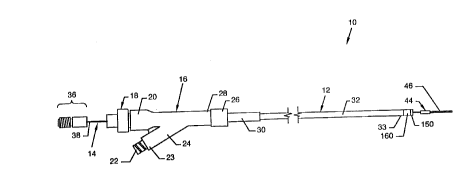
FIG. 1 illustrates a side view of a rheolytic
thrombectomy catheter 10 useful for the removal of thrombus,
and FIG. 2 illustrates a semi-exploded side view of the
rheolytic thrombectomy catheter 10. The rheolytic
thrombectomy catheter 10 includes two major assemblies:
namely, an outer assembly 12 and an inner assembly 14. The
inner assembly 14 aligns concentrically to and within the
outer assembly 12 and extends beyond the length of the outer
assembly 12. Externally visible components, or portions of
components, of the outer assembly 12 of the rheolytic
thrombectomy catheter 10, as illustrated in FIGS. 1 and 2,
include a manifold 16, also known as a Y-adapter, a hemostasis
nut 18 secured in the proximal end 20 of the manifold 16, a
Luer connection 22 located at the proximal end 23 of an angled
manifold branch 24 extending from the manifold 16, a Luer
fitting 26 secured to the distal end 28 of the manifold 16, a
strain relief 30 secured to the distal end 28 of the
manifold 16 by the Luer fitting 26, and a first tube or
catheter 32, having a distal end 33, secured to the
manifold 16 by the strain relief 30 and Luer fitting 26. The
externally visible components of the inner assembly 14,
illustrated in FIG. 2, include a high pressure second tube or
hypo-tube 34, a filter housing/high pressure connection
assembly 36 concentrically aligned to and secured over and
about the hypo-tube proximal end 38, a configured transitional
stop 40 concentrically aligned to and secured over and about
the hypo-tube 34 at a point near and adjacent to the hypo-tube
distal end 42, a jet cap 44 concentrically aligned to and
secured over and about the hypo-tube 34 at the hypo-tube
distal end 42, and a guidewire coil 46 concentrically aligned
POSSIS - CIP OF 09/019,728 - 2 ~ -
10-29-98 11:30 AM
MTFILES\PAT\P321
CA 02285668 1999-10-06
to and secured to one end of the jet cap 44. The high
pressure hypo-tube 34 is drawn and is tapered in incremental
steps to provide degrees of flexibility along its length. For
purposes of example and illustration, the hypo-tube 34 can
include a hypo-tube portion 34a at the hypo-tube proximal
end 38 having an outer diameter of .018 inch or smaller, and
can include a plurality of incrementally stepped down
hypo-tube portions 34b-34n each of lesser outer diameter,
where the last hypo-tube portion 34n is stepped down to an
outer diameter of .008 inch at the hypo-tube distal end 42.
The hypo-tube 34 becomes increasingly more flexible from the
hypo-tube proximal end 38 towards the hypo-tube distal end 42
due to the incremental diameter decrease along its length.
Increasing flexibility along the length of the hypo-tube 34
allows for easier flexed penetration into tortuous vascular
paths. Although the hypo-tube 34 is stepped down in
increments, the hypo-tube 34 can also be fashioned of a
constantly decreasing outer diameter to provide increasing
flexibility along its length and shall not be construed to be
limiting to the scope of the invention.
POSSIS - CIP OF 09/019,728 - 2 1 -
10-29-98 11:30 AM
MYFILES\PAT\P321
CA 02285668 1999-10-06
FIG. 3 illustrates a semi-exploded cross sectional
side view of the manifold 16 and adjacent components, where
all numerals correspond to those elements previously or
otherwise described. The manifold 16 includes a tapered
centrally located passage 48 aligned along the longitudinal
axis of the manifold 16 and a branch passage 50 extending
along the axis of the branch 24 which intersects and is
connected to the central passage 48. The manifold proximal
end 20 houses a mufti-radius cavity 52 including a round outer
cavity portion 54 and a connected round inner and smaller
cavity portion 56 having a threaded surface 58 on the proximal
portion thereof. The hemostasis nut 18 includes a body 62
having a grasping surface 64 extending thereabout, a threaded
surface 66 extending from the body 62, an annular surface 63
at the end of the threaded surface 66, and a passageway 68
aligned centrally to the longitudinal axis of the hemostasis
nut 18. The passageway 68 has a wide radius at the proximal
end which decreases toward the distal end. The initial wide
radius is helpful for insertion of the inner assembly I4 or
guidewires and the like. A seal 60 aligns to the distally
located annular surface 61 of the round inner cavity
portion 56 and bears against the annular surface 63 of the
hemostasis nut 18 to seal the central passage 48 of the
manifold 16 to the passageway 68 in the hemostasis nut 18.
The mufti-radius cavity 52 and its internal geometry
accommodate corresponding geometry of the hemostasis nut 18
and the seal 60. Luer connection 22 extends from the angled
manifold branch proximal end 23. A filter 72 aligns at the
mouth of the branch passage 50. The filter 72 and a Luer
fitting (not illustrated) can be used to prevent any
particulate outflow, to provide for metered outflow, or,
POSSIS - CIP OF 09/019,728 - 2 2 -
10-29-98 11:30 AM
NYFILES\PAT\P321
CA 02285668 1999-10-06
alternatively, to provide suction for fluid or particle
evacuation.
Luer fitting 26 is utilized to secure the strain
relief 30 and the catheter 32 to the distal manifold end 28.
The strain relief 30 is comprised of a tube 31, a central
bore 74 internal to the tube 31 which accommodates the
catheter 32, an annular flange 76 about the tube 31, and a
tapered proximal tube mouth end 78. It is noted that the
outer diameter of the tube 31 is constant from the annular
flange 76 to the distal tube end 80, and that the outer
diameter steadily decreases from the annular flange 76 to the
tapered proximal tube mouth end 78 to provide a tapered tube
surface 82 which conforms, for purpose of a proper fit, to the
taper of the tapered central passage surface 88 of the central
passage 48. The tapered proximal tube mouth end 78 allows for
easily accomplished alignment of guidewires and other
assemblies, such as inner assembly 14 and the like, with a
lumen 87 located in the catheter 32. The Luer fitting 26
includes threads 84 which threadingly engage corresponding
threads 86 at the distal end 28 of the manifold 16. The Luer
fitting 26 bears against the annular flange 76 of the strain
relief 30 to force the tapered tube surface 82 of the strain
relief 30 against the tapered central passage surface 88 of
the central passage 48 to effect a suitable seal.
POSSIS - CIP OF 09/019,728 - 2 3 -
10-29-98 11:30 A!1
lIYFILES\PAT\P321
CA 02285668 1999-10-06
FIG. 4 illustrates a longitudinal sectional view of
the filter housing/high pressure connection assembly 36
located at the hypo-tube proximal end 38 of the hypo-tube 34,
where all numerals correspond to those elements previously or
otherwise described. The filter housing/high pressure
connection assembly 36 includes a cylindrical-like body 90
having a threaded surface 92, a tubular cavity 94, fine and
course filters 96 and 98 residing in the tubular cavity 94, a
central passage 100 extending through the body 90 and
connecting to the tubular cavity 94, and a plug-like cap 102,
having a central bore 104, extending into the tubular
cavity 94 of the body 90. The hypo-tube 34 suitably secures
within the central bore 104 of the cap 102. The central
passage 100 communicates through fine and course filters 96
and 98 with the lumen 106 of the hypo-tube 34.
POSSIS - CIP OF 09/019,728 - 2 4 -
10-29-98 11:30 AN
RIfFILES\PAT\P321
CA 02285668 1999-10-06
FIG. 5 illustrates a side view of the transitional
stop 40, the jet cap 44 and the guidewire coil 46 aligned over
and about the hypo-tube 34 near or at the hypo-tube distal
end 42, where all numerals correspond to those elements
previously or otherwise described. The relative sizes of the
transitional stop 40 and the jet cap 44 with respect to each
other and with respect to the sizes of the lumen 87 of the
catheter 32 and a stationary stop 150 residing in the
catheter 32, as well as details of the transitional stop 40,
are discussed in detail below with relation to FIGB. 6, 12
and 13.
POSSIS - CIP OF 09/019,728 - 2 5 -
10-29-98 11:30 Ahl
MYFILES\PAT\P321
CA 02285668 1999-10-06
FIG. 6 illustrates an isometric view of the
transitional stop 40, where all numerals correspond to those
elements previously or otherwise described. The one-piece
transitional stop 40 includes a tubular body 108 having a
central bore 110 and a plurality of guide bars 112a-112n
extending radially from the tubular body 108. Guide
bars 112a-112n include angled leading edges 114a-114n
extending from the leading portion of the body 108 to arced
surfaces 116a-116n. The angled leading edges 114a-114n
contact a stationary stop 150 in the catheter 32, as later
described in detail. Preferably, and for purposes of example
and illustration, the arced surfaces 116a-116n describe arcs
centered on the longitudinal axis of the tubular body 108;
but, in the alternative, the arced surfaces 116a-116n could
describe arcs having other centers, or the surfaces could be
flat or be of other geometric design, and shall not be
construed to be limiting to the scope of the invention.
POSSIS - CIP OF 09/019,728 - 2 6 -
10-29-98 11:30 AN
NYFILES\PAT\P321
CA 02285668 1999-10-06
FIG. 7 illustrates a longitudinal sectional view,
taken along line 7-7 of FIG. 5, of the transitional stop 40,
the jet cap 44 and the guidewire coil 46 aligned and secured
over and about the hypo-tube 34 near or at the hypo-tube
distal end 42; and FIG 8 illustrates a view of the jet cap 44
looking in the direction of line 8-8 of FIG. 7, where all
numerals correspond to those elements previously or otherwise
described. The central bore 110 of the transitional stop 40
is aligned and appropriately secured over and about the last
hypo-tube portion 34n to affix the transitional stop 40 over
and about and near the hypo-tube distal end 42. The proximal
end of the transitional stop 40 juxtaposes and abuts the
shoulder-like transition 117 between the next to the last
hypo-tube portion 34g and the last hypo-tube portion 34n. The
jet cap 44 aligns over and about and is secured to the last
hypo-tube portion 34n at the hypo-tube distal end 42. As
shown in FIGS. 7 and 8, the jet cap 44 is tubular and includes
a circular peripheral wall 118 and a circular end wall 120
extending inwardly from one end of the circular peripheral
wall 118. Central to the circular end wall 120 is an
elongated hole 122 having arcuate ends and opposite sides each.
having an arcuate mid section and straight portions extending
oppositely from the arcuate mid section to the opposite
arcuate ends, as shown in FIG. 8. The arcuate mid sections of
the opposite sides of the elongated hole 122 are positioned at
the center of the elongated hole 122 and are defined by
opposing aligned arcuate portions 124 and 126 of common
radius. The last hypo-tube portion 34n aligns to and extends
through the center of the elongated hole 122 and is embraced
by the arcuate portions 124 and 126, thereby dividing the
elongated hole 122 into two jet orifices 128 and 130, the jet
POSSIS - CIP OF 09/019,728 - 2 7 -
10-29-98 11:30 AM
MYFILES\PAT\P321
CA 02285668 1999-10-06
orifice 128 being defined by the portion of elongated hole 122
to one side of the outer surface of the last hypo-tube
portion 34n, and the jet orifice 130 being defined by the
portion of elongated hole 122 to the other side of the outer
surface of the last hypo-tube portion 34n. At the distal end
of the circular peripheral wall 118 is a weld 132 which joins
together the circular peripheral wall 118, the extreme tip of
the distal end 42 of the hypo-tube 34, the guidewire coil 46
and a tapered core 134. A plurality of orifices including
orifices 136 and 138 in the distal end 42 of hypo-tube 34
align within the central cavity 140 of the jet cap 44 for
fluid communication from lumen 106 to the central cavity 140
and to the two jet orifices 128 and 130. A weld 142 is also
included at the distal end of the guidewire coil 46 to secure
the end of the tapered core 134 to the guidewire coil 46 and
to provide for smooth entry into a vessel or other body
cavity.
POSSIS - CIP OF 09/019,728 - 2 8 -
10-29-98 11:30 AM
MYFILES\PAT\P321
CA 02285668 1999-10-06
FIG. 9 illustrates a slightly modified version of
the jet cap 44, wherein two distinct jet orifices 144 and 146
are included in the circular end wall 120 in lieu of the
elongated hole 122 shown in FIG. 8, and wherein a bore 148 in
the circular end wall 120 accommodates the last hypo-tube
portion 34n.
POSSIS - CIP OF 09/019,728 - 2 9 -
10-29-98 11:30 AM
NYFILES\PAT\P321
CA 02285668 1999-10-06
FIG. 10 illustrates a longitudinal sectional view of
the catheter distal end 33 of the catheter 32 taken along
line 10-10 of FIG. 2, where all numerals correspond to those
elements previously or otherwise described. Illustrated in
particular is the multi-radiused stationary stop 150
frictionally engaging the lumen 87 at the catheter distal
end 33. One outer radius defines the cylindrical body 152,
which frictionally engages lumen 87, and another larger outer
radius defines a cap 153 at the end of the stationary
stop 150. A central bore 154 aligns coaxially within the
cylindrical body 152 and the cap 153. An annular shoulder 156
between the cap 153 and the cylindrical body 152 abuts and
aligns to the catheter distal end 33. An angled annular
surface 158, which is complementary to the angled leading
edges 114a-114n of the transitional stop 40 shown in FIG. 6,
is included at the proximal end of the cylindrical body 152.
An annular crimp sleeve 160 applied over and about the
catheter distal end 33 ensures a positive fixation of the
stationary stop 150 in the lumen 87.
POSSIS - CIP OF 09/019,728 - 3 ~ -
10-29-98 11:30 At4
MYFILES\PA7\P321
CA 02285668 1999-10-06
FIG. 11 illustrates a longitudinal sectional view of
the catheter distal end with the jet cap 44 transiting the
central bore 154 of the stationary stop 150 and with the
transitional stop 40 aligned within the lumen 87 of the
catheter 32, where all numerals correspond to those elements
previously or otherwise described.
POSSIS - CIP OF 09/019,728 - 3 1 -
10-29-98 11:30 A!1
NYFILES\PAT\P321
CA 02285668 1999-10-06
FIG. 12 illustrates a longitudinal sectional view of
the catheter distal end with the transitional stop 40 aligned
within the lumen 87 of the catheter 32 and in mutual
engagement with the stationary stop 150, where all numerals
correspond to those elements previously or otherwise
described. Mutual engagement of the stationary stop 150 with
the transitional stop 40 positions the jet cap 44 at a
desirable and finite distance from the stationary stop 150 at
the catheter distal end 33.
Tubular catheter 32 may be constructed of a flexible
polymer material and is characterized by an ability to follow
over a flexible guidewire through the vasculature of a patient
to be treated. Since the tubular catheter 32 may also be
subjected to reduced or vacuum pressures in some applications,
the tubular catheter 32 should be resistant to collapse or
bursting at the pressure differentials employed. Again, for
purposes of example and illustration, the catheter 32 can have
an outer diameter of about 0.040 inch or smaller, and an inner
diameter of about 0.028 inch which can also taper in diameter.
As is well known in the art, the catheter 32 may be advanced
and maneuvered through the vasculature such that the catheter
.distal end 33 may be selectively positioned adjacent to the
site of desired surgical action, for example, adjacent to a
thrombus obstructing a blood vessel.
The stationary stop 150 may be formed from a variety
of materials. Preferably, the stationary stop 150 is formed
of material identical to that of the catheter 32.
The transitional stop 40 is mounted in the
hypo-tube 34 at a location spaced apart from the hypo-tube
distal end 42 and distal from the hypo-tube portion 34g. The
transitional stop 40 has a cross sectional extent such that it
POSSIS - CIP OF 09/019,728 - 3 2 -
10-29-98 11:30 AM
MYFILES\PAT\P321
CA 02285668 1999-10-06
may not freely pass the stationary stop 150. The transitional
stop 40 has a substantially X-shaped cross section when viewed
axially, as in FIG. 13, which allows for fluid passage in a
proximal direction. However, as will be discussed
subsequently, numerous alternative shapes might be employed
for the transitional stop 40 provided that at least passage of
the transitional stop past the stationary stop 15o is
prevented. Preferably, the distal portion of the transitional
stop 40 includes tapered surfaces, such as angled leading
edges 114a-114n. The jet cap 44 presents a cross section
capable of passing through the central bore 154 of the
stationary stop 150. The angled leading edges 114a-114n
serve, in juxtaposition with the angled annular surface 158 of
the stationary stop 150, to desirably longitudinally position
the transitional stop 40 relative to the stationary stop 150.
The close longitudinal alignment of the plurality of guide
bars 112a-112n within the lumen 87 of the catheter 32
generates lateral spaced relations, such as, for example, a
concentric relationship between the first tube or catheter 32
and the second tube or hypo-tube 34, respectively.
Preferably, the cross sectional extent of the transitional
stop 40 is roughly about 0.010 inch to about 0.028 inch;
however, the critical consideration in cross sectional
dimensions of the transitional stop 40 is that it must pass
through the lumen 87 of the first tube or catheter 32 and yet
not pass the stationary stop 150.
The jet cap 44 is mounted at the distal end 42 of
the hypo-tube 34 and includes a guidewire coil 46 extending
distally from the jet cap 44. In a preferred embodiment, the
jet cap 44, guidewire coil 46 and transitional stop 40 are
radially symmetrical about the longitudinal extent of the
POSSIS - CIP OF 09/019,728 - 3 3 -
10-29-98 11:30 AH
HYFI~ES\PAT\P321
CA 02285668 1999-10-06
hypo-tube 34. In such an embodiment, the jet cap 44
preferably has a diameter of from about 0.010 inch to about
0.030 inch. The hypo-tube 34 preferably has an outer diameter
of about 0.008 inch to about 0.018 inch and also includes a
continuous high pressure lumen 106 extending from the
hypo-tube proximal end 38 to the hypo-tube distal end 42 and
continuing into the jet cap 44. When the hypo-tube distal
end 42 of the hypo-tube 34 is advanced through the lumen 87 of
the catheter 32, the guidewire coil 46 and the jet cap 44 and
any portion of the hypo-tube 34 distal from the transitional
stop 40 are free to pass the location of the stationary
stop 150. However, passage of the transitional stop 40 is
prevented by the partial obstruction of the lumen 87 of
catheter 32 by the stationary stop 150. Thus, when the distal
angled leading edges 114a-114n of the transitional stop 40
engage the angled annular surface 158 of the stationary
stop 150, a desired longitudinal relationship is dependably
generated between the jet cap 44 and the catheter distal
end 33 (at the cap 153) of the catheter 32. Most importantly,
the jet cap 44 is oriented and spaced apart and distally
situated at a desired relationship to the catheter distal
end 33 of the catheter 32.
The jet cap 44 is preferably rounded or tapered at
the distal end to facilitate advancement of the hypo-tube 34
and to avoid catching or snagging on the interior of the
catheter 32, on the stationary stop 150, or on a vessel wall
when advanced beyond the catheter distal end 33.
Fluid communication between the lumen 87 and the
central bore 154 of the stationary stop 150 is allowed
longitudinally and in a distal direction about the geometry of
the transitional stop 40. As partially shown in FIGB. 5 and 6
POSSIS - CIP OF 09/019,728 - 3 4 -
10-29-98 11:30 AM
MYFIIES\PAT\P321
CA 02285668 1999-10-06
i
and as fully shown in FIG. 13, longitudinally oriented
passages 162a-116n are formed. For example, passage 162a is
formed between guide bars 112a and 112b and a portion of the
periphery of transitional stop body 108 extending from the
proximal region of the transitional stop 40 distally toward
and including the angled leading edges 114a-114b.
Longitudinally oriented passages 162b-162n are formed in a
corresponding fashion. Note particularly that a portion of
the lumen 87 remains open where the transitional stop 40
interacts with the stationary stop 150 to allow passage of
liquid and small portions of suspended tissue proximally
through the catheter 32.
POSSIS - CIP OF 09/019,728 - 3 5 -
10-29-98 11:30 AM '
MYFILES\PAT\P321
CA 02285668 1999-10-06
FIG. 13 illustrates a cross sectional view of the
guide catheter distal end 33 taken along line 13-13 of
FIG. 12, where all numerals correspond to those elements
previously or otherwise described. Illustrated in particular
are the plurality of passages 162a-162n about the transitional
stop 40 which allow passage of liquid and small portions of
suspended tissue proximally through the lumen 87 of the
catheter 32. Although the guide bars 112a-112n include planar
side surfaces, other configurations having a rounded
intersection or even having non-planar intersecting walls or
other variations of longitudinal passages can be utilized and
shall not be construed to be limiting to the scope of the
invention.
POSSIS - CIP OF 09/019,728 - 3 6 -
10-29-98 11:30 AM
!1'f F I LES\PAT\P321
CA 02285668 1999-10-06
MODE OF OPERATION
FIG. 14 best illustrates the mode of operation of
the rhevlytic thrombectomy catheter 10 with particular
attention to the catheter distal end 33 and jet cap 44
positioned in a blood vessel 164, artery or the like at the
site of a thrombotic deposit and lesion 166.
A guidewire is first advanced percutaneously through
the vasculature to the site of the thrombotic deposit and
lesion 166. For a distal coronary vessel or a vessel of the
brain, typically the guidewire has a diameter of
0.010-0.016 inch. This invention can also be applied to
larger vessels which require larger diameter guidewires. Once
a guidewire has been advanced along the vessel 164 and has
reached the thrombotic deposit and lesion, catheter 32, the
first tube, which serves as a flexible evacuation tube, can be
advanced over the guidewire through tortuous turns to reach
the thrombotic deposit and lesion. With the catheter distal
end 33 of the catheter 32 positioned near the thrombotic
deposit and lesion 166, the guidewire can then be removed from
the catheter 32 and the patient's body. The jet cap 44 at the
terminus of the second tube or hypo-tube 34 is then advanced
within the lumen 87 of the catheter 32 until the transitional
stop 40 contacts the stationary stop 150 of the catheter 32.
The arced surfaces 116a-116n at the extremities of
the guide bars 112a-112n of the transitional stop 40 provide
for guidance of the transitional stop 40 along the lumen 87
and also center the jet cap 44 in the center of the
catheter 32 during initial transition and provide for
centering of the jet cap 44 in the central bore 154 of the
stationary stop 150 prior to engagement of the transitional
stop 40 with the stationary stop 150. Engagement of the
POSSIS - CIP OF 09/019,728 - 3 7 -
10-29-98 11:30 aM
MYFILES\PAT\P321
CA 02285668 1999-10-06
angled leading edges 114a-114n with the stationary stop 150
sets a predetermined gap or distance from the jet cap 44
proximal end to the stationary stop 150. The central bore 154
and lumen 87 of the catheter 32 serve as an evacuation tube at
the catheter distal end 33. The rheolytic thrombectomy
catheter 10 can then be activated by providing high pressure
liquid, preferably saline, to the proximal end of the
catheter 32 via the manifold 16.
High pressure saline, or other liquid, from the
manifold 16 is provided and f lows through the lumen 106 of the
hypo-tube 34 to exit orifices 136 and 138 leading to the
central cavity 140 of the jet cap 44. The high pressure
saline exits jet orifices 128 and 130 as retrograde jets 170
of high velocity saline being directed toward the open central
bore 154 in the stationary stop 150 at the catheter distal
end 33. The high velocity saline jets 170 dislodge tissue
from the thrombotic deposit and lesion 166 and entrain it into
the saline jets 170 where it is broken up into smaller
fragments. Impingement of the saline jets 170 onto the
catheter distal end opening creates a stagnation pressure
within the evacuation lumen 87 that drives the debris
particles of tissue from the thrombotic deposit and lesion 166
toward the proximal end of the catheter 32.
A positive displacement piston pump (not
illustrated) can be used to provide liquid, preferably saline,
under pressure to the proximal end of the hypo-tube 34. A
pressure ranging from 500-15,000 psi will provide the energy
to create a useful high velocity jet as the saline exits the
jet orifices 128 and 130 located at the circular end wall 120
of the jet cap 44. The flow rate of saline can be controlled
by adjusting the pumping rate of the positive displacement
POSSIS - CIP OF 09/019,728 - 3 8 -
10-29-98 11:30 AM
MYFILES\PAT\P321
CA 02285668 1999-10-06
pump. The proximal end of the catheter 32 interfaces with a
suction device through the Luer connection 22 at the manifold
branch 24, for example, a roller pump, prior to discharge of
the evacuated thrombotic debris into a collection bag for
disposal. The rate of evacuation can be controlled by
adjusting the rate of the roller pump. The rate of saline
inflow can be balanced with the rate of removal of thrombotic
debris by simultaneous adjustment of the piston pump and the
roller pump. The rate of saline inf low can be less than,
equal to, or greater than the rate of removal of thrombotic
debris. The rate of thrombus removal can be set to slightly
exceed the rate of saline inflow to reduce the likelihood for
distal embolization of thrombotic tissue.
POSSIS - CIP OF 09/019,728 _ 3 9 _
10-29-98 11:30 AM
hITFILES\PAT\P321
CA 02285668 1999-10-06
AhTERNATIVE EMBODIMENTB
FIG. 15, a first alternative embodiment, illustrates
a longitudinal sectional view of the transitional stop 40, an
alternative jet cap 180, in lieu of jet cap 44, and a
guidewire coil 46a aligned and secured over and about the
hypo-tube 34 near or at a hypo-tube distal end 42a; and
FIG. 16 illustrates a view of the jet cap 180 looking in the
direction of line 16-16 of FIG. 15, where all numerals
correspond to those elements previously or otherwise
described. The jet cap 180 includes several like components
as described previously. The jet cap 180 aligns over and
about and is secured to the last hypo-tube portion 34na, which
angles downwardly from the longitudinal axis of the
hypo-tube 34 at the hypo-tube distal end 42a. The jet cap 180
is tubular and includes a circular peripheral wall 118a and a
circular end wall 120a extending inwardly from one end of the
circular peripheral wall 118a. Located in the circular end
wall 120a are two holes 182 and 184 which support a U-shaped
hypo-tube portion 34x extending from the last hypo-tube
portion 34na. The U-shaped hypo-tube portion 34x aligns to
and extends through the holes 182 and 184 in the circular end
wall 120a, as well as through the jet cap central cavity 140a.
The free end portion of the U-shaped hypo-tube portion 34x
secures in the hole 184 flush with the circular end wall 120a
and is open, thereby defining an orif ice aligned to direct a
high velocity jet stream, preferably saline, in a proximal
direction in a manner and fashion such as previously
described. At the distal end of the circular peripheral
wall 118a is a weld 132a which joins together the circular
peripheral wall 118a, the bight of the U-shaped portion 34x of
the hypo-tube 34, the guidewire coil 46a and a tapered
POSSIS - CIP OF 09/019,728 - 4 0 -
10-29-98 11:30 AM
MYFILES\PAT\P321
(.
CA 02285668 1999-10-06
core 134a. A weld 142a is also included at the distal end of
the guidewire coil 46a to secure the end of the tapered
core 134a to the guidewire coil 46a and to provide for smooth
entry into a vessel or other body cavity.
POSSIS - CIP OF 09/019,728 - 4 1 -
10-29-98 11:30 AM
NYFILES\PAT\P321
CA 02285668 1999-10-06
FIG. 16 is a view of the proximal end of the first
alternative jet cap embodiment looking in the direction of
line l6-16 of FIG. 15, where all numerals correspond to those
elements previously or otherwise described.
POSSIS - CIP OF 09/019,728 - 4 2 -
10-29-98 11:30 AM
!11'F I LES\PAT\P321
CA 02285668 1999-10-06
FIG. 17, a second alternative embodiment,
illustrates a longitudinal sectional view of the transitional
stop 40, an alternative jet cap 200, in lieu of jet cap 44,
and a guidewire coil 46b aligned and secured over and about
the hypo-tube 34 near or at a hypo-tube distal end 42b; and
FIG. 18 illustrates a view of the jet cap 200 looking in the
direction of line 18-18 of FIG. 17, where all numerals
correspond to those elements previously or otherwise
described. The jet cap 200 includes several like components
as described previously. The jet cap 200 aligns over and
about and is secured to the last hypo-tube portion 34nb, which
angles downwardly from the longitudinal axis of the
hypo-tube 34 at the hypo-tube distal end 42b. The jet cap 200
is tubular and includes a circular peripheral wall 118b and.a
circular end wall 120b extending inwardly from one end of the
circular peripheral wall 118b. Located in the circular end
wall 120b is a hole 202, and, preferably, a centrally located
jet orifice 206. Preferably one jet orifice is included,
although more jet orifices can be utilized and shall not be
deemed as limiting to the scope of the invention. The last
hypo-tube portion 34nb aligns to and extends through the
hole 202 in the circular end wall 120b and has an open end or
orifice which ends in the jet cap central cavity 140b of the
jet cap 200 for fluid communication from lumen 106 to the
central cavity 140b and to the jet orifice 206 to direct a
high velocity jet stream, preferably saline, in a proximal
direction in a manner and fashion such as previously
described. At the distal end of the circular peripheral
wall 118b is a weld 132b which joins together the circular
peripheral wall 118b, the guidewire coil 46b and a tapered
core 134b. A weld 142b is also included at the distal end of
POSSIS - CIP OF 09/019,728 - 4 3 -
10-29-98 11:30 AM
lIYFILES\P0.T\P321
CA 02285668 1999-10-06
the guidewire coil 46b to secure the end of the tapered
core 134b to the guidewire coil 46b and to provide for smooth
entry into a vessel or other body cavity.
POSSIS - CIP OF 09/019,728 - 4 4 -
10-29-98 11:30 AN
MYFILES\PAT\P321
CA 02285668 1999-10-06
FIG. i8 is a view of the proximal end of the second
alternative jet cap embodiment looking in the direction of
line 18-18 of FIG. 17, where all numerals correspond to those
elements previously or otherwise described.
POSSIS - CIP OF 09/019,TL8 - 4 5 -
10-29-98 11:30 AN
MYFILES\PAT\P321
CA 02285668 1999-10-06
FIG. 19, a third alternative embodiment, illustrates
a longitudinal sectional view of a transitional stop 210, a
jet cap 212 being similar to the configuration of jet cap 180
of FIG. 15 and in lieu of jet cap 44, and a guidewire
coil 46c, being similar in configuration to guidewire
coil 46a, aligned and secured over and about the hypo-tube 34
near or at a non-angled hypo-tube distal end 42c; and FIG. 20
illustrates a view of the catheter distal end 33 looking in
the direction of line 20-20 of FIG. 19, where all numerals
correspond to those elements previously or otherwise
described. In this embodiment the jet cap 212 aligns over and
about and is secured to the last hypo-tube portion 34nc which
projects straight outwardly from the lumen 87 and from
transitional stop 210. The longitudinal axis of the
hypo-tube 34 and the last hypo-tube portion 34nc is offset
from the central axis of the transitional stop 210, at the
hypo-tube distal end 42c. Having the last hypo-tube
portion 34nc located off-center obviates the requirement of
having a last hypo-tube portion which angles downwardly from
the longitudinal axis of the hypo-tube 34 and also allows the
jet cap 212 to align with the central bore 154 of the
stationary stop 150 without having an angled last hypo-tube
portion. The transitional stop 210 is fashioned of a solid
material having a circular cross section, one end of which is
in the form of a truncated cone having an angled annular
surface 214 and also having a longitudinally oriented hole 216
distant from the central longitudinal axis of the transitional
stop 210 and, in addition, a longitudinally oriented lumen 218
distant from the central longitudinal axis of the transitional
stop 210. The transitional stop 210 is positioned as
illustrated to position the angled annular surface 214 against
POSSIS - CIP OF 09/019,728 - 4 6 -
10-29-98 11:30 AM
lIYFILES\PAT\P321
CA 02285668 1999-10-06
y
the angled annular surface 158 of the stationary stop 150 to
position the jet cap 212 at a desirable and finite distance
from the stationary stop 150 at the catheter distal end 33 so
that a high velocity jet stream, preferably saline, emanating
from the open end or orifice of the hypo-tube may be directed
in a proximal direction in a manner and fashion toward the
lumen 218 to dislodge, break up and carry away thrombotic
tissue debris, such as previously described.
POSSIS - CIP OF 09/019,728 - 4 f -
10-29-98 11:30 A!1
lIYFILES\PAT\P321
CA 02285668 1999-10-06
FIG. 20 illustrates a view of the catheter distal
end 33 looking in the direction of line 20-20 of FIG. 19,
where all numerals correspond to those elements previously or
otherwise described.
POSSIS - CIP OF 09/019,728 - 4 8 -
10-29-98 11:30 A!1
lIYFILES\PAT\P321
CA 02285668 1999-10-06
FIG. 21, a fourth alternative embodiment,
illustrates a longitudinal sectional view of a catheter distal
end 33a and having alternatively conf igured,stationary and
transitional stops, where all numerals correspond to those
elements previously or otherwise described. Located at the
catheter distal end 33a of the catheter 32 is a stationary
stop 230. The stationary stop 230 is permanently connected
to, molded to, or otherwise formed to the tubing wall of the
catheter 32 and projects into the lumen 87 of the catheter 32.
By projecting inward and into the lumen 87, the stationary
stop 230, being comprised of a plurality of arcuate
stops 230a-230n, partially obstructs the lumen 87. However,
the stationary stop 230 does not fully obstruct the lumen 87.
Moreover, the stationary stop 230 allows for free passage of
a standard guidewire through the lumen 87 in the region
adjacent the catheter distal end 33a of the catheter 32.
Preferably, and for purposes of example and illustration, the
arrangement and dimensions of the stationary stop 230 are such
that a coronary or neurological guidewire having a diameter of
at least 0.010 inch, more preferably 0.016 inch, can freely
pass the stationary stop 230. Most preferably, the
unobstructed diameter of the stationary stop 230 is from about
0.010 inch to about 0.030 inch. The catheter 32 has an outer
diameter of about 0.040 inch and an inner diameter of about
0.028 inch or smaller. As is well known in the art, the
catheter 32 may be advanced and maneuvered through the
vasculature such that the catheter distal end 33a may be
selectively positioned adjacent to the site of desired
surgical action, for example, adjacent to a thrombus
obstructing a blood vessel.
POSSIS - CIP OF 09/019,728 - 4 9 -
10-29-98 11:30 AM
MYFILES\PAT\P321
CA 02285668 1999-10-06
The stationary stop 230 has a plurality of arcuate
stops 230a-230n aligned parallel to the central axis of the
catheter 32, each having a proximal tapered surface 234a-24n
and a distal tapered surface 236a-236n. The stationary
stop 230 may be formed from a variety of materials.
Preferably, the stationary stop 230 is formed of material
identical to that of the catheter 32. Most preferably, the
stationary stop 230 is fabricated by a permanent deformation
and thickening of the wall of the catheter 32 at the desired
location. Alternatively, the stationary stop 230 might be
separately constructed and then fixed within the catheter 32.
The hypo-tube 34, or second tube, is fashioned as
previously described having a hypo-tube distal end 42d and a
proximal end (not shown). A transitional stop 238 is mounted
on the last hypo-tube portion 34nd at a location spaced apart
from a jet cap 240 and a guidewire coil 46d also mounted on
the last hypo-tube portion 34nd. The transitional stop 238
has a cross sectional extent such that it may not freely pass
the stationary stop 230. In one embodiment, the transitional
stop 238 has a rounded cross section when viewed axially.
However, numerous alternative shapes might be employed for the
transitional stop 238 provided that at least passage past the
stationary stop 230 is prevented. Preferably, the distal
surface 242 of the transitional stop 238 is tapered, such that
a distalmost extent of the transitional stop 238 presents a
cross section capable of passing the proximalmost extent of
the stationary stop 230, generally as represented by the
proximal tapered surfaces 234a-234n. Distal tapered
surface 242 serves a dual function by first facilitating
passage and advancement of the hypo-tube 34 by reducing any
tendency to catch or bind within the catheter 32, and second,
POSSIS - CIP OF 09/019,728 - 5 0 -
10-29-98 11:30 AM
MYFILES\PAT\P321
CA 02285668 1999-10-06
to desirably laterally position the transitional stop 238
relative to the stationary stop 230 and thereby generate
lateral relations, such as for example, a concentric
relationship between the catheter 32 and hypo-tube 34,
respectively. Preferably, the cross sectional extent of the
transitional stop 238 is roughly about 0.010 inch to about
0.028 inch; however, the critical consideration in cross
sectional dimensions of the transitional stop 238 is that it
must pass through the lumen 87 of the catheter 32 and yet not
pass the stationary stop 230.
As previously mentioned, a jet cap 240 is mounted at
the hypo-tube distal end 42d of the hypo-tube 34. A guidewire
coil 46d extends distally from the jet cap 240. The jet
cap 240, guidewire coil 46 and transitional stop 238 are
radially symmetrical about the longitudinal extent of the
hypo-tube 34. The jet cap 240 preferably has a diameter of
from about 0.010 inch to about 0.030 inch. The hypo-tube 34
preferably has an outer diameter of about 0.008 inch to about
0.018 inch and also includes a continuous high pressure
lumen 106 extending from the proximal end to the hypo-tube
distal end 42d and continuing into the jet cap 240. When the
end of the hypo-tube 34 is advanced through the lumen 87 of
the catheter 32, the guidewire coil 46d adjacent the jet
cap 240 and any portion of the hypo-tube 34 distal from the
transitional stop 238 are free to pass the location of the
stationary stop 230. However, passage of the transitional
stop 238 is prevented by the partial obstruction of the
lumen 87 of catheter 32 by the stationary stop 230. Thus,
when the distal tapered surface 242 of the transitional
stop 238 engages the proximal tapered surfaces 234a-234n of
the stationary stop 230, a desired longitudinal relationship
PaSSIS - CIP OF 09/019,T28 -51-
10-29-98 11:30 AH
!IY F I LES\PA1'\P321
CA 02285668 1999-10-06
is dependably generated between the jet cap 240 and the
catheter distal end 33a. Most importantly, the jet cap 240 is
oriented and spaced apart and distally situated at a desired
relationship to the distal end 33a of the catheter 32.
POSSIS - CIP OF 09/019,728 - 5 2 -
10-29-98 11:30 AM
NYFILES\PAT\P321
CA 02285668 1999-10-06
FIG. 22 illustrates a view of the catheter distal
end 33a looking in the direction of line 22-22 of FIG. 21,
where all numerals correspond to those elements previously or
otherwise described. Illustrated in particular are the
plurality of arcuate stops 230a-230n shown in contact with the
distal tapered surface 242 of the transitional stop 238.
Fluids containing thrombotic debris can pass between the
arcuate stops 230a-230n, along the inner wall of the
catheter 32 which is adjacent to and between the arcuate
stops 230a-230n, along the transitional stop 238, and into the
lumen 87 of the catheter 32 for passage to the manifold 16.
POSSIS - CIP OF 09/019,728 - 5 3 -
10-29-98 11:30 A!1
!1'lF I LES\P11T\P321
CA 02285668 1999-10-06
FIG. 23, a fifth alternative embodiment,
illustrates, in partial cross section, a side view of the
catheter distal end 33 where the hypo-tube 34 is fixed along
the longitudinal axis of the catheter 32, where all numerals
correspond to those elements previously or otherwise
described. In this embodiment of a one-piece catheter, the
hypo-tube 34 is appropriately aligned and secured in a central
bore 244 of a cylindrical fixture 246 which secures in the end
of the catheter 32 by a crimp sleeve 248. A jet cap 250 and
a guidewire coil 46e secure to the hypo-tube distal end 42e at
the last hypo-tube portion 34ne at a fixed distance from the
catheter distal end 33. In this embodiment, no transitional
or stationary stops are incorporated, as the entire catheter
system incorporating a longitudinally fixed hypo-tube 34 is
inserted into the body without use of a guidewire. The
cylindrical fixture 246 has passages with the same profile as
passages 162a-162n of the transitional stop 40 for connection
to lumen 87 in the catheter 32.
POSSIS - CIP OF 09/019,728 - 5 4 -
10-29-98 11:30 AM
MYFILES\PAT\P321
CA 02285668 1999-10-06 '
FIG. 24, a sixth alternative embodiment,
illustrates a side view of a rheolytic thrombectomy
catheter 310, useful for the removal of thrombus, and FIG. 25
illustrates a semi-exploded side view of the rheolytic
thrombectomy catheter 310. The rheolytic thrombectomy
catheter 310 includes two major assemblies: namely, an outer
assembly 312 and an inner assembly 314 as best shown in
FIG. 25. The majority of the components of the rheolytic
thrombectomy catheter 310 are comprised of tubular members as
described herein. The inner assembly 314 aligns
concentrically to and within the outer assembly 312 and
extends beyond the length of the outer assembly 312.
Externally visible components, or portions of components, of
the outer assembly 312 of the rheolytic thrombectomy
catheter 310, as illustrated in FIGS. 24 and 25, include a
manifold 316, also known as a Y-adapter, a hemostasis
nut/stop 318 secured to the proximal end 320 of the
manifold 316, a Luer connection 322 located at the proximal
end 323 of an angled manifold branch 324 extending from the
manifold 316 , a rotatable Luer fitting 326 (screw cap)
secured to the distal end 328 of the manifold 316, a Luer.
connection 327 having a strain relief 330 and opposing
manipulation tabs 329a and 329b which secures to the distal
end 328 of the manifold 316 by the rotatable Luer fitting 326,
and a first tube or catheter 332, having a distal end 333,
secured at the catheter proximal end 337 to the manifold 316
by the strain relief 330 and rotatable Luer fitting 326 and
the Luer connection 327. A radio-opaque marker 335 is
positioned and fixed over and about the catheter distal
end 333. The externally visible components of the inner
assembly 314, illustrated in FIG. 25, include a high pressure
POSSIS - CIP OF 09/019,728 - 5 5 -
10-29-98 11:30 AM
MYFIIES\PAT\P321
CA 02285668 1999-10-06
second tube or hypo-tube 334, a filter housing/high pressure
connection stop assembly 336, having opposing manipulation
tabs 339a and 339b, which concentrically aligns to and secures
over and about the proximal end 338 (FIG. 26) of the
hypo-tube 334, a flow director 340 having and being comprised
of a connecting expandable exhaust tube 342, a connecting
inner body 344, and an optional radio-opaque marker 346 which
passes over and about the hypo tube 334 near hypo-tube distal
end 348, a jet cap 350 secured to the hypo-tube distal
end 348, and a guidewire coil 352 concentrically aligned to
and secured to one end of the jet cap 350. The high pressure
hypo-tube 334 is drawn and is tapered in incremental steps to
provide degrees of flexibility along its length. For purposes
of example and illustration, the hypo-tube 334 can include a
hypo-tube portion 334a at the hypo-tube proximal end 338
having an outer diameter of .018 inch or smaller, and can
include a plurality of incrementally stepped down hypo-tube
portions 334b-334n each of lesser outer diameter, where the
last hypo-tube portion 334n is stepped down to an outer
diameter range of .006 to .012 inches at the hypo-tube distal
end 348. The hypo-tube 334 becomes increasingly more flexible
from the hypo-tube proximal end 338 towards the hypo-tube
distal end 348 due to the incremental diameter decrease along
its length. Increasing flexibility along the length of the
hypo-tube 334 allows for easier flexed penetration into
tortuous vascular paths. Although the hypo-tube 334 is
stepped down in increments, the hypo-tube 334 can also be
fashioned of a constantly decreasing outer diameter to provide
increasing flexibility along its length and shall not be
construed to be limiting to the scope of the invention.
POSSIS - CIP OF 09/019,728 - 5 6 -
10-29-98 11:30 AM
MYFILES\PAT\P321
CA 02285668 1999-10-06
FIG. 26 illustrates a longitudinal sectional view of
the filter housing/high pressure connection stop assembly 336
located at the proximal end 338 of the hypo-tube 334, where
all numerals correspond to those elements previously or
otherwise described. The proximally located filter
housing/high pressure connection stop assembly 336 includes a
filter housing 349 which mounts in a grasping assembly 341.
The filter housing 349 has a cylindrical-like body 360 having
a threaded surface 362 utilized for high pressure connection
extending therefrom, a tubular cavity 364, fine and course
filters 366 and 368 residing in the tubular cavity 364, a
central passage 370 concentric to and co-located with the
threaded surface 362 and extending through the proximal end of
the body 360 and connecting to the tubular cavity 364, and a
ferrule 374 residing in the tubular cavity 364 juxtaposing
fine filter 366. The grasping assembly 341 includes a tubular
body 343 terminating in manipulating tabs 339a and 339b and
includes a central bore 345 extending through the tubular
body 343 and partially into the region of the manipulating
tabs 339a and 339b to intersect bore 347. Bore 347 in the
grasping assembly 341 accommodates the body 360 of the filter
housing 349 which is suitably secured thereto and therein.
The hypo-tube proximal end 338 extends through bore 345 of the
tubular body 343 and suitably secures within and fractionally
engages the interior of the ferrule 374. The central
passage 370 communicates through fine and course filters 366
and 368 with the lumen 372 of the hypo-tube 334.
POSSIS - CIP OF 09/019,728 _ 5 7 -
10-29-98 11:30 AM
NYFILES\PAT\P321
CA 02285668 1999-10-06
FIG. 27 illustrates an isometric view of the flow
director 340, the jet cap 350 and the guidewire coil 352, and
FIG. 28 illustrates an exploded isometric view of the flow
director 340, the jet cap 350 and the guidewire coil 352,
where all numerals correspond to those elements previously or
otherwise described. The hypo-tube 334 extends proximally
through the flow director 340, and collectively the
hypo-tube 334 and the f low director 340 extend proximally
through the catheter 332. As illustrated in the unpressurized
mode, it is noted that an annulus 378 is formed between the
interior annular surface 384 of the catheter 332 and the outer
annular surface 386 of the expandable exhaust tube 342.
During normal pressurized operation, the expandable exhaust
tube 342 expands to cause the outer annular surface 386 of the
expandable exhaust tube 342 to expand and impinge the interior
annular surface 384 of the catheter 332, thereby eliminating
the annulus 378, as later described in detail. The guidewire
coil 352 includes a centrally located tapered core 380 of
decreasing taper distally to increase flexibility in a distal
direction. A weld 382 is also included at the distal end of
the guidewire coil 352 to secure the distal end of the tapered
core 380 to the guidewire coil 352 distal end and to provide
for smooth entry into a vessel or other body cavity. The
proximal end of the tapered core 380 suitably secures to a
bore 388 at one end of the jet cap 350. The proximal end of
the guidewire coil 352 suitably secures to the jet cap 350.
The inner body 344 includes a reduced radius neck 390 which is
accommodated by the distal end of the expandable exhaust
tube 342. The reduced radius neck 390 also includes a slotted
cutout 392 for mounting, such as by welding or other suitable
means, of the distal end 348 of the hypo-tube 334. Also
POSSIS - CIP OF 09/019,728 - 5 8 -
10-29-98 11:30 AM
hYFILES\PAT\P321
CA 02285668 1999-10-06
included, in the interior of the inner body 344, is a
passage 394 having a ramped annular surface 396.
As the hypo-tube 334 is positioned, during
pressurized or unpressurized operation, the f low director 340,
the jet cap 350, the guidewire coil 352, and tapered core 380,
along with the hypo-tube 334, move and position as a unit to
a desired position along a variable displacement distance 383
which is the distance from the distal end 333 of the
catheter 332 to the weld 382 at the distal end of the
guidewire coil 352. The variable displacement distance 383
can range from a minimum distance where the weld 382 at the
distal end of the guidewire coil 352 is positioned just inside
the distal end 333 of the catheter 332 to a distance where the
proximal end 342a of the expandable exhaust tube 342 is
positioned just inside the distal end 333 of the catheter 332,
whereby a major portion of the expandable exhaust tube 342,
the inner body 344, the space 406, the jet cap 350, the
guidewire coil 352, and the tapered core 380 are distally
located with reference to the distal end 333 of the
catheter 332. At this extended position, further distal
movement is prevented by impingement of the filter
housing/high pressure connection/stop assembly 336 with the
hemostasis nut/stop 318 shown in FIG. 24.
POSSIS - CIP OF 09/019,728 _ 5 9 _
10-29-98 11:30 AM
hIYFILES\PAT\P321
CA 02285668 1999-10-06
FIG. 29 illustrates a view in cross section of the
flow director 340, the jet cap 350 and the guidewire coil 352
along line 29-29 of FIG. 24 in the unpressurized mode, where
all numerals correspond to those elements previously or
otherwise described. Illustrated in particular is the
relationship of the interior annular surface 384 of the
catheter 332 and the outer annular surface 386 of the
expandable exhaust tube 342 which form the annulus 378. Also
illustrated is the jet cap 350 which secures over and about
the distal end 348 of the hypo-tube 334. A horizontally
aligned bore 398 in the upper region of the jet cap 350
accommodates the distal end 348 of the hypo-tube 334 which
suitably secures and seals therein. An orifice 400 located in
the distal end 348 of the hypo-tube 334 aligns with a
vertically aligned passage 402 to further communicate with a
horizontally aligned and rearwardly or proximally aimed
jet 404. A predetermined and suitable space 406, is located
between the proximal end of the jet 404, which is aligned with
the proximal end of the jet cap 350, and, in general, the
catheter distal end 333, and, more specifically, to distal end
of the tamped annular surface 396 of the inner body 344. The
maximum distal position of the space 406 with relation to the
catheter distal end 333 is determined by the relationship of
the distal end of the filter housing/high pressure connection
stop assembly 336 (FIG. 24) and the hemostasis nut/stop 3I8
which contact each other to limit the distal movement of the
hypo-tube 334. The location of space 406 can be determined by
observation of the relationship of one or more of the
following components, including the radio-opaque marker 335 at
the catheter distal end 333, the radio-opaque marker 346, the
inner body 344, the jet cap 350, and most commonly by the
POSSIS - CIP OF 09/019,728 - 6 ~ -
10-29-98 11:30 AM
MYFILES\PAT\P321
CA 02285668 1999-10-06 ..
guidewire coil 352 which is made of platinum, or other
components by known observation methods. The hypo-tube 334
can be fashioned of material such as, but not limited to,
stainless steel or nickel titanium alloys.
POSSIS - CIP OF 09/019,728 - 6 1 -
10-29-98 11:30 AR
lITFIIES\PAT\P321
CA 02285668 1999-10-06
FIG. 30 illustrates a view in cross section of the
flow director 340, the jet cap 350 and the guidewire coil 352
along line 29-29 of FIG. 24 in the pressurized mode, where all
numerals correspond to those elements previously or otherwise
described. Subsequent to proper positioning of the
appropriate component of the invention in a vessel or other
body member in the unpressurized mode, saline 410, under high
pressure, is injected through the inner assembly 314 and
through the hypo-tube 334 and delivered to the distal
hypo-tube portion 334n, orifice 400, passage 402 and thence to
jet 404 in the jet cap 350. The pressurized saline exits the
jet 404 as a saline jet flow 408 and is directed partially
into the ramped annular surface 396 and the passage 394 of the
inner body 344 and into the lumen 412 of the expandable
exhaust tube 342 to pressurize the expandable exhaust tube 342
causing the expandable exhaust tube 342 to expand and force
the expandable exhaust tube outer annular surface 386 to seal
against the catheter interior annular surface 384. The saline
jet flow 408 also flows to entrain thrombotic tissue adjacent
to or lying within the space 406 to break up and erode the
thrombotic tissue. Positive pressurized flow of the
pressurized saline and the entrained particles of thrombotic
tissue is prevented from back flowing out of the previously
open annulus 378 which has been subsequently closed by the
seal between the inner assembly 314 within the outer
assembly 312 and is allowed to travel under full pressurized
force along the lumen 412 of the expandable exhaust tube 342
and along a catheter lumen 385 to the manifold 316 and
outwardly through the angled manifold branch 324. The ability
to insert and maneuver the inner assembly 314 within the outer
assembly 312 freely and unhampered and then to subsequently
POSSIS - CIP OF 09/019,728 - 6 2 -
10-29-98 11:30 AM
IiYFILES\PAT\P321
CA 02285668 1999-10-06
t,
effect a seal between the inner assembly 314 and the outer
assembly 312 while maintaining maneuverability contributes to
the novelty and usefulness of the invention.
POSSIS - CIP OF 09/019,728 - 6 3 -
10-29-98 11:30 AM
lllf F I LES\PAT\P321
CA 02285668 1999-10-06 ,
FIG. 31 illustrates a view in cross section of the
jet cap 350 and the guidewire coil 352 along line 31-31 of
FIG. 27, where all numerals correspond to those elements
previously or otherwise described.
POSSIS - CIP OF 09/019,728 - 6 4 -
10-29-98 11:30 AM
RYFIIES\PAT\P321
CA 02285668 1999-10-06
FIG. 32 illustrates a view in cross section of the
junction of the inner body 344 and the expandable exhaust
tube 342 along line 32-32 of FIG. 30, where all numerals
correspond to those elements previously or otherwise
described. Illustrated in particular is the mounting and the
securing of the hypo-tube 334 to opposing sides of the slotted
cutout 392 in the reduced radius neck 390 of the inner
body 344 by welds 414 and 416. Positioning and securing of
the hypo-tube 334 in the upper region of the inner body 344
ensures alignment of the jet cap 350, and thus the jet 404,
with the inner body 344.
POSSIS - CIP OF 09/019,728 - 6 5 -
10-29-98 11:30 AM
MYFILES\PAT\P321
CA 02285668 1999-10-06 . .
FIG. 33 illustrates a view in cross section of the
distal end of the rheolytic thrombectomy catheter 310 along
line 33-33 of FIG. 29 in the unpressurized mode, where all
numerals correspond to those elements previously or otherwise
described. Illustrated in particular is the annulus 378
between the catheter interior annular surface 384 and the
expandable exhaust tube outer annular surface 386.
Annulus 378 allows for ready and adequate passage of the flow
director 340 through the catheter 332 during positioning of
the inner assembly 314 (FIG. 25).
POSSIS - CIP OF 09/019,T28 - 6 6 -
10-29-98 11:30 A!1
lIYFILES\PAT\P321
CA 02285668 1999-10-06
FIG. 3~1 illustrates a view in cross section of the
junction of the distal end of the rheolytic thrombectomy
catheter 310 along line 34-34 of FIG. 30 in the pressurized
mode, where all numerals correspond to those elements
previously or otherwise described. Illustrated in particular
is the closing or elimination of the annulus 378 (FIG. 33)
between the catheter interior annular surface 384 and the
expandable exhaust tube outer annular surface 386. Closing of
the annulus 378 allows for sealing of the flow director 340
against the catheter interior annular surface 384 to maintain
full pressurization (FIG. 25).
POSSIS - CIP OF 09/019,728 - 6 7 -
10-29-98 11:30 AM
MIfFIIES\PA1\P321
CA 02285668 1999-10-06
MODE OF OPERATION
FIG. 35 illustrates a view in cross section and in
partial cutaway of the distal end of the rheolytic
thrombectomy catheter 310 in operation in a blood vessel 418.
FIG. 35, with reference to elements previously or otherwise
described in relation to FIGB. 24-34, best illustrates the
mode of operation of the rheolytic thrombectomy catheter 310,
with particular attention to the catheter distal end 333, the
flow director 340 and the jet cap 350 positioned in a blood
vessel 418, artery or the like at the site of a thrombotic
deposit and lesion 420.
A guidewire is first advanced percutaneously through
the vasculature to the site of the thrombotic deposit and
lesion 420. For a distal coronary vessel or a vessel of the
brain, typically the guidewire has a diameter which can range
from 0.010-0.016 inches. This invention can also be applied
to larger vessels which require larger diameter guidewires.
Once a guidewire has been advanced along the vessel 418 and
has reached the thrombotic deposit and lesion 420,
catheter 332, the first tube, which serves as a flexible
evacuation tube, can be advanced over the guidewire through
tortuous turns to reach the thrombotic deposit and lesion 420.
With the catheter distal end 333 of the catheter 332
positioned near the thrombotic deposit and lesion 420, the
guidewire can then be removed from the catheter 332 and the
patient's body. The jet cap 350, which can have a lubricious
coating to aid in deployment through the lumen 385 of the
catheter 332, and guidewire coil 352, at the terminus of the
second tube or hypo-tube 334, is then advanced within the
lumen 385 of the catheter 332 to a position along the variable
deployment distance 383 where the tapered core 380, the
POSSIS - CIP OF 09/019,728 _ 6 8 _
10-29-98 11:30 AM
MYFILES\PAT\P321
CA 02285668 1999-10-06
guidewire coil 352, the space 406, the inner body 344, and the
expandable exhaust tube 342 are positioned as desired beyond
the distal end 333 of the catheter 332, whereby the expandable
exhaust tube 342 is aligned to the distal end 333 of the
catheter 332. The hypo-tube 334 can be of stainless steel or
nickel titanium alloy. The passage 394 of the inner body 344,
the lumen 412 of the expandable exhaust tube 342, and the
lumen 385 of the catheter 332 serve as an evacuation tube at
the catheter distal end 333. The rheolytic thrombectomy
catheter 310 can then be activated by providing high pressure
liquid, preferably saline, to the proximal end of the
catheter 332 via the manifold 316.
High pressure saline 410, or other liquid, from the
manifold 316 is provided and flows through the lumen 372 of
the hypo-tube 334 to enter orifice 400 and passage 402 leading
to the jet 404 of the jet cap 350 (FIG. 30). The high
pressure saline exits jet 404 as high velocity saline jet
flow 408 being directed toward the open ramped annular
surface 396 in the inner body 344 at the catheter distal
end 333:
(1) to close the annulus 378 to ensure positive
flow without leak-back through an annulus such
as annulus 378, as previously described; and,
(2) to dislodge tissue from the thrombotic deposit
and lesion 420 and entrain the tissue into the
saline jet flow 408 where it is broken up into
smaller fragments and carried proximally.
Impingement of the saline jet flow 408 into the flow
director 340 and the co-located catheter distal end 333
opening creates a stagnation pressure within the flow director
lumen 412 and catheter lumen 385 (evacuation lumen)that drives
POSSIS - CIP OF 09/019,728 - 6 9 -
10-29-98 11:30 AM
MYFILES\PAT\P321
CA 02285668 1999-10-06
the debris particles of thrombotic deposit tissue and
lesion 420 toward the proximal end of the catheter 332.
A positive displacement piston pump (not
illustrated) can be used to provide liquid, preferably saline,
under pressure to the proximal end of the hypo-tube 334. A
pressure ranging from 500-15,000 psi will provide the energy
to create a useful high velocity saline jet flow 408 as the
saline exits the jet 404 located at the proximal surface of
the jet cap 350. The flow rate of saline can be controlled by
adjusting the pumping rate of the positive displacement piston
pump. The proximal end of the catheter 332 interfaces with
a metering device through the Luer connection 327 at the
manifold branch 324, for example, a roller pump, prior to
discharge of the evacuated thrombotic debris into a collection
bag for disposal. The rate of evacuation can be controlled by
adjusting the rate of the roller pump. The rate of saline
inflow can be balanced with the rate of removal of thrombotic
debris by simultaneous adjustment of the piston pump and the
roller pump. The rate of saline inflow can be less than,
equal to, or greater than the rate of removal of thrombotic
debris. The rate of thrombus removal can be set to slightly
exceed the rate of saline inflow to reduce the likelihood for
distal embolization of thrombotic tissue.
Because numerous modifications may be made to this
invention without departing from the spirit thereof, the scope
of the invention is not to be limited to the embodiments
illustrated and described. Rather, the scope of the invention
is to be determined by the appended claims and their
equivalents. The tip at weld 382 can be radio-opaque. The
guidewire, coil and mandrel can also be radio-opaque material,
such as platinum, iridium or gold alloy. Alternatively, the
PaSSIS - CIP OF 09/019,728
10-29-98 11:30 A11
lIYFILES\PAT\P321
CA 02285668 1999-10-06
guidewire coil 352 can be a braided polymer or other suitable
material, such as a platinum coil. The guidewire core can be
tapered to provide a gradual transition from flexible to stiff
in order to negotiate the neuroanatomy. The length can be 0.2
to 2 cm. The exhaust tube portion can direct flow into the
exhaust tube and when pressurized can expand to provide a
seal.
POSSIS - CIP OF 09/019,728 _ 7 1 _
10-29-98 11:30 A!1
lIYFILES\PAT\P321
CA 02285668 1999-10-06
RHEOLYTIC THROMBECTOMY CATHETER
AND METHOD OF USING SAME
PARTS LI8T
rheolytic 42 hypo-tube distal end
thrombectomy catheter 42a hypo-tube distal end
42b hypo-tube distal end
12 outer assembly 42c hypo-tube distal end
14 inner assembly 42d hypo-tube distal end
16 manifold 42e hypo-tube distal end
18 hemoatasis nut 44 jet cap
manifold proximal end 46 guidewire coil
22 Luer connection 46a guidewire coil
23 proximal end (branch) 46b guidewire coil
24 branch 46c guidewire coil
26 Luer fitting 46d guidewire coil
28 distal manifold end 46e guidewire coil
strain relief 48 central passage
31 tube 50 branch passage
52 multi-radius cavity
32 first tube or catheter
33 catheter distal end 54 round outer cavity
33a catheter distal end portion
56 round inner cavity
34 second tube or portion
hypo-tube
58 threaded surface
34a-n hypo-tube portions 60 seal
34n last hypo-tube portion 61 distal annular
34na last hypo-tube portion surface
34nb last hypo-tube portion 62 body
34nc last hypo-tube portion 63 annular surface
34nd last hypo-tube portion 64 grasping surface
34ne last hypo-tube portion 66 threaded surface
68 passageway
34x U-shaped hypo-tube 72 filter
portion 74 central bore
76 annular flange
36 filter housing/high
pressure connection 78 tapered proximal tube
assembly mouth end
38 hypo-tube proximal 80 distal tube end
end 82 tapered tube surface
84 threads
transitional stop 86 threads
POSSIS - CIP OF 09/019,728
10-29-98 11:30 AH
HTF1LES\PAT\P321
CA 02285668 1999-10-06
87 lumen (of 32) 148 bore
150 stationary stop
88 tapered central 152 cylindrical body
passage surface 153 cap
154 central bore
90 body 156 shoulder
92 threaded surface 158 angled annular
94 tubular cavity surface
96 fine filter
98 course filter 160 crimp sleeve
100 central passage 162a-n passages
102 cap 164 blood vessel
104 central bore 166 thrombotic deposit
106 lumen (of 34) and lesion
108 body 170 saline jet
110 central bore 180 jet cap
112a-n guide bars 182 hole
114a-n angled leading edges 184 hole
116a-n arced surfaces 200 jet cap
117 shoulder-like 202 hole
transition 206 jet orifice
118 peripheral wall 210 transitional stop
118a peripheral wall 212 jet cap
118b peripheral wall
120 circular end wall 214 angled annular
120a circular end wall surface
120b circular end wall 216 hole
122 elongated hole 218 lumen
124 arcuate portion 230 sta~ionary stop
126 arcuate portion 230a-n arcuate stops
128 jet orifice 234a-n proximal tapered
130 jet orifice surfaces
132 weld 236a-n distal tapered
132a weld surfaces
132b weld 238 transitional stop
134 tapered core 240 jet cap
134a tapered core
136 orifice 242 distal tapered
138 orifice surface
140 central cavity 244 central bore
140a central cavity 246 cylindrical fixture
140b central cavity 248 crimp sleeve
142 weld 250 jet cap
142a weld
142b weld 310 rheolytic
144 jet orifice thrombectomy catheter
146 jet orifice
POSSIS - CIP OF 09/019,728 _ _
10-29-98 11:30 AM
MYFILES\PAT\P321
CA 02285668 1999-10-06
312 outer assembly 349 filter housing
314 inner assembly 350 jet cap
316 manifold 352 guidewire coil
318 hemostasis nut/stop 360 body
320 manifold proximal end 362 threaded surface
322 Luer connection 364 tubular cavity
323 proximal end (branch) 366 fine filter
324 branch 368 course filter
370 central passage
326 rotatable Luer fitting 372 lumen
(screw cap) 374 ferrule
378 annulus
327 luer connection 380 tapered core
328 distal end (manifold) 382 weld
329a-b manipulation tabs
330 strain relief 384 interior annular
332 first tube or catheter surface of catheter
333 distal end (catheter) 385 catheter lumen
334 second tube or 386 outer annular surface
hypo-tube of expandable exhaust
tube
334a-n hypo-tube portions
335 radio-opaque marker 388 bore
390 reduced radius neck
336 filter housing/high 392 slotted cutout
pressure 394 passage
connection/stop 396 ramped annular
assembly surface
337 proximal end 398 bore
(catheter) 400 orifice
402 passage
338 hypo-tube proximal end 404 jet
339a-b manipulation tabs 406 space
340 flow director 408 saline jet flow
341 grasping assembly 410 saline
412 lumen
342 expandable exhaust 414 weld
tube 416 weld
343 tubular body 418 blood vessel
344 inner body
345 central bore 420 thrombotic deposit
346 radio-opaque marker and lesion
347 bore
348 distal end
(hypo-tube)
POSSIS - CIP OF 09/019,728
10-29-98 11:30 AN
MYF1LES\PAT\P321
CA 02285668 1999-10-06
Various modifications can be made to the present
invention without departing from the apparent scope hereof.
IT I8 CLAIMED:
POSSIS - CIP OF 09/019,728 - 7 5 -
10-29-98 11:30 AM
NYFILES\PAT\P321