Note: Descriptions are shown in the official language in which they were submitted.
CA 02285702 1999-09-23
WO 98/42850
PCT/G1198/00850
-I -
ANTICOAGULANT FUSION PROTEIN ANCHORED TO CELL MEMBRANE
FIELD OF THE INVENTION
This invention relates to the inhibition of blood coagulation, especially
during organ rejection.
BACKGROUND TO THE INVENTION
The surgical technique of organ transplantation has now been successfully
practised for several
decades and, because of its success, the procedure has become widespread and,
arguably, routine.
However, the supply of suitable transplant organs is not able to match ever-
rising demands.
Because of the shortage of suitable human (ie. allogeneic) organs, the
possibility of using animal
(ie. xenogeneic) organs in human transplant operations ("xenografting" or
"xenotransplantation")
has been receiving increased attention in recent years (eg. Nature 1997; 385:
285). Porcine donor
organs are thought to be suitable candidates because pigs are anatomically and
physiologically
similar to humans and are in abundant supply.
Xenografting is currently hindered, however, by the severe and well-documented
problems of
rejection. This process can be divided into distinct stages, the first of
which occurs within
minutes of transplantation. This is known as the hyperacute response and is
caused by existing
antibodies in the recipient which recognise and react with foreign antigens on
the endothelial
cells (ECs) of the xenograft. This recognition triggers the complement cascade
which in turn
leads to lysis and death of ECs of the transplant.
This initial hyperacute rejection is then reinforced by the delayed vascular
response (also known
as acute vascular rejection or delayed xenograft rejection). The lysis and
death of ECs during the
hyperacute response is accompanied by oedema and the exposure of adventitial
cells, which
constitutively express tissue factor (TF) on their surface. Tissue factor is
thought to be pivotal in
the initiation of the in vivo coagulation cascade, and its exposure to plasma
triggers the clotting
reactions. Thrombin and TNF-a become localised around the damaged tissue and
this induces
further synthesis and expression of TF by ECs.
= The environment around resting ECs does not favour coagulation. Several
natural coagulation
inhibitors are associated with the extracellular proteoglycans of ECs, such as
tissue factor
pathway inhibitor, antithrombin III, and thrombomodulin. The recognition of
the foreign tissue
by xenoreactive natural antibodies (XNAs), however, causes the loss of these
molecules.
CA 02285702 1999-09-23
WO 98/42850 PCT/GB98/00850
-2-
Together with the exposure and induction of tissue factor, the anticoagulant
environment around
ECs thus becomes pro-coagulant.
The vascularised regions of the xenograft thus become sites of blood clots, a
characteristic of
damaged tissue. Blood flow is impaired and the transplanted organ becomes
ischaemic. A fuller
account of delayed vascular rejection can be found in Bach et al. (1996).
The use of xenogeneic organs in transplants is therefore hindered by an
initial hyperacute
rejection followed by a prolonged vascular rejection, possibly followed by 1-
cell mediated
rejection. Inhibition of the mechanisms responsible for these rejections could
facilitate the use of
xenografts.
The simple administration of suitable inhibitors, however, is not a
particularly suitable approach.
Completely inhibiting complement in a recipient animal is tantamount to
immunosuppression,
leaving the subject prone to opportunistic infections. Similarly, inhibiting
the coagulation
cascade in a recipient will leave the animal susceptible to uncontrolled post-
operative bleeding.
Therefore the inhibitors should desirably be localised in the recipient to the
site of the xenograft.
The prevention of hyperacute rejection is the subject of European patent
0495852 (Imutran). To
make tissues more suitable for xenografting this patent teaches that they
should be associated
with homologous complement restriction factors, which prevent the complete
activation of
complement in the xenogeneic organ recipient.
This approach has been developed and applied in order to produce transgenic
animals with
organs designed to survive hyperacute rejection (Squinto, 1996). Transgenic
mice expressing
human CD59, a complement regulator, on cardiac ECs have been produced
(Diamond, 1995).
The human CD59 retained biological activity and complement was inhibited when
transgenic
hearts were perfused with human plasma.
Transgenic pigs expressing human DAF and/or CD59 have been reported (McCurry,
1996).
Cardiac rejection took twice as long to occur with the transgenic xenografts
than with controls.
Inhibiting delayed vascular rejection has not received the same attention,
although inhibitors of
the coagulation cascade are well known in the art and many have been well
characterised.
For instance, tissue factor pathway inhibitor (TFPI) is known to inhibit the
function of the active
complex which is normally formed between tissue factor, factor Vila, and
factor Xa. TFPI is a
,
CA 02285702 1999-09-23
WO 98/42850 PCT/GB98/00850
-3-
276 residue soluble polypeptide whose positively charged C-terminus binds to
heparin sulphate
in the proteoglycan layer of ECs. It has been notionally divided into three
"Kunitz" domains:
Kunitz domain I is responsible for binding tissue factor and factor VIIa;
domain II binds factor
Xa; but the functions of domain III are less clear (Hamamoto, 1993).
Tick anticoagulant peptide (TAP) is a specific and potent inhibitor of factor
Xa. This 60 amino
acid polypeptide has been purified from the soft tick Ornithodoros moubata.
Many snake venoms also contain anticoagulant polypeptides. For instance, a 231
amino acid
protein C activator has been purified from the venom of the snake Agkistrodon
contortrix
contortrix (McMullen, 1989; Kisiel, 1987).
Hirudin is the anticoagulant protein utilised by the leech Hirudo medicinalis
when extracting
blood from its victim. It is highly potent and binds to thrombin at a 1:1
ratio with a dissociation
constant in the femtomolar range. The active site of thrombin is masked in the
stable complex
and so the hirudin prevents fibrinogen breakdown, thus inhibiting clot
formation.
One possible approach for localising anticoagulants to the site of rejection
is to link hirudin to
antibodies against E-selectin, which is expressed on the surface of ECs during
cell activation.
This approach has been shown to be effective in inhibiting clot formation in
vitro (Kiely, 1995).
Other possible strategies were recently reviewed by Bach et al. (1996).
P-selectin (also known as CD62) is also expressed on the surface of ECs during
cell activation.
During synthesis it is targeted to secretory storage granules in platelets and
endothelial cells by
sequences residing in its cytoplasmic domain (Disdier, 1992). In response to
cell agonists, such
as thrombin, the granules are rapidly redistributed and P-selectin is
expressed on the cell surface
(Green, 1994).
It is an object of the present invention to provide membrane-bound
anticoagulant proteins. These
proteins are suitable for inhibiting the clotting cascade at the surface of
ECs, thus inhibiting in
vivo mechanisms responsible for organ rejection.
It is a further object to provide regulated expression of such molecules on
the surface of ECs
such that coagulation inhibition occurs locally during conditions of organ
rejection. The rejection
might be xenogeneic or allogeneic.
It is yet a further object of the invention to provide biological tissue
suitable for transplantation,
particularly for xenotransplantation.
CA 02285702 2015-01-30
- 4 -
DESCRIPTION OF THE INVENTION
According to a first aspect of the present disclosure there is provided a
protein comprising a
region with anticoagulant activity and a region which can anchor said protein
to a cell
membrane. Preferably this is a chimeric protein, that is to say the anchor
region and
anticoagulant region are derived from different proteins.
In one disclosed embodiment, the anticoagulant region can comprise the
sequence of any
anticoagulant polypeptide. Examples of such anticoagulant polypeptides include
heparin,
TAPs, antithrombin, hirudins, and TFPIs, along with their functional
derivatives, such as
fragments and derivatives which retain anticoagulant activity. Anticoagulant
derivatives of
thrombin, normally a procoagulant, have also been reported (Dang, 1997).
Preferably the anticoagulant region comprises the sequence of a hirudin.
Hirudins include
hirudin, hirudin derivatives, analogs ("hirulogs"), and variants (eg.
hirudisins). For instance, it
has been reported that sulphation at Tyr-64 increases the anticoagulant
activity of hirudin,
and that hirudisin-2 is a more potent inhibitor of thrombin activity than
hirudin itself (eg.
Knapp, 1992; Skern, 1990).
As an alternative, the anticoagulant region might comprise the sequence of a
tissue factor
pathway inhibitor (TFPI). TFPIs include TFPI itself and derivatives or analogs
thereof which
retain inhibitory activity. Preferably the TFPI sequence comprises Kunitz
domains I and II of
TFPI itself.
As a further alternative, the anticoagulant region might comprise the sequence
of a tick
anticoagulant peptide (TAP). TAPs include TAP itself and derivatives or
analogs thereof
which retain inhibitory activity. For instance, the potency of FXa inhibition
by TAP has been
enhanced by site-directed mutagenesis (eg. Mao, 1995).
Further alternative anticoagulant regions could, for instance, comprise the
sequence of a
protein C activator, such as those isolated from snake venom (eg. McMullen,
1989; Kisiel,
1987), or the sequence of anticoagulants isolated from snake venoms which act
other than via
protein C activation, or their derivatives or analogs which retain
anticoagulant activity.
The anchor region can be any entity which can attach the protein to a cell
membrane. Suitable
examples include transmembrane sequences from membrane proteins and GPI
anchors.
CA 02285702 2015-01-30
- 5 -
Preferably the anchor region is a sequence capable of attaching the protein to
a lipid bilayer,
such as the transmembrane regions of the HLA class I or CD4 proteins. It may
also be desirable
for the protein to comprise the cytoplasmic domain which is usually associated
with said
transmembrane regions, such as the CD4 cytoplasmic domain, and/or the
extracellular domains
immediately juxtaposed with the cell membrane, such as CD4 domains 3 and 4.
Alternatively the
anchor region might be a sequence conferring on the protein the ability to
associate extracellularly
with a membrane protein without the protein itself being inserted into the
cell membrane.
According to a second aspect of the disclosure, there is provided a protein
according to the first
aspect further comprising a targeting sequence which prevents the protein from
being
constitutively expressed at the cell surface.
Preferably the targeting sequence is a polypeptide sequence which can target a
nascent
polypeptide to a secretory granule, and more preferably the secretory granule
is one which does
not fuse with the cell's plasma membrane until the cell is suitably
stimulated. For example,
Weibel-Palade bodies do not fuse with the plasma membrane until the
endothelial cell surface is
stimulated by a secretagogue, such as thrombin or fibrin (Wagner, 1993).
Preferably the
secretory granule fuses with the plasma membrane during EC activation which
occurs during
organ rejection.
Thus the targeting sequence is preferably one which targets a nascent
polypeptide to a Weibel-
Palade body, such as the relevant sequence from P-selectin. Most preferably
the protein
according to the second aspect of the disclosure comprises an anticoagulant
sequence and the
transmembrane and cytoplasmic domains of P-selectin. The domains from P-
selectin thus
provide both the anchor sequence and the targeting sequence.
According to a third aspect of the disclosure, there is provided a
polynucleotide encoding a
protein as disclosed herein. Preferably the polynucleotide is DNA.
Preferably the polynucleotide comprises sequences suitable for the regulation
of expression, of
protein according to the disclosure. This expression can preferably be
controlled, such as cell-
specific control, inducible control, or temporal control. For instance,
expression might be specific
for ECs, or might be regulated in response to cell activation.
According to a fourth aspect of the disclosure, there is provided a vector
comprising a
CA 02285702 2015-01-30
- 6 -
polynucleotide according to the third aspect.
The term "vector" signifies a molecule which is capable of transferring a
polynucleotide to a host
cell. Preferably the vector is a DNA vector and, more preferably, is capable
of expressing RNA
encoding a protein according to the invention. Numerous suitable vectors are
known in the art.
Preferably the vector is suitable for the production of a transgenic animal.
Vectors suitable for the
generation of transgenic pigs, for example, are described in Heck-Ostreicher
(1995), McCurry
(1996), White (1995), Yannoutsos (1995), and Langford (1996). Minigene vectors
suitable for
the generation of transgenic mice are described in Diamond (1995).
According to a fifth aspect of the disclosure, there is provided a delivery
system comprising a
molecule of the first, second, third, or fourth aspects and means to deliver
said molecule to a
target cell.
Certain vectors according to the fourth aspect may also function as suitable
delivery systems.
Likewise, certain delivery systems according to this fifth aspect may also
inherently be vectors,
but this is not always the case. For instance, a viral vector can also
function as a delivery system,
whereas a liposomal delivery system is not a vector.
The delivery system may be viral or non-viral. Non-viral systems, such as
liposomes, avoid some
of the difficulties associated with virus-based systems, such as the expense
of scaled production,
poor persistence of expression, and concerns about safety. Preferably the
delivery system is
suitable for use in gene therapy. Numerous appropriate delivery systems are
known in the art.
Preferably, the delivery system will be targeted so that molecules according
to the present
invention are taken up by cells suitable for transplantation, or cells which
have been transplanted.
More preferably the delivery system will be specific for these cells. For
example, the delivery
system may be targeted to a specific organ, such as the heart or the kidney,
or to a specific cell
type, such as endothelial cells.
To achieve this the delivery system may, for example, be a receptor-mediated
delivery system,
being targeted to receptors found on target cells. For example, the delivery
system may be
targeted to receptors found on heart cells, preferably to receptors found
exclusively on heart cells,
or it may be targeted to receptors found on endothelial cells, preferably to
receptors found
CA 02285702 2015-01-30
- 7 -
exclusively on endothelial cells, or to receptors found on activated
endothelial cells, such as E-
selectin or P-selectin.
The delivery system is preferably suitable for the generation of transgenic
animals. For
example, the delivery system may be targeted to a gamete, a zygote, or an
embryonic stem
cell.
According to a sixth aspect of the disclosure, there is provided a method of
transfecting a
cell with a vector according to the disclosure. This may involve the use of a
delivery system
according to the invention.
The cell type is not restricted and may be prokaryotic or eukaryotic.
Transfection can occur
in vivo or ex vivo.
Where the cell is for use in transplantation, the cell is preferably
eukaryotic, more preferably
an endothelial cell. The stable transfection of porcine endothelial cells, for
example, is
described in Heckl-Ostreicher (1995).
Preferably, the cell is suitable for the generation of a transgenic animal.
More preferably, the
cell is a gamete, a zygote, or an embryonic stem cell. The transfection of
murine ova by
microinjection to generate transgenic mice, for example, is described in
Diamond (1995),
and the microinjection of porcine zygotes, for instance, to generate
transgenic pigs is
described in Yannoutsos (1995), Langford (1996), and White (1995).
According to a seventh aspect of the disclosure, there is provided a cell
transfected
according to the sixth aspect.
To increase the efficacy of inhibition of the coagulation cascade, the cell is
preferably able to
express two or more different proteins according to the disclosure, each of
which inhibits the
coagulation cascade at a different stage. For example, the anticoagulant
region in one protein
might comprise a TFPI, whilst in the other it comprises a hirudin.
According to an eighth aspect of the disclosure, there is provided biological
tissue comprising
a cell according to the disclosure. The term "biological tissue" as used
herein includes
collections of cells, tissues, and organs. Accordingly the definition
includes, for example,
CA 02285702 2015-01-30
,
- 8 -
fibroblasts, a cornea, nervous tissue, a heart, a liver, or a kidney.
According to a ninth aspect of the disclosure, there is provided an animal
comprising a cell
and/or biological tissue according to the disclosure. Preferably the animal is
suitable for the
production of organs for transplantation into humans. Preferably the animal is
a mammal, and
more preferably it is a transgenic pig or a transgenic sheep.
The animal might be treated whilst alive such that it comprises transgenic
biological tissue
(ie. treated by gene therapy). Preferably, a live animal is transfected with a
vector according
to the invention in order to produce a transgenic animal. For example, a
vector according to
the invention could be specifically delivered to endothelial cells in a pig to
produce
transgenic organs suitable for xenotransplantation.
Alternatively, the animal might be born as a transgenic animal. Various
suitable approaches
for generating such transgenic animals are known in the art (eg. Bradley &
Liu, 1996;
Clarke, 1996; Wheeler, 1994). For example, direct manipulation of the zygote
or early
embryo, by microinjection of DNA for instance, is well known, as is the in
vitro
manipulation of pluripotent cells such as embryonic stem cells. Retroviral
infection of early
embryos has proved successful in a range of species, and adenoviral infection
of zona-free
eggs has been reported. Transgenesis and cloning of sheep by nuclear transfer
has also been
described (eg. W097/07668).
According to a tenth aspect of the disclosure, there is provided a method of
rendering
biological tissue suitable for transplantation, comprising expressing one or
more proteins
according to the present invention in said biological tissue, preferably in
its endothelial cells.
The biological tissue may be so rendered either in vivo or ex vivo. For
example, an animal
organ may be in vivo transfected with a vector according to the invention, or
an organ could
be transfected ex vivo before transplantation or in vivo after
transplantation.
According to an eleventh aspect of the disclosure, there is provided a method
of
transplantation comprising transplanting biological tissue from a donor animal
into a
recipient animal. Preferably the method is for xenotransplantation and the
donor biological
tissue is xenogeneic with respect to the recipient animal.
CA 02285702 2015-01-30
- 8a -
In one aspect, there is provided a cell expressing a protein, wherein the
expression of the
protein renders a tissue or organ suitable for transplantation, comprising a
region with
anticoagulant activity and a region which can anchor the protein to a cell
membrane,
wherein the anchor region comprises a transmembrane sequence and the anchor
region and
anticoagulant region of the protein are derived from different proteins, and
wherein the
anticoagulant region comprises the sequence of an anticoagulant polypeptide
selected from
the groups consisting of: (i) hirudin, tissue factor pathway inhibitor, tick
anticoagulant
peptide and protein C activator; (ii) functional derivatives, fragments or
analogues of i)
which retain anticoagulant activity; (iii) heparin and antithrombin; (iv)
functional derivatives
and fragments of iii) which retain anticoagulant activity; and (v)
anticoagulant derivatives of
thrombin, and wherein the cell is a mammalian cell.
BRIEF DESCRIPTION OF THE DRAWINGS
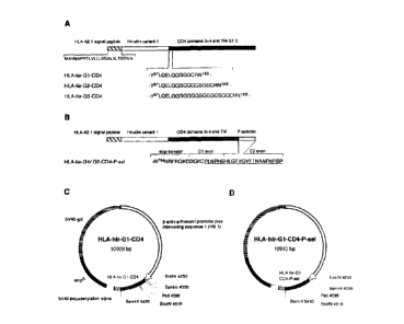
Figure 1 shows maps of hirudin-CD4 chimeric proteins and constructs according
to the
invention. (A) HLA-hirudin-CD4 constructs with glycine linkers. (B) HLA-
hirudin-CD4
construct with human P-selectin C-terminal, with the specific targeting
sequence underlined.
Transmembrane (TM), stop transfer (ST), and cytoplasmic (C) regions of CD4 are
indicated.
Figure 2 shows FACS profiles for HLA-hirudin-CD4 constructs expressed in DAP.3
fibroblasts.
Figure 3 shows FACS profiles for HLA-hirudin-CD4-P-selectin cDNA constructs
expressed
in CHO-Kl .
CA 02285702 1999-09-23
WO 98/42850
PCT/GB98/00850
-9-
Figure 4 shows that hirudin-CD4 expressing fibroblasts bind thrombin.
Figure 5 shows the specificity of thrombin binding to cells expressing hirudin-
CD4.
Figure 6 shows thrombin binding to CI-10-K1 cells transfected with HLA-hirudin
constructs.
Figure 7 shows that inactivation of thrombin abolishes thrombin binding to
hirudin-CD4 at the
cell surface. Cells expressing hirudin-G2-CD4 were incubated with thrombin or
inactivated
thrombin and stained for thrombin binding with anti-prothrombin or anti-
thrombin-hirudin
antibodies.
Figure 8 shows maps of TFPI-CD4 chimeric proteins and constructs according to
the invention.
Figure 9 shows flow cytometry profiles of DAP.3 cells expressing TFPI tethered
to the cell
surface.
Figure 10 shows specific FXa binding to cell surface bound TFPI1_276-CD4 and
TFPI1183-CD4.
Figure 11 shows the blocking of FXa binding by a polyclonal anti-TFPI
immunoglobulin
fraction.
Figure 12 shows the blocking of FXa binding by monoclonal antibodies directed
against Kunitz
domains I and II.
Figure 13 shows the inhibition of FXa by cells expressing TFPI1_276-CD4 and
TFPI1_183-CD4. The
mean time for a FXa-specific chromogenic substrate to reach 0D405-0.1 is shown
for transfected
DAP.3 cells incubated with FXa. Values for control cells were subtracted and
error bars indicate
standard deviations.
Figure 14 shows that an active TF1.219/FVIIa complex is required for maximal
binding to
TFPI-CD4 chimeric proteins.
Figure 15 shows the specificity of thrombin binding to immortalised porcine
endothelial cells
(IPEC) expressing hirudin-CD4, and also shows the effect of cell-surface
hirudin-CD4
=
expression on clotting times.
Figure 16 shows the distribution of ACTH and hirudin in D16/16 cells, as
revealed by
fluorescence.
Figure 17 shows the change in cellular distribution of hirudin-CD4-P-selectin
after PMA
CA 02285702 1999-09-23
WO 98/42850 PCT/GB98/00850
-10-
stimulation
Figure 18 shows that TFPI-CD4 expressed on IPEC retains its binding
properties.
Figure 19 shows the competitive binding of porcine and human tissue factors.
Figure 20 shows that TFPI-CD4 prolongs clotting times when expressed on IPEC
surface.
Figure 21 shows the anti-coagulant effect of co-expression of TFPI-CD4 and
hirudin-CD4.
DESCRIPTION OF EMBODIMENTS
1. Hirudin fused with HLA class I signal peptide and linked to domains 3 and 4
of human
CD4 is tethered to the cell membrane
To express heterologous hirudin constructs in mammalian cells, the cDNA for
the membrane-
targeting signal peptide leader sequence from human HLA class I A2.1, amino
acids ¨Ito ¨24
(Holmes, 1987), was fused to hirudin variant 1 (Dodt, 1984) using PCR with
overlapping
extension (Figure 1).
The HLA A2.1 leader sequence was amplified using primers:
5'-cagtgtcgacggatccatggccgtcatggcgccccga-3' [hla-1]
<SEQ ID 1>
(introducing Sall and BamHI restriction sites) and:
5'-gtcagtgtaaacaaccgcccaggtctgggtcagg-31
<SEQ ID 2>
The hirudin sequence was amplified using primers:
5'-acccagacctgggeggttgtttacactgactgcacc-3' and
<SEQ ID 3>
5'-gacgctgcagaattettgcaggtattatccgggatt-3' [hir-3]
<SEQ ID 4>
(introducing distal EcoRI and Pstl sites).
The resulting PCR products (108 and 228 bp) were purified by agarose gel
electrophoresis and
then used in a third PCR using flanking primers hla-1 and hir-3. The resulting
PCR product (300
bp) was digested with Sall and BamHI and subcloned into pBluescript SK(+)
(Stratagene).
An anchor consisting of a cDNA encoding for CD4 domains 3 and 4 (Maddon, 1985)
in
conjunction with the stop transfer sequence (ST), transmembrane and
cytoplasmic domains of
CA 02285702 1999-09-23
WO 98/42850 PCT/GB98/00850
-11-
CD4 (CD4166_435) was added to the HLA-hirudin cassette.
To ensure that hirudin stayed mobile and active when linked by its C-terminal
to the CD4
anchor, however, 3 different glycine linker lengths were made (designated G1
to G3 ¨ Fig. 1A):
¨ for glycine linker 1 (G1; GGSGG), the oligonucleotide pair consisted of
5'-aattaggaggttctggaggctgca-3' <SEQ ID 5> (containing a mutated EcoRI
recognition
sequence and a PstI site) and 5'-gcctccagaacctcct-3' <SEQ ID 6>;
¨ glycine linkers 2 (G2) and 3 (G3) consisted of the core sequence (GGSGG)
repeated
two or three times, respectively.
These linkers were introduced into the 3' end of the HLA-hirudin fragment.
The glycine linker oligonucleotides were annealed and each ligated into the
EcoRIIPstI site of
plasmids containing the HLA-hirudin cassette, prior to the insertion of the
CD4 anchor.
CD4166435 was amplified using primers:
5i-tgtctgcaggaaccagaagaaggtggaattca-3'
<SEQ ID 7>
(introducing PstI and EcoRI sites) and:
5'-gtgggatccgcctggcctcgtgcctcaa-3' <SEQ ID 8>
(containing a distal BamHI).
The resulting PCR product was cloned into pBluescript and sequenced. In CD4166-
435' v328 was
found to be mutated to A328. The PstIlBamHI CD4 fragment was subcloned into
HLA-hirudin
-G1, -G2, & -G3 plasmids, and these constructs were verified by DNA sequence
analysis.
Each of the three cDNA constructs were subcloned into the BamH1 site of the
mammalian
expression vector pHr3Actpr- 1 gpt (Gunning, 1987), containing the human 13-
actin enhancer and
promoter region in conjunction with an SV40 enhancer element driving the gpt
resistance gene,
allowing the selection of clones in the presence of mycophenolic acid (Figures
1C & 1D). The
orientation of the final constructs was verified by restriction endonuclease
mapping.
Vectors containing the individual HLA-hirudin-G1/2/3-CD4 constructs were
transfected into
mouse fibroblast cell line DAP.3 (Marguelies, 1983) with calcium-phosphate
according to
standard protocols. After 18 hours growth in DMEM medium (Gibco) supplemented
with 5%
CA 02285702 1999-09-23
WO 98/42850 PCT/GB98/00850
-12-
fetal calf serum, ampicillin, streptomycin, and glutamine, cells were glycerol
treated for 30
seconds. Cells were then washed twice with phosphate buffered saline (PBS),
and new medium
including xanthine, hypoxanthine, and mycophenolic acid to a final
concentration 12p.g/m1, was
added.
For a negative control, DAP.3 cells transfected with a human class II
construct expressing
HLA-DR (cell line 531) (Lechler, 1988) grown in identical mycophenolic acid-
containing
culture medium.
Surviving clones were tested for hirudin and CD4 expression by FACS using
murine monoclonal
antibodies 4158-81-7 (Schlaeppi, 1991) and OKT-4 (Reinherz, 1979)
respectively. 105 cells were
stained with the murine antibodies for 30 minutes on ice and a FITC-conjugated
sheep anti-
mouse polyclonal antibody was added as a secondary layer.
As shown in Figure 2, these hirudin-CD4 constructs were well expressed at the
cell surface of
DAP.3. No significant difference in expression levels was detected between
hirudin-CD4 with
the three different glycine linker lengths.
Therefore anticoagulant polypeptides can be stably expressed on the cell
surface.
2. Hirudin-CD4 with a targeting sequence from the C-terminal of P-selectin is
expressed
at the cell surface of CHO-Kl
In addition to the HLA-hirudin-G1/2/3-CD4 constructs, two more constructs were
synthesised
with targeting sequences derived from human P-selectin (Figure 1B). The
transmembrane region
from CD4 was used for these constructs, while the stop transfer sequence and C-
terminal were
replaced with the corresponding sequences from P-selectin (Johnston, 1989).
To fuse CD4 domains 3 and 4 plus the transmembrane region (CD4166.395) with
the stop transfer
sequence and cytoplasmic regions 1 and 2 of human P-selectin (P-se1754_789)
(McEver 1989), PCR
with overlapping extension was performed. For amplification of the CD4 part of
the molecule,
primers:
5'-tgtctgcaggaaccagaagaaggtggaattca-3' [CD4-5]
<SEQ ID 7>
(introducing Pstl and EcoRI restriction sites) and:
5'-gtctgancgctttctgaagaagatgcctagcccaatgaaaagcaggaggccg-3'
<SEQ ID 9>
CA 02285702 2008-12-08
-13-
were used. In parallel, to amplify the C-terminal region of P-selectin,
primers:
5t-tgggctaggcatcttatcagaaagcgmcagacaaaaaga-3' and
<SEQ ID 10>
5'-gaccaggatccggacaggtctetta-31 [P-selN3]
<SEQ ID 11>
(introducing a distal BamHI site) were used.
After purification of resulting PCR products from agarose gels, a third PCR
was run using the
flanking primers CD4-5 and P-selN3. The resulting PCR product (832 bp) was
digested with Pstl
and BamHI, subcloned into pBluescript, and sequenced. Thereafter, the CD4-P-
sel fragment
(CD416095-P-se1754,489.) was excised with PstIlBamH1 and subcloned into
plasmids containing
HLA-hirudin-G1 or -02.
The final HLA-hirudin-Gl/G2-CD4-P-selectin constructs were subcloned into the
BamHI site of
pHf3Actpr- I gpt and transfected into CHO-Kl cells (ATCC CCL61), grown in
RPM.! 1640
medium (Gibco) supplemented with 5% fetal calf serum, ampicillin,
streptomycin, and
glutamine.
Transfection was by electroporation according to standard protocols. Briefly,
5x10⁶ cells were
resuspended in 350 1 serum-free medium and transferred to a 1 ml
electroporation cuvette with
a 0.4 cm space between electrodes (Bio-Rad). After addition of 10 g plasmid
DNA in 150 I,
samples were gently shaken and kept on ice. Cells were subjected to
electroporation at infinite
resistance, 960 F and 350 V in a Gene Pulserrmapparatus (Bio-Rad). The day.
after
electroporation, cells were washed twice with PBS and new medium including
mycophenolic
acid, xanthine, and hypoxanthine was added.
Recently it was shown that when CHO-Kl cells were transfected with P-selectin
cDNA, P-
selectin protein was not accumulated intracellularly, but was expressed at the
cell surface
(Disdier, 1992). In the CHO-Kl transfectants produced above, both hirudin-G1-
CD4-P-selectin
and hirudin-G2-CD4-P-selectin were expressed at the surface as judged by
staining with OKT-4
and 4158-81-7 monoclonals (Figure 3). The negative control used was a CHO-K 1
cell line
expressing TFPI fused to CD4 domains 3 and 4 (TFPI-CD4166,435), grown in the
same
mycophenolic acid-containing medium.
As a positive control, CHO-Kl cells were transfected with full length human P-
selectin
(Johnston, 1989), which was subcloned as a 3142bp Sall fragment into
plifiActpr-Ineo
CA 02285702 1999-09-23
WO 98/42850 PCT/GB98/00850
-14-
containing an SV40-driven neomycin (G418) resistance gene. These cells were
treated with 400
pig/m1 G418 and after 2 weeks individual clones were picked with cotton swabs
and transferred
to 12-well plates. Surviving clones were analysed for hirudin and CD4
expression using
4158-81-7 at 10 jig/m1 and an undiluted OKT-4 hybridoma supernatant.
Human P-selectin was detected by anti-CD62 tnAb (Becton Dickinson), according
to the
manufacturer's recommendations. A similar FACS profile as with hirudin-CD4-P-
selectin was
observed for these CD62-labelled cells (Figure 3E), confirming that CHO-K 1
cells express
P-selectin at the plasma membrane.
Thus, chimeric proteins comprising the P-selectin targeting sequence remain
functional when
expressed at the cell surface.
3. Hirudin anchored to the cell surface binds thrombin as detected with
specific
antibodies
To test whether hirudin tethered in this way to the cell surface retains its
thrombin binding
activity, the following binding assay was used.
Stably transfected cells were grown in T75 culture flasks for 36 hours before
each experiment.
DAP.3 cells were detached using a cell scraper, whilst CHO-K 1 cells were
detached from the
plastic by treatment with PBS, 5 mM EDTA for 10 minutes at 37 C. After 4
washes with PBS
containing 0.1% BSA (w/v), 2.5x10⁵ cells in 150 1 were incubated for 1 hour at
37 C with
increasing concentrations of thrombin. The cells were washed four times with
PBS containing
0.1% BSA and further incubated for 30 minutes on ice with rabbit anti-human
prothrombin
immunoglobulins (1011g/m1 in 100u1) (Dakopatts). After two further washes,
cells were
incubated for 30 minutes with FITC-conjugated swine anti-rabbit
immunoglobulins (Dakopatts).
Finally, transfectants were washed three times and analysed by flow cytometry.
As shown in Figure 4, hirudin expressed at the cell surface retains the
ability to bind thrombin
and glycine linker length did not influence thrombin binding.
To assess the amount of thrombin needed to saturate the hirudin-CD4 expressing
cells, two
clones were incubated with thrombin up to 82 U/ml. When percentage positive
cells was
analysed, transfectants were saturated at 41 U/ml thrombin (Figure 4C).
According to the mean
fluorescence intensities (mfi), however, cells were not saturated even at 82
U/ml (Figure 4D). At
CA 02285702 1999-09-23
WO 98/42850
PCT/GB98/00850
-15-
these high experimental thrombin concentrations the background binding to
control cells
expressing HLA-DR increased significantly.
= To elucidate the specificity of thrombin binding to hirudin-CD4 further,
blocking experiments
were carried out. DAP.3 HLA-hirudin-G3-CD4 transfectants were pre-incubated on
ice for 30
minutes with 101.tg/m1 anti-hirudin mAb or appropriate controls (mouse IgG1
and IgG2a,
Dakopatts) for 30 minutes on ice, and washed twice in PBS containing 0.1% BSA
before
incubating with thrombin for 1 hour at 37 C as above. Thrombin binding was
analysed as above.
Pre-incubation with 4158-81-7 inhibited specific thrombin binding to hirudin-
CD4 (Figure 5A).
Thrombin binding by hirudin-CD4 was demonstrated by incubation with thrombin
and
comparing labelling with mAb 4107-76-1 (Schlaeppi, 1991) and anti-prothrombin
immtmoglobulins. 4107-76-1 is directed against the hirudin-thrombin complex
and detects
neither hirudin without thrombin nor thrombin bound to endogenous thrombin
receptors. As
shown in Figure 5B, thrombin binding detected with 4107-76-1 paralleled the
binding observed
with the anti-prothrombin immunoglobulin fraction.
Thus hirudin expressed on the surface of DAP.3 cells retains specific thrombin
binding.
Immortalised porcine epithelial cells (IPEC) were transfected with hirudin-CD4
in the same way.
As shown in Figure 15A, only the transfected cells bound thrombin, and this
was blocked by the
4158-81-7 in a dose-dependent manner (Figure 15B). A human plasma
recalcification test system
was used for further investigation of the functional effect of expressing
surface-tethered hirudin
on IPEC. As shown in Figure 15C, untransfected IPEC shortened the clotting
time of recalcified
plasma to approximately 170 seconds, compared with a control clotting time
370s in the absence
of cells. Preincubation with IL-1, which induces TF expression, further
reduced the clotting time
to below 100s. In contrast, clotting times for transfected IPEC were
prolonged, even after
preincubation with IL-1-induced TF expression. Incubation with 4158-81-7
reduced the
anticoagulant effect in a dose-dependent manner, indicating that the effect
was due to the
presence of cell-surface hirudin (Figure 15D).
Hirudin expressed on the surface of IPEC thus binds thrombin and also inhibits
the clotting of
human plasma.
CA 02285702 1999-09-23
WO 98/42850 PCT/GB98/00850
-16-
4. Hirudin-CD4-P-selectin expressed by CHO-Kl cells binds thrombin
To investigate whether hirudin-CD4-P-selectin also binds thrombin when
expressed at the
surface of CHO-K 1 cells, these cells were incubated with thrombin for 1 hour
at 37 C. After
staining with anti-prothrombin immunoglobulins and addition of a second FITC-
labelled
antibody layer, cells were analysed by flow cytometry.
A distinct binding profile was detected, as shown in Figure 6A. With anti-
prothrombin
immunoglobulins, background thrombin binding to CHO-K I cells expressing an
irrelevant
protein linked to CD4 was detectable after incubation with fairly low
concentrations of thrombin.
However, specific thrombin binding to hirudin was verified by staining with
the specific
anti-hirudin/thrombin mAb 4107-76-1 (Figure 6B). With this antibody,
background binding by
the control CHO-K 1 cells was undetectable. It is also evident from Figure 6
that clones
expressing hirudin appeared to bind thrombin non-specifically to a different
degree. implying
that they had different expression levels of endogenous thrombin receptors.
This variation in
non-specific binding was confirmed with several other clones.
For comparison, results from two CHO-K 1 transfectants expressing hirudin-G1-
CD4 and
hirudin-G2-CD4 (ie. no P-selectin sequence) are shown in Figures 6C and 6D.
Except for a
slightly increased thrombin binding due to better expressed chimeric proteins
(higher mfi's), no
major differences in binding profiles were detected compared to transfectants
expressing hirudin
linked to the CD4-P-selectin anchor.
5. Hirudin-CD4-P-selectin is stored in secretory granules and can be released
on
activation
To examine intracellular accumulation of hirudin and its route from secretory
granules to the
cell surface, a secretory murine pituitary cell line (D16/16) was transiently
transfected with
cDNA encoding either hirudin-CD4-P-selectin or hirudin-CD4. This cell line was
chosen for
two reasons. Firstly, these cells are known to express ACTH in specific
storage granules,
which are discharged at the cell surface on activation with phorbol esters.
Secondly,
endothelial cells (which would appear to be the ideal cell type to investigate
intracellular
targeting of the P-selectin construct) rapidly lose their Weibel-Palade
storage granules during
in vitro culture.
48 hours after transfection, D16/16 cells were stained with antibodies against
hirudin and
CA 02285702 1999-09-23
WO 98/42850 PCT/GB98/00850
-17-
ACTH. In cells transfected with hirudin-CD4-P-selectin, hirudin was detected
in granules
evenly distributed in the cytoplasm (Figure 16A). The same pattern of granule
distribution was
seen with ACTfl-specific staining, implying co-localisation with hirudin
(Figure 16B). This
was verified when both antibodies were used for staining (Figure 16C).
In contrast, D16/16 cells transfected with hirudin-CD4 did not accumulate
hirudin in
intracellular granules, but expressed high levels of hirudin at the cell
surface (Figure 16D).
Dual staining (Figure 16F) revealed only slight co-localisation of hirudin and
ACTH.
Cells expressing hirudin-CD4-P-selectin were activated with phorbol ester PMA,
and were
analysed by flow cytometry. 4158-81-7 did not detect any hirudin at the cell-
surface in
unstimulated cells (Figure 17A). After 30 minutes of PMA-stimulation, however,
hirudin was
detected at the cell-surface (Figure 17B). Furthermore, activated D16/16 cells
specifically
bound to thrombin, unlike non-activated cells (Figure 17C ¨ stained with 4107-
76-1).
Thus, by using the granule-containing pituitary cell line D16/16, it was
clearly demonstrated
that hirudin-CD4-P-selectin can be targeted to specific intracellular storage
granules, and that
functional chimeric molecules can be released and exposed at the cell surface
upon activation.
6. The interaction between thrombin and hirudin-CD4 is abolished when the
catalytic site
of thrombin is inactivated.
Specific thrombin binding to hirudin-CD4 with and without P-selectin targeting
sequence was
clear (Figures 4 and 6). To strengthen the specificity of the thrombin-hirudin
interaction further,
thrombin (210 nmol in 500 Tris-buffered saline (TBS), 0.1% BSA, pH 7.4) was
pre-incubated
for 1 hour at 37 C with either:
¨ native full-length hirudin (Biopharm) at a 10-fold molar excess;
¨ D-Phe-Pro-Arg chloromethyl ketone dihydrochloride ("PPACK-HC1") (Calbiochem)
at
100-fold molar excess; or
¨ a synthetic C-terminal hirudin dodecapeptide analog comprising hirudin
residues 53-64, with
sulfato-Tyr64 (American Diagnostica) at 100-fold molar excess.
The thrombin-dependent catalytic activity was analysed with a small
chromogenic oligopeptide
substrate (H-D-Phe-Pip-Arg-pNA-2HCI ("S-2238") (Quadratech).
CA 02285702 1999-09-23
WO 98/42850 PCT/GB98/00850
-18-
To ascertain whether thrombin was inactivated by PPACK-HC1 and hirudin, 5 ul
of each
reaction mixture were diluted with 95 ml TBS, 0.1% BSA and incubated with 50
ul 4mM
S-2238 for 10 minutes at 37 C.
As expected, no chromogenic conversion was observed with thrombin incubated
with
PPACK-HC1 or hirudin as compared to thrombin incubated without inhibitor,
whereas the
dodecapeptide did not influence thrombin-dependent catalytic activity as
measured by cleavage
of S-2238.
The three different preparations were added to transfectants expressing
hirudin tethered to the
cell surface. Using the procedure described above, thrombin binding was
investigated with the
anti-prothrombin or anti-hirudin-thrombin antibodies. As can be seen in Figure
7A. thrombin
inactivated with hirudin or PPACK-HC1 was not bound by hirudin expressed at
the cell surface
of DAP.3. In addition, only a partial thrombin-dodecapeptide complex binding
was observed. In
contrast to DAP.3 transfectants, CHO-K1 cells displayed a relatively high
thrombin-
PPACK-HC1 binding (Figure 7B). This interaction was found to be unspecific as
illustrated with
the anti-hirudin-thrombin rnAb 4107-76-1. No specific thrombin-PPACK-HC1-
hirudin binding
was detected.
This confirms that hirudin tethered to the cell surface specifically and
strongly binds thrombin at
its catalytic site.
7. Full length and truncated TFPI anchored to CD4 domains is expressed at the
cell
surface
In order to tether TFPI to the cell membrane, a fusion protein consisting of
human CD4166435
linked either to full length TFPI including all three Kunitz domains
(TFPII_276) or to a truncated
form of TFPI lacking Kunitz domain III and the C-terminal
(Wun, 1988) (Figure 8).
These were synthesized in a similar way to that described above for hirudin,
with the TFPI and
CD4 sequences being fused using a cassette cloning strategy, but unlike
hirudin, TFPI is a
mammalian protein and hence contains an endogenous signal peptide.
DNA encoding the N-terminal portion of TFPI including Kunitz domains I and 11
(675 bp) was
amplified using the primers:
5'-catcgtcgacggatcctagatgatttacacaatgaaganagtacatgcactttgggc-3'
<SEQ ID 12>
CA 02285702 1999-09-23
WO 98/42850 PCT/GB98/00850
-19-
(introducing Sall and BamHI restriction sites); and
5'-ggacctgcagaattcaananggctgg-3'
<SEQ ID 13>
(containing EcoRI and Pstl sites).
DNA encoding the third Kunitz domain together with the C-terminal end of TFPI
(315 bp) was
amplified using primers:
5 '-agcct ___________ tgaattccacggtccctcat-3 '
<SEQ ID 14>
(with an EcoRI site); and
5'-cattgctataavaactgcagatatttttaac-3'
<SEQ ID 15>
(containing a PstI site).
CD4166.435 was amplified as described above.
By the introduction of restriction sites into the 3' end of the TFPII_183 cDNA
and the 5' end of the
TFP1184-276 cDNA, HI84 and G'85
were mutated to C'84 and Rm in the recombinant fusion proteins
(Figure 8). Furthermore, 13186 was mutated to S'86. The stop codon of TFPI was
removed by
introducing a Pstl site, thus mutating M276 to I276, and the addition of amino
acid C277. In the
course of introducing a Pst1 site in the N-terminal end of CD4 domain 3, L164
and Q'65 were
mutated to C164 and R'65, respectively. In the TFP1,84_276 cDNA, K265 was
found to be mutated to
E265 and in CD4,66,35 V228 was mutated to A328 (as described above).
All PCR products were cloned into pBluescript SK(+).
The complete TFPI-CD4 cDNAs were ligated into the BamHI site of the plif3Actpr-
1gpt
expression vector.
As above, DAP.3 cells, maintained in supplemented DMEM were transfected with
calcium-
phosphate as above. Clones were analysed for TFPI and CD4 expression by FACS
using
murine anti-human TFPI mAbs 4903 or 4904 (American Diagnostica), both at 10
g/ml, and
an undiluted OKT-4 hybridoma supernatant (Reinherz, 1979). 4903 is directed
against Kunitz
domain I, whereas 4904 is directed against Kunitz domain II. 105 cells for
each sample were
analysed and, as above, cell line 531 was used as a control.
As shown in Figure 9, both TFPII_276-CD4 and TFPI1_183-CD4 can be expressed at
the cell surface.
CA 02285702 1999-09-23
WO 98/42850 PCT/GB98/00850
-20-
8. TFP11.183-CD4 and TFP1/.276-CD4 tethered to the cell surface confer FXa
binding
To test whether TFPI tethered in this way to the cell surface retains its FXa
binding activity, the
following binding assay was used.
Stably transfected DAP.3 cells were detached by treatment with PBS, 5 mM EDTA
for 10
minutes at 37 C. After 4 washes with excess PBS, 0.1 % BSA (w/v), 2.5x10⁵
cells in 100 p.1
were incubated for 1 hour at 37 C with increasing concentrations of FXa.
Cells were then washed twice and further incubated for 30 minutes on ice with
10 g/m1 rabbit
anti-human FXa immunoglobulins (RAFX-IG, Enzyme Research Laboratories) in 100
pl. After
two additional washes, cells were incubated for 30 minutes with FITC-
conjugated swine
anti-rabbit polyclonal immunoglobulins and analysed by flow cytometry.
As shown in Figure 10, DAP.3 cells expressing TFPII.276-CD4 and TFPI1_183-CD4
at the cell
surface strongly bound FXa in a dose-dependent fashion (Figure 10), with
significant binding
detected at 0.02 nIVI. No difference in FXa binding was detected between full
length and
truncated TFPI-CD4.
It was also possible to block FXa binding with a polyclonal anti-TFPI
immunoglobulin fraction
(4901) or with monoclonals 4903 and 4904.
Cells were incubated on ice for 30 minutes with 4901, 4903, or 4904 at
increasing
concentrations, using an anti-haemoglobin antiserum (Dakopatts) as a negative
control. The cells
were then washed twice in PBS, 0.1% BSA, and further incubated with 5 nM FXa
for one hour
at 37 C. The cells were then washed and incubated with RAFX-IG as above and
analysed for
FXa binding by FACS.
FXa binding to TFPII_276-CD4 decreased 27% and 55% at 10 and 80 11g/m1
polyclonal 4901,
respectively, compared with cells incubated with the irrelevant anti-
haemoglobin polyclonal
control (Figure 11A). Diminished FXa binding was also found for TFPII_183-CD4
cells
pre-incubated with 4901 (Figure 11B).
When TFP11_226-CD4 was blocked with either 4903 or 4904, 33% less FXa binding
was observed
at 401g/ml mAb, compared with isotype-matched mouse immunoglobulins (Fig. 12).
No
significant difference in blocking activity was detected between mAbs 4903 and
4904.
This demonstrates for the first time that TFPI retains its FXa binding
activity when expressed as
CA 02285702 1999-09-23
WO 98/42850 PCT/GB98/00850
-21-
a membrane-bound fusion protein.
9. TFPI1-CD4 and TFPI1_276-CD4 are both functionally active against FXa
To determine whether TFPI tethered to the cell surface retained its ability to
inhibit the function
of FXa, the proteolytic activity of FXa was analysed using the chromogenic
substrate
N-a-Z-D-Arg-Gly-Arg-pNA=2HC1 ("S-2765") (Quadratech).
Transfected DAP.3 cells were detached as described above and washed 4 times
with excess TBS,
pH 7.4, 0.1% BSA. 0.5x10⁶ cells (in 100 1) per well were incubated for 1 hour
at 37 C with
various concentrations of FXa. 50 1.11 of 4 mM S-2765 were added and cells
were further
incubated for 2 hours at 37 C. OD405 was measured every 30 seconds and the
time required to
reach OD405=0.1 was determined, showing remaining active FXa.
FXa activity was inhibited by expressed TFPI-CD4 in a dose dependent manner
with the greatest
inhibition noted when low concentrations of FXa (0.16 nM) were added (Figure
13). In a series
of experiments, no significant difference in FXa inhibition was observed
between cells
expressing TFPI1.183-CD4 or TFPI1_276-CD4.
Thus, Kunitz domain II retains its function when tethered to the cell surface
in both
TFPII_183-CD4 and TFPI1_276-CD4.
10. TF1_20/FV1Ia complex binds irrespectively of the presence of the third
Kunitz domain
Binding of tissue factor and factor Vila can be used to confirm whether Kunitz
domain I also
retains its function.
Recombinant human TF1_219 and FVIIa were produced in E. coli and CHO-K1,
respectively
(O'Brien, 1994). These were mixed in equimolar concentrations and incubated at
25 C for 15
minutes to obtain a TF1.219/FVIIa complex.
Polyclonal rabbit immunoglobulins against human TF were produced according to
standard
methods.
DAP.3 cells expressing either TFP11_276-CD4 or TFPI1_183-CD4 were incubated
with 5 nM FXa for
1 hour at 37 C. Cells were washed twice and TF1_20/FVI1a complex was added to
2.5x10⁵ cells in
100 pl. After 1 hour at 37 C transfectants were washed twice and incubated
with 50 p.1
polyclonal rabbit anti-TF immunoglobulins (2.5 g/ml) for 30 minutes on ice
followed by 2
CA 02285702 1999-09-23
WO 98/42850
PCT/GB98/00850
-22-
washes, and further incubation with FITC-conjugated swine anti-rabbit
immunoglobulins.
Positive cells were analysed by flow cytometry.
TF1õ19/FVIIa bound equally efficient to both TFPI1_276-CD4 (Fig. 14A) and
TFPI1_183-CD4 (Fig.
14B), while no binding at all was detected to control cell line 531.
To confirm specific binding to Kunitz domain I by the TF,,,,/FVIIa complex,
FVIIa was
inactivated by pre-incubation with 1,5-dansyl-Glu-Gly-Arg-chloromethyl ketone,
dihydrochloride ("1,5-DNS-GGACK-HC1"). This binds to the active site of FVIIa
and inhibits
binding to TFPI whilst not affecting the formation of the TF1_219/FVIIa
complex (Bajaj, 1992).
FVIIa was first incubated with a 100-fold molar excess of 1,5-DNS-GGACK.1-10
for 18 hours at
20 C and repurified by ion-exchange chromatography. Active-site inhibited
FVIIa (FVIIai) was
incubated with an equimolar concentration of TF,-219 at 25 C for 15 minutes
and then added to
2.5x10⁵ cells in 100 ill. Subsequent steps were as described above.
As can be seen from figure 14, significantly less TF1õ19/FVIIai complex bound
to TFPI-CD4
expressing cells as compared to bound "active" TF1_21,/FVIIa. No difference
was observed
between DAP.3 transfected with TFPI1276-CD4 or TFPI1_183-CD4.
Thus Kunitz domain I also retains its function when tethered to the cell
surface in TFPI1_183-CD4
and TFPI1276-CD4. It is therefore apparent that TFPI tethered at the cell
surface is functionally
active as a whole.
11. TFPI-CD4 expressed on IPEC binds relevant human clotting factors and
porcine TF
As shown in Figure 18, the TFPI-CD4 fusion protein can be expressed on IPEC
and retains the
ability to bind FXa and FVIIa. To demonstrate that TFPI can physically
interact with porcine
TF, a competitive inhibition approach using soluble human TF was adopted. As
shown in
Figure 19A, in the presence of saturating concentrations of FXa and FVIIa, the
binding of
soluble human TF to TFPI-transfected IPEC (pre-treated with IL-1a) was
significantly reduced
compared to the binding by TF-negative control transfectants (not IL-1 a
activated). This
suggests that porcine TF was competing with soluble human TF for Vila, and
therefore for
TFPI binding. Figure 19B supports this, showing that binding of soluble human
IF to
TFPI-CD4-transfected IPEC (IL-la pre-activated) was increased if the
transfectants were
incubated with increasing concentrations of antibody against porcine TF. The
effect of this
antibody could reflect inhibition of the interaction between porcine TF and
FVIIa, or between
CA 02285702 1999-09-23
WO 98/42850
PCT/GB98/00850
-23-
porcine TF-VIIa complexes and TFPI-CD4. Either way, the results suggest that
the
TFPI-CD4-fusion protein expressed on the surface of IPEC physically interacts
with porcine
TF-FVIIa.
12. TFPI-CD4 expressed on IPEC inhibits TF-dependent fibrin generation
Figure 20A shows the results of a single representative experiment to
illustrate the
procoagulant phenotype of TFPI-CD4-transfected IPEC. The presence of the
fusion protein on
transfected cells consistently prolonged clotting times when compared with
control IPEC. This
effect was only observed, however, after IL-la activation ¨ TFPI-CD4
expression had no
influence on clotting times when TF-negative IPEC were used. Thus, the TFPI-
CD4, as
expected, inhibited TF-dependent, but not TF-independent fibrin generation. An
anti-TFPI
antibody, used in increasing concentrations during a pre-incubation step, was
able to normalise
clotting times back to those seen with untransfected IL-1 a-activated control
IPEC (Figure
20B), indicating that the prolongation of clotting times in the presence of
the transfected cells
was due entirely to the specific inhibitory action of TFPI.
13. Expression of a protein C activator at the cell membrane
To express heterologous constructs comprising the protein C activator isolated
from the venom
of Agkistrodon contortrix contortrix (McMullen, 1989; Kisiel, 1987), a cDNA
encoding the
protein was synthesised. The protein sequence is <SEQ ID 16>:
/IGGDECNINEHRFLALVYANGSLCG
GTLINQEWVLTARHCDRGNMRIYLGM
HNLKVLNKDALRRFPKEKYFCLNTRN
DT IWDKDIMLIRLNRPVRNSAHIAPL
SLPSNPPSVGSVCRIMGWGTITSPNA
TLPDVPHCANINILDYAVCQAAYKGL
AATTLCAGILEGGKDTCKGDSGGPLI
CNGQFQGILSVGGNPCAQPRKPGIYT
KVFDYTDWIQSIISGNTDATCPP
In accordance with porcine codon-usage bias (which is applicable to most, if
not all, mammalian
cells), the following single stranded DNA was synthesised <SEQ ID 17>:
GTG ATC GGC GGC GAO GAG TGC AAC ATC AAC GAG CAC CGC
TTC CTG GCC CTG GTG TAC GCC AAC GGC AGO CTG TGC GGC
GGC ACC CTG ATC AAC CAG GAG TGG GTG CTG ACC GCC CGC
CAC TGC GAO CGC GGC AAC ATG CGC ATC TAO CTG GGC ATG
CA 02285702 1999-09-23
WO 98/42850
PCT/GB98/00850
-24-
CAC AAC CTG AAG GTG CTG AAC AAG GAC GCC CTG CGC CGC
TTC CCC AAG GAG AAG TAO TTC TGC CTG AAC ACC CGC AAC
GAC ACC ATC TGG GAC AAG GAC ATC ATG CTG ATC CGC CTG
AAC CGC CCC GTG CGC AAC AGC GCC CAC ATC GCC CCC CTG
AGC CTG CCC AGC AAC CCC CCC AGC GTG GGC AGC GTG TGC
CGC ATC ATG GGC TGG GGC ACC ATC ACC AGC CCC AAC GCC
ACC CTG CCC GAC GTG CCC CAC TGC GCC AAC ATC AAC ATC
CTG GAC TAO GCC GTG TGC CAG GOO GCC TAO AAG GGC CTG
GOO GCC ACC ACC CTG TGC GCC GGC ATC CTG GAG GGC GGC
AAG GAC ACC TGC AAG GGC GAC AGC GGC GGC CCC CTG ATC
TGC AAC GGC CAG TTC CAG GGC ATC CTG AGC GTG GGC GGC
AAC CCC TGC GCC CAG CCC CGC AAG CCC GGC ATC TAO ACC
AAG GTG TTC GAC TAO ACC GAC TGG ATC CAG AGC ATC ATC
AGC GGC AAC ACC GAC GCC ACC TGC CCC CCC
This single-stranded DNA was annealed to complementary oligonucleotides to
give a
double-stranded molecule. Restriction sites are included at either end of the
double-stranded
DNA, to which is ligated a CD4 anchor and a P-selectin signal sequence in a
similar way to that
described above. The resulting molecule was ligated, as before, into the p1-
113Actpr-1 gpt vector.
As an alternative DNA source, a snake cDNA library could be screened on the
basis of the
known protein sequence.
14. Co-expression of TFPI-CD4 and hirudin-CD5 causes inhibition of IF-
dependent and
TF-independent clotting
Stable transfectants expressing both TFPI-CD4 and hirudin-CD4 were generated.
As shown in
Figure 21A, the primary transfectants expressed variable levels of hirudin and
low levels of
TFPI. Despite this modest expression by the majority of transfectants,
however, the
procoagulant phenotype of these cells was significantly reduced compared to
controls (Figure
2IB). The cell-surface presence of both anticoagulant molecules on IL-la-
activated IPEC
markedly prolonged the time to clot plasma to approximately 300 seconds, which
is
approaching the time taken for recalcified human plasma to clot spontaneously.
Blocking
studies with anti-hirudin and anti-TFPI antibodies confirmed that the altered
phenotype of
these double transfectants was due to specific inhibition of coagulation by
the expressed
hirudin and TFPI.
It will be understood that the invention is described above by way of example
only and
modifications may be made whilst remaining within the scope and spirit of the
invention.
CA 02285702 1999-09-23
WO 98/42850
PCT/GB98/00850
-25-
REFERENCES (THE CONTENTS OF WHICH ARE INCORPORATED HEREIN)
Bach FH, Winkler H, Ferran C, Hancock WW, Robson SC. Delayed xenograft
rejection.
Immunology Today 1996; 17(8); 379-384.
Bajaj SP, Sabharvval AK, Gorka J, Birktoft JJ. Antibody-probed conformational
transitions in the
protease domain of human factor IX upon calcium binding and zymogen
activation: putative
high-affinity Ca2+-binding site in the protease domain. PNAS USA 1992; 89: 152-
156.
Bradley A, Liu P. Target practice in transgenics. Nature Genet. 1996; 14: 121-
123.
Clarke AR. The adenovirus and the egg: a new approach to transgenesis. Nature
Biotech. 1996;
14: 942.
Dang QD, Guinto ER, Di Cera E. Rational engineering of activity and
specificity in a serine
protease. Nature Biotech. 1997; 15: 146-149.
Diamond LE, McCurry KR, Oldham ER, Tone M, Waldmann H. Platt JL, Logan JS.
Human
CD59 expressed in transgenic mouse hearts inhibits the activation of
complement. Transplant
lmmunol. 1995; 3: 305-312.
Disdier M, Morrissey JH, Fugate RD, Bainton DF, McEver RP. Cytoplasmic domain
of P-
selectin (CD62) contains the signal for sorting into the regulated secretory
pathway. Molee. Biol.
Cell. 1992; 3: 309-321.
Dodt J, Kohler S, Baici A. Interaction of site specific hirudin variants with
alpha-thrombin. FEBS
Lett.1988; 229: 87-90
Green SA, Setiadi H, McEver RP, Kelly RB. The cytoplasmic domain of P-selectin
contains a
sorting determinant that mediates rpaid degradation in lysosomes. J. Cell.
Biol. 1994; 124: 435-
448.
Gunning P, Leavitt J, Muscat G, Ng SY, Kedes L. A human beta-actin expression
vector system
directs high-level accumulation of antisense transcripts. PNAS USA.1987; 84:
4831-5
Hamamoto T, Yamamoto M, Nordfang 0, Petersen JGL, Foster DC, Kisiel W.
Inhibitory
properties of full-length and truncated recombinant TFPI variants expressed in
Saccharomyces
cereviaiae. Biol, Chem. 1993; 268: 13344-13351.
Heckl-Ostreicher B, Binder R, Kirschfink M. Functional activity of the
membrane-associated
complement inhibitor CD59 in a pig-to-human in vtiro model for hyperacture
xenogran
CA 02285702 1999-09-23
WO 98/42850
PCT/GB98/00850
-26-
rejection. Clin. Exp. ImmunoL 1995;102:589-595.
Holmes N, Ennis P, Wan AM, Denney DW, Parham P. Multiple genetic mechanisms
have
contributed to the generation of the HLA-A2/A28 family of class I MHC
molecules. J
ImmunoL 1987; 139: 936-41
Johnston GI, Cook RG, McEver RP. Cloning of GMP-140, a granule membrane
protein of
platelets and endothelium: sequence similarity to proteins involved in cell
adhesion and
inflammation. CelL 1989; 56: 1033-44
Kiely J-M, Cybulsky MI, Luscinskas FW, Gimborne MA. Immunoselective targeting
of an anti-
thrombin agent to the surface of cytokine-activated vascular endothelial
cells. Arterioscler.
Thromb. Vasc. Biol. 1995; 15: 1211-1218.
Kisiel E, Kondo S, Smith KJ, McMullen BA, Smith LF. Characterization of a
protein C activator
from Agkistrodon contortrix contortrix venom. J.BioL Chem. 1987;262:12607-13.
Knapp A, Degenhardt T, Dodt J. Hirudisins. J. Biol. Chem. 1992; 34: 24230-
24234.
Langford GA, Cozzi E, Yannoutsos N, Lancaster R, Elsome K, Chen P, White DGJ.
Production
of pigs transgenic for human regulators of complement activation using YAC
technology.
Transplant Proc. 1996; 28: 862-863.
Lechler RI, Bal V, Rothbard JB, Germain RN, Sekaly R, Long EO, Lamb J.
Structural and
functional studies of HLA-DR restricted antigen recognition by human helper T
lymphocyte
clones by using transfected murine cell lines. J. ImmunoL 1988; 141: 3003-
3009.
McCurry KR, Diamond LE, Kooyman DL, Byrne GW, Martin MJ, Logan JS, Platt JL.
Human
complement regulatory proteins expressed in transgenic swine protect swine
xenografts from
humoral injury. Transplant Proc. 1996; 28: 758.
McMullen BA, Fujikawa K, Kisiel W. Primary structure of a protein C activator
from
Agkistrodon contortrix contortrix venom. Biochemistry 1989; 28: 674-679.
Maddon PJ, Littman DR, Godfrey M, Maddon DE, Chess L, Axel R. The isolation
and
nucleotide sequence of a cDNA encoding the T cell surface protein T4: a new
member of the
immunoglobulin gene family. Cell. 1985; 42: 93-104
Mao SS, Huang J, Welebob C, Neeper MP, Garsky VM, Shafer JA. Identification
and
characterization of variants of tick anticoagulant peptide with increased
inhibitory potency
CA 02285702 1999-09-23
WO 98/42850
PCT/GB98/00850
-27-
toward human factor Xa. Biochemistry 1995; 34: 5098-5103.
Merkenschlager M, Altmann DM, Ikeda H. T cell alloresponses against HLA-DQ and
-DR
products involve multiple epitopes on the CD4 molecule. Distinct mechanisms
contribute to the
inhibition of HLA class II-dependent and -independent T cell responses by
antibodies to CD4.
Ilmmuno1.1990;145: 3181-7.
O'Brien DP, Kemball-Cook G, Hutchinson AM, Martin DM, Johnson DJ, Byfield PG.
Takamiya
0, Tuddenham EG, McVey JH. Surface plasmon resonance studies of the
interaction between
factor VII and tissue factor. Biochemistry 1994; 33:14162-9.
Reinherz EL, Kung PC, Goldstein G, Schlossman SF. Separation of functional
subsets of human
T cells by a monoclonal antibody. PNAS USA .1979; 76: 4061-4065
Schlaeppi JM. Preparation of monoclonal antibodies to the thrombin/hirudin
complex. Thromb
Res. 1991; 62: 459-470
Skern T, Bischoff R, Jallat S, Dott K, Ali-Hadji D, Clesse D, Kieny MP,
Courtney M. Sulphation
of hirudin in BHK cells. FEBS. 1990; 1: 36-38.
Squinto SP. Xenogeneic organ transplantation. Curr. Opin. Biotech. 1996; 7:
641-645.
Wagner DD. The Weibel-Palade body: the storage granule for von Willebrand
factor and P-
selectin. Thrombosis & Haemostasis. 1993; 70: 105-110.
Wheeler MB. Development and validation of swine embryonic stem cells: a
review. Reprod
Fertil. Dec. 1994; 6:563-568.
White D, Cozzi E, Langford G, Oglesby T, Wang M, Wright L, Wallwork J. The
control of
hyperacute rejection by genetic engineering of the donor species. Eye 1995; 9:
185-189.
Wun IC, Kretzmer KK, Girard TJ, Miletich JP, Broze GJ. Cloning and
characterization of a
cDNA coding for the lipoprotein-associated coagulation inhibitor shows that it
consists of three
tandem Kunitz-type inhibitory domains. I Biol. Chem.1988; 263: 6001A
Yarmoutsos N, Langford GA, Cozzi E, Lancaster R, Elsome K, Chen P, White DJG.
Production
of pigs transgenic for human regulators of complement activation. Transplant
Proc. 1995; 27:
324-325.
CA 02285702 2000-01-05
1 / 6
SEQUENCE LISTING
(1) GENERAL INFORMATION:
(i) APPLICANT:
(A) NAME: RPMS Technology Limited
(B) STREET: Commonwealth Building
(C) CITY: Du Cane Road
(D) STATE: London
(E) COUNTRY: United Kingdom
(F) POSTAL CODE (ZIP): W12 ONN
(ii) TITLE OF INVENTION: Coagulation inhibition
(iii) NUMBER OF SEQUENCES: 17
(iv) COMPUTER READABLE FORM:
(A) MEDIUM TYPE: Floppy disk 720K
(B) COMPUTER: IBM PC compatible
(C) OPERATING SYSTEM: PC-DOS/MS-DOS
(D) SOFTWARE: PatentIn Ver 2.0/Microsoft Word 97
On CURRENT APPLICATION DATA:
APPLICATION NUMBER: PCT/GB98/00850
(2) INFORMATION FOR SEQ ID NO: 1
(i) SEQUENCE CHARACTERISTICS:
(A) LENGTH: 37 nucleotides
(B) TYPE: nucleic acid
(C) STRANDEDNESS: single
(D) TOPOLOGY: linear
(ii) MOLECULE TYPE: DNA
(xi) SEQUENCE DESCRIPTION FOR SEQ ID NO: 1
cagtgtcgac ggatccatgg ccgtcatggc gccccga 37
(2) INFORMATION FOR SEQ ID NO: 2
(i) SEQUENCE CHARACTERISTICS:
(A) LENGTH: 34 nucleotides
(B) TYPE: nucleic acid
(C) STRANDEDNESS: single
(D) TOPOLOGY: linear
(ii) MOLECULE TYPE: DNA
(xi) SEQUENCE DESCRIPTION FOR SEQ ID NO: 2
gtcagtgtaa acaaccgccc aggtctgggt cagg 34
(2) INFORMATION FOR SEQ ID NO: 3
CA 02285702 2000-01-05
2 / 6
(i) SEQUENCE CHARACTERISTICS:
(A) LENGTH: 36 nucleotides
(B) TYPE: nucleic acid
(C) STRANDEDNESS: single
(D) TOPOLOGY: linear
(ii) MOLECULE TYPE: DNA
(xi) SEQUENCE DESCRIPTION FOR SEQ ID NO: 3
acccagacct gggcggttgt ttacactgac tgcacc 36
(2) INFORMATION FOR SEQ ID NO: 4
(i) SEQUENCE CHARACTERISTICS:
(IQ LENGTH: 37 nucleotides
(B) TYPE: nucleic acid
(C) STRANDEDNESS: single
(D) TOPOLOGY: linear
(ii) MOLECULE TYPE: DNA
(xi) SEQUENCE DESCRIPTION FOR SEQ ID NO: 4
gacgctgcag aattcttgca ggtattcttc cgggatt 37
(2) INFORMATION FOR SEQ ID NO: 5
(i) SEQUENCE CHARACTERISTICS:
(A) LENGTH: 24 nucleotides
(B) TYPE: nucleic acid
(C) STRANDEDNESS: single
(D) TOPOLOGY: linear
(ii) MOLECULE TYPE: DNA
(xi) SEQUENCE DESCRIPTION FOR SEQ ID NO: 5
aattaggagg ttctggaggc tgca 24
(2) INFORMATION FOR SEQ ID NO: 6
(i) SEQUENCE CHARACTERISTICS:
(10 LENGTH: 16 nucleotides
(B) TYPE: nucleic acid
(C) STRANDEDNESS: single
(D) TOPOLOGY: linear
(ii) MOLECULE TYPE: DNA
(xi) SEQUENCE DESCRIPTION FOR SEQ ID NO: 6
CA 02285702 2000-01-05
3 / 6
gcctccagaa cctcct 16
(2) INFORMATION FOR SEQ ID NO: 7
(i) SEQUENCE CHARACTERISTICS:
(A) LENGTH: 32 nucleotides
(B) TYPE: nucleic acid
(C) STRANDEDNESS: single
(D) TOPOLOGY: linear
(ii) MOLECULE TYPE: DNA
(xi) SEQUENCE DESCRIPTION FOR SEQ ID NO: 7
tgtctgcagg aaccagaaga aggtggaatt ca 32
(2) INFORMATION FOR SEQ ID NO: 8
(i) SEQUENCE CHARACTERISTICS:
(A) LENGTH: 28 nucleotides
(B) TYPE: nucleic acid
(C) STRANDEDNESS: single
(D) TOPOLOGY: linear
(ii) MOLECULE TYPE: DNA
(xi) SEQUENCE DESCRIPTION FOR SEQ ID NO: 8
gtgggatccg cctggcctcg tgcctcaa 28
(2) INFORMATION FOR SEQ ID NO: 9
(i) SEQUENCE CHARACTERISTICS:
(A) LENGTH: 53 nucleotides
(B) TYPE: nucleic acid
(C) STRANDEDNESS: single
(D) TOPOLOGY: linear
(ii) MOLECULE TYPE: DNA
(xi) SEQUENCE DESCRIPTION FOR SEQ ID NO: 9
gtctgaaacg ctttctgaag aagatgccta gcccaatgaa aagcaggagg ccg 53
(2) INFORMATION FOR SEQ ID NO: 10
(i) SEQUENCE CHARACTERISTICS:
(A) LENGTH: 42 nucleotides
(B) TYPE: nucleic acid
(C) STRANDEDNESS: single
(D) TOPOLOGY: linear
CA 02285702 2000-01-05
4 / 6
(ii) MOLECULE TYPE: DNA
(xi) SEQUENCE DESCRIPTION FOR SEQ ID NO: 10
tgggctaggc atcttcttca gaaagcgttt cagacaaaaa ga 42
(2) INFORMATION FOR SEQ ID NO: 11
(i) SEQUENCE CHARACTERISTICS:
(A) LENGTH: 25 nucleotides
(B) TYPE: nucleic acid
(C) STRANDEDNESS: single
(D) TOPOLOGY: linear
(ii) MOLECULE TYPE: DNA
(xi) SEQUENCE DESCRIPTION FOR SEQ ID NO: 11
gaccaggatc cggacaggtc tctta 25
(2) INFORMATION FOR SEQ ID NO: 12
(i) SEQUENCE CHARACTERISTICS:
(A) LENGTH: 57 nucleotides
(B) TYPE: nucleic acid
(C) STRANDEDNESS: single
(D) TOPOLOGY: linear
(ii) MOLECULE TYPE: DNA
(xi) SEQUENCE DESCRIPTION FOR SEQ ID NO: 12
catcgtcgac ggatcctaga tgatttacac aatgaagaaa gtacatgcac tttgggc 57
(2) INFORMATION FOR SEQ ID NO: 13
(i) SEQUENCE CHARACTERISTICS:
(A) LENGTH: 26 nucleotides
(B) TYPE: nucleic acid
(C) STRANDEDNESS: single
(D) TOPOLOGY: linear
(ii) MOLECULE TYPE: DNA
(xi) SEQUENCE DESCRIPTION FOR SEQ ID NO: 13
ggacctgcag aattcaaaaa ggctgg 26
(2) INFORMATION FOR SEQ ID NO: 14
(i) SEQUENCE CHARACTERISTICS:
CA 02285702 2000-01-05
/ 6
(A) LENGTH: 28 nucleotides
(B) TYPE: nucleic acid
(C) STRANDEDNESS: single
(D) TOPOLOGY: linear
(ii) MOLECULE TYPE: DNA
(xi) SEQUENCE DESCRIPTION FOR SEQ ID NO: 14
agcctttttg aattccacgg tccctcat 28
(2) INFORMATION FOR SEQ ID NO: 15
(i) SEQUENCE CHARACTERISTICS:
(A) LENGTH: 31 nucleotides
(B) TYPE: nucleic acid
(C) STRANDEDNESS: single
(D) TOPOLOGY: linear
(ii) MOLECULE TYPE: DNA
(xi) SEQUENCE DESCRIPTION FOR SEQ ID NO: 15
cattgctata acaactgcag atatttttaa c 31
(2) INFORMATION FOR SEQ ID NO: 16
(i) SEQUENCE CHARACTERISTICS:
(A) LENGTH: 231 amino acids
(B) TYPE: protein
(C) STRANDEDNESS: single
(D) TOPOLOGY: linear
(ii) MOLECULE TYPE: protein
(vi) ORIGINAL SOURCE:
(A) ORGANISM: Agkistrodon contortrix contortrix
(xi) SEQUENCE DESCRIPTION FOR SEQ ID NO: 16
Val Ile Gly Gly Asp Glu Cys Asn Ile Asn Glu His Arg Phe Leu Ala
1 5 10 15
Leu Val Tyr Ala Asn Gly Ser Leu Cys Gly Gly Thr Leu Ile Asn Gln
20 25 30
Glu Trp Val Leu Thr Ala Arg His Cys Asp Arg Gly Asn Met Arg Ile
35 40 45
Tyr Leu Gly Met His Asn Leu Lys Val Leu Asn Lys Asp Ala Leu Arg
50 55 60
Arg Phe Pro Lys Glu Lys Tyr Phe Cys Leu Asn Thr Arg Asn Asp Thr
65 70 75 80
CA 02285702 2000-01-05
6 / 6
Ile Trp Asp Lys Asp Ile Met Leu Ile Arg Leu Asn Arg Pro Val Arg
85 90 95
Asn Ser Ala His Ile Ala Pro Leu Ser Leu Pro Ser Asn Pro Pro Ser
100 105 110
Val Gly Ser Val Cys Arg Ile Met Gly Trp Gly Thr Ile Thr Ser Pro
115 120 125
Asn Ala Thr Leu Pro Asp Val Pro His Cys Ala Asn Ile Asn Ile Leu
130 135 140
Asp Tyr Ala Val Cys Gin Ala Ala Tyr Lys Gly Leu Ala Ala Thr Thr
145 150 155 160
Leu Cys Ala Gly Ile Leu Glu Gly Gly Lys Asp Thr Cys Lys Gly Asp
165 170 175
Ser Gly Gly Pro Leu Ile Cys Asn Gly Gin Phe Gin Gly Ile Leu Ser
180 185 190
Val Gly Gly Asn Pro Cys Ala Gin Pro Arg Lys Pro Gly Ile Tyr Thr
195 200 205
Lys Val Phe Asp Tyr Thr Asp Trp Ile Gin Ser Ile Ile Ser Gly Asn
210 215 220
Thr Asp Ala Thr Cys Pro Pro
225 230
(2) INFORMATION FOR SEQ ID NO: 17
(i) SEQUENCE CHARACTERISTICS:
(A) LENGTH: 693 nucleotides
(B) TYPE: nucleic acid
(C) STRANDEDNESS: single
(D) TOPOLOGY: linear
(ii) MOLECULE TYPE: DNA
(xi) SEQUENCE DESCRIPTION FOR SEQ ID NO: 17
gtgatcggcg gcgacgagtg caacatcaac gagcaccgct tcctggccct ggtgtacgcc 60
aacggcagcc tgtgcggcgg caccctgatc aaccaggagt gggtgctgac cgcccgccac 120
tgcgaccgcg gcaacatgcg catctacctg ggcatgcaca acctgaaggt gctgaacaag 180
gacgccctgc gccgcttccc caaggagaag tacttctgcc tgaacacccg caacgacacc 240
atctgggaca aggacatcat gctgatccgc ctgaaccgcc ccgtgcgcaa cagcgcccac 300
atcgcccccc tgagcctgcc cagcaacccc cccagcgtgg gcagcgtgtg ccgcatcatg 360
ggctggggca ccatcaccag ccccaacgcc accctgcccg acgtgcccca ctgcgccaac 420
atcaacatcc tggactacgc cgtgtgccag gccgcctaca agggcctggc cgccaccacc 480
ctgtgcgccg gcatcctgga gggcggcaag gacacctgca agggcgacag cggcggcccc 540
ctgatctgca acggccagtt ccagggcatc ctgagcgtgg gcggcaaccc ctgcgcccag 600
ccccgcaagc ccggcatcta caccaaggtg ttcgactaca ccgactggat ccagagcatc 660
atcagcggca acaccgacgc cacctgcccc ccc 693
____