Note: Descriptions are shown in the official language in which they were submitted.
CA 02286272 2005-08-17
-1-
SURGICAL RETRACTOR
Numerous devices have been used to position tissue at
a surgical site to aid in the performing of surgical
procedures. Retractors, for example, have been used to
hold an artery in position during operations adjacent to
the heart to prevent movement of the artery. This serves
to minimize the risk of injury to the artery and adjacent
tissue and can facilitate the desired anastomosis.
A recently developed procedure, referred to as the
minimally invasive direct coronary artery bypass procedure,
has been used to graft onto a coronary artery without
cardiopulmonary-bypass. This procedure involves the
grafting of the left internal mammary artery (LIMA) onto
the left anterior descending (LAD) or other artery. As
this procedure does not require the use of a heart lung
machine to oxygenate and pump blood, the morbidity and
mortality associated with this procedure is substantially
CA 02286272 1999-10-05
WO 98/48704 PCT/US98/08348
-2-
lower than previous bypass techniques. A problem
associated with the minimally invasive procedure, however,
is that while the heart continues to pump during the
procedure, the motion of the heart can interfere with the
surgeon's task of attaching the LIMA to the LAD. There is
also a need to stop blood flow in the area of the graft to
maintain a clear'field of view and provide precise suture
placement.
Two basic strategies have been employed to address the
problem of operating on a moving site, one being the use of
pharmacological agents to limit heart motion, and the other
being mechanical, such as a two prong retractor that is
pushed down against the heart on both sides of the artery,
or alternatively, upward traction away from the moving
heart by traction tape or suture thread. Both of these
options, however, have problems associated with them. Both
options are susceptible to some movement of the vessel
grafting site. The use of pharmacological agents is
undesirable and impairs circulatory function. Traction by
compression of the heart against the spine does serve to
immobilize the site but can compromise the ability of the
heart to maintain circulation and result in hypotension.
Upward traction can involve circumferential compression of
the artery to occlude the artery and prevent blood flow,
however upward traction that is sufficient to immobilize
the site can cause injury, stenosis or occlusion of the
vessel.
There is a continuing need however for improvement in
devices and methods for retaining tissue at surgical sites
to further reduce the risks associated with surgical
, , . ..
CA 02286272 2005-08-17
-3-
procedures where the devices and methods are inexpensive,
safe and reliable.
According to one aspect of the present invention
there is provided a surgical retractor comprising: a
retaining element having an aperture defining an operative
site; and a holder on the retaining element, characterized
in that the holder is positioned to receive a connector
including a cord that is attached to the holder and that
compresses tissue at the operative site against a planar
compression surface on the retaining element.
According to a further aspect of the present
invention there is provided a surgical retractor for a
coronary bypass procedure comprising: a retaining base
having an aperture that exposes an operative site, the
aperture extending along a longitudinal axis of the base; a
plurality of holders on the retaining base such that a
first holder is positioned on a first side of the aperture
and a second holder is positioned on a second side of the
aperture; and an arm attached to the base and extending
above the base such that a user can position the base at
the operative site with a coronary artery exposed through
the aperture.
The present invention relates to a surgical retractor
for immobilizing tissue at a surgical site and to a method
of using the retractor during a surgical procedure. A
preferred embodiment of the retractor includes a retaining
element having an aperture that exposes the surgical site
and a holder that is used to position tissue at the
surgical site relative to the retaining element. A handle
can be attached to or fabricated with the retaining element
or platform so that the user can manipulate the position of
the retractor as needed.
CA 02286272 2005-08-17
-3a-
In a preferred embodiment of the invention a connector
such as elastic tape or thread is used to position tissue
at the surgical site within the retractor aperture and to
prevent movement of the tissue during the procedure. The
connecting cord, thread or tape also aids in the
compression of the artery in a grafting procedure to
occlude flow on one or both sides of the surgical site.
The cord is attached to the holder on the retaining
element. A preferred embodiment of the holder can be a
plurality of slits or openings positioned on both sides of
the retractor that receive and frictionally secure the cord
on both sides of the aperture. In another preferred
embodiment a mechanical fastener is used to grip both'sides
of the cord. The fastener can be a spring mounted valve,
for example, that allows the user to adjust the tension in
CA 02286272 1999-10-05
WO 98/48704 PCT/US98/08348
-4-
the cord.
A preferred embodiment of the invention comprises a
retaining element or base having two sections that can be
separated after the procedure is complete to permit removal
of the retractor from under the grafted artery. Another
preferred embodiment uses a side opening in the platform of
the retractor that extends to the aperture so that the
grafted artery slips through the side opening during
removal. During minimally invasive direct coronary artery
bypass operations, one or more surface sections of the
retractor platform can be positioned against the inner
surface or posterior aspect of one or both ribs adjacent to
the surgical site. Thus, the size and geometry of the
platform are selected to utilize the adjoining ribs where
the upper surface of the platform frictionally engages the
inner surface one or more ribs to hold the retractor in a
fixed position. The retractor can be beneficial in any
procedure where it is necessary to stabilize a surgical
site. For example, the retractor can also be used for
grafting onto the diagonal, right or other coronary
arteries without altering the heart's pumping function.
The coronary arteries are about 1-2mm in diameter, and
the pumping heart can move these arteries over distances of
several millimeters during each heartbeat. As the movement
of even 1 or 2 millimeters can result in a displacement of
the grafting site that can substantially interfere with
effective anastomosis, it is desirable to restrain movement
of the artery at the surgical site in any direction to less
than lmm. The retractor of the present invention restrains
CA 02286272 1999-10-05
WO 98/48704 PCT/US98/08348
-5-
movement in the plane of the base to less than 0.5 mm, and
preferably less than 0.2 mm.
In a preferred embodiment of the invention, the handle
or articulating arm that is secured to the platform can be
held in position by the user, attached to a frame that is
fixed around the operative site or simply clipped to a
drape around the site.
When used in a minimally invasive coronary bypass
procedure, the retractor is positioned to expose the left
anterior descending (LAD) artery grafting site after
incision, removal of the rib section and dissection of the
left internal mammary artery (LIMA) from the chest wall. A
pair of cords, for example, sialastic tape (i.e. a silicon
elastomer) or suture thread, are passed through the
myocardium at two locations flanking the artery grafting
site with blunt needles. The four ends of the two cords
are connected to the platform holder with sufficient
tension to occlude blood flow on both sides of the
operative site. The tapes compress the artery against the
bottom surface of the platform while they hold the artery
grafting site in a fixed position relative to the aperture.
The coronary artery is opened longitudinally and the end of
the mammary artery is sewn to the graft opening with
multiple fine sutures. The cords are released, blood flow
is restored and the anastomosis is inspected for hemostatis
and other defects and the wound is closed.
The platform can include tabs or cord retainers that
extend into the aperture to provide a surface against which
the arteries can be compressed.
CA 02286272 1999-10-05
WO 98/48704 PCTIUS98/08348
-6-
BRIEF DESCRIPTION OF THE DRAWINGS
Figure 1 is a perspective view of a surgical retractor
in accordance with a preferred embodiment of the invention.
Figure 2 is a perspective view of a surgical site
illustrating a surgical procedure.
Figure 3 is a perspective view of a surgical retractor
for a grafting procedure in accordance with the invention.
Figure 4 is a bottom perspective view of a surgical
retractor in accordance with the invention.
Figure 5 is a cross-sectional view of a surgical
retractor during a surgical procedure.
Figures 6A and 6B are partial cross-sectional views of
a holder in accordance with the invention.
Figure 7 is a top view of a two piece retainer in
accordance with the invention.
Figure 8 is a top perspective view of another
preferred embodiment of a surgical retractor in accordance
with the invention.
Figure 9 is a top perspective view of another
preferred embodiment of a surgical retractor in accordance
with the invention.
Figure 10 is a schematic diagram illustrating a
surgical procedure in accordance with the invention.
Figure 11 is a perspective view of a frame supporting
a retractor in accordance with the invention.
Figure 12A and 12B are enlarged detailed views of a
surgical retractor in accordance with the invention.
_. .. .. ~_.~...~___._. ~ ,
CA 02286272 1999-10-05
WO 98/48704 PCT/US98/08348
-7-
DETAILED DESCRIPTION
A preferred embodiment of the invention is illustrated
in connection with Figure 1. A retractor 10 includes a
retaining element or base 12 having an aperture 16 that is
positioned to expose tissue at a surgical site. The base
12 can be made with a metal or a molded plastic material.
The retractor 10 can be sterilized after each use, or
alternatively, can be disposable after one procedure. A
handle 30 or articulating arm can be permanently attached
to the base 12, or as described below in connection with
other preferred embodiments, can be detachable.
A suction tube 32 can be attached to the handle 30 or
integrated therein and is used to remove material such as
blood from the operative site. In this particular
embodiment the tube 32 is connected at one end to a tube 34
from a suction pump and connected at a second end to a port
36 in fluid communication with a channel within tube 28
that extends around the periphery of base 12. The
peripheral tube can have small openings 38 positioned on
the sides or top thereof through which fluid such as blood
or other debris can be suctioned from the surgical site to
maintain a clear field.
A preferred.ernbodiment of the invention can be used at
a surgical site 50 such as the example illustrated in
Figure 2. In this particular procedure for a coronary
graft without cardiopulmonary bypass, a section of the 4th
costal cartilage or rib 56 is removed to expose a section
of the LAD artery 61.
A proximal portion of the LIMA 62 is dissected from
CA 02286272 1999-10-05
WO 98/48704 PCT/US98/08348
-8-
the chest wall to expose an end 65 to be grafted onto a
grafting site 60 on artery 61. Blood flow in vessel 62 can
be occluded with a clamp 64.
In this example, a connector such as a pair of cords
or sialastic tapes 70, 72 are threaded through myocardium
surface 78 under the artery 61 at two locations 74, 76 on
opposite sides of the grafting site 60. Note that the
exposed surface 78 of heart 52 is undergoing substantial
movement during the procedure.
As seen in the reverse perspective view of Figure 3 in
which the retractor 10 has been inserted and positioned
during the procedure, the retractor 10 serves to immobilize
the grafting site 60 using connecting tapes 70, 72 which
are stretched and attached to a holder mechanism including
slots 20a-20d in the peripheral edge of base 12. As
described in greater detail below, the slots 20A-20d can be
manually opened or closed using actuators 22a-22d,
respectively, to allow the user to adjust the tension in
the tapes or threads.
The aperture 16 extends longitudinally along the axis
of artery 61. The site 60 is preferably located in the
plane of the upper surface of base 12. The tapes 70, 72
exert a compressive force on the artery 61 which is pressed
against a bottom surface 40 as seen in Figure 4. More
particularly, the tapes 70, 72 extend in a direction that
is substantially perpendicular to the artery 61 axis
exposed in the aperture 16. The aperture can have a first
pair of lateral sections 18a and 18b which are aligned to
accommodate the positioning of tape 70 and the aperture can
CA 02286272 1999-10-05
WO 98/48704 PCT/US98/08348
-9-
also have a second pair of lateral sections 18c and 18d to
accommodate the positioning of tape 72. Alternatively,
holes extending through the base 12 that are separated from
the aperture can be used. The holes are large enough to
provide easy feed through and can be angled towards the
bottom center to provide compression of the artery at lower
tension of the cord.
The size of the aperture can be in the range of 1-3cm
in length and 5-15mm in width. The aperture can be
narrower in the center and wider at the opposite ends to
accommodate the openings or sections 18a-18d.
Between each pair of sections 18a-18b and 18c-18d, a
sidewall section of the aperture, namely tabs 24, 26 extend
on opposite ends of aperture 16. The tapes 70, 72 compress
respective portions of artery 61 on opposite sides of site
60 against tabs 26, 24. As seen in Figure 4, those
portions 42, 44 of the bottom surface 40 are in contact
with artery 61 and compress it. The bottom surface that
surrounds the artery and is in contact with the heart wall
can be roughened or abraded to frictionally engage the
heart wall around the artery and thereby locally restrict
heart motion around the surgical site.
In a preferred embodiment of the invention opposite
ends 82 and 84 can be positioned under adjacent ribs 54 and
58, respectively. This eliminates any substantial movement
of the base 12 while the heart is pumping so that
anastomosis 80 of the end 65 onto site 60 can be quickly
completed. The opposite ends 82, 84 can be slightly raised
relative to the plane of the remainder of the base 12 to
CA 02286272 1999-10-05
WO 98/48704 PCT/US98/08348
-10-
provide a concave structure to enhance the frictional
engagement of sections 82, 84 to ribs 54, 58, respectively.
The platform has a substantially rectangular shape with
each side having a length in the range between 3.5 cm and 6
cm. Thus the surface area of the platform is between 12cm2
and 25cm2, preferably between 14cm2 and 20cmZ. This size
fits readily in the incision between the ribs and can be
positioned with both ends extending under the 3rd and 5th
ribs. This structure exerts little downward force on the
heart or upward force on the artery while immobilizing the
artery at the surgical site. Also the anterior-posterior
compression of the artery avoids trauma to the artery due
to circumferential compression. By engaging the ribs, the
retractor is self retaining providing for easier use and
manipulation.
As seen in Figure 5, the tape 76 under the bottom
surface 94 of the tab 24 lifts the artery 60 to form an
occlusion 86. This view also shows the optional channel 92
extending around the periphery of base 12 that is used to
irrigate or suction around the site.
The fastening mechanism is illustrated in the partial
cross-sectional views of Figures 6A and 6B. The closed
position 110 is illustrated in Figure 6A where spring 112
has expanded to move slot 116 in element 115 out of
alignment with slot 114 in the outer tube. The cord 72 is
displaced and frictionally grasped by the sliding movement
of element 115. The user can manually displace 118 to
align slot 114 with slot 116 while compressing spring 112.
In the "open" position 120, the cord 72 can be easily
... _._...w.._..._..._... , , ,
CA 02286272 1999-10-05
WO 98/48704 PCT/US98/08348
-11-
removed or pulled through to increase tension.
After the procedure is complete the retractor 10 needs
to be removed from the site. In the embodiment of Figure
1, the base 12 can be formed with two sections or plates
14a, 14b. As seen in Figure 7, these components can be
separated at joint 25 to allow removal of the retractor 10.
The two halves 14a, 14b can be connected with a frictional
tube section 96.
In the preferred embodiment illustrated in Figure 8,
the retractor 100 can have a plurality of handle attachment
sites 102, 104, 106, 108 so that the user can attach the
handle 105 at any site to provide the most convenient
access to the aperture and facilitate immobilization of
other arteries. The handle can alternatively be positioned
between the two cords at an orthogonal angle relative to
the aperture axis and extending above the top surface of
the base.
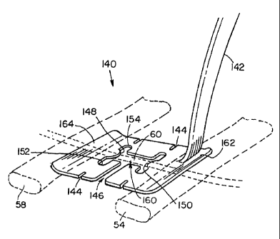
In another preferred embodiment of the invention
illustrated in the perspective view of Figure 9, a
retractor 140 has a handle 142, slots 144 located in the
plane of the aperture 160 to secure the cords, end sections
162, 164 that engage the ribs 54, 58, tabs 148, 150 for
compression of both sides of the artery at the site 60 and
a side opening 146 so that the retractor can be removed.
In this embodiment, the LIMA slides out through
opening 146 during removal of the retractor after
cornpletion of the procedure. This unitary retractor
structure 140 can also include various features described
previously in connection with the embodiment of Figure 1
CA 02286272 1999-10-05
WO 98/48704 PCT/US98/08348
-12-
including the attached or integrated suction tube, the
detachable handle, the irrigation or suction channel with
ports or the mechanically actuated fasteners.
A preferred method of stabilizing tissue during a
coronary bypass procedure 200 is illustrated in the process
flow sequence of Figure 10. A 5-8 cm sized incision is
made over the 4th rib and a section of the 4th costal
cartilage is removed 202. The LIMA is dissected from the
chest wall 204 and divided distally. After blood flow
assessment the LIMA can be temporarily closed with a spring
loaded clip.
A self-retaining wound retractor is used to distract
the edges of the incision and a "trap door" incision is
made in the pericardium and the cut edge sewn to the skin
to pull the pericardial sack and heart anteriorly. The LAD
is exposed and a site suitable for anastomosis is selected
for grafting 206. Tapes are inserted in the myocardium
with blunt needles approximately 1-2 cm apart 208 and the
retractor is inserted 210 with the tapes being pulled
through the aperture and positioned in the lateral sections
thereof. The tapes are connected to the holder 212 to
compress the artery 214 and occlude blood flow on both
sides of the grafting site. The tension in the tapes can
optionally be adjusted during the procedure to minimize
blood loss at the site.
The retractor is secured 216 at the site by
positioning one or both ends under adjoining ribs, or
alternatively, attaching the handle or arm to the wound
retractor or other implement. The grafting site undergoes
CA 02286272 1999-10-05
WO 98/48704 PCTIUS98/08348
-13-
less than 0.1 mm of movement in any direction during this
example procedure.
The site is suctioned or irrigated 218 during
anastomosis, the grafting site is inspected, the tapes are
released from the holders, and the retractor is removed
either by sliding the LIMA through a side opening in the
retractor or detaching a section of the retractor to
accommodate removal of the LIMA from the aperture. After
blood flow is restored, the site is inspected and closed
220.
Although the use of the retractor has been described
in connection with a particular bypass procedure, it can
also be used in other procedures such as bypass operations
involving the diagonal, right or other coronary artery
where movement at the site can interfere with the
procedure.
Alternative embodiments involve opening of the chest
and positioning the retractor at any exposed site on the
heart wall or surrounding areas to immobilize the operative
site. The retractor serves to isolate the site and limits
or stops motion at the site due to respiratory movement of
the lungs or the pumping motion of the heart.
In another preferred embodiment, a stabilizer system
or frame 240 manufactured by Genzyme Surgical Products is
illustrated in Figure 11 to support a surgical retractor
260 in accordance with the invention.
The frame 240 used with the invention includes a bar
242 having an arm 244 extending orthogonally from a first
end and attached to a second arm 246 with a thumb screw at
CA 02286272 1999-10-05
WO 98/48704 PCTIUS98/08348
-14-
a second end. Each arm 244, 246 has a pair of mounting
elements 252, 255 on which a pivot rod 256 can be mounted.
This rod 256 can be rotated 360 degrees to any desired
position such that mounting arm 245 can oriented relative
to the surgical site as needed to position the retractor
260. Each arm 244, 246 has a pair of grippers 248, 250
that engage anatomical features such as neighboring ribs at
the site to stabilize the frame 240.
The mounting arm 245 supports the handle or support
arm 262 with a friction fitting 258 which the user tightens
with knob 268 to grip arm 262 at region 266. The support
arm 262 has a knob 264 at one end that can be turned by the
user to engage a post 276 shown in Figure 12A. A ball on
the post 276 can be slipped through an opening 265 in the
second end of arm 262 and locked into position using knob
264.
The post 276 can be pivoted relative to arm 262 by
loosening the knob 264, thus allowing the user to orient
the retractor 260 at the site for fine positioning. The
post 276 is mounted on a plastic retaining element 270 in
this embodiment. The element 270 can be a transparent or
opaque molded device that can be separated into two
components 272, 274 as described previously. The two
components can be attached by friction fit rods 294 that
are inserted into holes in element 272. Element 270 can be
made with a transparent material to enhance visability at
the site.
Both components have raised holder elements 284, 286.
Element 284 has a pair of slots 288, 289 that each
_.,....____......__._.~. ~.Y - __ _ .. . ... _ ... _ _ _ ~
CA 02286272 1999-10-05
WO 98/48704 PCT/US98/08348
-15-
frictionally grip an end of a cord which extends through
the aperture 278 to attach tissue to the retractor. The
second end of each cord is gripped by corresponding slots
290, 292 in element 286.
Tabs or cord retainers 280, 282 are integrally formed
with component 274 and function as described previously.
In the detailed partial view of Figure 12B, the front
inclined surface can be formed at a shallower angle such
that the top ridge 279 is narrower. This embodiment of
cord retainer 281 affords easier insertion of cords into
the aperture.
This embodiment can also be formed with integral
suction channels or openings in the top surface of the
element 270. A suction tube can be attached through or
with the arm 262 or attached to a suction port on element
270.
CA 02286272 1999-10-05
WO 98/48704 PCTIUS98/08348
-16-
EQUIVALENTS
While the invention has been described in connection
with specific methods and apparatus, it is to be understood
by those skilled in the art that the description is by way
of example and not as a limitation on the scope of the
invention as set forth in the appended claims.
_.,.
_ ~~._ ~.._.a~.. . .