Note: Descriptions are shown in the official language in which they were submitted.
CA 02286397 1999-10-06
WO 98/45332 PCT/US98/06724
HUMANIZED ANTIBODIES AND METHODS FOR
FORMING HUMANIZED ANTIBODIES
FIELD OF THE INVENTION
The present invention is directed at humanized antibodies and methods for
preparing
humanized antibodies. In particular, the present invention is directed at
methods for
preparing humanized antibodies using a monovalent phage display system and
antibody
mutants produced by random mutagenesis of a small set of critical framework
residues made
to a single human framework. More particularly, this invention is directed at
the
humanization of a murine antibody which binds to vascular endothelial growth
factor
(VEGF).
BACKGROUND OF THE INVENTION
Monoclonal antibodies (mAbs) have enormous potential as therapeutic agents,
particularly
when they can be used to regulate defined systems. For example, in some
circumstances it
would be desirable to regulate a system such as angiogenesis, where new blood
capillaries
are formed from the walls of existing small vessels. Angiogenesis is generally
important after
infliction of a wound or infection so that a burst of capillary growth can be
stimulated in the
neighborhood of the damaged tissue. However, angiogenesis is also important in
tumor
growth since, for continued growth, a tumor must induce the formation of a
capillary
network that invades the tumor mass.
Certain growth factors have been identified which regulate angiogenesis. Of
particular
interest is the vascular endothelial growth factor (VEGF), which seems to be
the agent by
which some tumors acquire their rich blood supply. Molecular Biology of the
Cell, 3rd Ed.,
Alberts et al., Garland Publishing, page 1154 (1994). Therefore, mAbs to VEGF,
for
example, can be useful for a variety of reasons, including for use in the
regulation of
angiogenesis and more particularly, as an anti-tumor agent. A murine anti-VEGF
mAb
A4.6.1 which blocks VEGF receptor binding has been previously described. This
antibody
has been shown to inhibit mitogenic signaling. Kim et al., Growth Factors 7,
53 (1992); Kim
et al., Nature 362, 841 (1993).
1
CA 02286397 1999-10-06
WO 98/45332 PCTNS98/06724
Most mAbs including.the anti-VEGF described above are derived from murine or
other
non-human sources which limits clinical efficacy. In particular, the body
often reacts with
an immunogenic response to non-human antibodies whereby the antibody is
rapidly cleared
from the system before any therapeutic effect can occur. In addition to the
immunogenicity
of non-human mAbs invoked when administered to humans, further limitations
arise from
weak recruitment of effector function.
As a means of circumventing these deficiencies, the antigen binding properties
of non-human
mAbs can be conferred to human antibodies through a process known as antibody
"humanization". A humanized antibody contains the amino acid sequence from the
six
complementarily-determining regions (CDRs) (the antigen-binding site of the
antibody
molecule) of the parent or corresponding non-human mAb, grafted onto a human
antibody
framework. Therefore, humanization of non-human antibodies is commonly
referred to as
CDR grafting. The low content of non-human sequence in such humanized
antibodies (-5%)
has proven effective in reducing the immunogenicity and prolonging the serum
half-life of the
antibodies administered to humans. Inter alia, humanized monoclonal antibodies
("chimeric
immunoglobulins") are disclosed in U.S. Patent No. 4,816,567.
Unfortunately, simple grafting of CDR sequences often yields humanized
antibodies which
bind antigen much more weakly than the parent non-human mAb. In order to
restore high
affinity, the antibody must be further engineered to fine-tune the structure
of the antigen
binding loops. This is achieved by replacing key residues in the framework
regions of the
antibody variable domains with the matching sequence from the parent murine
antibody.
These framework residues are usually involved in supporting the conformation
of the CDR
loops, although some framework residues may themselves directly contact the
antigen.
Studies have been conducted which note the importance of certain framework
residues to
CDR conformation and a comprehensive list of all the framework residues which
can affect
antigen binding has been compiled. Chothia et al., J. Mol. Biol. 224, 487
(1992); Foote et
al., J. Mo! Biol. 224, 489 (1992). The comprehensive list includes some thiry
"vernier"
residues which can potentially contribute to CDR structure. Although higher
antigen affinity
would likely result from editing the entire set of vernier residues within a
humanized antibody
so as to match the corresponding parent non-human sequence, this is not
generally desirable
given to
2
CA 02286397 1999-10-06
increased risk of immunogenicity imposed by adding further elements of non-
human
sequence. Thus, from a therapeutic standpoint, it is preferable to confine
framework
changes to the minimum set which affords a high affinity humanized antibody.
Therefore, it is desirable to identify a small set of changes which suffice to
optimize
binding, however, the required changes are expected to differ from one
humanized
antibody to the next. To achieve the desired result, one approach has been to
identify
the proper combination of mutations by constructing a po-e1 of mutants having
"suspect" framework residues replaced by their murine counterpart. These
variants are
each individually formed and tested for antigen and then combined with other
variants
found to have favorable binding affinities. However, this method involves
cycles of
individual site-directed mutagenesis, isolation and screening, and is
therefore
undesirable because it is time consuming and tedious.
As a means of simplifying antibody humanization, a number of different
approaches
have been developed. See, for example, Queen et al., PNAS USA 86, 10029
(1989);
Kettleborough et at, Protein Eng. 4, 773 (1991); Tempest et al., Biotechnology
9, 266
(1991); Padlan, Mol. Immunol. 28, 489 (1991); Roguska et al., PNAS LISA 91,
969
(1994); Studnicka et al., Protein Eng. 7, 805 (1994); Allen et at, J. Immunol.
135. 368
(1985); Carter et al., PNAS USA 89, 4285 (1992); Presta et al., J. Immunol.
151, 2623
(1993); Eigenbrot et at, Proteins 18, 49 (1994); Shalaby et al., J. Exp. Med.
175, 217
(1992); Kabat et al., S~uences ofProteins of Tmmunoiogiral ~nrer i, (5th),
Public
Health Service, NIH, Bethesda, MD (1991); Rosok et al., J. Biol. Chem. 271,
22611
(1996); WO-A-92122653, GB-A-2 268 744, and WO 94,104679.
It is an object of the present invention to provide a general means of rapidly
selecting
framework mutations which improve the binding of humanized antibodies to their
cognate antigens wherein the current methods of framework optimization based
on
cycles of individual site-directed mutagenesis and screening are eliminated.
It is also an object to provide rapid methods of humanizing antibodies which
provide
antibodies with low immunogenecity and which utilize a single human framework
as a
generic scaffold.
3
AMENDED SFtEET
CA 02286397 1999-10-06
WO 98/45332 PCT/US98/06724
It is a further object of the present invention to provide humanized
antibodies which are
mutated to have enhanced affinity for antigen relative to the initial
humanized antibody with
no framework changes.
It is additionally a further object of the present invention to provide
humanized antibodies
that have a reduced clearance rate and hence longer retention within the body
after systemic
administration such that lower doses of the material are available for
systemic administration
for therapeutic effect.
It is also a further object of the present invention to provide humanized
monoclonal
antibodies to VEGF.
SUMMARY OF THE INVENTION
The present invention provides a humanized antibody to vascular endothelial
growth factor
(VEGF). The initial humanized anti-VEGF has a framework derived from consensus
sequences of the most abundant human subclasses, namely VLK subgroup I (VViI)
and VH
subgroup III (VHIII) wherein the CDRs from non-human anti-VEGF are grafted
thereon.
Random mutagenesis of critical framework residues on the initial construct
produced the
humanized anti-VEGF described herein which has 125 fold enhanced affinity for
antigen
relative to the initial humanized antibody with no framework changes. A single
additional
mutation gave a further six fold improvement in binding. This humanized anti-
VEGF can be
reproduced by the method described herein or by traditional recombinant
techniques given
the sequence information provided herein.
Also provided herein is a method for rapidly producing and identifying
framework mutations
which improve the binding of humanized antibodies to their cognate antigens.
In a preferred
embodiment, non-human CDRs are grafted onto a human V,xI- VHIII framework.
Random
mutagenesis of a small set of critical framework residues is also performed
followed by
monovalent display of the resultant library of antibody molecules on the
surface of
filamentous phage. The optimal framework sequences are then identified by
affinity-based
selection. Optionally, the selected antibodies can be further mutated so as to
replace vernier
4
CA 02286397 1999-10-06
WO 98/45332 PCT/US98/06724
residues which sit at the VL-VH interface with residues which match the non-
human parent
antibody.
The methods described herein can be applied to any non-human antibody.
Accordingly,
humanized antibodies are provided by the present invention.
BRIEF DESCRIPTION OF THE DRAWINGS
Figure 1 depicts the amino acid sequences of murine A4.6.1 (SEQ ID NO: 6 and 9
for the
VL and VH domains, respectively), humanized A4.6.1 variant hu2.0, (SEQ ID NO:
7 and 10
for the VL and VH domains, respectively), and humanized A4.6.1 variant hu2. 10
(SEQ ID
NO: 8 and 11 for the VL and VH domains, respectively). Sequence numbering is
according
to Kabat et al., Sequences of Proteins of Immunological Interest, (5th),
Public Health
Service, NIH, Bethesda, MD (1991) and mismatches are indicated by asterisks
(murine
A4.6.1 vs hu2.0) or bullets (hu2.0 vs hu2.10). Variant hu2.0 contains only the
CDR
sequences (bold) from the murine antibody grafted onto a human light chain K
subgroup I,
heavy chain subgroup III framework. Variant hu2.10 is the consensus humanized
clone
obtained from phage sorting experiments described herein.
Figure 2 depicts the framework residues targeted for randomization.
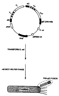
Figure 3 depicts the phagemid construct for surface display of Fab-pIII
fusions on phage. The
phagemid construct encodes a humanized version of the Fab fragment for
antibody A4.6.1
fused to a portion of the M13 gene III coat protein. The fusion protein
consists of the Fab
joined at the carboxyl terminus of the heavy chain to a single glutamine
residue (from
suppression of an amber codon in supE E. coli), then the C-terminal region of
the gene III
protein (residues 249-406). Transformation into F+ E. coli, followed by
superinfection with
M13KO7 helper phage, produces phagemid particles in which a small proportion
of these
display a single copy of the fusion protein.
Detailed Description of the Invention:
A. Definitions
"Antibodies" (Abs) and "immunoglobulins" (Igs) are glycoproteins having the
same structural
5
CA 02286397 1999-10-06
WO 98/45332 PCT/US98/06724
characteristics. While antibodies exhibit binding specificity to a specific
antigen,
immunoglobulins include both antibodies and other antibody-like molecules
which lack
antigen specificity. Polypeptides of the latter kind are, for example,
produced at low levels
by the lymph system and at increased levels by myelomas.
"Native antibodies" and "native immunoglobulins" are usually heterotetrameric
glycoproteins
of about 150,000 daltons, composed of two identical light (L) chains and two
identical heavy
(H) chains. Each light chain is linked to a heavy chain by one covalent
disulfide bond, while
the number of disulfide linkages varies between the heavy chains of different
immunoglobulin
isotypes. Each heavy and light chain also has regularly spaced intrachain
disulfide bridges.
Each heavy chain has at one end a variable domain (VH) followed by a number of
constant
domains. Each light chain has a variable domain at one and (VL) and a constant
domain at
its other end; the constant domain of the light chain is aligned with the
first constant domain
of the heavy chain, and the light chain variable domain is aligned with the
variable domain of
the heavy chain. Particular amino acid residues are believed to form an
interface between the
light and heavy chain variable domains. Clothia et al., J. Mol. Biol. 186, 651
(1985);
Novotny et al., PNAS USA 82, 4592 (1985).
The term "variable" refers to the fact that certain portions of the variable
domains differ
extensively in sequence among antibodies and are used in the binding and
specificity of each
particular antibody for its particular antigen. However, the variability is
not evenly
distributed through the variable domains of antibodies. It is concentrated in
three segments
called "complementarily determining regions" (CDRs) or "hypervariable regions"
both in the
light chain and the heavy chain variable domains. The more highly conserved
portions of
variable domains are called the framework (FR). The variable domains of native
heavy and
light chains each comprise four FR regions, largely adopting a p-sheet
configuration,
connected by three CDRs, which form loops connecting, and in some cases
forming part of,
the a-sheet structure. The CDRs in each chain are held together in close
proximity by the
FR regions and, with the CDRs from the other chain, contribute to the
formation of the
antigen binding site of antibodies. Kabat et al., supra. The constant domains
are not
involved directly in binding an antibody to an antigen, but exhibit various
effector functions,
such as participation of the antibody in antibody-dependent cellular toxicity.
6
CA 02286397 1999-10-06
WO 98/45332 PCT/US98/06724
Papain digestion of antibodies produces two identical antigen binding
fragments, called Fab
fragments, each with a single antigen binding site, and a residual "Fc"
fragment, whose name
reflects its ability to crystallize readily. Pepsin treatment yields an
F(ab')2 fragment that has
= two antigen combining sites and is still capable of cross linking antigen.
"Fv" is the minimum antibody fragment which contains a complete antigen
recognition and
binding site. This region consists of a dimer of one heavy and one light chain
variable domain
in tight, non-covalent association. It is in this configuration that the three
CDRs of each
variable domain interact to define an antigen binding site on the surface of
the VH-VL dimer.
Collectively, the six CDRs confer antigen binding specificity to the antibody.
However, even
a single variable domain (or half of an Fv comprising only three CDRs specific
for an antigen)
has the ability to recognize and bind antigen, although at a lower affinity
than the entire
binding site.
A "Fab" fragment contains the constant domain of the light chain and the first
constant
domain (CHI) of the heavy chain. Fab' fragments differ from Fab fragments by
the addition
of a few residues at the carboxy terminus of the heavy chain CHI domain
including one or
more cysteines from the antibody hinge region. Fab'-SH is the designation
herein for Fab'
in which the cysteine residue(s) of the constant domains bear a free thiol
group. F(ab')2
antibody fragments originally were produced as pairs of Fab' fragments which
have hinge
cysteines between them. Other, chemical couplings of antibody fragments are
also known.
The light chains of antibodies (immunoglobulins) from any vertebrate species
can be assigned
to one of two clearly distinct types, called kappa (ic)and lambda (A), based
on the amino acid
sequences of their constant domains.
Depending on the amino acid sequence of the constant domain of their heavy
chains,
immunoglobulins can be assigned to different classes. There are five major
classes of
immunoglobulins: IgA, IgD, IgE, IgG and IgM, and several of these may be
further divided
into subclasses (isotypes), e.g. IgG-1, IgG-2, IgG-3, and IgG-4; IgA-1 and IgA-
2. The heavy
chain constant domains that correspond to the different classes of
immunoglobulins are called
a, delta, epsilon, y, and, /.c, respectively. The subunit structures and three-
dimensional
7
CA 02286397 1999-10-06
WO 98/45332 PCT/US98/06724
configurations of different classes of immunoglobulins are well known.
The term "antibody" is used in the broadest sense and specifically covers
single monoclonal
antibodies (including agonist and antagonist antibodies), antibody
compositions with
polyepitopic specificity, as well as antibody fragments (e.g., Fab, F(ab')2,
and Fv), so long
as they exhibit the desired biological activity.
The term "monoclonal antibody" as used herein refers to an antibody obtained
from a
population of substantially homogeneous antibodies, i.e., the individual
antibodies comprising
the population are identical except for possible naturally occurring mutations
that may be
present in minor amounts. Monoclonal antibodies are highly specific, being
directed against
a single antigenic site. Furthermore, in contrast to conventional (polyclonal)
antibody
preparations which typically include different antibodies directed against
different
determinants (epitopes), each monoclonal antibody is directed against a single
determinant
on the antigen. In addition to their specificity, the monoclonal antibodies
are advantageous
in that they are synthesized by the hybridoma culture, uncontaminated by other
immunoglobulins. The modifier "monoclonal" indicates the character of the
antibody as
being obtained from a substantially homogeneous population of antibodies, and
is not to be
construed as requiring production of the antibody by any particular method.
For example,
the monoclonal antibodies to be used in accordance with the present invention
may be made
by the hybridoma method first described by Kohler et al., Nature 256, 495
(1975), or may
be made by recombinant DNA methods, see, e.g. U.S. Patent No. 4,816,567.
"Chimeric" antibodies (immunoglobulins) are antibodies wherein a portion of
the heavy
and/or light chain is identical with or homologous to corresponding sequences
in antibodies
derived from a particular species or belonging to a particular antibody class
or subclass, while
the remainder of the chain(s) is identical with or homologous to corresponding
sequences in
antibodies derived from another species or belonging to another antibody class
or subclass,
as well as fragments of such antibodies, so long as they exhibit the desired
biological activity.
U.S. Patent No. 4,816,567.
8
CA 02286397 1999-10-06
WO 98/45332 PCT/US98/06724
"Humanized" forms of non-human (e.g. murine) antibodies are chimeric
immunoglobulins,
immunoglobulin chains or fragments thereof (such as Fv, Fab, Fab', F(at )2 or
other
antigen-binding subsequences of antibodies) which contain minimal sequence
derived from
non-human immunoglobulin. For the most part, humanized antibodies are human
immunoglobulins (recipient antibody) in which residues from a complementary
determining
region (CDR) of the recipient are replaced by residues from a CDR of a non-
human species
(donor antibody) such as mouse, rat or rabbit having the desired specificity,
affinity and
capacity. In some instances, Fv framework residues of the human immunoglobulin
are
replaced by corresponding non-human residues. Furthermore, humanized antibody
may
comprise residues which are found neither in the recipient antibody nor in the
imported CDR
or framework sequences. These modifications are made to further refine and
optimize
antibody performance. In general, the humanized antibody will comprise
substantially all of
at least one, and typically two, variable domains, in which all or
substantially all of the CDR
regions correspond to those of a non-human immunoglobulin and all or
substantially all of
the FR regions are those of a human immunoglobulin consensus sequence. The
humanized
antibody optimally also will comprise at least a portion of an immunoglobulin
constant region
(Fe), typically that of a human immunoglobulin. For further details see: Jones
et al., Nature
321, 522 (1986); Reichmann et al., Nature 332, 323 (1988); and Presta, Curr.
Op. Struct.
Biol. 2, 593 (1992).
"Non-immunogenic in a human" means that upon contacting the humanized antibody
in a
therapeutically effective amount with appropriate tissue of a human, a state
of sensitivity or
resistance to the humanized antibody is not substantially demonstratable upon
administration.
As used herein, "vascular endothelial cell growth factor," or "VEGF," refers
to a mammalian
growth factor as defined in U.S. Patent 5,332,671, including the human amino
acid sequence
of Fig. 1. The biological activity of native VEGF is shared by any analogue or
variant thereof
that is capable of promoting selective growth of vascular endothelial cells
but not of bovine
corneal endothelial cells, lens epithelial cells, adrenal cortex cells, BHK-21
fibroblasts, or
keratinocytes, or that possesses an immune epitope that is immunologically
cross-reactive
with an antibody raised against at least one epitope of the corresponding
native VEGF.
9
CA 02286397 1999-10-06
WO 98/45332 PCT/US98/06724
"Site-directed mutagenesis" is a technique standard in the art, and is
conducted using a
synthetic oligonucleotide primer complementary to a single-stranded phage DNA
to be
mutagenized except for limited mismatching, representing the desired mutation.
Briefly, the
synthetic oligonucleotide is used as a primer to direct synthesis of a strand
complementary
to the phage, and the resulting double-stranded DNA is transformed into a
phage-supporting
host bacterium. Cultures of the transformed bacteria are plated in top agar,
permitting plaque
formation from single cells that harbor the phage. Theoretically, 50% of the
new plaques will
contain the phage having, as a single strand, the mutated form; 50% will have
the original
sequence. The plaques are hybridized with kinased synthetic primer at a
temperature that
permits hybridization of an exact match, but at which the mismatches with the
original strand
are sufficient to prevent hybridization. Plaques that hybridize with the probe
are then
selected and cultured, and the DNA is recovered.
"Expression system" refers to DNA sequences containing a desired coding
sequence and
control sequences in operable linkage, so that hosts transformed with these
sequences are
capable of producing the encoded proteins. To effect transformation, the
expression system
may be included on a vector; however, the relevant DNA may then also be
integrated into
the host chromosome.
As used herein, "cell," "cell line," and "cell culture" are used
interchangeably and all such
designations include progeny. Thus, "transformants" or "transformed cells"
includes the
primary subject cell and cultures derived therefrom without regard for the
number of
transfers. It is also understood that all progeny may not be precisely
identical in DNA
content, due to deliberate or inadvertent mutations. Mutant progeny that have
the same
functionality as screened for in the originally transformed cell are included.
Where distinct
designations are intended, it will be clear from the context.
"Plasmids" are designated by a lower case p preceded and/or followed by
capital letters
and/or numbers. The starting plasmids herein are commercially available, are
publicly
available on an unrestricted basis, or can be constructed from such available
plasmids in
accord with published procedures. In addition, other equivalent plasmids are
known in the
art and will be apparent to the ordinary artisan.
CA 02286397 1999-10-06
WO 98/45332 PCT/US98/06724
"Affinity binding" refers to the strength of the sum total of noncovalent
interactions between
a single antigen-binding site on an antibody and a single epitope. Low-
affinity antibodies bind
antigen weakly and tend to dissociate readily, whereas high-affinity
antibodies bind antigen
= more tightly and remain bound longer.
"Transformation" means introducing DNA into an organism so that the DNA is
replicable,
either as an extrachromosomal element or by chromosomal integration. Depending
on the
host cell used, transformation is done using standard techniques appropriate
to such cells.
The calcium treatment employing calcium chloride, as described by Cohen, Proc.
Nall. Acad.
Sci. USA 69, 2110 (1972) and Mandel et al., J. Mol. Biol. 53, 154 (1970), is
generally used
for prokaryotes or other cells that contain substantial cell-wall barriers.
For mammalian cells
without such cell walls, the calcium phosphate precipitation method of Graham
and van der
Eb, Virology 52, 456 (1978) is preferred. General aspects of mammalian cell
host system
transformations have been described by Axel in U.S. Pat. No. 4,399,216 issued
August 16,
1983. Transformations into yeast are typically carried out according to the
method of Van
Solingen et al., J. Bact. 130, 946 (1977) and Hsiao et al., Proc. Nall. Acad.
Sci. USA 76,
3829 (1979). However, other methods for introducing DNA into cells such as by
nuclear
injection, electroporation or by protoplast fusion may also be used.
"Recovery" or "isolation" of a given fragment of DNA from a restriction digest
means
separation of the digest on polyacrylamide or agarose gel by electrophoresis,
identification
of the fragment of interest by comparison of its mobility versus that of
marker DNA
fragments of known molecular weight, removal of the gel section containing the
desired
fragment, and separation of the gel from DNA. This procedure is known
generally. For
example, see Lawn et al., Nucleic Acids Res. 9, 6103 (1981) and Goeddel et
al., Nucleic
Acids Res. 8, 4057 (1980).
"Ligation" refers to the process of forming phosphodiester bonds between two
double
stranded nucleic acid fragments. Unless otherwise provided, ligation may be
accomplished
using known buffers and conditions with 10 units of T4 DNA ligase ("ligase")
per 0.5 mg of
approximately equimolar amounts of the DNA fragments to be ligated.
11
CA 02286397 1999-10-06
WO 98/45332 PCT/US98/06724
The term "control sequences" refers to DNA sequences necessary for the
expression of an
operably linked coding sequence in a particular host organism. The control
sequences that
are suitable for prokaryotes, for example, include a promoter, optionally an
operator
sequence, a ribosome binding site, and possibly, other as yet poorly
understood sequences.
Eukaryotic cells are known to utilize promoters, polyadenylation signals, and
enhancers.
Nucleic acid is "operably linked" or "operatively linked" when it is placed
into a functional
relationship with another nucleic acid sequence. For example, DNA for a
presequence or a
secretory leader is operably linked to DNA for a polypeptide if it is
expressed as a preprotein
that participates in the secretion of the polypeptide; a promoter or enhancer
is operably linked
to a coding sequence if it affects the transcription of the sequence; or a
ribosome binding site
is operably linked to a coding sequence if it is positioned so as to
facilitate translation.
Generally, "operably linked" or "operatively linked" means that the DNA
sequences being
linked are contiguous and, in the case of a secretory leader, contiguous and
in reading phase.
However, enhancers do not have to be contiguous. Linking is accomplished by
ligation at
convenient restriction sites. If such sites do not exist, then synthetic
oligonucleotide adaptors
or linkers are used in accord with conventional practice.
As used herein, "representatively numbered" refers to a position number of a
residue in a
particular sequence and corresponding position numbers in different sequences.
Corresponding position numbers are those positions within sequences, generally
human
antibody framework sequences, which are functionally equivalent to the
respresentatively
numbered position when used in the construction of a humanized antibody.
Ordinarily, the terms "amino acid" and "amino acids" refer to all naturally
occurring L-a-
amino acids. In some embodiments, however, D-amino acids may be present in the
polypeptides or peptides of the present invention in order to facilitate
conformational
restriction. The amino acids are identified by either the single-letter or
three-letter
designations:
12
CA 02286397 2002-03-20
WO 98/45332 PCT/US98/06724
Asp D aspartic acid He I isoleucine
Thr T threonine Leu L leucine
Ser S serine Tyr Y tyrosine
Glu E glutamic acid Phe F phenylalanine
Pro P proline His H histidine
Gly G glycine Lys K lysine
Ala A alanine Arg R arginine
Cys C cysteine. Tip W tryptophan
Val V val Gin Q glutamine
Met M nt_thionine Asn N asparagine
The term "am no acid sequence variant" refers to molecules with some
differences in their
t
amino acid sequences as compared to a native amino acid sequence-
Substitutional variants are those that have at least one amino acid residue in
a native sequence
removed and a different amino acid inserted in its place at the same position.
The
substitutions may be single, where only one amino acid in the molecule has
been substituted,
or they may be multiple, where two or more amino acids have been substituted
in the same
molecule.
Hybridization is preferably performed under "stringent conditions" which means
(1)
employing low ionic strength and high temperature for washing, for example,
0.015 sodium
chloride/0.0015 M sodium citrate/0,1% sodium dodecyl sulfate at 50 C, or (2)
employing
during hybridization a denaturing agent, such as formamide, for example, 50%
(vol/vol)
formamide with 0.1% bovine serum albumin/0.1% Ficoltt/0.1%
polyvinylpyrrolidone/50 nM
sodium phosphate buffer at pH 6.5 with 750 mM sodium chloride, 75 mM sodium
citrate at
42 C. Another example is use of 500K formamide, 5 x SSC (0.75 M NaCl, 0.075 M
sodium
citrate), 50 mM sodium phosphate (pH 618), 0,1% sodium pyrophosphate, 5 x
Denhardt's
solution, sonicated salmon sperm DNA (50 jig/ml), 0.1 % SbS, and 10% dextran
sulfate at
42 C, with washes at 42 C in 0.2 x SSC and 0,1% SDS. Yet another example is
hybridization using a buffer of 101/6 dextran sulfate, 2 x SSC (sodium
chloride/sodium citrate)
and 50% formamide at 55 C, followed by a high-stringency wash consisting of
0.1 x SSC
*-trademark
13
CA 02286397 1999-10-06
WO 98/45332 PCT/US98/06724
containing EDTA at 55 C. When a nucleic acid sequence of a nucleic acid
molecule is
provided, other nucleic acid molecules hybridizing thereto under the
conditions described
above are considered within the scope of the sequence.
Where amino acid sequences are described it is understood that these sequences
can be
reproduced by reconstructing the amino acid sequence synthetically or by
mutation.
Alternatively, it is understood that recombinant techniques can be used such
that the DNA
encoding the amino acid sequences is recovered. The DNA is recovered by
forming a library
from the DNA encoding the desired amino acid sequences. Probes are then
generated based
on the amino acid sequences. DNA hybridizing to the probes is then isolated
and analyzed
to determine whether the product encoded by the DNA is the desired product.
Generally,
cells are transformed with the DNA (or RNA) and expression studies are
performed.
B. General Methodology for Humanizing Antibodies
The methods described herein can be used to humanize any antibody. Similarly,
it is
understood that the humanized antibody specifically described herein,
humanized anti-VEGF,
can be reproduced by the methods described herein or by traditional DNA
recombinant
techniques. Specifically, since the critical framework residue mutations are
described herein,
the humanized antibody can be reproduced to have the same mutations without
being
reproduced using the monovalent phage display system. Rather, the DNA encoding
the
described amino acid sequences can be synthesized or reproduced by traditional
DNA
recombinant techniques. The DNA product can then be expressed, identified and
recovered.
Alternatively, site-directed mutagenesis can be performed on the antibody by
methods known
in the art, or the antibody can be synthesized so as to have the mutations
described herein.
A particularly preferred method for producing the humanized antibodies
described herein
involves the following: preparing an antibody phagemid vector for monovalent
display of Fab
fragments having CDR sequences transplanted by site-directed mutagenesis onto
a vector
which codes for a human VLKI-CKI light chain and human 'j, III-q ly heavy
chain Fd;
constructing the antibody Fab phagemid library by random mutagenesis of a
small set of
selected critical framework residues; expressing and purifying the humanized
Fab fragments;
selecting humanized Fab variants; and, determining binding affinities. These
steps do not
14
CA 02286397 1999-10-06
WO 98/45332 PCf/US98/06724
have to be performed in any particular order. These steps are specifically
described below
in the "specific example" but are generally performed as follows:
Preparation of antibody phagemid vector for monovalent display of Fab
fragments
First an antibody to be humanized is selected and the complementary
determining regions
(CDRs) identified. The CDR sequences of the antibody can be identified
according to the
sequence definition of Kabat et al., supra. The CDR sequences are transplanted
by
site-directed mutagenesis onto a vector which codes for a human VLKI-Cx, light
chain and
human VHIII-CHIy, heavy chain Fd. The Fab encoding sequence can then be
subcloned into
a phagemid vector. This construct encodes an initial humanized antibody
wherein the
C-terminus of the heavy chain is fused precisely to the carboxyl portion of a
phage coat
protein. Perferably, a phagemid vector is selected which provides expression
of both secreted
heavy chain or heavy chain-gene III fusions in supE suppressor strains of E.
coli.
Constriction of the antibody Fab phagemid library
Based on the cumulative results from humanizing a number of non-human
antibodies onto
a human VLKI-VHIII framework, it was considered that framework changes
required to
optimize antigen binding are limited to some subset of the residues. See,
Carter et al., PNAS
USA 89, 4285 (1992); Presta et al., J. Immnnol. 151, 2623 (1993); Eigenbrot et
al., Proteins
18, 49-62 (1994); Shalaby et al., J. Exp. Med. 175, 217 (1992). Accordingly, a
novel group
of residues was selected for randomization. Randomizing these identified key
framework
residues provides the desired library of Fab variants to be displayed on the
surface of
filamentous phage. Specifically, VL residues 4 and 71 pnd V residues 24,
37,67,69,71,71,75,76,78,93 and 94 have been selected as key framework residues
important
for antigen binding and targeted for randomization.
Expression and purification of humanized Fab fragments
Various methods are known in the art to express and purify fragments. As
described herein,
an E. coli strain 34B8, a nonsuppressor, was transformed with phagemid pMB419,
or
variants thereof Single colonies were grown overnight at 37 C in 5 mL 2YT
containing 50
pg/mL carbenicillin. These cultures were diluted into 200 mL AP5 medium,
described in
Chang et al., Gene 55, 189 (1987), containing 20 pg/mL carbenicillin and
incubated for 26
CA 02286397 1999-10-06
WO 98/45332 PCT/US98/06724
hours at 30 C. The cells were pelleted at 4000 x g and frozen at -20 C for at
least 2 hours.
Cell pellets were then resuspended in 5 mL of 10 mM Tris-HCI (pH 7.6)
containing 1 mM
EDTA, shaken at 4 C for 90 minutes and centrifuged at 10,000 x g for 15
minutes. The
supernatant was applied to a 1 mL streptococcal protein G-SEPHAROSE column (a
column
produced by Pharmacia) and washed with 10 mL of 10 mM MES (pH 5.5). The bound
Fab
fragment was eluted with 2.5 mL 100 mM acetic acid and immediately neutralized
with 0.75
mL IM TrisHCl, pH 8Ø Fab preparations were buffer-exchanged into PBS and
concentrated using CENTRICON-30 concentrators (produced by Amicon). Typical
yields
of Fab were approximately 1 mg/L culture, post-protein G purification.
Purified Fab samples
were characterized by electrospray mass spectrometry, and concentrations were
determined
by amino acid analysis.
Selection of humanized Fab variants
Purified labeled antigen is coated onto a microtiter plate. The coating
solution is discarded,
the wells blocked, and phagemid stock is added. After a period, the wells are
washed and
the bound phage eluted and titered. The remaining phage eluted from the VEGF-
coated well
are propagated for use in the next selection cycle. This process can be
repeated several times
to obtain the desired number of clones. For example, a few dozen individual
clones can be
selected and sequenced.
Determination of VEGF binding affinities
Association and dissociation rate constants for binding of the humanized
variants to VEGF
are measured. Binding profiles are analyzed and those variants showing the
highest affinities
are selected.
Administration of the humanized anti-VEGF
Administration of the humanized anti-VEGF can be extrapolated from the data
presented on
the murine anti-VEGF described in Kim et al., Growth Factors 7, 53 (1992); Kim
et al.,
Nature 362, 841 (1993). In particular, Kim et al. demonstrates that as little
as 10 pg twice
weekly of the VEGF antibody resulted in significant inhibition of tumor
growth. Maximal
effects were achieved with antibody doses of 50-100 g.
16
CA 02286397 1999-10-06
WO 98/45332 PCT/US98/06724
The following example is intended merely to illustrate the best mode now known
for
practicing the invention but the invention is not to be considered as limited
to the details of
this example.
Specific Example I
Construction of the phagemid vector and the initial humanized anti-VEGF
The murine anti-VEGF mAb A4.6.1 has been previously described by Kim et al,
Growth
Factors, 7, 53 (1992); Kim et al., Nature, 362, 841 (1993). The first Fab
variant of
humanized A4.6. 1, hu2.0, was constructed by site-directed mutagenesis using a
deoxyuridine-containing template of plasmid pAK2 which codes for a human VLKI-
Cx, light
chain and human VHIII-CHIYl heavy chain Fd fragment. Carter et al., PNAS USA
89, 4285
(1992). The transplanted A4.6.1 CDR sequences were chosen according to the
sequence
definition of Kabat et al., Sequences of Proteins of Immunological Interest
(5th), Public
Health Service, National Institutes of Health, Bethesda, MD. (1991), except
for CDR-H1
which we extended to encompass both sequence and structural definitions, viz
VH residues
26-35, Chothia et al., J. Mol. Biol. 196, 901 (1987). The Fab encoding
sequence was
subcloned into the phagemid vector phGHamg3. Bass and Wells, Proteins, 8, 309
(1990);
Lowman et al., Biochem. 30, 10832 (1991). This construct, pMB4-19, encodes the
initial
humanized A4.6.1 Fab, hu2.0, with the C-terminus of the heavy chain fused
precisely to the
carboxyl portion of the M13 gene III coat protein. pMB4-19 is similar in
construction to
pDH188, a previously described plasmid for monovalent display of Fab
fragments. Garrard
et al., Biotechn. 9: 1373-1377 (1991). Notable differences between pMB4-19 and
pDHI88
include a shorter M13 gene III segment (codons 249-406) and use of an amber
stop codon
immediately following the antibody heavy chain Fd fragment. This permits
expression of
both secreted heavy chain or heavy chain-gene III fusions in sripE suppressor
strains of E.
coli.
The initial humanized A4.6.1 Fab fragment (hu2.0) in which the CDRs from
A4.6.1 were
grafted onto a human VL,I-V},III framework is shown in Figure 1. The VL domain
of hu2.0
is set forth in SEQ ID NO: 7 and the VH domain of hu2.0 is set forth in SEQ ID
NO: 10.
All residues other than the grafted CDRs were maintained as the human
sequence. Binding
17
CA 02286397 1999-10-06
WO 98/45332 PCT/US98/06724
of this initial humanized antibody to VEGF was so weak as to be undetectable.
Based on
the relative affinity of other weakly-binding humanized A4.6.1 variants (data
not shown), the
K. for binding of hu2.0 was estimated at >7 M. This contrasts with an
affinity of 1.6 nM
for a chimeric Fab construct consisting of the intact VL and VH domains from
murine A4.6.1
and human constant domains. Thus, binding of hu2.0 to VEGF was at least 4000-
fold
reduced relative to the chimera.
Design of the anti-VEGF Fab phagemid library
The group of framework changes required to optimize antigen binding when using
human
VLKI-V},III framework were selected as shown in Table 1 and Figure 2. The
humanized
A4.6.1 phagemid library was constructed by site-directed mutagenesis according
to the
method of Kunkel et al., Methods Enzymol. 204, 125 (1991). A derivative of
pMB4-19
containing TAA stop triplets at V,., codons 24, 37, 67 and 93 was prepared for
use as the
mutagenesis template (all sequence numbering according to Kabat et al., supra.
This
modification was to prevent subsequent background contamination by wild type
sequences.
The codons targeted for randomization were 4 and 71 (light chain) and 24, 37,
67, 69, 71,
73, 75, 76, 78, 93 and 94 (heavy chain).
18
CA 02286397 1999-10-06
WO 98/45332 PCT/US98/06724
Table 1: Key framework residues important for antigen binding and targeted
for randomization
Framework residue Human VKLI, VHIII Murine A4.6.1 Randomization!
consensus residue residue
VL: 4 Met Met Met,Leu
71 Phe Tyr Phe, Tyr
VH: 24 Ala Ala Ala, Val, Thr
37 Val Val Val, Ile
67 Phe Phe Phe, Val, Thr,
Leu, Ile, Ala
69 Ile Phe Ile, Phe
71 Arg Leu Argb, Leub
73 Asp Thr Aspb, Thrb
75 Lys Ala Lysb, Alab
76 Asn Ser Asnb, Serb
78 Leu Ala Leu, Ala, Val,
Phe
93 Ala Ala Ala, Val, Leu,
Ser, Thr
94 Arg Lys Arg, Lys
' Amino acid diversity in phagemid library
b VH 71, 73, 75, 76 randomized to yield the all-murine (L71/T73/A75/S76) or
all-human
(R71/D73/K75/N76) V11III tetrad
A concern in designing the humanized A4.6.1 phagemid library was that residues
targeted
for randomization were widely distributed across the VL and VH sequences.
Limitations in
the length of synthetic oligonucleotides requires that simultaneous
randomization of each of
these framework positions can only be achieved through the use of multiple
oligonucleotides.
However, as the total number of oligonucleotides increases, the efficiency of
mutagenesis
decreases (i.e. the proportion of mutants obtained which incorporate sequence
derived from
all of the mutagenic oligonucleotides). To circumvent this problem, two
features were
incorporated into the library construction. The first was to prepare four
different
mutagenesis templates coding for each of the possible VL framework
combinations. This was
simple to do given the limited diversity of the light chain framework (only 4
different
sequences), but was beneficial in that it eliminated the need for two
oligonucleotides from
the mutagenesis strategy. Secondly, two 126 base oligonucleotides were
preassembled from
smaller synthetic fragments. This made possible randomization of V11 codons
67, 69, 71, 73,
19
CA 02286397 1999-10-06
WO 98/45332 PCT/US98/06724
75, 76, 93 and 94 with a single long oligonucleotide, rather than two smaller
ones. The final
randomization mutagenesis strategy therefore employed only two
oligonucleotides
simultaneously onto four different templates.
More specifically, in order to randomize heavy chain codons 67, 69, 71, 73,
75, 76, 78, 93
and 94 with a single mutagenic oligonucleotide, two 126-mer oligonucleotides
were first
preassembled from 60 and 66-mer fragments by template-assisted enzymatic
ligation.
Specifically, 1.5 nmol of 5' phosphorylated oligonucleotide GAT TTC AAA CGT
CGT NYT
ACT VV7T TCT AGA GAC AAC TCC AAA AAC ACA B TY TAC CTG CAG ATG AAC
(SEQ ID NO: 12) or GAT TTC AAA CGT CGT NYT ACT WTT TCT T TA GAC ACC
TCC GCA AGC ACA B YT TAC CTG CAG ATG AAC (SEQ ID NO: 1) were combined
with 1.5 nmol of AGC CTG CGC GCT GAG GAC ACT GCC GTC TAT TAC TGT DYA
ARG TAC CCC CAC TAT TAT GGG (SEQ ID NO: 2). The randomized codons are
underlined and N represents A/G/T/C; W represents A/T; B represents G/T/C; D
represents
G/A/T; R represents A/G; and Y represents C/T ("/" represents "or"). Then, 1.5
nmol of
template oligonucleotide CTC AGC GCG CAG GCT GTT CAT CTG CAG GTA (SEQ ID
NO: 3), with complementary sequence to the 5' ends of SEQ ID NOS: 12 and I and
the 3'
end of SEQ ID NO: 3 was added to hybridize to each end of the ligation
junction. To this
mixture, Taq ligase (thermostable ligase from New England Biolabs) and buffer
were added,
and the reaction mixture was subjected to 40 rounds of thermal cycling, (95'C
for 1.25
minutes and 50 C for 5 minutes) so as to cycle the template oligonucleotide
between ligated
and unligated junctions. The product 126-mer oligonucleotides were purified on
a 6%
urea/TBE polyacrylamide gel and extracted from the polyacrylamide in buffer.
The two
126-mer products were combined in equal ratio, ethanol precipitated and
finally solubilized
in 10 mM Tris-HCI,1 mM EDTA. The mixed 126-mer oligonucleotide product was
labeled
504-01.
Randomization of select framework codons (VL 4, 71; VH 24, 37, 67, 69, 71, 73,
75, 76, 93,
94) was thus effected in two steps. First, VL randomization was achieved by
preparing three
additional derivatives of the modified pMB4-19 template. Framework codons 4
and 71 in
the light chain were replaced individually or pairwise using the two mutagenic
oligonucleotides GCT GAT ATC CAG TTG ACC CAG TCC CCG (SEQ ID NO: 13) and
CA 02286397 1999-10-06
WO 98/45332 PCTIUS98/06724
TCT GGG ACG GAT JAC ACT CTG ACC ATC (SEQ ID NO: 4). Deoxyuridine
containing template was prepared from each of these new derivatives. Together
with the
original template, these four constructs coded for each of the four possible
light chain
framework sequence combinations (see Table 1).
Oligonucleotides 504-01, the mixture of two 126-mer oligonucleotides, and CGT
TTG TCC
TGT GCA RYT TCT GGC TAT ACC TTC ACC AAC TAT GGT ATG AAC TGG RTC
CGT CAG GCC CCG GGT AAG (SEQ ID NO: 5) were used to randomize heavy chain
framework codons using each of the four templates just described. The four
libraries were
electroporated into E. coli XL-l BLUE CELLS (marker cells produced by
Stratagene) and
combined. The total number of independent transformants was estimated at >1.2
x 108,
approximately 1,500-fold greater than the maximum number of DNA sequences in
the library.
From this strategy, each of residues 4 and 71 in the light chain and 24, 37,
67, 78 and 93
from the heavy chain were partially randomized to allow the selection of
either the murine
A4.6. 1, human VLKI-Vt,III sequence, or sequences commonly found in other
human and
murine frameworks (Table I). Note that randomization of these residues was not
confined
to a choice between the human VLKI-VHIII consensus or A4.6.1 framework
sequences.
Rather, inclusion of additional amino acids commonly found in other human and
murine
framework sequences allows for the possibility that additional diversity may
lead to the
selection of tighter binding variants.
Some of the heavy chain framework residues were randomized in a binary fashion
according
to the human VHIII and murine A4.6.1 framework sequences. Residues V. 71, 73,
75 and
76 are positioned in a hairpin loop adjacent to the antigen binding site. The
side chains of VH
71 and 73 are largely buried in canonical antibody structures and their
potential role in
shaping the conformation of CDR-H2 and CDR-H3 is well known. Kettleborough et
al.,
Protein Eng. 4, 773 (1991); Carter et al., PNAS USA 89, 4285 (1992); Shalaby
et al., J.
Exp. Med. 175, 217 (1992). On the other hand, although the side chains of VH
75 and 76
are solvent exposed (Figure 2), it has nevertheless been observed that these
two residues can
also influence antigen binding (Eigenbrot, Proteins 18, 49 [1994)), presumably
due to direct
antigen contact in some antibody-antigen complexes. Because of their proximity
in sequence
21
CA 02286397 1999-10-06
WO 98/45332 PCT/US98/06724
and possible interdependence, V,., 71, 73, 75 and 76 were randomized en bloc
such that only
two possible combinations of this tetrad could be selected; either all human
VHIII or all
murine A4.6.1 sequence. Finally, VH residues 69 and 94 were randomized, but
only to
represent the VHIII and A4.6.1 sequences. The V}, 69 and 94 were not replaced
in previous
antibody humanizations, but because they differ between the V11III consensus
and A4.6.1
sequences (Figure 1) and have been noted as potentially important for proper
CDR
conformation (Foote et al., J. Mol. Biol. 224, 487 [1992]), they were included
in this
randomization strategy.
Humanized A4.6.1 Fab library displayed on the surface of phagemid
A variety of systems have been developed for the functional display of
antibody fragments
on the surface of filamentous phage. Winter et al., Ann. Rev. Immnmol. 12, 433
(1994).
These include the display of Fab or single chain Fv (scFv) fragments as
fusions to either the
gene III or gene VIII coat proteins of M13 bacteriophage. The system selected
herein is
similar to that described by Garrard et al., Biotechn. 9, 13 73 (1991) in
which a Fab fragment
is monovalently displayed as a gene III fusion (Figure 3). This system has two
notable
features. In particular, unlike scFvs, Fab fragments have no tendency to form
dimeric
species, the presence of which can prevent selection of the tightest binders
due to avidity
effects. Additionally, the monovalency of the displayed protein eliminates a
second potential
source of avidity effects that would otherwise result from the presence of
multiple copies of
a protein on each phagemid particle. Bass and Wells, Proteins 8, 309 (1990);
Lowman et
al., Biochemistry 30, 10832 (1991).
Phagemid particles displaying the humanized A4.6.1 Fab fragments were
propagated in E.
coli XL-1 Blue cells. Briefly, cells harboring the randomized pMB4-19
construct were
grown overnight at 37 C in 25 mL 2YT medium containing 50 pg/mL carbenicillin
and
approximately 1010 M13KO7 helper phage (Viera and Messing, Methods Enzymol.
153, 3
[1987]). Phagemid stocks were purified from culture supernatants by
precipitation with a
saline polyethylene glycol solution, and resuspended in 100 L PBS
(approximately 1014
phagemid/mL).
22
CA 02286397 1999-10-06
WO 98/45332 PCTIUS98/06724
Selection of humanized A4.6.1 Fab variants
Purified VEGF121 (100 L at 10 gg/mL in PBS) was coated onto a microtiter
plate well
overnight at 4 C. The coating solution was discarded and this well and an
uncoated well
were blocked with 6% skim milk for 1 hour and washed with PBS containing 0.05%
TWEEN-20 (detergent). Then, 10 gL of phagemid stock, diluted to 100 gL with 20
mM Tris
(pH 7.5) containing 0.1% BSA and 0.05% TWEEN-20, was added to each well. After
2
hours, the wells were washed and the bound phage eluted with 100 gL of 0.1 M
glycine (pH
2.0), and neutralized with 25 gL of IM Tris pH 8Ø An aliquot of this was
used to titer the
number of phage eluted. The remaining phage eluted from the VEGF-coated well
were
propagated for use in the next selection cycle. A total of 8 rounds of
selection was performed
after which time 20 individual clones were selected and sequenced (Sanger et
al., PNAS USA
74, 5463 [1977]).
Variants from the humanized A4.6.1 Fab phagemid library were thusly selected
based on
binding to VEGF. Enrichment of functional phagemid, as measured by comparing
titers for
phage eluted from a VEGF-coated versus uncoated microtiter plate well,
increased up to the
seventh round of affinity panning. After one additional round of sorting, 20
clones were
sequenced to identify preferred framework residues selected at each position
randomized.
These results, summarized in Table 2, revealed strong consensus amongst the
clones selected.
Ten out of the twenty clones had the identical DNA sequence, designated hu2.
10. Of the
thirteen framework positions randomized, eight substitutions were selected in
hu2. 10 (VL 71;
VH 37, 71, 73, 75, 76, 78 and 94). Interestingly, residues VH 37 (Ile) and 78
(Val) were
selected neither as the human VI1III or murine A4.6.1 sequence. This result
suggests that
some framework positions may benefit from extending the diversity beyond the
target human
and parent murine framework sequences.
23
CA 02286397 1999-10-06
WO 98/45332 PCT/US98/06724
Table 2: Sequences selected from the humanized A4.6.1 phagemid Fab library
Variant Residue substitutions
VL VH
4 71 24 37 67 69 71 73 75 76 78 93 94
murine A4.6.1 M Y A V F F L T A S A A K
hu2.0 (CDR-graft) M F A V F I R N K N L A R
Phage-selected
clones:
hu2.1 (2) - Y - I - - - - - - V - K
hu2.2 (2) L Y - I - - - - - - V - K
hu2.6 (1) L - - I T - L T A S V - K
hu2.7(1) L - - I T - - - - - V - K
hu2.10 (10) - Y - I - - L T A S V - K
Differences between hu2.0 and murine A4.6.1 antibodies are underlined. The
number of
identical clones identified for each phage-selected sequence is indicated in
parentheses.
Dashes in the sequences of phage-selected clones indicate selection of the
human VLKI-VHIII
framework sequence (i.e. as in hu2.0).
There were. four other unique amino acid sequences among the remaining ten
clones
analyzed: hu2.1, hu2.2, hu2.6 and hu2.7. All of these clones, in addition to
hu2.10,
contained identical framework substitutions at positions Võ 37 (Ile), 78 (Val)
and 94 (Lys),
but retained the human V11III consensus sequence at positions 24 and 93. Four
clones had
lost the light chain coding sequence and did not bind VEGF when tested in a
phage ELISA
assay (Cunningham et al., EMBO J. 13, 2508 [1994]). We have occasionally noted
the loss
of heavy or light chain sequence with other Fab phagemid libraries
(unpublished data), and
these clones are presumably selected for on the basis of enhanced expression.
Such artifacts
can often be minimized by reducing the number of sorting cycles or by
propagating libraries
on solid media.
24
CA 02286397 2002-03-20
WO 98/45332 PCT/US96/06724
Aeterminatign of VEGF indiniZ affinities
Association (kaõ) and dissociation (koa) rate constants for binding of
humanized A4.6.1 Fab
variants to VEGF1, were measured by surface plasmon resonance (Karlsson et al,
J. Immun.
Methods 145, 229 [1991]) on a Pharmacia BIAcoreInstrument. VEGF,=, was
covalently
S immobilized on the biosensor chip via primary amino groups. Binding of
humanized A4.6.i
Fab variants was measured by flowing solutions of Fab in PBS/0.05% TWEEN-20
(detergent) over the chip at a flow rate of 20 liL/min Following each binding
measurement,
residual Fab was stripped from the immobilized ligand by washing with 5 L of
50 mM
aqueous HCI at S pUmin. Binding profiles were analyzed by nonlinear regression
using a
simple monovalent binding model (BlAevaluation software v2.0; Pharmacia).
Phage-selected variants hu2.1, hu2.2, hu2.6, hu2.7 and hu2.10 were expressed
in E. coil
using shake flasks and Fab fragments were purified from periplasmic extracts
by protein G
affinity chromatography. Recovered yields of Fab for these five clones ranged
from 0.2
(hu2.6) to 1.7 mg/L (hu2_ i)_ The affinity of each of these variants for
antigen (VEGF)
measured by surface plasmon resonance on a BlAcore instrument as shown in
Table 3.
Table 3 VE F binding affinity of humanized A4.6.1 Fab variants.
Variant ko,T K[) ED (A4.6.1)
Ko (mut)
WS-1/101 101s' nM
A4.6.1 chimera 5.4 0.85 l.f
hu2.0 ND ND >7000** >4000
Fhage selected
clones:
hu2.1 0.70 18 260 170
hu2.2 0.47 16 340 210
hu2.6 0.67 4.5 67 40
hu2.7 0.67 24 360 230
hu2.10 0.63 3.5 55 35
*hu2.1OV 2,0 1.8 9.3 5.8
*hu2 IOV = hu2.10 with mutation VL Leu46 -> Val; Estimated errors in the
Biacore binding
measurements are +/- 25%; **Too weak to measure, estimate of lower bound
*-trademark
CA 02286397 1999-10-06
WO 98/45332 PCT/US98/06724
Analysis of this binding data revealed that the consensus clone hu2. 10
possessed the highest
affinity for VEGF out of the five variants tested. Thus our Fab phagemid
library was
selectively enriched for the tightest binding clone. The calculated KD for
hu2.10 was 55 nM,
at least 125-fold tighter than for hu2.0- which contains no framework changes
(KD >7 M).
The other four selected variants all exhibited weaker binding to VEGF, ranging
down to a
KD of 360 nM for the weakest (hu2.7). Interestingly, the KD for hu2.6, 67 nM,
was only
marginally weaker than that of hu2. 10 and yet only one copy of this clone was
found among
20 clones sequenced. This may have due to a lower level of expression and
display, as was
the case when expressing the soluble Fab of this variant. However, despite the
lower
expression rate, this variant is useful as a humanized antibody.
Additional improvement of humanized variant hu2.10
Despite the large improvement in antigen affinity over the initial humanized
variant, binding
of hu2. 10 to VEGF was still 35-fold weaker than a chimeric Fab fragment
containing the
murine A4.6.1 VL and V,Idomains. This considerable difference suggested that
further
optimization of the humanized framework might be possible through additional
mutations.
Of the vernier residues identified by Foote et al., J. Mol. Biol. 196, 901
(1992), only residues
VL 46, V}, 2 and VH 48 differed in the A4.6.1 versus human V,iI-V},III
framework (Figure
1) but were not randomized in our phagemid library. A molecular model of the
humanized
A4.6.1 Fv fragment showed that VL 46 sits at the VL-VH interface and could
influence the
conformation of CDRH3. Furthermore, this amino acid is almost always leucine
in most VLK
frameworks (Kabat et al., supra.), but is valine in A4.6.1. Accordingly, a Leu
-> Val
substitution was made at this position in the background of hu2. 10. Analysis
of binding
kinetics for this new variant, hu2. I OV, indicated a further 6-fold
improvement in the KD for
VEGF binding. The KD for hu2. I OV (9.3 nM) was thus within 6-fold that of the
chimera.
In contrast to VL 46, no improvement in the binding affinity of hu2.10 was
observed for
replacement of either VH 2 or Võ 48 with the corresponding residue from murine
A4.6. 1.
Interestingly, part of the improvement prior to the last change in affinity
was due to an
increase in the association rate constant (koõ ), suggesting that \( 46 may
play a role in
preorganizing the antibody structure into a conformation more suitable for
antigen binding.
Other mutations which affected antigen affinity were primarily due to changes
in the
26
CA 02286397 2002-03-20
WO 98/45332 PC fUS98106724
dissociation rate constant (k,,) for binding. Comparison of hu2. I and hu2.10
reveals a 5-fold
improvement in affinity for substitution of Vn residues 71, 73, 75, 76 with
the A4.6.1
sequence. Conversion of VL- '71 to the A4.6.1 sequence (Phe -> Tyr) had
negligible effect
on binding (hu2.2 vs hu2.7), while variants with leucine at VL 4 bound
marginally worse
(<2-fold) than those with methionine, the naturally occurring residue in both
the A4.6.1 and
human VKLI frameworks (hu22 vs hu2.1). Comparison of other humanized A4.6.1
variants
not shown here revealed that the Võ 94 Arg -> Lys change resulted in a 5-fold
improvement
in K0, either due to direct antigen contact by this residue, or to a
structural role in
maintaining the proper conformation of CDR-H3_ Variant hu2.6 has three
sequence
differences relative to the consensus clone hu2. 10, but nevertheless has a
similar KO, thereby
suggesting that these three substitutions have little effect on antigen
binding. The negligible
effect of conservative changes at V1 4 and 71 concurs with binding data for
other variants,
yet the change at V, 67 (Phe -> Thr) had little effect on binding.
Concluding Remarks
The foregoing description details specific methods which can be employed to
practice the
present invention. Having detailed such specific methods, those skilled in the
art will well
enough know how to devise alternative reliable methods at arriving at the same
information
by using the fruits of the present invention. Thus, however detailed the
foregoing may appear
in text, it should not be construed as limiting the overall scope thereof;
rather, the ambit of
the present invention is to be determined only by the lawful construction of
the appended
claims.
27
CA 02286397 1999-10-06
WO 98/45332 PCT/US98/06724
SEQUENCE LISTING
(1) GENERAL INFORMATION:
(i) APPLICANT: Genentech, Inc.
(ii) TITLE OF INVENTION: HUMANIZED ANTIBODIES AND METHODS FOR
FORMING HUMANIZED ANTIBODIES
(iii) NUMBER OF SEQUENCES: 14
(iv) CORRESPONDENCE ADDRESS:
(A) ADDRESSEE: Flehr, Hohbach, Test, Albritton & Herbert
(B) STREET: Four Embarcadero Center, Suite 3400
(C) CITY: San Francisco
(D) STATE: California
(E) COUNTRY: United States
(F) ZIP: 94111
(v) COMPUTER READABLE FORM:
(A) MEDIUM TYPE: Floppy disk
(B) COMPUTER: IBM PC compatible
(C) OPERATING SYSTEM: PC-DOS/MS-DOS
(D) SOFTWARE: Patentln Release #1.0, Version #1.30
(vi) CURRENT APPLICATION DATA:
(A) APPLICATION NUMBER: PCT HEREWITH
(B) FILING DATE: 02-APR-1998
(C) CLASSIFICATION:
(vii) PRIOR APPLICATION DATA:
(A) APPLICATION NUMBER:08/833,504
(B) FILING DATE: 07-APR-1997
(viii) ATTORNEY/AGENT INFORMATION:
(A) NAME: Dreger, Walter H.
(B) REGISTRATION NUMBER: 24,190
(C) REFERENCE/DOCKET NUMBER: A-64254
(ix) TELECOMMUNICATION INFORMATION:
(A) TELEPHONE: (415) 781-1989
(B) TELEFAX: (415) 398-3249
(2) INFORMATION FOR SEQ ID NO:1:
(i) SEQUENCE CHARACTERISTICS:
(A) LENGTH: 66 base pairs
(B) TYPE: nucleic acid
(C) STRANDEDNESS: unknown
(D) TOPOLOGY: unknown
(ii) MOLECULE TYPE: DNA (genomic)
(xi) SEQUENCE DESCRIPTION: SEQ ID NO:1:
GATTTCAAAC GTCGTNYTAC TWTTTCTTTA GACACCTCCG CAAGCACABY TTACCTGCAG 60
ATGAAC 66
28
CA 02286397 1999-10-06
WO 98/45332 PCT/US98/06724
(2) INFORMATION FOR SEQ ID NO:2:
(i) SEQUENCE CHARACTERISTICS:
(A) LENGTH: 60 base pairs
(B) TYPE: nucleic acid
(C) STRANDEDNESS: unknown
(D) TOPOLOGY: unknown
(ii) MOLECULE TYPE: DNA (genomic)
(xi) SEQUENCE DESCRIPTION: SEQ ID NO:2:
AGCCTGCGCG CTGAGGACAC TGCCGTCTAT TACTGTDYAA RGTACCCCCA CTATTATGGG 60
(2) INFORMATION FOR SEQ ID NO:3:
(i) SEQUENCE CHARACTERISTICS:
(A) LENGTH: 30 base pairs
(B) TYPE: nucleic acid
(C) STRANDEDNESS: unknown
(D) TOPOLOGY: unknown
(ii) MOLECULE TYPE: DNA (genomic)
(xi) SEQUENCE DESCRIPTION: SEQ ID NO:3:
CTCAGCGCGC AGGCTGTTCA TCTGCAGGTA 30
(2) INFORMATION FOR SEQ ID NO:4:
(i) SEQUENCE CHARACTERISTICS:
(A) LENGTH: 27 base pairs
(B) TYPE: nucleic acid
(C) STRANDEDNESS: unknown
(D) TOPOLOGY: unknown
(ii) MOLECULE TYPE: DNA (genomic)
(xi) SEQUENCE DESCRIPTION: SEQ ID NO:4:
TCTGGGACGG ATTACACTCT GACCATC 27
(2) INFORMATION FOR SEQ ID NO:5:
(i) SEQUENCE CHARACTERISTICS:
(A) LENGTH: 75 base pairs
(B) TYPE: nucleic acid
(C) STRANDEDNESS: unknown
(D) TOPOLOGY: unknown
(ii) MOLECULE TYPE: DNA (genomic)
(xi) SEQUENCE DESCRIPTION: SEQ ID NO:5:
CGTTTGTCCT GTGCARYTTC TGGCTATACC TTCACCAACT ATGGTATGAA CTGGRTCCGT 60
CAGGCCCCGG GTAAG 75
29
CA 02286397 1999-10-06
WO 98/45332 PCT/US98/06724
(2) INFORMATION FOR SEQ ID NO:6:
(i) SEQUENCE CHARACTERISTICS:
(A) LENGTH: 107 amino acids
(B) TYPE: amino acid
(C) STRANDEDNESS: unknown
(D) TOPOLOGY: unknown
(ii) MOLECULE TYPE: protein
(xi) SEQUENCE DESCRIPTION: SEQ ID NO:6:
Asp Ile Gln Met Thr Gln Thr Thr Ser Ser Leu Ser Ala Ser Leu Gly
1 5 10 15
Asp Arg Val Ile Ile Ser Cys Ser Ala Ser Gln Asp Ile Ser Asn Tyr
25 30
Leu Asn Trp Tyr Gln Gln Lys Pro Asp Gly Thr Val Lys Val Leu Ile
20 35 40 45
Tyr Phe Thr Ser Ser Leu His Ser Gly Val Pro Ser Arg Phe Ser Gly
50 55 60
Ser Gly Ser Gly Thr Asp Tyr Ser Leu Thr Ile Ser Asn Leu Glu Pro
65 70 75 80
Glu Asp Ile Ala Thr Tyr Tyr Cys Gln Gln Tyr Ser Thr Val Pro Trp
85 90 95
Thr Phe Gly Gly Gly Thr Lys Leu Glu Ile Lys
100 105
(2) INFORMATION FOR SEQ ID NO:7:
(i) SEQUENCE CHARACTERISTICS:
(A) LENGTH: 107 amino acids
(B) TYPE: amino acid
(C) STRANDEDNESS: unknown
(D) TOPOLOGY: unknown
(ii) MOLECULE TYPE: protein
(xi) SEQUENCE DESCRIPTION: SEQ ID NO:7:
Asp Ile Gln Met Thr Gln Ser Pro Ser Ser Leu Ser Ala Ser Val Gly
1 5 10 15
Asp Arg Val Thr Ile Thr Cys Ser Ala Ser Gln Asp Ile Ser Asn Tyr
20 25 30
Leu Asn Trp Tyr Gln Gln Lys Pro Gly Lys Ala Pro Lys Leu Leu Ile
35 40 45
Tyr Phe Thr Ser Ser Leu His Ser Gly Val Pro Ser Arg Phe Ser Gly
50 55 60
Ser Gly Ser Gly Thr Asp Phe Thr Leu Thr Ile Ser Ser Leu Gln Pro
65 70 75 80
Glu Asp Phe Ala Thr Tyr Tyr Cys Gin Gin Tyr Ser Thr Val Pro Trp
85 90 95
CA 02286397 1999-10-06
WO 98/45332 PCT/US98/06724
Thr Phe Gly Gln Gly Thr Lys Val Glu Ile Lys
100 105
(2) INFORMATION FOR SEQ ID NO:8:
(i) SEQUENCE CHARACTERISTICS:
(A) LENGTH: 107 amino acids
(B) TYPE: amino acid
(C) STRANDEDNESS: unknown
(D) TOPOLOGY: unknown
(ii) MOLECULE TYPE: protein
(xi) SEQUENCE DESCRIPTION: SEQ ID NO:8:
Asp Ile Gln Met Thr Gln Ser Pro Ser Ser Leu Ser Ala Ser Val Gly
1 5 10 15
Asp Arg Val Thr Ile Thr Cys Ser Ala Ser Gln Asp Ile Ser Asn Tyr
20 25 30
Leu Asn Trp Tyr Gln Gln Lys Pro Gly Lys Ala Pro Lys Leu Leu Ile
35 40 45
Tyr Phe Thr Ser Ser Leu His Ser Gly Val Pro Ser Arg Phe Ser Gly
50 55 60
Ser Gly Ser Gly Thr Asp Tyr Thr Leu Thr Ile Ser Ser Leu Gln Pro
65 70 75 80
Glu Asp Phe Ala Thr Tyr Tyr Cys Gln Gln Tyr Ser Thr Val Pro Trp
85 90 95
Thr Phe Gly Gln Gly Thr Lys Val Glu Ile Lys
100 105
(2) INFORMATION FOR SEQ ID NO:9:
(i) SEQUENCE CHARACTERISTICS:
(A) LENGTH: 123 amino acids
(B) TYPE: amino acid
(C) STRANDEDNESS: unknown
(D) TOPOLOGY: unknown
(ii) MOLECULE TYPE: protein-
(xi) SEQUENCE DESCRIPTION: SEQ ID NO:9:
Glu Ile Gin Leu Val Gln Ser Gly Pro Glu Leu Lys Gln Pro Gly Glu
1 5 10 15
Thr Val Arg Ile Ser Cys Lys Ala Ser Gly Tyr Thr Phe Thr Asn Tyr
20 25 30
Gly Met Asn Trp Val Lys Gln Ala Pro Gly Lys Gly Leu Lys Trp Met
35 40 45
Gly Trp Ile Asn Thr Tyr Thr Gly Glu Pro Thr Tyr Ala Ala Asp Phe
50 55 60
Lys Arg Arg Phe Thr Phe Ser Leu Glu Thr Ser Ala Ser Thr Ala Tyr
70 75 80
31
CA 02286397 1999-10-06
WO 98/45332 PCT/US98/06724
Leu Gln Ile Ser Asn Leu Lys Asn Asp Asp Thr Ala Thr Tyr Phe Cys
85 90 95
Ala Lys Tyr Pro His Tyr Tyr Gly Ser Ser His Trp Tyr Phe Asp Val
100 105 110
Trp Gly Ala Gly Thr Thr Val Thr Val Ser Ser
115 120
(2) INFORMATION FOR SEQ ID NO:10:
(i) SEQUENCE CHARACTERISTICS:
(A) LENGTH: 123 amino acids
(B) TYPE: amino acid
(C) STRANDEDNESS: unknown
(D) TOPOLOGY: unknown
(ii) MOLECULE TYPE: protein
(xi) SEQUENCE DESCRIPTION: SEQ ID NO:10:
Glu Val Gln Leu Val Glu Ser Gly Gly Gly Leu Val Gln Pro Gly Gly
1 5 10 15
Ser Leu Arg Leu Ser Cys Ala Ala Ser Gly Tyr Thr Phe Thr Asn Tyr
20 25 30
Gly Met Asn Trp Val Arg Gln Ala Pro Gly Lys Gly Leu Glu Trp Val
35 40 45
Gly Trp Ile Asn Thr Tyr Thr Gly Glu Pro Thr Tyr Ala Ala Asp Phe
50 55 60
Lys Arg Arg Phe Thr Ile Ser Arg Asp Asn Ser Lys Asn Thr Leu Tyr
65 70 75 80
Leu Gln Met Asn Ser Leu Arg Ala Glu Asp Thr Ala Val Tyr Tyr Cys
85 90 95
Ala Arg Tyr Pro His Tyr Tyr Gly Ser Ser His Trp Tyr Phe Asp Val
100 105 110
Trp Gly Gln Gly Thr Leu Val Thr Val Ser Ser
115 120
(2) INFORMATION FOR SEQ ID NO:11:
(i) SEQUENCE CHARACTERISTICS:
(A) LENGTH: 123 amino acids
(B) TYPE: amino acid
(C) STRANDEDNESS: unknown
(D) TOPOLOGY: unknown
(ii) MOLECULE TYPE: protein
(xi) SEQUENCE DESCRIPTION: SEQ ID NO:11:
Glu Val Gin Leu Val Glu Ser Gly Gly Gly Leu Val Gln Pro Gly Gly
1 5 10 15
Ser Leu Arg Leu Ser Cys Ala Ala Ser Gly Tyr Thr Phe Thr Asn Tyr
20 25 30
32
CA 02286397 1999-10-06
WO 98/45332 PCTIUS98/06724
Gly Met Asn Trp Ile Arg Gln Ala Pro Gly Lys Gly Leu Glu Trp Val
35 40 45
Gly Trp Ile Asn Thr Tyr Thr Gly Glu Pro Thr Tyr Ala Ala Asp Phe
50 55 60
Lys Arg Arg Phe Thr Ile Ser Leu Asp Thr Ser Ala Ser Thr Val Tyr
65 70 75 80
Leu Gln Met Asn Ser Leu Arg Ala Glu Asp Thr Ala Val Tyr Tyr Cys
85 90 95
Ala Lys Tyr Pro His Tyr Tyr Gly Ser Ser His Trp Tyr Phe Asp Val
100 105 110
Trp Gly Gln Gly Thr Ser Val Thr Val Ser Ser
115 120
(2) INFORMATION FOR SEQ ID NO:12:
(i) SEQUENCE CHARACTERISTICS:
(A) LENGTH: 66 base pairs
(B) TYPE: nucleic acid
(C) STRANDEDNESS: unknown
(D) TOPOLOGY: unknown
(ii) MOLECULE TYPE: DNA (genomic)
(xi) SEQUENCE DESCRIPTION: SEQ ID NO:12:
GATTTCAAAC GTCGTNYTAC TWTTTCTAGA GACAACTCCA AAAACACABY TTACCTGCAG 60
ATGAAC 66
(2) INFORMATION FOR SEQ ID NO:13:
(i) SEQUENCE CHARACTERISTICS:
(A) LENGTH: 27 base pairs
(B) TYPE: nucleic acid
(C) STRANDEDNESS: unknown
(D) TOPOLOGY: unknown
(ii) MOLECULE TYPE: DNA (genomic)
(xi) SEQUENCE DESCRIPTION: SEQ ID NO:13:
GCTGATATCC AGTTGACCCA GTCCCCG 27
(2) INFORMATION FOR SEQ ID NO:14:
(i) SEQUENCE CHARACTERISTICS:
(A) LENGTH: 6072 base pairs
(B) TYPE: nucleic acid
(C) STRANDEDNESS: unknown
(D) TOPOLOGY: unknown
(ii) MOLECULE TYPE: DNA (genomic)
(ix) FEATURE:
(A) NAME/KEY: misc feature
(B) LOCATION: 459._460
33
CA 02286397 1999-10-06
WO 98/45332 PCT/US98/06724
(D) OTHER INFORMATION: /note= "Light chain begins at base
no. 459."
(ix) FEATURE:
(A) NAME/KEY: misc feature
(B) LOCATION: 1101-1102
(D) OTHER INFORMATION: /note= "Light chain terminates at
base no. 1101."
(ix) FEATURE:
(A) NAME/KEY: misc feature
(B) LOCATION: 1254_.1255
(D) OTHER INFORMATION: /note= "Heavy chain begins at base
no. 1254."
(ix) FEATURE:
(A) NAME/KEY: misc feature
(B) LOCATION: 2424_.2425
(D) OTHER INFORMATION: /note= "Heavy chain terminates at
base no. 2424."
(xi) SEQUENCE DESCRIPTION: SEQ ID NO:14:
GAATTCAACT TCTCCATACT TTGGATAAGG AAATACAGAC ATGAAAAATC TCATTGCTGA 60
GTTGTTATTT AAGCTTTGGA GATTATCGTC ACTGCAATGC TTCGCAATAT GGCGCAAAAT 120
GACCAACAGC GGTTGATTGA TCAGGTAGAG GGGGCGCTGT ACGAGGTAAA GCCCGATGCC 180
AGCATTCCTG ACGACGATAC GGAGCTGCTG CGCGATTACG TAAAGAAGTT ATTGAAGCAT 240
CCTCGTCAGT AAAAAGTTAA TCTTTTCAAC AGCTGTCATA AAGTTGTCAC GGCCGAGACT 300
TATAGTCGCT TTGTTTTTAT TTTTTAATGT ATTTGTAACT AGAATTCGAG CTCGGTACCC 360
GGGGATCCTC TAGAGGTTGA GGTGATTTTA TGAAAAAGAA TATCGCATTT CTTCTTGCAT 420
CTATGTTCGT TTTTTCTATT GCTACAAACG CGTACGCTGA TATCCAGATG ACCCAGTCCC 480
CGAGCTCCCT GTCCGCCTCT GTGGGCGATA GGGTCACCAT CACCTGCAGC GCAAGTCAGG 540
ATATTAGCAA CTATTTAAAC TGGTATCAAC AGAAACCAGG AAAAGCTCCG AAAGTACTGA 600
TTTACTTCAC CTCCTCTCTC CACTCTGGAG TCCCTTCTCG CTTCTCTGGA TCCGGTTCTG 660
GGACGGATTA CACTCTGACC ATCAGCAGTC TGCAGCCAGA AGACTTCGCA ACTTATTACT 720
GTCAACAGTA TAGCACCGTG CCGTGGACGT TTGGACAGGG TACCAAGGTG GAGATCAAAC 780
GAACTGTGGC TGCACCATCT GTCTTCATCT TCCCGCCATC TGATGAGCAG TTGAAATCTG 840
GAACTGCTTC TGTTGTGTGC CTGCTGAATA ACTTCTATCC CAGAGAGGCC AAAGTACAGT 900
GGAAGGTGGA TAACGCCCTC CAATCGGGTA ACTCCCAGGA GAGTGTCACA GAGCAGGACA 960
GCAAGGACAG CACCTACAGC CTCAGCAGCA CCCTGACGCT GAGCAAAGCA GACTACGAGA 1020
AACACAAAGT CTACGCCTGC GAAGTCACCC ATCAGGGCCT GAGCTCGCCC GTCACAAAGA 1080
GCTTCAACAG GGGAGAGTGT TAAGCTGATC CTCTACGCCG GACGCATCGT GGCCCTAGTA 1140
CGCAACTAGT CGTAAAAAGG GTATCTAGAG GTTGAGGTGA TTTTATGAAA AAGAATATCG 1200
CATTTCTTCT TGCATCTATG TTCGTTTTTT CTATTGCTAC AAACGCGTAC GCTGAGGTTC 1260
34
CA 02286397 1999-10-06
WO 98/45332 PCT/US98/06724
AGCTGGTGGA GTCTGGCGGT GGCCTGGTGC AGCCAGGGGG CTCACTCCGT TTGTCCTGTG 1320
CAGCTTCTGG CTATACCTTC ACCAACTATG GTATGAACTG GATCCGTCAG GCCCCGGGTA 1380
AGGGCCTGGA ATGGGTTGGA TGGATTAACA CCTATACCGG TGAACCGACC TATGCTGCGG 1440
ATTTCAAACG TCGTTTTACT ATTTCTTTAG ACACCTCCGC AAGCACAGTT TACCTGCAGA 1500
TGAACAGCCT GCGCGCTGAG GACACTGCCG TCTATTACTG TGCAAAGTAC CCCCACTATT 1560
ATGGGAGCAG CCACTGGTAT TTCGACGTCT GGGGTCAAGG AACCCTGGTC ACCGTCTCCT 1620
CGGCCTCCAC CAAGGGCCCA TCGGTCTTCC CCCTGGCACC CTCCTCCAAG AGCACCTCTG 1680
GGGGCACAGC GGCCCTGGGC TGCCTGGTCA AGGACTACTT CCCCGAACCG GTGACGGTGT 1740
CGTGGAACTC AGGCGCCCTG ACCAGCGGCG TGCACACCTT CCCGGCTGTC CTACAGTCCT 1800
CAGGACTCTA CTCCCTCAGC AGCGTGGTGA CCGTGCCCTC CAGCAGCTTG GGCACCCAGA 1860
CCTACATCTG CAACGTGAAT CACAAGCCCA GCAACACCAA GGTCGACAAG AAAGTTGAGC 1920
CCAAATCTTG TGACAAAACT CACCTCTAGA GTGGCGGTGG CTCTGGTTCC GGTGATTTTG 1980
ATTATGAAAA GATGGCAAAC GCTAATAAGG GGGCTATGAC CGAAAATGCC GATGAAAACG 2040
CGCTACAGTC TGACGCTAAA GGCAAACTTG ATTCTGTCGC TACTGATTAC GGTGCTGCTA 2100
TCGATGGTTT CATTGGTGAC GTTTCCGGCC TTGCTAATGG TAATGGTGCT ACTGGTGATT 2160
TTGCTGGCTC TAATTCCCAA ATGGCTCAAG TCGGTGACGG TGATAATTCA CCTTTAATGA 2220
ATAATTTCCG TCAATATTTA CCTTCCCTCC CTCAATCGGT TGAATGTCGC CCTTTTGTCT 2280
TTAGCGCTGG TAAACCATAT GAATTTTCTA TTGATTGTGA CAAAATAAAC TTATTCCGTG 2340
GTGTCTTTGC GTTTCTTTTA TATGTTGCCA CCTTTATGTA TGTATTTTCT ACGTTTGCTA 2400
ACATACTGCG TAATAAGGAG TCTTAATCAT GCCAGTTCTT TTGGCTAGCG CCGCCCTATA 2460
CCTTGTCTGC CTCCCCGCGT TGCGTCGCGG TGCATGGAGC CGGGCCACCT CGACCTGAAT 2520
GGAAGCCGGC GGCACCTCGC TAACGGATTC ACCACTCCAA GAATTGGAGC CAATCAATTC 2580
TTGCGGAGAA CTGTGAATGC GCAAACCAAC CCTTGGCAGA ACATATCCAT CGCGTCCGCC 2640
ATCTCCAGCA GCCGCACGCG GCGCATCTCG GGCAGCGTTG GGTCCTGGCC ACGGGTGCGC 2700
ATGATCGTGC TCCTGTCGTT GAGGACCCGG CTAGGCTGGC GGGGTTGCCT TACTGGTTAG 2760
CAGAATGAAT CACCGATACG CGAGCGAACG TGAAGCGACT GCTGCTGCAA AACGTCTGCG 2820
ACCTGAGCAA CAACATGAAT GGTCTTCGGT TTCCGTGTTT CGTAAAGTCT GGAAACGCGG 2880
AAGTCAGCGC CCTGCACCAT TATGTTCCGG ATCTGCATCG CAGGATGCTG CTGGCTACCC 2940
TGTGGAACAC CTACATCTGT ATTAACGAAG CGCTGGCATT GACCCTGAGT GATTTTTCTC 3000
TGGTCCCGCC GCATCCATAC CGCCAGTTGT TTACCCTCAC AACGTTCCAG TAACCGGGCA 3060
TGTTCATCAT CAGTAACCCG TATCGTGAGC ATCCTCTCTC GTTTCATCGG TATCATTACC 3120
CCCATGAACA GAAATTCCCC CTTACACGGA GGCATCAAGT GACCAAACAG GAAAAAACCG 3180
CA 02286397 1999-10-06
WO 98/45332 PCT/US98/06724
CCCTTAACAT GGCCCGCTTT ATCAGAAGCC AGACATTAAC GCTTCTGGAG AACCTCAACG 3240
AGCTGGACGC GGATGAACAG GCAGACATCT GTGAATCGCT TCACGACCAC GCTGATGAGC 3300
TTTACCGCAG GATCCGGAAA TTGTAAACGT TAATATTTTG TTAAAATTCG CGTTAAATTT 3360
TTGTTAAATC AGCTCATTTT TTACCCAATA GGCCGAAATC GGCAAAATCC CTTATAAATC 3420
AAAAGAATAG ACCGAGATAG GGTTGAGTGT TGTTCCAGTT TGGAACAAGA GTCCACTATT 3480
AAAGAACGTG GACTCCAACG TCAAAGGGCG AAAAACCGTC TATCAGGGCT ATGGCCCACT 3540
ACGTGAACCA TCACCCTAAT CAAGTTTTTT GGGGTCGAGG TGCCGTAAAG CACTAAATCG 3600
GAACCCTAAA GGGAGCCCCC GATTTAGAGC TTGACGGGGA AAGCCGGCGA ACGTGGCGAG 3660
AAAGGAAGGG AAGAAAGCGA AAGGAGCGGG CGCTAGGGCG CTGGCAAGTG TAGCGGTCAC 3720
GCTGCGCGTA ACCACCACAC CCGCCGCGCT TAATGCGCCG CTACAGGGCG CGTCCGGATC 3780
CTGCCTCGCG CGTTTCGGTG ATGACGGTGA AAACCTCTGA CACATGCAGC TCCCGGAGAC 3840
GGTCACAGCT TGTCTGTAAG CGGATGCCGG GAGCAGACAA GCCCGTCAGG GCGCGTCAGC 3900
GGGTGTTGGC GGGTGTCGGG GCGCAGCCAT GACCCAGTCA CGTAGCGATA GCGGAGTGTA 3960
TACTGGCTTA ACTATGCGGC ATCAGAGCAG ATTGTACTGA GAGTGCACCA TATGCGGTGT 4020
GAAATACCGC ACAGATGCGT AAGGAGAAAA TACCGCATCA GGCGCTCTTC CGCTTCCTCG 4080
CTCACTGACT CGCTGCGCTC GGTCGTTCGG CTGCGGCGAG CGGTATCAGC TCACTCAAAG 4140
GCGGTAATAC GGTTATCCAC AGAATCAGGG GATAACGCAG GAAAGAACAT GTGAGCAAAA 4200
GGCCAGCAAA AGGCCAGGAA CCGTAAAAGG GCCGCGTTGC TGGCGTTTTT CCATAGGCTC 4260
CGCCCCCCTG ACGAGCATCA CAAAAATCGA CGCTCAAGTC AGAGGTGGCG AAACCCGACA 4320
GGACTATAAA GATACCAGGC GTTTCCCCCT GGAAGCTCCC TCGTGCGCTC TCCTGTTCCG 4380
ACCCTGCCGC TTACCGGATA CCTGTCCGCC TTTCTCCCTT CGGGAAGCGT GGCGCTTTCT 4440
CATAGCTCAC GCTGTAGGTA TCTCAGTTCG GTGTAGGTCG TTCGCTCCAA GCTGGGCTGT 4500
GTGCACGAAC CCCCCGTTCA GCCCGACCGC TGCGCCTTAT CCGGTAACTA TCGTCTTGAG 4560
TCCAACCCGG TAAGACACGA CTTATCGCCA CTGGCAGCAG CCACTGGTAA CAGGATTAGC 4620
AGAGCGAGGT ATGTAGGCGG TGCTACAGAG TTCTTGAAGT GGTGGCCTAA CTACGGCTAC 4680
ACTAGAAGGA CAGTATTTGG TATCTGCGCT CTGCTGAAGC CAGTTACCTT CGGAAAAAGA 4740
GTTGGTAGCT CTTGATCCGG CAAACAAACC ACCGCTGGTA GCGGTGGTTT TTTTGTTTGC 4800
AAGCAGCAGA TTACGCGCAG AAAAAAAGGA TCTCAPLGAAG ATCCTTTGAT CTTTTCTACG 4860
GGGTCTGACG CTCAGTGGAA CGAAAACTCA CGTTAAGGGA TTTTGGTCAT GAGATTATCA 4920
AAAAGGATCT TCACCTAGAT CCTTTTAAAT TAAAPATGAA GTTTTAAATC AATCTAAAGT 4980
ATATATGAGT AAACTTGGTC TGACAGTTAC CAATGCTTAA TCAGTGAGGC ACCTATCTCA 5040
GCGATCTGTC TATTTCGTTC ATCCATAGTT GCCTGACTCC CCGTCGTGTA GATAACTACG 5100
36
CA 02286397 1999-10-06
WO 98/45332 PCT/US98/06724
ATACGGGAGG GCTTACCATC TGGCCCCAGT GCTGCAATGA TACCGCGAGA CCCACGCTCA 5160
CCGGCTCCAG ATTTATCAGC AATAAACCAG CCAGCCGGAA GGGCCGAGCG CAGAAGTGGT 5220
CCTGCAACTT TATCCGCCTC CATCCAGTCT ATTAATTGTT GCCGGGAAGC TAGAGTAAGT 5280
AGTTCGCCAG TTAATAGTTT GCGCAACGTT GTTGCCATTG CTGCAGGCAT CGTGGTGTCA 5340
CGCTCGTCGT TTGGTATGGC TTCATTCAGC TCCGGTTCCC AACGATCAAG GCGAGTTACA 5400
TGATCCCCCA TGTTGTGCAA AAAAGCGGTT AGCTCCTTCG GTCCTCCGAT CGTTGTCAGA 5460
AGTAAGTTGG CCGCAGTGTT ATCACTCATG GTTATGGCAG CACTGCATAA TTCTCTTACT 5520
GTCATGCCAT CCGTAAGATG CTTTTCTGTG ACTGGTGAGT ACTCAACCAA GTCATTCTGA 5580
GAATAGTGTA TGCGGCGACC GAGTTGCTCT TGCCCGGCGT CAACACGGGA TAATACCGCG 5640
CCACATAGCA GAACTTTAAA AGTGCTCATC ATTGGAAAAC GTTCTTCGGG GCGAAAACTC 5700
TCAAGGATCT TACCGCTGTT GAGATCCAGT TCGATGTAAC CCACTCGTGC ACCCAACTGA 5760
TCTTCAGCAT CTTTTACTTT CACCAGCGTT TCTGGGTGAG CAAAAACAGG AAGGCAAAAT 5820
GCCGCAAAAA AGGGAATAAG GGCGACACGG AAATGTTGAA TACTCATACT CTTCCTTTTT 5880
CAATATTATT GAAGCATTTA TCAGGGTTAT TGTCTCATGA GCGGATACAT ATTTGAATGT 5940
ATTTAGAAAA ATAAACAAAT AGGGGTTCCG CGCACATTTC CCCGAAAAGT GCCACCTGAC 6000
GTCTAAGAAA CCATTATTAT CATGACATTA ACCTATAAAA ATAGGCGTAT CACGAGGCCC 6060
TTTCGTCTTC AA 6072
37