Note: Descriptions are shown in the official language in which they were submitted.
CA 02287112 1999-10-22
METHOD AND APPARATUS
FOR STRENGTHENING VERTEBRAL BODIES
Field of the Invention
The present invention relates to vertebroplasty and a method and apparatus for
strengthening vertebral bodies.
Background of the Invention
Percutaneous vertebroplasty is a well-known procedure involving the injection
of a bone
cement or suitable biomaterial into a vertebral body via percutaneous route
under X-ray
guidance, typically lateral projection fluoroscopy. The cement is injected as
a semi-liquid
substance through a needle that has been passed into the vertebral body,
generally along a
transpedicular or posterolateral approach. The three main indications are
benign osteoporotic
fractures, malignant metastatic disease and benign tumours of the bone.
Percutaneous vertebroplasty is intended to provide structural reinforcement of
avertebral
body through injection, by a minimally invasive percutaneous approach, of bone
cement into the
vertebral body. See, for example, Cotton A., et al "Percutaneous
vertebroplasty: State of the
Art." Radiograhics 1998 March-April; 18(2):311-20; discussion at 320-3.
Percutaneous
vertebroplasty can result in increased structural integrity, decreased
micromotion at the fracture
site, and possibly a destruction of pain fibres due to the heat of the bone
cement as it polymerizes
and sets. Complete pain relief can be achieved in up to eighty percent of
patients. As known to
those of skill in the art, the cement should have properties that, when
injected, can increase
vertebral body stiffness and compressive strength. It is generally preferred
that the cement is
liquid enough to flow into fracture planes and to fuse them. There is some
debate about the
appropriate thermal properties, but it is believed by some that the heating
effect can be beneficial
and cause death to local nerve endings involved in pain stimulation. It is
generally accepted that
most pain relief is achieved due to increased structural integrity.
1
CA 02287112 2007-05-16
The general steps for performing a vertebroplasty are as follows. The patient
is placed
in the prone position and the skin overlying the fractured vertebrae is
prepped and draped. A
suitable local anaesthetic such as 1 /a Lidocaine is injected into the skin
underlying fat and into
the periosteum of the pedicle to be entered. Next, a skin incision of about
five millimetres is
made with a No.11 scalpel blade or other suitable surgical implement. The
decision regarding
which pedicle to use is made based on CT (computed tomography) and MR
(magnetic resonance)
images. A needle of an appropriate gauge (such as eleven gauge or thirteen
gauge in a smaller
vertebral body) is passed down the pedicle until it enters the vertebral body
and reaches the
junction of the anterior and middle thirds. This area is the region of maximum
mechanical
moment and usually the area of greatest compression. At this point a
vertebrogram can be
performed, if desired, by the injection of non-ionic X-ray contrast into the
vertebral body to
look for epidural draining veins.
Next, a cement is prepared. One suitable cement is a mixture of barium powder
with
methyl methacrylate powder and.with a monomer liquid added to the mixture.
Known cement
TM
products include Howmedica Simplex from Stryker Corporation 6300 Sprinkle
Road,
TM
Kalamazoo, MI 49001, Osteobond from Zimmer Inc., 1800 West Center Street,
Warsaw Indiana
TM
46580, and Codman Cranioplastic from Codman, A Johnson & Johnson Company, 325
Paramount Drive, Raynham, MA 02767. From the moment that the monomer liquid is
added
to the powder there are generally about four to eleven minutes, with an
average of about five to
six minutes, before the cement thickens and becomes unworkable. Cement is
injected under
lateral X-Ray projection fluoroscopy imaging. The posterior aspect of the
vertebral body is an
important area to observe for posterior extension of cement, and it is
generally accepted that this
should be watched constantly during the injection. The injection is stopped as
the cement starts
to extend into some unwanted location such as the disc space or towards the
posterior quarter of
the vertebral body, where the risk of epidural venous filling and hence spinal
cord compression
is greatest. The injection is also discontinued if adequate vertebral filling
is achieved. On
average, about four to five cubic-centimetres of cement can be injected on
each side, and it is
known to inject up to about eight to nine cubic-centimetres per side. -
About forty percent of the time it is possible to adequately fill a vertebral
body from a
single injection. If the vertebral body is not adequately filled during the
first injection, it is
2
CA 02287112 1999-10-22
necessary to pass a second needle down the other pedicle and inject more
cement. The second
side is anaesthetized, and a second skin incision is made. A second needle is
passed down the
other pedicle and advanced into the vertebral body to the junction of the
anterior and middle third
of the vertebral body. Again, cement is mixed and injected under lateral
projection fluoroscopy
imaging.
However, as is known to those of skill in the art, it can be very difficult to
visualize the
injection of cement against the existing cement from the first injection.
Under the lateral
projection fluoroscopy imaging, the second injection is visualized as a
gradual subtle increase
in the overall density or when the cement therefrom flows beyond the
boundaries of the existing
cement. In other words, the second injection cement is often not clearly
identified until it enters
an area, as viewed using lateral projection fluoroscopy, that the first
injection did not fill. Due
to the difficultly of observing the flow of cement during the second
injection, the risk of spinal
cord compression, unwanted venous filling and/or related damage, is increased.
In particular,
it is a challenge to observe when the cement extends posteriorly towards the
epidural venous
plexus, a group of veins that lie anterior to the spinal cord. Over injection
can lead to the filling
of these veins with cement and result in paraplegia or severe nerve
compression. Pulmonary
embolism is also possible.
Some of the foregoing difficulties may be overcome using a biplane
angiography, where
it is possible to see the cement in two planes simultaneously. However, it is
not possible to judge
the depth by the anterior-posterior projection and does not overcome the issue
of control on the
lateral proj ection of the posterior extent ofthe degree of cementfilling.
Furthermore, the expense
and rarity of biplane angiographic equipment can make this an inaccessible
option for many
patients.
Summary of the Invention
It is therefore an obj ect of the present invention to provide a novel method
and apparatus
for strengthening vertebral bodies which obviates or mitigates at least one of
the disadvantages
of the prior art.
3
_ ....... _..,.~.. .~....~. ~~.~...__ _ _ __. _. _~..-- -~..._ . . _. _ _
_.._...._ . _ _ _
CA 02287112 1999-10-22
In one aspect of the invention there is provided a kit for use in performing
vertebroplasty
comprising a first bone cement for strengthening a vertebral body, the first
bone cement having
a first imaging property and a second bone cement for strengthening the
vertebral body having
a second imaging property. When the vertebral body is exposed to an imaging
device, an
injection of the second bone cement is distinguishable from an injection of
the first bone cement.
An apparatus and method for performing vertebroplasty are provided. In one
embodiment of the invention, there is provided a kit for performing
vertebroplasty having two
packs, each pack having the necessary components for performing vertebroplasty
via each
pedicle of the damaged vertebrae. Each pack is sterilized, so that if the
vertebroplasty performed
via one pedicle is sufficient, then the second pack can be saved for later
use. In another
embodiment, each pack within the kit has two cements, each cement having
different imaging
properties, such that each cement will appear different when viewed with an
imaging device in
the lateral plane and/or will be viewable when overlapping. A presently
preferred embodiment
involves two packs having methylmethacrylate powder but each pack has a
different amount of
opacifier to be added to the powder, such that when each cement is mixed and
injected, each
cement is visible when exposed to X-ray lateral fluoroscopy. A method is also
provided that
utilizes the kit, and which allows a medical professional to monitor a second
injection of cement
via the second pedicle and thus reduce the risk of spinal cord compression due
to unwanted flow
of cement into the spinal cord, or nerve compression.
Brief Description of the Drawings
The present invention will now be explained, by way of example only, with
reference to certain
embodiments and the attached Figures in which:
Figure 1 is a lateral view of three normal vertebrae;
Figure 2 is a lateral view of three vertebrae wherein the middle vertebral
body has a condition
suitable for treatment by vertebroplasty;
Figure 3 is an axial view of the compressed vertebral body through line III-
III of Figure 2;
4
_ __ . ..__.a._.~._~-.._. __..._.. ,.._._......~.~..._.,._.-.~ _..._...._ . _
.__.4.. .-._.~.._.~_~~~___.___.._.~.._.._._._. __~
CA 02287112 1999-10-22
Figure 4 is a schematic representation of a kit for vertebroplasty in
accordance with an
embodiment of the invention;
Figure 5 is a schematic representation of an operating table and an imaging
device forperforming
vertebroplasty, showing a patient lying prone and prepped for vertebroplasty
and an imaging
display showing the lateral view of Figure 2;
Figure 6 is the axial view of Figure 3 showing the insertion of a
vertebroplasty needle through
the left pedicle;
Figure 7 is the lateral view of Figure 6, as projected by the imaging device;
Figure 8 is the axial view of Figure 6 showing the injection of a first cement
having a first
imaging property into the vertebral body;
Figure 9 is the lateral view of Figure 8, as projected by the imaging device;
Figure 10 is the axial view of Figure 8 showing the insertion of a
vertebroplasty needle through
the right pedicle;
Figure 11 is the lateral view of Figure 10, as projected by the imaging
device;
Figure 12 is the axial view of Figure 10 showing the injection of a second
cement having a
second imaging property into the vertebral body; and,
Figure 13 is the lateral view of Figure 12, as projected by the imaging
device.
Detailed Description of the Invention
5
CA 02287112 1999-10-22
Before discussing embodiments ofthe present invention, various components
ofvertebrae
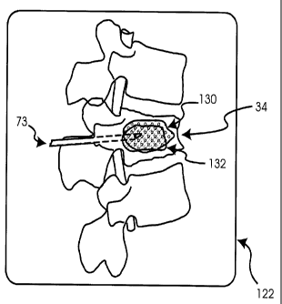
and the spine will be discussed. Figure 1 is a right lateral view of a segment
20 of a normal
spine. Segment 20 includes three vertebrae 22. The spinal cord 28 and epidural
veins 29 run
through the spinal canal of each vertebrae 22. In contrast to segment 20 of
Figure 1, Figure 2
shows a right lateral view of a segment 30 of a spine wherein at least one of
the vertebra has a
condition suitable for treatment by vertebroplasty. Segment 30 includes a
first vertebra 32, a
compressed middle vertebra 34 and a third vertebra 36. Spinal cord 38 and
epidural veins 39 run
through the spinal canal of each vertebrae 32, 34 and 36.
As shown in Figures 2 and 3, vertebra 34 has a right and left transverse
process 40R, 40L,
a right and left superior articular process 42R, 42L, and a spinous process 44
at the posterior of
vertebra 34. Right and left lamina 45R, 45L lie intermediate spinous process
44 and superior
articular processes 42R, 42L, respectively. Right and left pedicles 46R, 46L
and lamina 45R,
45L cooperate to form the vertebral arch 48. The vertebral body 49 is located
at the anterior of
vertebra 34, and is joined to arch 48 at pedicles 46R, 46L. Arch 48 and
vertebral body 49 define
the spinal canal 50 through which spinal cord 38 passes. Epidural veins 39 lie
between spinal
cord 38 and vertebral body 48. As seen in Figure 2, vertebral body 49 is
compressed as a result
of any condition suitable for treatment by vertebroplasty. Such conditions
generally include
benign osteoporotic fractures, malignant metastatic disease and benign tumours
of the bone.
Referring now to Figure 4, a kit, in accordance with an embodiment of the
invention, for
the strengthening of vertebral body 44 is indicated generally at 55. As will
be understood by
those of skill in the art, the individual items in Figure 4 are known, and are
drawn in a simplified
form and kit 55 should not be construed as limited to the representation of
Figure 4. Kit 55
includes a first pack 57 and a second pack 59. Kit 55 and each pack 57, 59
therein are housed
within a sterile packaging suitable for refrigeration and/or storage until
use. Each pack 57, 59
has a local anaesthesia assembly 61, including a vial of local anaesthesia 63,
a syringe 65 for
administering the anaesthesia, a needle 67 for anaesthesia aspiration and a
long needle 69 for
anaesthesia injection. It is presently preferred that vial 63 has ten cubic-
centimetres of 1%
lidocaine without adrenaline, and accordingly that syringe 65 is a ten cc
syringe. It is also
presently preferred that needle 67 is sixteen gauge, while long needle 69 is
twenty-two gauge.
It will be understood that anaesthesia assembly 61 consists of well-known
components, and that
other components can be substituted, as desired.
6
CA 02287112 1999-10-22
Each pack 57, 59 also includes a scalpel 71 suitable for making an incision to
perform
vertebroplasty. It is presently preferred that scalpel 71 is disposable and
has a number-eleven
blade. It will be understood that any scalpel or other functionally equivalent
surgical tool
suitable for vertebroplasty can be used, as will occur to those of skill in
the art.
Packs 57, 59 also include at least one vertebroplasty needle 73. Where kit 55
is for use
on a lumbar vertebral body, then it is generally preferred that an eleven
gauge vertebroplasty
needle 73 is included. Where kit 55 is for use on a thoracic vertebral body,
then it is generally
preferred that a thirteen gauge vertebroplasty needle 73 is included. It will
be understood,
however, that various sizes and combinations of vertebroplasty needles 73 can
be included into
each pack 57, 59 to offer greater flexibility for each kit 55, as desired
and/or required for a
particularvertebroplasty operation. One suitable vertebroplasty needle is the
Cook needle, model
DBBN-10(11)(13)-10.0(15.0)-m1(m2) from Cook Inc., Bloomington Indiana.
Pack 57 also includes the ingredients for a first bone cement for
strengthening a vertebral
body and which has a first imaging property. In a present embodiment, the
ingredients and
mixing devices for the first bone cement are provided in a first cement
assembly 80. First
cement assembly 80 includes monomer liquid 82 in a vial, a monomer compatible
aspiration
syringe 84, a monomer aspiration needle 85, a mixing bowl 86, a mixing spatula
88, a polymer
powder 90, and a first opacifier 92.
It is believed that the vial of monomer liquid 82 should contain from about
five cubic-
centimetres to about twenty cubic-centimetres of monomer. Any monomer that is
intended for
use with a corresponding polymer powder 90 can be used. For example, in
Osteobond there is
a liquid component of 99.25% methylmethacrylate monomer, 0.75% N, N-dimethyl-p-
toluidine
and 75 10 ppm hydroquinone in Osteobond Copolymer Bone Cement from Zimmer
Inc., 1800
West Center Street, Warsaw Indiana 46580. Other suitable monomer liquids are
included in
other bone cements as offered by the various bone cement suppliers.
Preferably, the vial contains
from about seven cubic-centimetres to about fifteen cubic-centimetres of
monomer liquid 82.
More preferably, the vial contains from about ten cubic-centimetres to about
thirteen cubic-
centimetres of monomer liquid 82. It is presently preferred, however, that the
vial contains from
about twelve cubic-centimetres of monomer liquid 82. Overall, it will be
understood that any
composition and/or quantity of monomer liquid 82 can be provided that allows a
radiologist or
other vertebroplasty professional to prepare polymer powder 90 with a desired
consistency.
7
_ __ ..~_,__..~.._..._..._._...._......_....,_.....~...~u...~.~w....~ ...
CA 02287112 2007-05-16
Monomer aspiration syringe 84 is accordingly sized to accommodate the volume
of
monomer liquid 82. Preferably, syringe 84 is DMSO (dimethylsulphoxide)
compatible, which
is designed so that the plunger does not swell when it contacts monomer liquid
82. A suitable
source for syringe 84 is MTI, Micro Therapeutics Inc., 2 Goodyear, Irvine CA
92618.
~
A suitable mixing bowl 86 is a small disposable plastic bowl, such the "gent-l-
kare"
sterile one quart single-use disposable utility bowl made by Premium Plastics
Inc., Chicago
Illinois 60616. Both mixing bowl 86 and mixing spatula 88 are made from a
material that is
suitable for use in the mixing of the bone cement, as is known to those of
skill in the art. Other
suitable mixing devices, such as the closed mixing system known as the "vacuum
cement
mixing" device supplied by Howmedica can be used, as will occur to those of
skill in the art.
Polymer powder 90 is packaged in any suitable sterile sachet. However, any bag
or
storage means can be used and which are suitable for holding from about five
grams to about
forty grams of methylmethacrylate. Preferably, polymer powder 90 is from about
ten grams to
about thirty grams of methylmethacrylate. More preferably, polymer powder 90
is from about
twelve grams to about twenty grams of methylmethacrylate. It is presently
prefeired, however,
that polymer powder 90 is about eighteen grams of methylmethacrylate.
It will now be apparent that the foregoing monomer liquid 82 and polymer
powder 90 is
obtainable in Osteobond. Other suitable bone cements include calcium
carbonate, calcium
phosphate, zirconium, or oxalate or hydroxyappatite derivatives, as will be
understood by those
of skill in the art.
First opacifier 92 is packaged in a sterile satchet or equivalent storage
means. In the
present embodiment, where polymer powder 90 is methylmethacrylate then first
opacifier 92 is
barium powder. It is believed that there should be a mass of barium of from
about ten percent
to about fifty percent of the mass of the methylmethacrylate. Preferably,
there should be a mass
of barium of from about fifteen percent to about forty-five percent of the
mass of
methylmethacry late. More preferably, there should be a mass of barium powder
of from about
twenty percent to about forty percent of the mass of methylmethacrylate. It is
presently
preferred, however, that there should be a mass of barium of about one-third
of the mass of
methylmethacrylate, and thus, where there is eighteen grams of
methylmethacrylate there should
be about six grams of barium. In general, it will be understood that a
sufficient amount of barium
should be added to the first cement that provides a suitable radio-opacity
without degrading the
8
CA 02287112 1999-10-22
physical properties of the first cement. Other suitable opacifiers, such as
calcium phosphate,
calcium carbonate, tantalum, tungsten or zirconium can be used, as will occur
to those of skill
in the art.
Pack 59 includes the ingredients for a second bone cement for strengthening a
vertebral
body that is compatible and/or usable with the first bone cement and which has
a second imaging
property. In a present embodiment, the ingredients and mixing devices for the
second bone
cement are provided in a second cement assembly 100. Second cement assembly
100 includes
monomer liquid 102 in a vial, a monomer aspiration syringe 104, a monomer
aspiration needle
105 a mixing bow1106, a mixing spatula 108, polymer powder 110, and a second
opacifier 112.
It is presently preferred that monomer liquid 102, syringe 104, needle 105,
bowl 106,
spatula 108 and powder 110 are the same as liquid 82, syringe 84, bowl 86,
spatula 88 and
powder 90, respectively, from pack 57.
However, second opacifier 112 has a different composition and/or quantity from
first
opacifier 92, so that when it is mixed into a second bone cement the second
bone cement has a
different imaging property from the first bone cement. Second opacifier 112 is
packaged in a
bag, similar to first opacifier 92. In the present embodiment, second
opacifier 112 is also barium
but has a different, preferably higher, quantity than first opacifier 92. It
is believed that there
should be about fifteen percent to about three-hundred percent more barium in
second opacifier
112 than first opacifier 92. Preferably, there should be about there should be
about thirty percent
to about two-hundred-and-fifty percent more barium in second opacifier 112
than first opacifier
92. More preferably, there should be about forty percent to about two-hundred
percent more
barium in second opacifier 112 than first opacifier 92. It is presently
preferred, however, that
there should be about one-hundred-and-eighty percent more barium powder in
second opacifier
112 than first opacifier 92. Thus, in a presently preferred embodiment, there
is about eleven
grams of barium powder in second opacifier 112 to contrast the six grams of
barium powder in
first opacifier 92. In general, it will be understood that a sufficient amount
of barium should be
added to polymer powder 110 to provide a radio-opacity that differs from the
radio-opacity of
the first cement, but without degrading the physical properties of the second
cement.
A method for performing vertebroplasty in accordance with an embodiment of the
invention will now be discussed, utilizing kit 55 and performed on a patient
having vertebrae 34.
Referring now to Figure 5, the patient is placed in the prone position so that
vertebrae 34 is
9
CA 02287112 1999-10-22
within the field of an imaging device 120, which in a present embodiment is an
X-Ray projection
fluoroscopy imaging device. Other imaging devices can be used, as will occur
to those of skill
in the art. When imaging device 120 is 'on', vertebrae 34 is projected onto
display 122. For
purposes of explaining the present embodiment, vertebrae 34 is projected onto
display 122 from
the same lateral view as shown in Figure 2. The skin overlying vertebrae 34 is
prepped and
draped in the usual manner with sterile technique. Next, the seal on kit 55 is
broken, and the seal
on pack 57 is broken. Anaesthesia assembly 61 is opened and utilized so that
anaesthesia 63 is
injected into the skin underlying fat and into the periosteum of the pedicle
to be entered. For
purposes of explaining the present method, it will be assumed that left
pedicle 46L will be
entered first. Next, using scalpel 71, a skin incision of about five
millimetres is made with
scalpel 73. As shown in Figures 6 and 7, at this point vertebroplasty needle
73 is inserted into
the incision and passed down left pedicle 46L, preferably until it enters the
vertebral body and
reaches the junction of the anterior and middle thirds.
At this point, first cement assembly 80 is opened. Powder 90 and first
opacifier 92 are
placed in mixing bow186 and monomer 82 is injected into mixing bowl 86 using
syringe 84. A
first cement for strengthening a vertebral body and having a first imaging
property is thus
prepared by mixing the contents of mixing bow186 with spatula 88. In the
present embodiment,
the first imaging property is determined by the quantity of first opacifier 92
within the first
cement. As shown in Figures 8 and 9, the first cement is injected into
vertebral body 49 via left
pedicle 46L through needle 73, the first cement being indicated at 130.
Opacifier 92 allows first
cement 130 to be detected by imaging device 120 and is thus viewable on
display 122 as having
a first imaging property. The first imaging property is represented in first
cement 130 as a
pattern of small circles. Accordingly, the quantity and flow-route of first
cement 130 is
monitored on display 122, as shown in Figure 9.
At this point, a decision can be made as to whether a sufficient quantity of
first cement
130 that has been injected. This decision is made using known criteria and is
typically made by
the radiologist, physician or other vertebroplasty professional who is
performing the method.
If it is determined that enough cement has been injected to provide the
desired strength to
vertebral body 34, then the treatment method is complete and the patient is
prepared for removal
from the X-ray room and transferred to the observation area. Pack 59 is still
sterile and can be
placed back into refrigeration or storage for use on another patient at a
later date.
CA 02287112 1999-10-22
If, however, it is determined that another injection is required along right
pedicle 46R,
then pack 59 is opened and anaesthesia assembly 61 therein is opened and
anaesthesia 63 is
injected into the skin posterior to right pedicle 46R. Scalpel 71 of pack 59
is then used to make
the appropriate incision, and vertebroplasty needle 73 of pack 59 is inserted
into vertebral body
49 via right pedicle 46R, as shown in Figures 10 and 11.
At this point, second cement assembly 100 is opened. Powder 110 and second
opacifier
112 are placed in mixing bowl 106 and monomer 102 is aspirated into mixing
bowl 106 using
syringe 104. A second cement for strengthening a vertebral body that has a
second imaging
property, (which in the present embodiment has different radio-opacity), is
thus prepared by
mixing the contents of mixing bowl 106 with spatula 108. In the present
embodiment, the
second imaging property is determined by the quantity of second opacifier 112
within the second
cement. As shown in Figures 12 and 13, the second cement is injected into
vertebral body 49
via right pedicle 46R through needle 73, the second cement being indicated at
132. Opacifier
112 allows second cement 132 to be detected by imaging device 120 and is thus
viewable on
display 122 as having a second imaging property. The second imaging property
is represented
in second cement 132 as a pattern of diagonal lines. Accordingly, the quantity
and flow-route of
second cement 132 is monitored on display 122, as shown in Figure 13. In
particular, the
quantity and flow-route of second cement 132 can be monitored in contrast to
the first cement
130, due to the contrasting or different imaging properties of first cement
130 and second cement
132. By monitoring the flow-route of second cement 132, the injection of
second cement 132
can be terminated before it reaches spinal cana150 and thus reduce the
likelihood of spinal cord
compression and/or related damage. Once a sufficient amount of second cement
132 has been
injected, the method is complete and the patient is prepared for discharge.
It should be understood that the opacifier in at least one of the first and
second cements
can be in the form of particles dispersed throughout the respective cement. In
certain
circumstances, the motion of such particles can increase the ability to detect
cement motion and
filling. For example, when using methylmethacrylate powder with barium, the
barium can be
in the form of powder and/or particles. It is presently preferred that the
barium particles are
about one millimetre in size, however, other particle sizes will occur to
those of skill in the art.
When used with an X-ray imaging system, other particles can include, for
example, calcium
11
CA 02287112 1999-10-22
phosphate, oxalate, zirconium, tantalum and/or tungsten. Other types of
particles will occur to
those of skill in the art.
It can be desired to use a first cement with a first density of radio-opaque
particles, and
a second cement having a second density of radio-opaque particles. Generally,
it is preferred that
the second cement injection has a greater density of particles than the first
cement injection.
Furthermore, previously discussed, it is generally preferred that the method
is performed so that
second cement injection appears in front of the first cement injection, as
displayed on the
imaging display.
It can be desired that only one of the cements has radio-opaque particles,
while the other
cement has a radio-opaque powder. While not necessary, it is generally
preferred that the cement
with the particles is used for the second injection of cement.
Other variations of how to provide two different opacifiers that will make
each respective
injection of each cement appear contrasting and/or different under an imaging
system will occur
to those of skill in the art, and are within the scope of the invention.
While the embodiments discussed herein are directed to particular
implementations of
the present invention, it will be apparent that the sub-sets and variations to
these embodiments
are within the scope of the invention. For example, kit 55 can include drapes,
disinfectant and/or
sponge tipped disinfectant applicators foruse in the preparation of the
patient prior to performing
the operation. Other items of assistance during a vertebroplasty can be added
to kit 55, as
desired.
It is to be understood that the individual packs 57, 59 of kit 55 need not
include
anaesthesia assemblies, vertebroplasty needles, scalpels etc. and that it is
contemplated that each
pack 57, 59 need only include a first cement for strengthening a vertebral
body that has a first
imaging property, and a second bone cement for strengthening a vertebral body
that has a second
imaging property, whereupon injectionthereof each cement is visible by an
imaging system, such
as a X-ray or other radiographic imaging system. The other items in packs 57,
59 can be obtained
and/or assembled from other sources prior to performing the method.
The ability to detect motion of cement during injection can be increased where
radiopaque vertebroplasty needles are used, thus allowing the detection of
motion of cement as
the cement travels along the length of the needle.
12
CA 02287112 1999-10-22
It will be understood that each pack 57, 59 can be packaged and/or sold
separately, and/or
need not be included in an entire kit 55. Furthermore, kit 55 can be sold as
having two of pack
59, two of pack 57, or, as previously discussed, one pack 57 and one pack 59.
By offering
different combinations of kit 57 and packs 57, 59 vertebroplasty professionals
can be offered kits
having cements with imaging properties that are personally preferred by the
professional, and/or
allow the purchase of additional individual packs that complement left-over
individual packs
from procedures that only required the use of one pack.
It is contemplated that the first and second bone cements can also be bone
cements that
are bioactive, integratable, stimulate bone growth and/or are resorbable.
OrthocompTM cement
by Orthovita of 45 Great Valley Parkway, Malvern PA 19355 is one such cement.
Other
suppliers of such cements include Howmedica/Stryker of 6300 Sprinkle Road,
Kalamazoo, MI
49001, and Codman/Depuy of 325 Paramount Drive Raynham MA 02767. It is
contemplated
that as other suitable cements are developed and/or approved, the contents of
the kit can vary to
suit the surgical procedure used to inject the first and second cements.
It is also contemplated that the mechanism for injecting the cement can be
automatically
controlled via a computer or other controller that receives the image from the
imaging device and
has an output connected to the vertebroplasty needle injection mechanism. Such
a controller can
be programmed to determine, based on the received image, when to commence,
stop or otherwise
control the flow of the injection of each cement.
While the present invention teaches first and second cements having differing
densities
and/or distributions of particles of barium to provide different imaging
properties when exposed
to lateral X-ray fluoroscopy, other opacifiers and/or imaging technologies can
be used, as will
occur to those of skill in the art. Other imaging technologies can include,
for example, magnetic
resonance imaging, and computed tomography.
It will be further understood that packs 57 and 59 within kit 55 can each have
completely
identical components, and, optionally kit 55 can further include a separate
package of opacifier
to be mixed with one of the cement assemblies to provide two different cements
that will have
different imaging properties. Alternatively, the extra opacifier need not be
provided with kit 55,
but can be obtained separately by a user of kit 55. The other permutations and
combinations of
kit 55 will now be apparent, and are within the scope of the invention.
13
CA 02287112 1999-10-22
It will also be understood that polymer powder 90 and first opacifier 92 can
be premixed
and packaged in a single sachet within first pack 57. Similarly, polymer
powder 110 and second
opacifier 112 can also be premixed.
While the present invention is generally suitable for known conditions that
are treatable
with vertebroplasty, it is contemplated that the present invention can be
suitable for other
conditions that require similar treatment. For example, prophylactic
vertebroplasty can be
performed in patients with critically low bone density.
The present invention provides a novel method and kit for increasing strength
of vertebral
bodies. In one embodiment, there is provided a first cement for strengthening
a vertebral body
which has a first imaging property, and a second bone cement for strengthening
a vertebral body
that is compatible with the first bone cement and which has a second imaging
property, such that
each cement is visible during injection under the guidance of an imaging
system, such as lateral
X-ray fluoroscopy. By providing these two cements, vertebroplasty can be
performed by
injection into both pedicles of the vertebra and allow a radiologist,
physician or other
vertebroplasty professional to observe the flow of the cements and thus more
safely and/or
effectively inject cement in the vertebral body and reduce the likelihood of
spinal cord
compression and/or related damage. The present invention also provides a kit
for performing
vertebroplasty, having a first pack for performing vertebroplasty through one
pedicle, and a
second pack for performing vertebroplasty through a second pedicle. Each pack
can have a
variety of different combinations, as desired. Each pack can be used
independently. Unused
packages can be stored for later use and a second conventional vertebroplasty
procedure can be
carried out using of the second pack.
14