Note: Descriptions are shown in the official language in which they were submitted.
CA 02287206 1999-10-20
WO 98156324 PCT/US98/07976
1
ELECTROSUI~GICAL SYSTEMS AND METHODS FOR
s RECANALIZ~~TION OF OCCLUDED BODY LUMENS
CROSS-REF)E;RENCE TO RELATED APPLICATIONS
The present application is a continuation-in-part of U.S. Application No.
08/874,173, filed June 13, 1997 (Attorney Docket No. 016238-005600), and also
derives
to priority from U.S. Application No. 09/002,315, filed January 2, 1998
(Attorney Docket
No. C-9), the complete disclosures of which are incorporated herein by
reference for all
purposes.
The present inve~,ntion is related to commonly assigned co-pending U.S.
Patent Application No. 081990,374, filed December 15, 1997 (Attorney Docket
No. E-3),
1s which is a continuation-in-part of U.S. Patent Application No. 081485,219,
filed on June
7, 1995, now U.S. Patent No. 5,697,281 (Attorney Docket 16238-000600),
Provisional
Patent Application No. 60/075,059, filed on February 18, 1998 (Attorney Docket
No. CB-
2P), U.S. Patent Application 1'10. 09/010,382, filed January 21, 1998
(Attorney Docket A-
6), and U.S. Patent Application No. 09/032,375, February 27, 1998 (Attorney
Docket No.
2o CB-3), U.S. Patent Applicaticn Nos. 08/977,845, filed on November 25, 1997
(Attorney
Docket No. D-2), 08/942,580, filed on October 2, 1997 (Attorney Docket No.
16238-
001300), 09/026,851, filed Fe'oruary 20, 1998 (Attorney Docket No. S-2), U.S.
Application No. 08/753,227, filed on November 22, 1996 (Docket 16238-002200),
U.S.
Application No. 08/687792, filed on July 18, 1996 (Docket No. 16238-001600),
and PCT
2s International Application, U.S. National Phase Serial No. PCTIUS94/05168,
filed on May
10, 1994, now U.S. Patent No. 5,697,909 (Attorney Docket 16238-000440), which
was a
continuation-in-part of U.S. Patent Application No. 08/059,681, filed on May
10, 1993
(Attorney Docket 16238-0004:!0), which was a continuation-in-part of U.S.
Patent'
r Application No. 071958,977, filed on October 9, 1992 (Attorney Docket 16238-
000410)
3o which was a continuation-in-port of U.S. Patent Application No. 07/817,575,
f"sled on
January 7, 1992 (Attorney Do~:ket 16238-000400), the complete disclosures of
which are
incorporated herein by reference for all purposes. The present invention is
also related to
CA 02287206 1999-10-20
WO 98156324 PCTIUS98/07976
2
commonly assigned U.S. Patent No. 5,683,366, filed November 22, 1995 (Attorney
Docket 16238-000700), the complete disclosure of which is incorporated herein
by
reference for all purposes.
BACKGROUND OF THE INVENTION
The present invention relates generally to apparatus and methods for
maintaining patency in body passages and more particularly to a catheter
system capable.of
selectively ablating occlusive media within a body lumen. The present
invention is
particularly useful for the electrosurgical cutting or ablation of invasive
tissue growth in
1o and around a stmt anchored in the body lumen to help reduce or eliminate
restenosis of the
body lumen.
When a patient is suffering from atherosclerosis, significant occlusions or
blockages are formed on the interior wall of the artery. As a result of these
occlusions, the
organ or extremity to which blood is to be supplied is compromised and the
patient may
1s experience a myocardial infarction or stroke. In less severe cases, it is
often sufficient to
treat the symptoms with pharmaceuticals and lifestyle modification to lessen
the underlying
causes of the disease. In more severe cases, a coronary artery blockage can
often be
treated using endovascular techniques such as balloon angioplasty,
atherectomy, laser or
hot tip ablation, placement of stents, and the Like.
2o Percutaneous transluminal balloon angioplasty (PTBA) has become a
recognized method of reducing the occlusion of blood vessels. The procedure
involves
routing a catheter having an inflatable balloon at the distal end thereof
through the vascular
system until the balloon is positioned at the site of the stenotic lesion to
be treated. The
balloon is then inflated to compress tile atherosclerotic plaque into the wall
of the blood
2s vessel, thus increasing the size of the opening and enhancing blood flow
through the
affected artery. However, this successful procedure is overshadowed by the
occurrence of
restenosis, a re-narrowing of the artery. Studies have shown that 30-40
percent of
angioplasty patients experience restenosis within 3-6 months of the
angioplasty procedure.
When restenosis occurs, patients may be treated with cardiovascular
medications,
3o additional angioplasty procedures or bypass surgery.
Restenosis often occurs because the wall of the dilated artery tends to spring
back to its original shape following deflation of the dilation balloon.
Arterial stenting has
._ , ,
CA 02287206 1999-10-20
WO 98/56324 PCT/US98/07976
3
been introduced as a solution to the recoil of the vessel wall. Arterial
stenting involves the
placement of an expandable coil spring or wire-mesh tube within the occluded
artery to
reopen the Iumen of the blood vessel. One example of an arterial stmt is
disclosed in U.S.
' Pat. No. 4,739,792 to Julio Paimaz. The Palmaz device comprises an
expandable wire-
s mesh graft or prosthesis which is mounted upon an inflatable balloon
catheter. The
catheter assembly, including tl: a graft, is delivered to the occluded area
and is then inflated
to radially force the graft into ~.ontact with the occlusion. As the graft
expands, the lumen
of the blood vessel is opened and blood flow is restored. After complete
expansion of the
graft, the balloon catheter is deflated and removed, leaving behind the graft
to buttress and
to prevent elastic recoil of the blood vessel wall.
Although this method is successful in preventing recoil of the vessel wall,
restenosis will often still occur. Smooth muscle cells which form the vessel
wall tend to
proliferate and build-up in the newly stented area of the blood vessel. This
cellular build-
up may eventually become larl;e enough to block the lumen of the blood vessel.
t5 It has recently been determined that localized heating of the blood vessel
wall may inhibit the proliferation of smooth muscle cells which are believed
to cause
restenosis. One example of localized blood vessel heating is disclosed in U.S.
Pat. No.
4,799,479 to Spears. The Spears patent discloses an apparatus for angioplasty
having an
inflatable balloon catheter whi~:h is provided with a meshwork of electrical
wires to supply
2o heat to a vessel wall. Following balloon angioplasty, the external surface
of the balloon is
heated to fuse together disrupted tissue elements and to kill smooth muscle
cells which are
believed to lead to restenosis. Unfortunately, the Spears device does not
adequately
prevent the spontaneous elastic: recoil of the arterial wall. Immediately
following
angioplasty, the arterial wall begins to spring back to its original shape.
25 Thus stenting in and of itself is ineffective in preventing restenosis due
to
the occurrence of cellular prol iferation. Likewise, balloon dilation in
combination with
localized heating does not adequately prevent restenosis since the vessel wall
tends to
spontaneously return to its ori;;inal occluded shape.
Other technique s have recently been developed to help reduce incidences of
3o restenosis. For example, procedures for irradiating the angioplasty site
with UV light to
reduce the proliferation of smooth muscle cells at the site have been
disclosed. In addition,
techniques have been disclosed for the controlled application of thermal
and/or electrical
CA 02287206 1999-10-20
WO 98/56324 PCT/US98/07976
4
energy directly to the stem by, for example, including resistive or inductive
heating
elements that may include radiofrequency electrodes within the stmt. The
radiofrequency
energy is then applied to the stent to disrupt~the cellular growth in or
around the stmt.
One major disadvantage of these procedures is that it is difficult to
selectively apply the
energy to the invasive tissue without causing thermal damage to the body lumen
wall. In
particular, methods that apply energy, such as RF energy, directly to the stmt
will often
cause thermal damage to the surrounding body lumen in which the stmt is
anchored.
SUMMARY OF THE INVENTION
to The present invention comprises apparatus and methods for maintaining
patency in body passages subject to occlusion by invasive tissue growth. The
apparatus
and methods of the present invention may be used to open and maintain patency
in
virtually any hollow body passage which may be subject to occlusion by
invasive cellular
growth or invasive solid tumor growth. Suitable hollow body passages include
ducts,
Is orifices, lumens, and the like, with exemplary body passages including the
coronary
arteries. The present invention is particularly useful for reducing or
eliminating the effects
of restenosis in coronary arteries by selectively removing tissue ingrowth in
or around
stents anchored therein.
The principles of the present invention are generally applicable to any body
20 lumen which becomes partially or totally occluded. Methods of the present
invention
comprise advancing an electrosurgical catheter within the body passage such
that an
electrode terminal is positioned near the occlusive media. High frequency
voltage is
applied to one or more electrode terminals) at the distal end of the catheter
such that an
electrical current flows from the electrode terminal(s), through the region of
the occlusive
25 media, and to a return electrode to volumetrically remove the occlusive
media in situ. In
exemplary embodiments, the high frequency voltage is sufficient to effect
molecular
dissociation or disintegration of the occlusive media, thus converting the
solid media into
non-condensable gases.
The present invention is particularly useful in a lumen containing a lumenal
3o prosthesis, such as a stmt, stmt-graft or graft, which may be metallic, non-
metallic or a
non-metallic coated metallic structure. Restenosis often occurs when
arthermateous media
or thrombus moves or grows through or around the cylindrical wall of the
prosthesis to
~ , T
CA 02287206 1999-10-20
WO 98/56324 PCT/US98/07976
partially occlude the body pass age. Methods of the present invention comprise
advancing
an electrosurgical catheter within the body passage such that an electrode
terminal is
positioned near the occlusive media. High frequency voltage is applied to one
or more
electrode terminals) at the distal end of the catheter such that an electrical
current flows
5 from the electrode terminal(s), through the region of the occlusive media,
and to a return
electrode to selectively remove the occlusive media without directly applying
thermal or
electrical energy to the prosthesis or the iumenal wall. The electrode
terminal may then be
advanced through the vacancy left by the removed occlusive media to recanalize
the vessel.
By selectively removing the occlusive media without passing energy directly to
the stmt,
1o thermal damage to the surrounding lumenal wall is minimized.
In an exemplary embodiment, the return electrode is located on the catheter
so that the current flow paths are confined between the return electrode and
one or more
electrode terminals in the vicinity of the working end of the catheter. This
confinement of
current flow paths minimizes tl~e undesired flow of current through portions
or all of the
~ s stmt, which may otherwise ins uce non-specific tissue injury beyond the
site of
recanalization of the occluded 1 umen. In one configuration, the return
electrode is a
movable guide wire positioned radially inward from the electrode terminal such
that the
electrical current flows from tf a electrode terminal radially inward to the
return electrode,
thereby inhibiting current flow through the prosthesis. In another embodiment,
the return
2o electrode is an annular band positioned proximal of the electrode
terminal(s).
In preferred embodiments, the high frequency voltage is applied in the
presence of electrically conducting fluid such that a current flow path is
generated between
the electrode terminals) and tre return electrode through the electrically
conducting fluid.
Preferably, the electrically conductive fluid is delivered through an internal
lumen in the
2s catheter (or through a separate instrument) to a region around the
occlusive media to
displace naturally occurring bodily fluids. This region may be fluidly
isolated to confine
the electrically conducting fluid around the tissue ablation site. In one
embodiment, the
region is isolated by advancing proximal and distal balloons to either side of
the region,
and inflating these balloons to effect a seal with the interior wall of the
body passage.
Once the target site is isolated from the rest of the vasculature, the supply
of
electrically conductive fluid is continuously delivered to the region and
balanced with the
aspiration of fluid from the sitf~ of intended recanalization. The electrode
terminals) are
CA 02287206 1999-10-20
WO 98/56324 PCTIUS98/07976
6
energized by applying a high frequency voltage between electrode terminals}
and the
return electrode, which can be a movable guide wire. A high electric field is
created at the
surface of the electrodes) which causes the volumetric removal or ablation or
target tissue
in close proximity with the electrode terminal{s). As the occlusive media is
ablated,
s gaseous products are generated which are entrained in the electrically
conducting fluid and
removed through the aspiration lumen in the catheter. The current flux lines
are generally
confined to the central portion of tissue ablation region because they
generally flow inward
towards the return electrode and because the occlusive media generally shields
the outer
region of the body passage (including the stent) from the current flux lines.
This
to minimizes undesirable interaction between the electrical current and the
stmt. In an
exemplary embodiment, the distal portion of the catheter body is reciprocally
rotated as the
electrode terminal is energized to selectively ablate the occlusive media. The
catheter
body is then advanced through the vacancy left by the ablated occlusive media
to recanalize
the vessel.
1 s In a specific configuration, the occlusive media is removed by molecular
dissociation or disintegration processes. In these embodiments, the high
frequency voltage
applied to the electrode terminals) is sufficient to vaporize an electrically
conductive fluid
(e.g., saline or blood) between the electrode terminals) and the occlusive
media. Within
the vaporized fluid, a ionized plasma is formed and charged particles (e.g.,
electrons) are
2o accelerated towards the target media to cause the molecular breakdown or
disintegration of
several cell layers of the media. This molecular dissociation is accompanied
by the
volumetric removal of the media. The short range of the accelerated charged
particles
within the plasma layer confines the molecular dissociation process to the
surface layer to
minimize damage and necrosis to the surrounding blood vessel walls. This
process can be
2s precisely controlled to effect the volumetric removal of tissue or media as
thin as 10 to 150
microns with minimal heating of, or damage to, surrounding or underlying
tissue
structures. A more complete description of this phenomena is described in
commonly
assigned U.S. Patent No. 5,683,366, the complete disclosure of which is
incorporated
herein by reference.
3o Apparatus of the present invention comprise a catheter shaft having a
flexible body with a proximal end portion and a distal end portion with one or
more
electrode terminal(s), and a connector extending through the body for coupling
the
T ,,f
CA 02287206 1999-10-20
WO 98/56324 PCT/US98107976
7
electrode terminals) to a sourv;e of high frequency voltage. Upon the
application of
sufficient high frequency voltage to the electrode terminal(s), the occlusive
media is
volumetrically removed from the body lumen to recanalize the body lumen. In
same
embodiments, the apparatus w iII further include one or more fluid delivery
elements) for
s delivering electrically conducting fluid to the electrode terminals) and the
target site. The
fluid delivery elements) may lie located on the catheter, e.g., one or more
fluid lumen(sj
or tube(s), or they may be part of a separate instrument. In an exemplary
embodiment, the
electrically conducting fluid will preferably generate a current flow path
between the
electrode terminals) and one or more return electrode(s). In an exemplary
embodiment,
1o the return electrode{s) are located on the catheter and spaced a sufficient
distance from the
electrode terminals) to substa:utially avoid or minimize current shorting
therebetween and
to shield the return electrodes) from tissue at the target site.
Alternatively, the return
electrodes) may comprise a d.spersive pad located on the outer surface of the
patient (i.e.,
a monopolar modality).
1 s In a specific configuration, the apparatus includes a plurality of
electrically
isolated electrode terminals extending from the distal end of the catheter
shaft. The
electrode terminals are each mounted within an electrically insulating support
member, and
spaced peripherally around the: distal opening of the catheter body. In these
embodiments,
the catheter may include a single, annular return electrode located proximal
of the distal
20 opening, or a plurality of electrode terminals mounted to the support
members proximal of
the electrode terminals. In this embodiment, the catheter body also includes
one or more
fluid delivery lumens spaced F eripherally around the central lumen for
delivering
electrically conductive fluid to the electrode terminals. In addition, the
catheter body will
preferably include one or more suction lumens spaced peripherally around the
central
2s lumen, and suitably coupled tc> an external suction source for aspirating
fluid, tissue andlor
gaseous products of ablation (~:.g., non-condensable gases) from the target
site.
In an exemplar:r embodiment, the working end portion of the catheter has an
adjustable outer diameter to facilitate advancement of this portion of the
catheter through a
variable diameter body lumen or stent. In one configuration, the working end
of the
3o catheter will taper in the distadirection (e.g., in a series of steps) so
that the surgeon can
advance the catheter through a. severely occluded body lumen. The catheter may
include a
series of axially spaced electrode terminals) that are electrically isolated
from each other
CA 02287206 1999-10-20
WO 98/56324 PCT/US98/07976
8
to allow for each set to be independently activated. By way of example, in a
severely
occluded body lumen, the surgeon may activate the distal set of electrode
terminals) to
remove the innermost occlusive media, advance these distal electrode
terminals) through
the vacancy left by the removed occlusive media, and then activate a more
proximal, and
radially outward, set of electrode terminals) to remove occlusive media
radially outward
from the initially removed media. -
In another configuration, the working end of the catheter may be radially
expandable and compressible so that the diameter of the working end can be
varied as the
catheter is advanced through the lumen. In some instances, scents will not
expand
o uniformly resulting in portions of the stem having smaller inner diameters.
In other
instances, vessel wall pressure may cause portions of the stmt to spring back
to its original
shape or partially back to this shape so that the overall inner diameter of
the stent varies in
the axial direction. Accordingly, the present invention allows the diameter of
the working
end of the catheter to vary (either automatically in response to the body
lumen or stmt
inner diameter, or through activation by the surgical team) to facilitate
advancement
through non-uniform stents or body lumens.
In another embodiment of the invention, the catheter system includes a high
frequency power supply configured to reduce or interrupt power when the
electrode
terminals) contact a low impedance object, such as a stent within the body
lumen. In one
2o embodiment, the power supply includes a spark prevention device for
eliminating or
reducing sudden pulses in current when an instrument powered by the power
supply
contacts a low impedance source. The spark limiting device is coupled to one
or more
current sensors on the electrode terminals) to substantially continuously
monitor current
output, interrupting current output from the output driver when current output
from the
output current sensor exceeds a predetermined threshold level. The spark
prevention
mechanism, which may be used in conjunction with other power limiting devices,
preferably turns off output from the power supply when output current from the
supply
exceeds a predetermined current level.
BRIEF DESCRIPTION OF THE DRAWINGS
CA 02287206 1999-10-20
WO 98156324 PCT/US98/07976
9
Fig. 1 schematically illustrates a lumen recanalization catheter system
according to the present invent.on;
Figs. 2A-2C illustrate a method of recanalizing an obstructed lumen
according to the present inventuon;
s Figs. 3A and 3E are transverse and longitudinal cross-sectional views,
respectively, of a first embodiment of the distal portion of the catheter;
Figs. 4A and 4~ are transverse and longitudinal cross-sectional views,
respectively, of a second embo3iment of the distal portion of the catheter;
Figs. SA and SL are transverse and longitudinal cross-sectional views,
to respectively, of the second embodiment of the distal portion of the
catheter further
illustrating the inflow of conductive liquid and aspiration of conductive
liquid and gaseous
products;
Figs. 6A and 6E. are transverse and longitudinal cross-sectional views,
respectively, of a third embodiment of the distal portion of the catheter;
15 Figs. 7A and 7E. are transverse and longitudinal cross-sectional views,
respectively, of a fourth embodiment of the distal portion of the catheter;
Figs. 8A and 8E~ are transverse and longitudinal cross-sectional views,
respectively, of a fifth embodiment of the distal portion of the catheter;
Figs. 9A and 91=; are transverse and longitudinal cross-sectional views,
2o respectively, of a sixth embodiment of the distal portion of the catheter;
Figs. l0A and lOB are transverse and longitudinal cross-sectional views,
respectively, of a seventh embodiment of the distal portion of the catheter;
Figs. 11 and 12 illustrate another embodiment of an electrosurgical catheter
incorporating a radially expan~ ible working end;
25 Fig. 13 is a block diagram of a power limiting device according to the
present invention;
Fig. 14 is a graph of the power output of the power supply during normal
operations and standby mode;
Fig. 15 is a graph of the power output of the power supply in a low power,
3o pulsatile mode;
Figs. 16A-16C show various embodiments of a current sensor;
CA 02287206 1999-10-20
WO 98/56324 PCT/US98/07976
Fig. 17 is a circuit schematic of an exemplary embodiment of a power
limiting device;
Fig. 18 is a block diagram of a spark limiting device according to the
present invention;
5 Fig. 19 is a chart of the current output of a spark limiting device
according
to the present invention;
Fig. 20 is a circuit schematic of an exemplary embodiment of a spark
limiting device;
Fig. 2I is a block diagram of the relationship between power limiting and
o spark limiting devices;
Fig. 22 is a circuit schematic showing both the power limiting and spark
limiting devices;
Fig. 23 illustrates a method of volumetrically removing media in a body
passage having a total occlusion; and
~5 Figs. 24A and 24B illustrate the volumetric removal of occlusive media in
more detail.
DESCRIPTION OF SPECIFIC EMBODIMENTS
2o The present invention relates generally to the field of electrosurgery,
and more particularly to surgical devices, systems and methods which employ
high
frequency electrical energy to remove or ablate tissue attached to implanted
objects within
the body. The systems and methods of the present invention are particularly
useful for
removing atheromatous material which partially or fully occludes the body
lumen, such as
25 a blood vessel or for removing stems or other implanted objects. Moreover,
other body
lumens that may be treated by the method and apparatus of the present
invention include
the urinary tract (which for example may be occluded by an enlarged prostrate
in males),
the fallopian tubes (which may be occluded and cause infertility), and the
like. In fact, the
methods and apparatus disclosed herein may be used in a wide variety of
procedures,
30 including open procedures, intravascular procedures, urology, laparascopy,
arthroscopy,
thoracoscopy or other cardiac procedures, dermatology, orthopedics,
gynecology,
otorhinolaryngology, spinal and neurologic procedures, oncology and the like.
For
,,
CA 02287206 1999-10-20
WO 98/56324 PCT/US98107976
11
convenience, the remaining disclosure will be directed specifically to the
removal of
occlusive media within body h.mens.
The stenotic ma.erial in blood vessels will be, by way of example but not
limited to, atheroma or atherolnatous plaque. It may be relatively soft
(fresh) or it may be
calcified and hardened. The invention applies heat selectively to the stenotic
material to
remove this material while limiting unwanted heating of the blood, the
surrounding vessel
wall and the stmt anchored therein. In some embodiments, the present invention
confines
the current flow paths between the return electrode and electrode terminals to
the vicinity
of the tissue ablating region. 'this confinement of current flow paths
minimizes the
~o undesired flow of current through the walls of the body passage, or
portions or all of the
stent, which may otherwise induce non-specific tissue injury beyond the site
of
recanalization of the occluded lumen.
In the present invention, high frequency (RF) electrical energy is applied to
one or more electrode termina.s (usually in the presence of electrically
conductive fluid) to
is remove and/or modify body structures. Depending on the specific procedure,
the present
invention may be used to: (1) ,volumetrically remove body structures (i.e.,
ablate or effect
molecular dissociation of the structure); (2) cut or resect body structures;
(3) vaporize,
cauterize or desiccate structurc;s andlor (4) coagulate and seal severed blood
vessels.
In the preferred method of the present invention, occlusive media within
2o body lumens is volumetrically removed or ablated. In this procedure, a high
frequency
voltage difference is applied between one or more electrode terminals) and one
or more
return electrodes) to develop high electric field intensities in the vicinity
of the target
tissue. The high electric field intensities lead to electric field induced
molecular
breakdown of target tissue thr~~ugh molecular dissociation (rather than
thermal evaporation
25 or carbonization). Applicant believes that the tissue structure is
volumetrically removed
through molecular disintegration of larger organic molecules into smaller
molecules andlor
atoms, such as hydrogen, oxides of carbon, hydrocarbons and nitrogen
compounds. This
molecular disintegration comF letely removes the tissue structure, as opposed
to
dehydrating the tissue material by the removal of liquid within the cells of
the tissue, as is
3o typically the case with electro;urgical desiccation and vaporization.
The high electric field intensities may be generated by applying a high
frequency voltage that is suffi dent to vaporize an electrically conducting
fluid over at least
ICA' 02287206 1999-10-20
WO 98/56324 PCT/US98/07976
12
a portion of the electrode terminals) in the region between the distal tip of
the electrode
terminals) and the target tissue. The electrically conductive fluid may be a
liquid, such as
isotonic saline or blood, delivered to the target site, or a viscous fluid,
such as a gel,
applied to the target site. Since the vapor layer or vaporized region has a
relatively high
electrical impedance, it increases the voltage differential between the
electrode terminal tip
and the tissue and causes ionization within the vapor layer due to the
presence of an
ionizable species (e.g., sodium when isotonic saline is the electrically
conducting fluid).
This ionization, under optimal conditions, induces the discharge of energetic
electrons and
photons from the vapor layer and to the surface of the target tissue. This
energy may be in
to the form of energetic photons (e.g., ultraviolet radiation), energetic
particles (e.g.,
electrons) or a combination thereof. A more detailed description of this
phenomena,
termed Coblation~' can be found in commonly assigned U.S. Patent No. 5,683,366
the
complete disclosure of which is incorporated herein by reference.
The present invention applies high frequency (RF) electrical energy in an
t 5 electrically conducting fluid environment to remove (i. e. , resect, cut
or ablate) a body
structure, and to seal transected vessels within the region of the target
tissue. The present
invention is particularly useful for sealing larger arterial vessels, e.g., on
the order of 1
mm or greater. In some embodiments, a high frequency power supply is provided
having
an ablation mode, wherein a first voltage is applied to an electrode terminal
sufficient to
2o effect molecular dissociation or disintegration of the tissue, and a
coagulation mode,
wherein a second, lower voltage is applied to an electrode terminal (either
the same or a
different electrode) sufficient to achieve hemostasis of severed vessels
within the tissue. In
other embodiments, an electrosurgical probe is provided having one or more
coagulation
electrodes) configured for sealing a severed vessel, such as an arterial
vessel, and one or
2s more electrode terminals configured for either contracting the collagen
fibers within the
tissue or removing (ablating) the tissue, e.g., by applying sufficient energy
to the tissue to
effect molecular dissociation. In the latter embodiments, the coagulation
electrodes) may
be configured such that a single voltage can be applied to coagulate with the
coagulation
electrode(s), and to ablate with the electrode terminal(s). In other
embodiments, the power
3o supply is combined with the coagulation probe such that the coagulation
electrode is used
when the power supply is in the coagulation mode (low voltage), and the
electrode
terminals) are used when the power supply is in the ablation mode (higher
voltage).
i T
CA 02287206 1999-10-20
WO 98/56324 PCT/US98/07976
13
The electrosurgi gal catheter will comprise a flexible body having a proximal
end and a distal end which supports one or more electrode terminals. The
catheter shaft
may be rigid or flexible, with flexible shafts optionally being combined with
a generally
rigid external tube for mechani gal support. Flexible shafts may be combined
with pull
s wires, shape memory actuators, and other known mechanisms for effecting
selective
deflection of the distal end of the shaft to facilitate positioning of the
electrode or electrode
array. The shaft will usually include a plurality of wires or other conductive
elements
running axially therethrough to permit connection of the electrode or
electrode array and
the return electrode to a connecaor at the proximal end of the shaft. The
catheter may
to include a guide wire for guiding the catheter to the target site, or the
catheter may
comprise a steerable guide catheter. The catheter may also include a
substantially rigid
distal end portion to increase tl~e torque control of the distal end portion
as the catheter is
advanced further into the patient's body. Specific shaft designs will be
described in detail
in connection with the figures Hereinafter.
is The electrode terminals) are preferably supported by an inorganic
insulating support positioned n,:ar the distal end of the catheter body. The
return electrode
may be part of the catheter body, part of a separate movable guide wire or on
another
instrument. In the preferred embodiments, the return electrode comprises a
separate
movable guide wire positioned within an internal lumen of the catheter body.
The
2o proximal end of the catheter w.ll include the appropriate electrical
connections for coupling
the return electrode and the electrode terminals) to a high frequency power
supply, such
as an electrosurgical generator
The catheter will also include other internal lumens for providing separate
functions, such as delivering fluid and aspirating products of ablation from
the target site.
2s Preferably, the catheter will have a fluid delivery lumen for delivering
electrically
conducting fluid to the target s ite, and an aspiration lumen coupled to a
vacuum source for
aspirating non-condensable gases and other products of ablation from the site.
The catheter will also preferably include an isolation system for fluidly
isolating the region around the target site. In one embodiment, the isolation
system
3o includes proximal and distal balloons that are movable to portions of the
body passage
proximal and distal to the regicra of the target site. The distal balloon, by
way of example,
may be formed on a hollow guide wire that is fluidly coupled to an inflation
source, such
ICA' 02287206 1999-10-20
WO 98156324 PCTNS98/07976
14
as a syringe. The proximal balloon, for example, may be coupled to the
catheter body
proximal to the active and return electrodes.
The invention typically includes guiding apparatus for guiding the catheter
along a pathway approximating the central region of the occluded blood vessel.
The
guiding apparatus is usually an electrically conducting wire that may serve as
the return
electrode. The electrically conducting wire is extensible from the tip of the
catheter and is
located within and concentric to the catheter conveniently being in the form
of a movable
or fixed guidewire, usually being a movable guidewire.
The current flow path between the electrode terminals) and the return
to electrodes) may be generated by submerging the tissue site in an electrical
conducting
fluid (e.g., within a viscous fluid, such as an electrically conductive gel)
or by directing an
electrically conducting fluid along a fluid path to the target site (i.e., a
liquid, such as
isotonic saline, or a gas, such as argon). The conductive gel may also be
delivered to the
target site to achieve a slower more controlled delivery rate of conductive
fluid. In
addition, the viscous nature of the gel may allow the surgeon to more easily
contain the gel
around the target site (e.g., rather than attempting to contain isotonic
saline). A more
complete description of an exemplary method of directing electrically
conducting fluid
between the active and return electrodes is described in U.S. Patent No.
S,b97,281,
previously incorporated herein by reference. Alternatively, the body's natural
conductive
2o fluids, such as blood, may be sufficient to establish a conductive path
between the return
electrodes) and the electrode terminal(s), and to provide the conditions for
establishing a
vapor layer, as described above.
In some procedures, it may also be necessary to retrieve or aspirate the
electrically conductive fluid and/or the non-condensable gaseous products of
ablation. In
2s addition, it may be desirable to aspirate small pieces of tissue or
occlusive media that are
not completely disintegrated by the high frequency energy, or other fluids at
the target site,
such as blood, mucus, the gaseous products of ablation, etc. Accordingly, the
system of
the present invention will usually include one or more suction lumens) in the
catheter, or
on another instrument, coupled to a suitable vacuum source for aspirating
fluids from the
3o target site.
The present invention may use a single active electrode terminal or an array
of electrode terminals spaced around the distal surface of the catheter. In
the latter
. ,
CA 02287206 1999-10-20
WO 98/56324 PCT/US98/07976
embodiment, the electrode array usually includes a plurality of independently
current-
limited and/or power-controlle 3 electrode terminals to apply electrical
energy selectively to
the target tissue while limiting the unwanted application of electrical energy
to the
surrounding tissue and environment resulting from power dissipation into
surrounding
5 electrically conductive fluids, <~uch as blood, normal saline, and the like.
The electrode
terminals may be independentl:,~ current-limited by isolating the terminals
from each other
and connecting each terminal t~~ a separate power source that is isolated from
the other .
electrode terminals. Alternatively, the electrode terminals may be connected
to each other
at either the proximal or distal ends of the catheter to form a single wire
that couples to a
t o power source.
In one configuration, each individual electrode terminal in the electrode
array is electrically insulated fi~om all other electrode terminals in the
array within said
probe and is connected to a po wer source which is isolated from each of the
other electrode
terminals in the array or to cir~:uitry which limits or interrupts current
flow to the electrode
15 terminal when low resistivity material (e.g., blood, electrically
conductive saline irrigant
or electrically conductive gel) pauses a lower impedance path between the
return electrode
and the individual electrode terminal. The isolated power sources for each
individual
electrode terminal may be sepmate power supply circuits having internal
impedance
characteristics which limit pov~er to the associated electrode terminal when a
low
2o impedance return path is encountered. By way of example, the isolated power
source may
be a user selectable constant current source. In this embodiment, lower
impedance paths
will automatically result in loner resistive heating levels since the heating
is proportional
to the square of the operating ~:urrent times the impedance. Alternatively, a
single power
source may be connected to each of the electrode terminals through
independently
actuatable switches, or by independent current limiting elements, such as
inductors,
capacitors, resistors and/or combinations thereof. The current limiting
elements may be
provided in the probe, connectors, cable, controller or along the conductive
path from the
controller to the distal tip of tl~e probe. Alternatively, the resistance
and/or capacitance
may occur on the surface of the active electrode terminals) due to oxide
layers which form
3o selected electrode terminals (e.g., titanium or a resistive coating on the
surface of metal,
such as platinum).
CA 02287206 1999-10-20
WO 98156324 PCT/US98/07976
16
The tip region of the probe may comprise many independent electrode
terminals designed to deliver electrical energy in the vicinity of the tip.
The selective
application of electrical energy to the conductive fluid is achieved by
connecting each
individual electrode terminal and the return electrode to a power source
having
s independently controlled or current limited channels. The return electrodes)
may
comprise a single tubular member of conductive material proximal to the
electrode array at
the tip which also serves as a conduit for the supply of the electrically
conducting fluid
between the active and return electrodes. Alternatively, the probe may
comprise an array
of return electrodes at the distal tip of the probe (together with the active
electrodes) to
1o maintain the electric current at the tip. The application of high frequency
voltage between
the return electrodes) and the electrode array results in the generation of
high electric field
intensities at the distal tips of the electrode terminals with conduction of
high frequency
current from each individual electrode terminal to the return electrode. The
current flow
from each individual electrode terminal to the return electrodes) is
controlled by either
15 active or passive means, or a combination thereof, to deliver electrical
energy to the
surrounding conductive fluid while minimizing energy delivery to surrounding
(non-target)
tissue.
The application of a high frequency voltage between the return electrodes)
and the electrode terminals) for appropriate time intervals effects cutting,
removing,
2o ablating, shaping, contracting or otherwise modifying the target tissue.
The tissue volume
over which energy is dissipated (i.e., a high current density exists) may be
precisely
controlled, for example, by the use of a multiplicity of small electrode
terminals whose
effective diameters or principal dimensions range from about 5 mm to O.Oi mm,
preferably
from about 2 mm to 0.05 mm, and more preferably from about i mm to 0.1 mm.
2s Electrode areas for both circular and non-circular terminals will have a
contact area (per
electrode terminal} below 25 mm2, preferably being in the range from 0.0001
mm2 to
1 mm2, and more preferably from 0.005 mmz to .5 mm2. The circumscribed area of
the
electrode array is in the range from 0.25 mmz to 75 mm2, preferably from 0.5
mm2 to 40
mm2, and will usually include at least two isolated electrode terminals,
preferably at least
3o five electrode terminals, often greater than 10 electrode terminals and
even 50 or more
electrode terminals, disposed over the distal contact surfaces on the shaft.
The use of
small diameter electrode terminals increases the electric field intensity and
reduces the
i,r
CA 02287206 1999-10-20
WO 98/56324 PCTIUS98/07976
17
extent ar depth of tissue heatin;; as a consequence of the divergence of
current flux lines
which emanate from the exposf;d surface of each electrode terminal.
The area of the tissue treatment surface can vary widely, and the tissue
treatment surface can assume a variety of geometries, with particular areas
and geometries
being selected for specific applications. Active electrode surfaces can have
areas in the
range from 0.25 mm2 to 75 mm2, usually being from about 0.5 mm2 to 40 mm2. The
geometries can be planar, concave, convex, hemispherical, conical, linear "in-
line" array
or virtually any other regular or irregular shape. Most commonly, the active
electrodes)
or electrode terminals) will be formed at the distal tip of the
electrosurgical probe shaft,
to frequently being planar, disk-shaped, or hemispherical surfaces for use in
reshaping
procedures or being linear arrays for use in cutting. Alternatively or
additionally, the
active electrodes) may be formed on lateral surfaces of the electrosurgical
probe shaft
(e.g., in the manner of a spatula), facilitating access to certain body
structures in
endoscopic procedures.
is In some embodiments, the electrode support and the fluid outlet may be
recessed from an outer surface of the catheter to confine the electrically
conductive fluid to
the region immediately surrounding the electrode support. In addition, the
shaft may be
shaped so as to form a cavity around the electrode support and the fluid
outlet. This helps
to assure that the electrically conductive fluid will remain in contact with
the electrode
2o terminals) and the return electrodes) to maintain the conductive path
therebetween. In
addition, this will help to maintain a vapor or plasma layer between the
electrode
terminals) and the tissue at thf: treatment site throughout the procedure,
which reduces the
thermal damage that might oth~:rwise occur if the vapor layer were
extinguished due to a
lack of conductive fluid. Prov ision of the electrically conductive fluid
around the target
25 site also helps to maintain the tissue temperature at desired levels.
The electrically conducting fluid should have a threshold conductivity to
provide a suitable conductive Bath between the return electrode and the
electrode
terminal(s). The electrical conductivity of the fluid (in units of
milliSiemans per
centimeter or mSlcm) will usu;~lly be greater than 0.2 mS/cm, preferably will
be greater
3o than 2 mS/cm and more preferably greater than 10 mSlcm. In an exemplary
embodiment,
the electrically conductive fluid is isotonic saline, which has a conductivity
of about I7
mSlcm.
ICA' 02287206 1999-10-20
WO 98/56324 PCT/US98/07976
18
The voltage difference applied between the return electrodes) and the
electrode terminal{s) will be at high or radio frequency, typically between
about 5 kHz and
20 MHz, usually being between about 30 kHz and 2.5 MHz, preferably being
between
about 50 kHz and 500 kHz, more preferably less than 350 kHz, and most
preferably
s between about 100 kHz and 200 kHz. The RMS (root mean square) voltage
applied will
usually be in the range from about 5 volts to 1000 volts, preferably being in
the range from
about 10 volts to 500 volts depending on the electrode terminal size, the
operating
frequency and the operation mode of the particular procedure or desired effect
on the tissue
(i.e., contraction, coagulation or ablation). For removal of occlusive media
within body
to lumens, the voltage will usually be in the range of about 100 to 300 Vrms.
Typically, the
peak-to-peak voltage will be in the range of 10 to 2000 volts and preferably
in the range of
20 to 500 volts and more preferably in the range of about 40 to 450 volts
(again,
depending on the electrode size, the operating frequency and the operation
mode).
As discussed above, the voltage is usually delivered in a series of voltage
t s pulses or alternating current of time varying voltage amplitude with a
sufficiently high
frequency (e.g., on the order of 5 kHz to 20 MHz) such that the voltage is
effectively
applied continuously (as compared with e.g., lasers claiming small depths of
necrosis,
which are generally pulsed about 10 to 20 Hz). In addition, the duty cycle
(i.e.,
cumulative time in any one-second interval that energy is applied) is on the
order of about
20 50 % for the present invention, as compared with pulsed lasers which
typically have a duty
cycle of about 0.0001 % .
The preferred power source of the present invention delivers a high
frequency current selectable to generate average power levels ranging from
several
milliwatts to tens of watts per electrode, depending on the volume of target
tissue being
2s heated, andlor the maximum allowed temperature selected for the probe tip.
The power
source allows the user to select the voltage level according to the specific
requirements of a
particular cardiac surgery, arthroscopic surgery, dermatological procedure,
ophthalmic
procedures, open surgery or other endoscopic surgery procedure. For cardiac
procedures,
the power source may have an additional filter, for filtering leakage voltages
at frequencies
3o below 100 kHz, particularly voltages around 60 kHz. A description of a
suitable power
source can be found in U.S. Provisional Application No. 60/062,997, filed on
October 23,
1997 (Attorney Docket No. 16238-007400).
r ° i r
CA 02287206 1999-10-20
WO 98156324 PCT/US98/07976
19
The power source may be current limited or otherwise controlled so that
undesired heating of the target tissue or surrounding (non-target) tissue does
not occur. In
a presently preferred embodiment of the present invention, current limiting
inductors are
placed in series with each independent electrode terminal, where the
inductance of the
inductor is in the range of lOuH to 50,OOOuH, depending on the electrical
properties of the
target tissue, the desired tissue: heating rate and the operating frequency.
Alternatively,
capacitor-inductor {LC) circui t structures may be employed, as described
previously in
U.S. Patent No. 5,697,909, tre complete disclosure of which is incorporated
herein by
reference. Additionally, currf:nt limiting resistors may be selected.
Preferably, these
to resistors will have a large pos.tive temperature coefficient of resistance
so that, as the
current level begins to rise for any individual electrode terminal in contact
with a low
resistance medium (e.g., saline irrigant or blood), the resistance of the
current limiting
resistor increases significantly, thereby minimizing the power delivery from
said electrode
terminal into the low resistance medium {e.g., saline irrigant or blood).
t5 In yet another aspect of the invention, the control system is "tuned" so
that
it will not apply excessive power to the blood (e.g., in the ventricle), once
it crosses the
wall of the heart and enters the chamber of the left ventricle. This minimizes
the
formation of a thrombus in the: heart (i. e. , will not induce thermal
coagulation of the
blood). The control system rr;ay include an active or passive architecture,
and will
2o typically include a mechanism for sensing resistance between a pairs) of
active electrodes
at the distal tip, or between one or more active electrodes and a return
electrode, to sense
when the electrode array has entered into the blood-filled chamber of the left
ventricle.
Alternatively, current limiting means may be provided to prevent sufficient
joulean heating
in the lower resistivity blood ro cause thermal coagulation of the blood. In
another
25 alternative embodiment, an ultrasound transducer at the tip of the probe
can be used to
detect the boundary between the wall of the heart and the blood filled Left
ventricle
chamber, turning off the electrode array just as the probe crosses the
boundary.
It should be clearly understood that the invention is not limited to -
electrically isolated electrode terminals, or even to a plurality of electrode
terminals. For
3o example, the array of active electrode terminals may be connected to a
single lead that
extends through the catheter shaft to a power source of high frequency
current.
Alternatively, the catheter may incorporate a single electrode that extends
directly through
CA 02287206 1999-10-20
WO 98/56324 PCT/US98/07976
the catheter shaft or is connected to a single lead that extends to the power
source. The
active electrodes) may have ball shapes (e.g., for tissue vaporization and
desiccation),
twizzle shapes (for vaporization and needle-like cutting), spring shapes (for
rapid tissue
debulking and desiccation), twisted metal shapes, annular or solid tube shapes
or the like.
5 Alternatively, the electrodes) may comprise a plurality of filaments; rigid
or flexible
brush electrodes) {for debulking a tumor, such as a fibroid, bladder tumor or
a prostate
adenoma), side-effect brush eiectrode(s) on a lateral surface of the shaft,
coiled electrodes)
or the like.
In one embodiment, an electrosurgical catheter comprises a single active
electrode terminal that extends from an insulating member, e.g., ceramic, at
the distal end
of the shaft. The insulating member is preferably a tubular structure that
separates the
active electrode terminal from a tubular or annular return electrode
positioned proximal to
the insulating member and the active electrode. In another embodiment, the
catheter
includes a single active electrode that can be rotated relative to the rest of
the catheter
15 body, or the entire catheter may be rotated relative to the body lumen.
Referring to the drawings in detail, wherein like numerals indicate like
elements, a lumen recanalization catheter system 2 is shown constructed
according to the
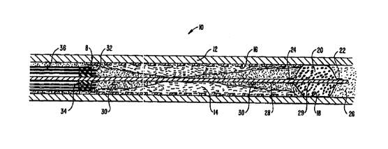
principles of the present invention. Catheter system 2 generally comprises an
electrosurgical catheter 6 connected to a power supply 80 by an
interconnecting cable 86
2o for providing high frequency voltage to a target tissue and an irrigant
reservoir ar source
100 for providing electrically conducting fluid to the target site. Catheter 6
generally
comprises an elongate, flexible shaft body 12 including a tissue ablating
region 8 at the
distal end of body 12, and a proximal balloon 40 positioned on body 12
proximal to region
8. In a specific embodiment, a guide wire 28 (which may also serve as a return
electrode)
2s includes a distal balloon 18 which may be axially translated relative to
region 8 and
proximal balloon 40, as discussed in further detail below.
The proximal portion of catheter 6 includes a multi-lumen fitment I 14
which provides for interconnections between lumens and electrical leads within
catheter 6
and conduits and cables proximal to fltment 114. By way of example, a catheter
electrical
3o connector 96 is removably connected to a distal cable connector 94 which,
in turn, is
removably connectable to generator 80 through connector 92. One or more
electrically
conducting lead wires (not shown) within catheter 6 extend between one or more
active
i.r
CA 02287206 1999-10-20
WO 98/56324 PCT/US98/0797b
21
electrodes at tissue ablating region 8 and one or more corresponding
electrical terminals
(also not shown) in catheter corrector 96 via active electrode cable branch
87. In the
illustrative embodiment, hollom guide wire 28 functions as the return
electrode, and is
electrically attached within a contact housing 111 by a sliding electrical
contact (not
shown}. A return electrode cable branch 89 couples the sliding electrical
contact to
catheter connector 96. Electrical leads within cable 86 allow connection
between terminals
corresponding to return electrode 28 and one or more active electrodes 32 in
distal cable.
connector 94 and generator 80.
Power supply 8U has an operator controllable voltage level adjustment 82 to
1o change the applied voltage level, which is observable at a voltage level
display 84. Power
supply 80 also includes a foot 1>edal 88 and a cable 90 which is removably
coupled to
power supply 80 for remotely adjusting the energy level applied to electrode
terminals. In
an exemplary embodiment, power supply 80 includes three such foot pedals (not
shown),
wherein the first foot pedal is used to place the power supply into the
"ablation" mode and
the second foot pedal places pcwer supply 80 into the "subablation" mode. The
third foot
pedal allows the user to adjust the voltage level within the "ablation" mode.
In the
ablation mode, a sufficient voltage is applied to the electrode terminals to
establish the
requisite conditions for molecular dissociation of the tissue (i.e.,
vaporizing a portion of
the electrically conductive fluid, ionizing charged particles within the vapor
layer and
2o accelerating these charged particles against the tissue). As discussed
above, the requisite
voltage level for ablation will ~~ary depending on the number, size, shape and
spacing of
the electrodes, the distance in which the electrodes extend from the support
member, etc.
Once the surgeon places the power supply in the "ablation" mode, voltage level
adjustment
82 or the third foot pedal may be used to adjust the voltage level to adjust
the degree or
aggressiveness of the ablation.
Of course, it wi Ll be recognized that the voltage and modality of the power
supply may be controlled by ocher input devices. However, applicant has found
that foot
pedals are convenient methods of controlling the power supply while
manipulating-the
probe during a surgical procedure.
3o In the subablatic>n mode, the power supply 80 applies a low enough voltage
to the electrode terminals to avoid vaporization of the electrically
conductive fluid and
subsequent molecular dissociation of the tissue. The surgeon may automatically
toggle the
CA 02287206 1999-10-20
WO 98/56324 PCT/US98/07976
22
power supply between these modes by alternatively stepping on the foot pedals.
This
allows the surgeon to quickly move between subablation (e.g., coagulation) and
ablation in
situ, without having to remove his/her concentration from the surgical field
or without
having to request an assistant to switch the power supply. By way of example,
as the
s surgeon is sculpting soft tissue in the ablation mode, the probe typically
will
simultaneously seal and/or coagulation small severed vessels within the
tissue. However,
larger vessels, or vessels with high fluid pressures (e.g., arterial vessels)
may not be sealed
in the ablation mode. Accordingly, the surgeon can simply step on the
appropriate foot
pedal, automatically lowering the voltage level below the threshold level for
ablation, and
to apply sufficient pressure onto the severed vessel for a sufficient period
of time to seal
andlor coagulate the vessel. After this is completed, the surgeon may quickly
move back
into the ablation mode by stepping on a foot pedal.
Referring now to Figs. 13-22, an exemplary power supply will be
described. The power supply 28 of the present invention may include power
limiting
15 devices to protect attached electrosurgical catheters from excessive power
delivery and to
sustain controlled probe operation. Power is the time rate of transferring or
transforming
energy, and for electricity, power is measured in watts, where one watt is the
power to
create energy at the rate of one joule per second. Referring to Fig. 13, the
power limiting
device 300 is designed to reduce the power drawdown from the power supply 28
when an
2o attached device such as a monopolar or bipolar surgical instrument is not
engaging body
tissue or draws excessive power. For example, excessive power is delivered
from the
power supply 28 if the RF catheter is in saline or blood and is not engaging
target media.
Device 300 conveniently conserves power used in the probe without completely
deactivating the power supply 28 or requiring the user to manually reduce
power.
2s Excessive power draw will overheat the power supply and corrupt power
supply
performance. Device 300 also acts as a safety feature by reducing the stray
emission of
energy when the probe is in transit through the body to a target site.
In general terms, the power limiting device 300 operates on a continuous
basis to detect excessive power output. The device 300 is responsive to the
"total power"
3o delivered by the device. Pig. 14 illustrates the power output of the power
supply 28 when
an excessive power is detected. Device 300 limits the overall output power
from the
controller to be lower than about 240-360 watts, preferably about 300 watts.
Once power
..r ,. , ,
CA 02287206 1999-10-20
WO 98/5b324 PCT/US98/07976
23
output exceeds a predetermined threshold level, the device 300 then operates
on a duty
cycle or periodic detection cycle; 301 between about 50 and 300 ms, where the
device 300
checks every cycle to determine if it is safe to resume power output.
Preferably, the
device 300 has a fixed duty cycle wave form and includes a fixed periodical
pulsing circuit
which is about 10 ms on and 90 ms off. Once the fault condition is gone, power
output
returns to operating levels.
In one embodiment (Fig. 13), the device 300 uses a current sensor 302
attached to the output electrodes to derive the power output of the power
supply 28. The
current limit, which may be set at any desire level, is about 5 amps for a 300
watt power
to limit when voltage is set at about 60V. When current output reaches 5 amps,
the device
300 reduces the output of the power supply to a standby mode. Once in standby
mode,
the power supply preferably hay a pulsatile power output. As shown in Fig. 16,
the device
300 allows the current output to be activated during each duty cycle to
determine if the
power supply may return to normal operation.
t5 When in the stan3by mode, the pulsatile power output may be described as
shown in Fig. 15. In the pulsatile mode, the duty cycle is about 10-15 ms on,
preferably
about 12 ms on, and about 85-90 ms off, preferably about 88 ms off. This
creates a cycle
of about 100 ms, during which time, power is increased and then reduced if the
probe
senses that it is not in the vicinity of body tissue or other higher impedance
material. This
2o sensing step is the initial portio;i of the duty cycle where current is
activated for a period of
time, described as being between 10-15 ms. If current again reaches the 5 amp
level or
some other predetermined level, the output is reduced and the device 300 waits
for the next
duty cycle. The total power output during this short period is only about 10
watts.
However, the current output is sufficient to show that the fault condition
still exists.
25 Thus, when in the standby mode, the device 300 tests for potentially
excessive power
output with a fault condition that occurs without actually reaching the power
level against
which the device is protecting. This pulsatile power output continues until
power
drawdown returns to within acceptable ranges (Fig. 14). The power limiting
reduces
power output on a fault conditim that is current based (so long as there is
constant
3o voltage).
Alternatively, the power limiting device 300 in the standby mode checks the
impedance (instead of current) encountered by the probe every 100ms or over
some other
CA 02287206 1999-10-20
WO 98/56324 PCT/US98/07976
24
interval selected by the user. As long as the probe is in a low impedance
environment and
impedance is below a predetermined level, the power supply will operate in the
pulsatile
mode, never fully activating to therapeutic power levels such as for ablation
or
coagulation. The low impedance is indicative of a potential over power
scenario. In
s alternative embodiments, the device 300 may check the impedance over
variable time
intervals that change as desired. When the probe reaches a target site or
comes in the
vicinity of higher impedance tissue, in one embodiment, a higher impedance is
noted by a
drop in current draw (i.e. power draw) from the probe, signaling the regulator
or logic
unit 310 to increase power on the current or the next duty cycle. This brings
the power
o supply out of the pulsatile mode. The power limiting device 300, however,
will continue
to check the impedance encountered every duty cycle.
Referring to Figs. 16A-16C, a preferred embodiment of the device 300
comprises of at least one current sensor 302 detecting the current output from
DCIDC
converter 304. The current sensor 302 may be configured as one sensor for one
electrode
is or one sensor for a plurality of electrodes. In the present embodiment, one
sensor 302 is
used for six electrodes on the probe, although more preferably one sensor is
used for three
electrodes. Typically, the sensors 302 (noted as T1, T4, T5, etc.) are
configured to wrap
around the electrodes as shown in Fig. 16. Signals from sensors 302 are passed
through a
plurality of rectifying diodes and capacitors which filter and condition the
typically analog
2o signal from the current sensor. In the block diagram of Fig. 13, these
diodes and
components are represented by signal conditioner 306. The conditioned signal
from the
sensor 302 is then passed to a voltage comparator 308. The comparator 308
determines if
the current output has exceed the predetermined threshold level. A logic unit
310 then
determines power of output drive 312 based on the value of the output current
compared to
2s a predetermined current value. In the standby or power limited mode, the
logic unit 310
of the device 300 will preferably duty cycle the output from output drive 312.
Although
the logic unit 310 is preferably an integrated circuit such as a Field
Programmable Gate
Array (FPGA) to maximize cost efficiency, it should be understood that other
devices such
as computers or microprocessors may also be used to perform the required logic
functions.
3o Fig. 17 shows an exemplary embodiment of power limiting device 300.
The circuit diagram shows that overcurrent is sensed by T5. It is rectified
and filtered by
DI6, D17, D18, D19, R33, and C19 etc. The rectified and filtered signals are
fed into
CA 02287206 1999-10-20
WO 98/56324 PCT/US98/07976
voltage comparator which determines if power threshold has been reached. The
output of
the comparator is fed into the F PGA which controls the power supply 28 to the
power
limiting mode (e.g. it turns of 1)CIDC converter lOms on and 90 ms offj.
Device 300
includes an converter of a full-wave bridge arrangement with all four
switching element
s driven by a single transformer. It is capable, through the antiparallel
diodes within the
MOSFETs, of four-quadrant of>eration, returning reactive load energy to the
power supply
for self protection. 100kHz sync arrives as 500 nanosecond pull-up pulses from
a
differentiation network connected to the FPGA. The FPGA also exerts direct
on/off
control via DC EN. The outFut smoothly ramps to regulation when allowed by the
to FPGA. Options for current limiting are provided. Both linear and digital
(pulsatile)
limiting are possible. Current limits may also respond to FPGA commands and
change
under logic control. The inverter is running at zero voltage switching mode to
reduce EMI
and indirectly reduces leakage current. A cycle-by-cycle current limit circuit
serves to
protect the switching elements from energy stored in filter and bypass
capacitors. Cycle-
ts by-cycle current limit control i~ applied by the FPGA removing the gate
drive. The
inverter runs at a fixed 50% dl,ty cycle whenever drive (of about 100 kHz or
other) from
the FPGA is available. The inverter is running at zero voltage switching mode
to reduce
EMI and indirectly reduces leakage current.
The power supply 28 of the present invention may also include a spark
20 limiting device 330 to prevent sudden current spikes which may char or
otherwise damage
the RF probe and surgical target site. For example, when an RF probe attached
to the
power supply touches a metallic object, the impedance encountered by the probe
(relative
to human tissue) decreases suddenly and this undesirably draws a large amount
of current
from the power supply. This sudden current increase may create sparks between
the
25 probe and the metal object. Tlle large amount of current passing through
the probe will
likely char items along the elecarical pathway and may melt electrodes on the
electrosurgical probe.
Referring now to Fig. 18, the spark limiting device 330 will be described in
detail. In general terms, the spark limiting device 330 will reduce current
output to zero
3o when an extremely low imped;mce source such as a metal screw or a rnetai
cannula creates
a high current drawdown. Th~; spark limiting device 330 is located much closer
to the
output electrode. This reduce:; the delay of the device 330 and allows the
device to
ICAI 02287206 1999-10-20
WO 98/56324 PCT/US98/07976
26
respond more quickly. The spark limiting device 330 is directed to reduce
current output
to prevent sparking, not total power output. Although the block diagram of
Fig. 10
appears similar to that of the power limiting device 300, the spark limiting
device 330 is
not a fixed periodical pulsing circuit of the type described in Fig. 17. The
spark limiting
device 330 preferably processes continuous signals, such as analog signals,
from the
current sensor 332. The spark limiting device 330 continuously monitors
current
fluctuations of the power output of converter 334 (typically an AC/DC
converter). The
continuous flow of signal in the spark limiting device 330 allows it to detect
the sudden
increase in current almost instantaneously and almost certainly before the
isolated, power
limiting device 300. Current output is preferably turned off after an
overcurrent is
detected.
The current output during normal therapeutic operation may be in the range
of 0.2 amperes or less. The spark limiting device 330 preferably interrupts
output when
current exceeds about 1.0 to 3.0 amperes. These current levels are
insufficient to cause
sparking, but enough to warrant concern over potential sparking. When current
exceeds
levels higher than those stated, the device 330 will preferably prevent any
current output
from the probe. The output of the power supply 28 is similar to that of Fig.
19. In one
embodiment, the spark limiting device 330 has a built-in delay device that
turns off current
output for a duration of 2-90 ms. Preferably, the delay is programmed into the
FPGA.
2o At the end of the delay period, the device 330 will allow current to flow
through the
probe, albeit at extremely low power, to detect if the extremely low impedance
state still
exists. If current again exceeds the threshold level of about 1.0 to 3.0
amperes (Fig. 19),
the device 330 will zero the output of the power supply and pause for the
built-in delay.
This delay acts in some ways to give the spark limiting device a duty cycle-
like operation.
It should be understood that although no current, preferably, is being
emitted from the probe during the delay period, the power supply does not
shutoff. This is
particularly useful as this eliminates down time associated with restarting
the power supply
from poweroff. As soon as the probe is removed from the area of extremely low
impedance, the spark limiting device 330 will allow power to flow from the RF
probe as
3o usual. Preferably, as long as the probe is exposed to the low impedance
source, the device
330 will not allow power to be transmitted. Of course, it may be possible to
configure the
CA 02287206 1999-10-20
WO 98/56324 PCT/US98/07976
27
spark limiting device 330 to all ow a low level of current to be emitted,
versus shutting off
the power output completely.
The block diagr;~m of Fig. 18 shows that the spark limiting device 330
includes a signal conditioner 3:36, a level detector 338, a regulator or logic
unit 340, and
an output driver 342 (such as an RF source known in the art). Although the
preferred
embodiment of the spark limiting device 330 is based on analog signals, it
should be
understood that the device 330 may be adapted to used analog signals or
digital signals
with extremely short duty cycl~a to approximate a continuous system. The logic
device
340, level detector 338, and si,;nal conditioner 336 may all be combined into
a single
1o device or processor as indicated by the dotted line 344. The same may also
apply to the
power limiting device 300 whi:.h has components that may be integrated
together.
Referring to Fig. 20, a circuit diagram of the spark limiting device 330 is
shown. The
current sensors 332 are denoted by elements T8, T9, T12, and T13. The diodes
D36-D76
and resistors/capacitors RI31-:21331C73 etc. are used to condition the analog
signal to
remove noise and other undesirable qualities. A voltage comparator U8D and the
FPGA,
corresponding to level detector 338 and logic device 340, are used to
determine if the
output current detected by sensor 332 is above a predetermined level.
Although the power limiting device 300 and the spark limiting device 330
may be used individually, it is understood that the two devices may also be
used
2o concurrently in the power supply. In a preferred embodiment, the power
supply of the
present invention has the powf;r limiting device 300 and the spark limiting
device 330
arranged in a serial configuration as shown in Fig. 21-22. This configuration
provides for
the circuit isolation mandated by safety regulations for medical device power
supplies. As
shown in Fig. 14, the power s upply 28 has P/O primary isolation, PIO
secondary
isolation, and patient isolation, Using devices 300 and 330 also provides
protection for
both converters (DC/DC and l~C/AC) used to provide stability of the power
output. Due
to the various isolation barriers required to meet safety and regulatory
standards for
medical device power supplies , the spark limiting device 330 is typically
located closer to
the electrode output while the power limiting device 300 is more isolated from
the
3o electrode output. The additional amount of isolation circuitry to
introduces a lag time into
the responsiveness of the pow ~r limiting device. Thus, one device reacts
slower and while
CA 02287206 1999-10-20
WO 98/56324 PCT/US98/07976
28
the device closer to the electrode reacts faster. In one embodiment, there is
about a 200
ms delay in order for the current to reach the power limiting device 300.
As an example of how the devices would function together, when an
attached RF catheter touches a metallic object, such as a stmt within a body
passage, the
spark limiting device 330 activates to reduce the current output from the
power supply to
zero. The current output may be reduced to some nonzero value so long as
sparks are not
generated. The spark limiting device 330 introduces a delay and then checks to
see if it
can power up. During this time, the power limiting device 300 also continues
to check
about every duty cycle to see if power should be increased. In one embodiment,
the power
to limiting device 300 introduces more delay into the system since its duty
cycle is longer
than the 2-90 ms delay of the spark limiting device. As soon as the probe is
removed from
the extremely low impedance site and current drawdown stays within acceptable
ranges,
the probe resumes normal operations. If the catheter is no longer in contact
with target
tissue, then the generator will most likely be in pulsatile mode while
awaiting to be
is repositioned.
Referring again to Fig. 1, conductive fluid 30 is provided to tissue ablation
region 8 of catheter 6 via a lumen (not shown in Fig. 1 ) within catheter 6.
Fluid is
supplied to lumen from the source along a conductive fluid supply line 102 and
a conduit
103, which is coupled to the inner catheter lumen at rnulti-lumen fitment 114.
The source
20 of conductive fluid (e.g., isotonic saline) may be an irrigant pump system
(not shown) or a
simple gravity-driven supply, such as an irrigant reservoir 100 positioned
several feet
above the level of the patient and tissue ablating region 8. A control valve
104 may be
positioned at the interface of fluid supply line 102 and conduit 103 to allow
manual control
of the flow rate of electrically conductive fluid 30. Alternatively, a
metering pump or flow
25 regulator may be used to precisely control the flow rate of the conductive
fluid.
System 2 further includes an aspiration or vacuum system (not shown) to
aspirate liquids and gases from the target site, and syringes 106, 108 for
inflating distal
and proximal balloons 18, 40, respectively. By way of example, as the plunger
of'syringe
108 is depressed, fluid in the syringe chamber is displaced such that it flows
through a
3o conduit 107 and an internal lumen 57 within catheter 6(not shown in Fig. 1)
to expand and
inflate balloon 40. Likewise, syringe 106 is provided at the proximal end of
guide wire 28
for inflating distal balloon 18, as shown by translation vectors 116, 118.
Also, guidewire
_,
CA 02287206 1999-10-20
WO 98/56324 PCT/US98/07976
29
28 can be advanced or retracted relative to tissue ablation region 8 of
catheter 6 as shown
by translation vectors 116, 118 such that, for each increment of relative
displacement 116
at the proximal end of catheter 6, there is a corresponding displacement 118
of the hollow
guidewire 28 relative to the tis:~ue ablating region 8 of catheter 6.
Referring now tc~ Figs. 2A-2C, one embodiment of the method and
apparatus of the present invention will be described in detail. As shown,
tissue ablating
region 8 of catheter 6 progresses through occlusive media 14, such as
atheromatous media
or thrombus within a body lumen 10, e.g., a blood vessel. The principles of
the present
invention are also applicable to any body lumen which becomes partially or
totally
0 occluded. The present invention is particularly useful in a lumen containing
a lumenal
prosthesis, such as a stmt 16, ;dent-graft or graft, which may be metallic,
non-metallic or a
non-metallic coated metallic structure. A particular advantage of the present
invention is
the confinement of current flour paths (not shown) between the return
electrode (hollow
guide wire 28 in the present example) and one or more active electrodes 32 to
the vicinity
of tissue ablating region 8. This confinement of current flow paths minimizes
the
undesired flow of current through portions or all of stmt 16, which may
otherwise induce
non-specific tissue injury beyo Zd the site of recanalization of the occluded
lumen 10.
Referring to Fig . 2A, tissue ablating region 8 of catheter 6 is positioned
proximal to the occlusive media 14 within lumen 10. The distal region of
hollow guide
2o wire 28 is positioned distal to the occlusive media 14 either before or
after the initial
positioning of tissue ablation r~:gion 8. Once hollow guide wire 28 is
positioned as shown
in Fig. 2A, proximal balloon 40 (not shown in Fig. 2A) is inflated to effect a
seal between
catheter shaft 42 and interior vrall 12 of lumen 10 to minimize the flow of
bodily fluid 26
(e.g., blood) from regions pro:Kimal to the tissue ablating region 8 of
catheter 6.
2s Electrically conductive and biologically compatible fluid 30 (e.g.,
isotonic saline) is
delivered into lumen 10 for a ; ufficient period of time to displace naturally
occurring
bodily fluid 26 in the region b~aween the tissue ablating region and the
distal tip of guide
wire 28. After the bodily fluid has been displaced, distal balloon 18 is
inflated to effect a
seal between balloon 18 and t)~.e interior wall 12 of lumen 10.
3o Once the target site is isolated from the rest of the vasculature, the
supply of
electrically conductive fluid 3(> is continuously delivered to region 8 and
balanced with the
aspiration of fluid from the sits of intended recanalization. The active
electrodes) 32 is
ICA' 02287206 1999-10-20
WO 98/56324 PCT/US98/07976
(are) then energized by applying a high frequency voltage between active
electrodes) 32
and return electrode or guide wire 28. A high electric field is created at the
surface of
active electrodes) 32 which causes the volumetric removal or ablation or
target tissue in
close proximity with active electrodes) 32. The flow of electrical current
between return
5 electrode 28 and active electrodes) 32 is shown by current flux lines 62 in
Fig. 2B. As
the occlusive media 14 is ablated, gaseous products are generated (not shown)
which are
entrained in the electrically conducting fluid 30 and removed through
aspiration lumen 58
(not shown). The current flux lines 62 are generally confined to the central
portion of
tissue ablation region 8 because they generally flow inward towards return
electrode 28
1o and because the occlusive media 14 generally shields the outer region of
lumen (including
stent 16) from flux lines 62. This minimizes undesirable interaction between
the electrical
current and stmt 16.
Referring to Fig. 2C, this ablation procedure is continued until the desired
length of the lumen containing occlusive media is recanalized. During the
recanalization
1 s process, the products of ablation are confined between proximal balloon 40
and distal
balloon 18 to minimize, for example, the injection of any non-condensable
gaseous
products of ablation into the blood stream which could otherwise lead to the
formation of
injurious or life-threatening emboli. Once the occlusive media 14 has been
volumetrically
removed (i.e., ablated), the energy application is suspended, the valve on the
aspiration
20 lumen is closed, control valve 104 is closed and balloons 18, 40 are
deflated. The time
period from the initial inflation of balloons 18, 40 to the deflation of these
balloons is
typically about 15-45 seconds, depending on the length and the extent of
occlusion in the
vessel. For longer occlusions, the above process may be repeated several times
with
intervals of no balloon inflation so that vital oxygen-bearing blood can be
reperfused
25 through the zone of intended recanalization to preserve the tissue distal
to the
recanalization zone.
A first embodiment of tissue ablation region 8 of catheter 6 is shown in
Figs. 3A and 3B. As shown, two active electrodes SOa and SOb are secured
within an
electrically insulating support member 34. The electrodes SOa, SOb are
preferably
3o composed of a refractory, electrically conductive metal or alloy, such as
platinum,
titanium, tantalum, tungsten, stainless steel, gold, copper, nickel and the
like. The support
member 34 is secured to the distal end of catheter 6 with a biocompatible
adhesive 60
r
CA 02287206 1999-10-20
WO 98/56324 PCT/US98/07976
31
between support member 34 and outer sleeve 36. An inorganic electrically
insulating
sleeve 54 preferably extends move the distal plane of active electrodes 50a,
50b by a
distance HS. A central lumen .n support member 34 provides a passageway for
guide wire
28 that permits axial displacement and rotation of tissue ablating region 8
relative to guide
wire 28.
In an exemplary embodiment, the support member 34 will comprise an
inorganic insulator, such as ceramic, glass, glass/ceramic or a high
resistivity material,
such as silicon or the like. An inorganic material is generally preferred for
the
construction of the support member 34 since organic or silicone based polymers
are known
1o to rapidly erode during sustained periods of the application of high
voltages between
electrodes 50 and the return electrode 28 during tissue ablation. However, for
situations in
which the total cumulative time of applied power is less than about one
minute, organic or
silicone based polymers may be used without significant erosion and loss of
material of the
support member 34 and, therefore, without significant reduction in ablation
performance.
~s As shown in Fig. 3A, an irrigation lumen 56 and an aspiration lumen 58 are
provided to inject electrically conducting fluid 30 and remove gaseous
products of ablation
48 from the site of recanalization. An additional fluid lumen 57 provides
fluid
communication between inflation syringe 108 and proximal balloon 40. This
fluid lumen
57 is filled with a sealant in tLose portions of the catheter distal to
proximal balloon 40.
2o In use with the present invention, catheter 6 is rotated about 180 degrees
clockwise and then about 180 degrees counter clockwise as the electrodes 50
are energized
by generator 80 (Fig. 1) to ef~ect ablation of the occlusive media. Using a
reciprocating
rotational motion combined with a small pressure to advance tissue ablation
region 8
through the longitudinal lengt 1 of the occlusive media 14 allow
recanalization of the
25 occluded vessel as described with reference to Figs. 2A-2C. The cross-
sectional shape of
the active electrodes may be round wires as shown in Fig. 3B, or they may have
shaped
surfaces to enhance the electri c field intensity at the distal surfaces of
the active electrodes
50. Suitable electrode designs for use with the present invention may be found
in~co-
pending, commonly assigned application Serial No. 08/687,792, filed July 19,
1996
30 (Attorney Docket No. 16238-001600), the complete disclosure of which is
incorporated
herein by reference for all pu;-poses.
CA 02287206 1999-10-20
WO 98/56324 PCT/US98/07976
32
Return electrode 28 comprises an electrically conducting material, usually
metal, which is selected from the group consisting of stainless steel alloys,
platinum or its
alloys, titanium or its alloys, molybdenum or its alloys, and nickel or its
alloys. The
return electrode 28 may be composed of the same metal or alloy which forms the
active
s electrodes 50 to minimize any potential for corrosion or the generation of
electrochemical
potentials due to the presence of dissimilar metals contained within an
electrically
conductive fluid 30, such as isotonic saline (discussed in greater detail
below).
Referring now to Figs. 4A and 4B, a second embodiment of tissue ablation
region 8 of catheter 6 will now be described. In this embodiment, four active
electrodes
32a, 32b, 32c, 32d are secured within an inorganic electrically insulating
support member
34. Similar to the previous embodiment, support member 34 is secured to the
distal end of
catheter 6 with a biocompatible adhesive 60 between support member 34 and
outer sleeve
36. An inorganic electrically insulating sleeve 54 preferably extends above
the distal plane
of active electrodes SOa, SOb by a distance HS. A central lumen in support
member 34
~ 5 provides a passageway for guide wire 28 that permits axial displacement
and rotation of
tissue ablating region 8 relative to guide wire 28. As shown in Fig. 4A, an
irrigation
lumen 56 and an aspiration lumen 58 are provided to inject electrically
conducting fluid 30
and remove gaseous products of ablation 48 from the site of recanalization. An
additional
fluid lumen 57 provides fluid communication between inflation syringe 108 and
proximal
2o balloon 40. This fluid lumen 57 is filled with a sealant in those portions
of the catheter
distal to proximal balloon 40.
In use, catheter b is rotated about 180 degrees clockwise and then about 180
degrees counter clockwise as the electrodes 32 are energized by generator 80
(Fig. 1) to
effect ablation of the occlusive media. Using a reciprocating rotational
motion combined
25 with a small pressure to advance tissue ablation region 8 through the
longitudinal length of
the occlusive media 14 allow recanalization of the occluded vessel as
described with
reference to Figs. 2A-2C. The cross-sectional shape of the active electrodes
may be round
wires as shown in Fig. 4B, or they may have shaped surfaces to enhance the
electric field
intensity at the distal surfaces of the active electrodes 32 as described co-
pending,
3o commonly assigned application Serial No. 08/687,792, filed July 19, 1996
(Attorney
Docket No. 16238-001600), the complete disclosure of which has previously been
incorporated herein by reference.
r . i . t
CA 02287206 1999-10-20
WO 98156324 PCT/US98/07976
33
The second embodiment of Figs. 4A and 4B is illustrated in greater detail in
Figs. 5A and SB. As shown, electrically conductive fluid flows through
irrigation lumen
56 of catheter 6 to and through irrigation port 44 and subsequently surrounds
the target
tissue site (i.e., occlusive media 14). When high frequency voltage is applied
between the
s return electrode 28 and active ~:lectrodes 32, a vapor layer 64 forms at and
around active
electrodes 32 with concomitant volumetric removal (ablation) of the occlusive
media 14.
A more detailed description of this phenomena can be found in commonly
assigned, co-
pending application Serial No. 081561,958, filed on November 22, 1995
(Attorney Docket
16238-000700), the complete c.isclosure of which has previously been
incorporated herein
1o by reference. The occlusive media 14 is decomposes into gaseous products of
ablation 48
which are entrained in electrically conducting fluid 30 and evacuated through
aspiration
port 46 and to the proximal eml of catheter 6 via aspiration lumen 58.
A third embodiment of tissue ablation region 8 is illustrated in Figs. 6A and
6B. Many of the elements of this embodiment are the same as previous
embodiments, and
15 therefore will not be repeated. As shown, a single active electrode 200 is
secured within
support member 34. Active electrode 200 preferably has an L-shaped distal end
so that a
distal portion 202 of electrode 200 extends radially outward along the distal
surface of
support member 34. As before:, electrode 200 is rotated in both directions, as
the region 8
is advanced through the lumen to recanalize the lumen.
2o A fourth embodiment of tissue ablation region 8 is illustrated in Figs. 7A
and 7B. Many of the element. of this embodiment are the same as previous
embodiments,
and therefore will not be repeated. As shown, six active electrodes 66a-66f
are secured
within inorganic support memi>er 34. An annular irrigation lumen 55 and an
aspiration
lumen 59 are provided to inject electrically conducting fluid 30 and remove
gaseous
25 products of ablation 48 from the site of recanalization. When high
frequency voltage is
applied between the return ele~arode 28 and active electrodes 66, a vapor
layer 64 forms at
and around active electrodes 65 with concomitant volumetric removal (ablation)
of the
occlusive media 14. For this f;mbodiment and that shown in Figs. 8A and 8B,
rotation
may be limited to +- 30 degrees due to the greater number and circumferential
distribution
30 of active electrodes. The power or current supplied to each electrode may
be individually
controlled by active or passive mechanisms as previously described in commonly
assigned,
co-pending application Serial lJo. 08/561,958, filed on November 22, 1995
(Attorney
CA 02287206 1999-10-20
WO 98/56324 PCT/US98/07976
34
Docket 16238-000700). The occlusive media 14 is decomposed into gaseous
products of
ablation 48 which are entrained in electrically conducting fluid 30 and
evacuated through
aspiration port 46 and onto the proximal end of catheter 6 via aspiration
lumen 59. As
shown in Fig. 7b, the current flux lines 62 are confined to the central
portions of tissue
s ablation region 8.
Figs. 8A and 8B illustrate a fifth embodiment of the present invention. This
embodiment is similar to the fourth embodiment in that six active electrodes
66a-66f are
secured within inorganic support member 34. A return electrode 70 (e.g., metal
sleeve) is
positioned proximal to the active electrodes 66a-66f by a distance HX. In this
embodiment,
t o current flux lines 62 travel proximally from the distal tips of electrodes
66 to the
proximally spaced return electrode 70.
Referring to Figs. 9A and 9B, a sixth embodiment of the invention will now
be described. As shown, a single active electrode 72 is secured within
inorganic support
member 34. In this embodiment, active electrode 72 comprises a coiled wire
having a
15 plurality of concentric coils tightly and helically wrapped and secured on
support member
34 (Fig. 9B). Preferably, the helical coil extends around return electrode 28
in concentric
configuration, as shown in Fig. 9A.
A seventh embodiment of the invention is shown in Figs. l0A and lOB.
This embodiment is similar to the sixth embodiment except that the single
active electrode
20 73 defines a series of concentric machined grooves 75 to form concentric
circular
electrodes 78 surrounding return electrode 28. The distal edges of electrodes
78 generate
regions of high electric field intensities when high frequency voltage is
applied between
return electrode 28 and concentric active electrodes 78. A vapor layer 64
forms at and
around active electrodes 78 with concomitant volumetric removal (ablation) of
the
25 occlusive media. The embodiments of Figs. 9 and 10 are usually advanced
through the
occlusive media without rotation.
As an alternative to the irrigation lumens shown above, the irrigant or
electrically conductive fluid may be supplied through the lumen of tubular
electrodes (not
shown). This may be advantageous in ensuring that electrically conductive
fluid is injected
3o into close proximity to the site of tissue ablationlcutting. Further, the
tubing can be filed
to expose additional edges to enhance the tissue cutting effect.
During the percutaneous introduction and removal of catheter body 62,
CA 02287206 1999-10-20
WO 98/56324 PCTIUS98/07976
measures should be taken to pr~:vent iatrogenic injury to the walls of the
body lumen as the
other tissues encountered along the pathway to the target site. In one
embodiment,
catheter 60 includes a compliar, t, atraumatic safety sheath {not shown) which
extends over
the working end of the catheter. In use, the sheath is advanced forward during
5 introduction and removal of tis~~ue ablation region 64. Once the target site
has been
accessed, the compliant, atraunlatic safety sheath is retracted (e.g., a
distance of 1.5 to 2.0
cm) exposing the electrode terr,ainal(s). The safety sheath is preferably
constructed using
thin-walled plastic tubing selecv:ed to provide biocompatability, compliance
and low friction
during insertion and removal. A number of plastic materials are available for
this purpose
o and include Teflon, polypropylene and polyvinyl chloride. The activation
mechanism may
be (1) the thin-walled plastic tubing moved relative to the catheter body at a
location
external to the patient's body or (2) a drive rod or wire {not shown) within
the catheter
body which actuates a short sel;ment of the safety sheath {e.g., 4 to 8 cm)
located at the
distal end of the catheter body .
15 In another aspect of the invention, the catheter includes a radially
expandable portion for allowin; the diameter of the electrode terminals to be
varied
according to the diameter of the body lumen. In some instances, stems will not
expand
uniformly resulting in portions of the stmt having smaller inner diameters. In
other
instances, vessel wall pressure may cause portions of the stmt to spring back
to its original
2o shape or partially back to this ;,hape so that the overall inner diameter
of the stmt varies in
the axial direction. According ~y, the present invention allows the diameter
of the working
end of the catheter to vary (either automatically in response to the body
lumen or stent
inner diameter, or through activation by the surgical team) to facilitate
advancement
through non-uniform stems or body lumens.
25 Referring now t~ Figs. 11 and 12, a catheter 500 includes a catheter body
501 and a radially expansible corking end portion 502 (e.g., a balloon or
similar
expansible member) supporting a plurality of electrode terminals 504
circumferentially
spaced around the working end 502. Working end 502 preferably comprises an
elastic
material that will allow the working end 502 to expand up to at least 25 % of
its original
3o diameter, usually at least 100 r~ of the original diameter, and often at
least 200% of the
original diameter (see Fig. 12). As shown, electrode terminals 504 are loops
512 formed
by a pair of elongate wires SOf~ extending through tubular support members
510. This
ICA' 02287206 1999-10-20
WO 98/56324 PCTIUS98107976
36
configuration provide the distal end of electrode terminals 504 with
sufficient flexibility to
expand outward (the loop straightens in the expanded configuration), as shown
in Fig. 12.
Of course, electrode terminals 504 may comprise a variety of other
configurations. In
addition, catheter 500 may include a single annular electrode terminal, as
discussed above.
Tubular support members 510 preferably comprise an inorganic material, such as
ceramic
or glass. Support members 510 are loosely coupled to each other with a
flexible sheath
520. As the balloon 502 expands (Fig. 12), tubular support members 510 expand
away .
from each other.
In the embodiment of Figs. 11 and 12, the guide wire (not shown) functions
to as the return electrode. However, it will be understood that the return
electrode may be
positioned on the catheter proximal to electrode terminals, and may be part of
the
expandible working end 502 of catheter 500. Alternatively, one or more of the
electrode
terminals 504 may serve as the return electrodes) by applying the opposite
polarity to
these electrode terminals 504.
1 s In other configurations (not shown in the figures), the working end of the
catheter will taper in the distal direction (e.g., in a series of steps) so
that the surgeon can
advance the catheter through a severely occluded body lumen. The catheter may
include a
series of axially spaced electrode terminals) that are electrically isolated
from each other
to allow for each set to be independently activated. By way of example, in a
severely
20 occluded body lumen, the surgeon may activate the distal set of electrode
terminals) to
remove the innermost occlusive media, advance these distal electrode
terminals) through
the vacancy left by the removed occlusive media, and then activate a more
proximal, and
radially outward, set of electrode terminals) to remove occlusive media
radially outward
from the initially removed media.
25 Referring now to Fig. 23, a method for recanalizing a severe occlusion 402
in a body passage 400 will be described. As shown, the occlusion 402
completely blocks
the body passage, making it extremely difficult to recanalize with
conventional catheter
techniques. In these circumstances, it is necessary to at least partially
recanalize (creating
an opening through) the occlusion before conventional catheter procedures can
begin.
3o Conventional methods for recanalizing severe occlusions include hot-tipped
catheters, laser
catheters, and drill-tipped catheters. These approaches rely on very
aggressive treatment
... , , ,
CA 02287206 1999-10-20
WO 98/56324 PCT/US98/07976
37
of the stenotic material, which ran expose the blood vessel wall 404 to
significant injury,
for example, vessel perforation
According to the present invention, the working end 406 of an
electrosurgical catheter 408 is advanced through the body passage 400 to the
site of
recanalization. The catheter 408 may be advanced with a variety of techniques,
such as a
guidewire, steerable catheter and the like. Once the surgeon has reached the
point of major
blockage, electrically conductive fluid is delivered through one or more
internal lumen(s).
409 within the catheter to the ti sue. In some embodiments, the catheter may
be
configured to operate with a na~:urally occurring body fluid, e.g., blood, as
the conductive
1o medium. The fluid flows past the return electrode 420 to the electrode
terminals 422 at the
distal end of the catheter shaft. The rate of fluid flow is controlled with a
valve (not
shown) such that the zone betw,~en the occlusion and electrode terminals) 422
is constantly
immersed in the fluid. The power supply 28 is then turned an and adjusted such
that a
high frequency voltage difference is applied between electrode terminals 422
and return
t 5 electrode 420. The electrically conductive fluid provides the conduction
path (see current
flux lines) between electrode tevminals 422 and the return electrode 420.
Figs. 24A and 2~EB illustrate the volumetric removal of occlusive media in
more detail As shown, the high frequency voltage is sufficient to convert the
electrically
conductive fluid (not shown) between the occlusion 402 and electrode
terminals) 422 into
2o an ionized vapor layer 412 or plasma. As a result of the applied voltage
difference
between electrode terminals) 4Z2 and the occlusive media 402 (i.e., the
voltage gradient
across the plasma layer 412), charged particles 415 in the plasma {viz. ,
electrons) are
accelerated towards the ocelusicm. At sufficiently high voltage differences,
these charged
particles 415 gain sufficient energy to cause dissociation of the molecular
bonds within
25 tissue structures. This molecul;~r dissociation is accompanied by the
volumetric removal
(i.e., ablative sublimation) of tissue and the production of low molecular
weight gases 414,
such as oxygen, nitrogen, carbon dioxide, hydrogen and methane. The short
range of the
accelerated charged particles 415 within the tissue confines the molecular
dissociation
process to the surface layer to n:linimize damage and necrosis to the
surrounding vessel
3o wall 404. During the process, he gases 414 may be aspirated through
catheter 408. In
addition, excess electrically conductive fluid, and other fluids (e.g., blood)
may be
aspirated from the target site to facilitate the surgeon's view.