Note: Descriptions are shown in the official language in which they were submitted.
CA 02288133 1999-10-25
WO 98/48702 PCT/US98/08581
1
Suturing Instrument With Rotatably Mounted Offset
Needle Holder and Method of Using the Same
BACKGROUND OF THE INVENTION
Field of the Invention:
The present inv~tion pertains to suturing of bodily or anatomical tissue and,
more particularly, to an apparatus and method for suturing anatomical tissue
during
endoscopic and open surgical procedures.
Discussion of the Related Art:
Suturing of bodily tissue, that is, the practice of using lengths of suture
material to ligate or approximate tissue, is a time consuming part of most
surgical
~xocedures including both open surgery and endoscopic or closed surgery. "Open
surgery" refers to surgery wherein the surgeon gains access to the surgical
site by
a relatively large incision and "endoscopic surgery" refers to minimally
invasive
surgery wherein the surgeon gains access to the surgical site via one or more
portals
through which endoscopes are introduced to view the surgical site and through
which
instruments, such as forceps, cutters, needle holders and the like, are
introduced to
the surgical site.
In the past, suturing has been accomplished with the use of a sharp suture
needle carrying a length of suture material, the suture needle being caused to
penetrate and pass through the tissue pulling the suture material through the
tissue.
CA 02288133 1999-10-25
WO 98/48702 PCTlUS98/08581
2
Once the suture material has been pulled through the tissue, the surgeon ties
a knot
in the suture material. The knotting procedure allows the surgeon to adjust
the
tension on the suture material to accommodate the particular tissue being
sutured
and to cor~trof approximation, occlusion, attachment or other conditions of
the tissue.
The process of tissue penetration and knotting of the suture material can be
time consuming and tedious work, particularly when performed in connection
with
microsurgery and endoscopic surgery and can unduly prolong the duration of
surgery
and therefore the period in which the patient is under anesthesia.
Nevertheless,
endoscopic surgery is preferred over open surgery due to the greatly reduced
trauma
and wound healing time for the patient and due to cost savings associated with
shorter hospital stays and performing surgery in non-hospital or out-patient
surgery
sites: Accordingly, there has been much effort to develop techniques for
facilitating
the suturing normally performed by use of a suture needle and a length of
suture
material. Alternative techniques proposed have included electrical
coagulation,
mechanical devices such as clips, clamps and staples, and lasers. However, no
alternative technique has yet been well accepted by surgeons to produce the
results
obtained by suturing and knotting. Thus, there is a great need for suturing
techniques useful in endoscopic surgery to permit surgeons to suture
anatomical
tissue using suture needles and lengths of suture material in a time
efficient,
consistent and precise manner.
The performance of an endoscopic procedure typically involves creation of
one or more puncture sites through a wall of an anatomical cavity using a
penetrating
instrument including an obturator, such as a trocar, disposed within a portat
sleeve.
After the penetrating instrument has penetrated into the anatomical cavity,
the
obturator is withdrawn leaving the sleeve in place to form a portal in the
cavity wall
for the introduction of instruments such as endoscopes, scissors, forceps,
needle
holders and the like (known generally as "end effectors") into the anatomical
cavity.
CA 02288133 1999-10-25
WO 98/48702 PCTNS98/08581
3
Suturing is typically performed with a needle holding instrument, or needle
holder, having a pair of jaws adapted to hold the body of a suture needle. The
jaws
of the needte holding instrument are inserted through the portal sleeve and
are
positioned at the operative site by manipulation of a handle at the proximal
end of the
instrument outside the body. With a suture needle held between the jaws of the
needle holding instrument, the handle is manipulated to cause a tip of the
needle to
be pushed through the tissue being sutured. Once the tip of the suture needle
has
been pushed through the tissue, the jaws of the needle holding instrument are
opened to release the suture needle so that the tip of the needle can be
grasped and
pulled through the tissue therewith, or, after opening the jaws, a second
needle
holding instrument is introduced at the operative site through another portal
to grasp
the tip of the suture needle after it has emerged from the tissue being
sutured.
These techniques require difficult manipulation of the needle holder of the
suture
needle within the jaws of the needle holder before another stitch can be made.
U.S. Patent Application Serial No. 08/758,648, the disclosure of which is
incorporated herein by reference, discloses a suture device having two needle
holders, i.e. a needle driver and a needle catcher, in a single endoscopic
instrument.
However, the device disclosed in this pending application has a working span
that
is confined within the diametrical limitations of the endoscopic device. This
can
present difficulties when large portions of tissue are to be sutured. U.S.
Patent No.
5,582,617 discloses an endoscopic instrument having an end effector that can
move
from a position within the diameter of the barrel of the device to a position
outside the
diameter. However, this device must pivot about an axis that is transverse to
the axis
of the barrel and an axis that is coincident with the axis of the barrel and
thus
requires a complex movement and linkage to accomplish the disclosed functions.
Accordingly, this device falls short of providing a needle holder that can be
utilized
over a large working span.
Of course, it is also generally desirable to minimize the size of each
puncture
site. Further, in order to permit a wide range of tissue sizes to be sutured,
it is
i
CA 02288133 1999-10-25
WO 98/48702 PCTIUS98108581
4
desirable to provide a needle holder that moves through a path having a large
radius
of curvature, i.e. a large working span. These objectives, small size of
punctures,
and a large working span, are seemingly contradictory. Conventional devices
have
not achieved the above-noted objectives in a satisfactory manner.
SUMMARY OF THE INVENTION
Accordingly, it is a primary object of the present invention to overcome the
above-mentioned disadvantages of the prior art and to improve suturing
instruments
and methods of suturing anatomical tissue.
It is a further object of the present invention to permit a suturing
instrument as
well as other medical instruments and devices to be introduced through a
single
portal in an endoscopic procedure without having to withdraw the suturing
instrument
from the portal.
It is another object of the invention to increase the working span of an
endoscopic suturing device and to reduce the insertion diameter while
replicating the
natural motion of needle passage.
It is another object of the invention to easily manipulate a needle holder
during
suturing.
Finally, it is an object of the invention to control an endoscopic or open
surgical suturing procedure with standard proximal end controls.
The present invention allows suturing of anatomical tissue to be accomplished
in a time efficient, consistent and precise manner. Also, suturing can be
accomplished using standard suture needles and filamentous suture materials
without the need for additional instruments at the operative site.
A first aspect of the present invention is generally characterized in an
instrument for suturing anatomical tissue with a suture needle including a
barrel, a
needle holder having a shaft that is mounted in the barrel for rotation about
an axis.
The needle holder shaft has needle holding jaw members offset from the axis
and
selectively operable to grasp and release the suture needle. The jaw members
are
CA 02288133 1999-10-25
WO 98/48702 PCTIE1S98/08581
coupled to the shaft by arms or connecting portions extending from a distal
end of the
shafts. When the jaw members of the needle holder are operated to grasp the
suture needle, the needle holder can be rotated to drive the suture needle
through
a path having a large radius of curvature to penetrate anatomical tissue.
During
insertion into an anatomical cavity through a portal or the like, the jaw
members are
contained within a diametrical dimension of the device. However during
suturing, the
jaw members can extend beyond this dimension due to the offset configuration.
Another aspect of the present invention is generally dlaraderized in a method
of suturing anatomical tissue using a length of suture material attached to a
suture
needle. The method indudes the steps of grasping the suture needle with offset
jaw
members of a needle holder, rotating the needle holder in a first direction to
cause
the tip of the needle to penetrate the anatomical tissue, releasing the suture
needle
from the needle holder, rotating the needle holder in a second direction to
grasp the
needle tip, and rotating the needle holder in the first direction again to
pull the needle
and the suture material through the anatomical tissue.
fn another aspect of the invention, a needle holder having offset jaw members
is combined with a ligating instrument in a single endoscopic device. The
ligating
instrument carries one or more loops of suture material that can be drawn
tightly
around a knotting element or the like to secure suture material after the
suture
material has been passed through the tissue by the needle driver. The ligating
instrument is inserted through an operating channel fomned in the shaft of the
needle
holder.
Other objects and advantages of the present invention will become apparent
from the following description of the preferred embodiments taken in
conjunction with
the accompanying drawings, wherein like parts in each of the several figures
are
ident~ed by the same reference numerals.
BRIEF DESCRIPTION OF THE DRAWINGS
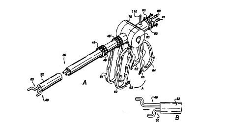
Fig. 1 is a side view of the first preferred embodiment;
CA 02288133 1999-10-25
WO 98/48702 PCT/US98/08581
6
Fig. 2 is a perspective view of a distal end of the preferred embodiment in an
operating position;
Fig. 3A is an end view of the first preferred embodiment in the insertion
position;
Fig. 3B is a perspective view of the distal end of the first preferred
embodiment in the insertion position;
Fig. 4A is a sectional view taken along line 4-4 of Fig. 1 illustrating the
inner
mechanism of the proximal controls in an operative position;
Fig. 4B is a sectional view taken along line 4-4 of Fig. 1 illustrating the
inner
mechanism of the proximal controls in an adjusting position;
Fig. 5A illustrates the needle driver removed from the barrel for illustrative
purposes;
Fig. 5B illustrates an alternative needle driver;
Fig. 5C illustrates an alternative needle driver;
Fig. 5D illustrates an alternative needle driver;
Fig. 5E shows a distal end of the preferred embodiment with a modified jaws
in the open position;
Fig. 5F shows a distal end of the preferred embodiment with a modified jaws
in the closed position;
Fig. 5G shows a distal end of the preferred embodiment with another modified
jaws in the closed position;
Fig. 5H shows a distal end of the preferred embodiment with another modified
jaws in the open position;
Fig. 5K illustrates a distal end of the preferred embodiment with another
modified jaws;
Fig. 5L illustrates a distal end of the prefer-ed embodiment with another
modified jaws;
Fig. 6A is an end view of the distal end of the first preferred embodiment
illustrating pushing a needle through tissue;
CA 02288133 1999-10-25
WO 98/48702 PCT/US98/08581
7
Fig. 6B is an end view of the distal end of the first preferred embodiment
illustrating pulling a needle through tissue;
Fig. 7A is a perspective view of the distal end of the first preferred
embodiment used in combination with a ligator for suturing;
Fig. 7B is a perspective view of the distal end in combination with an
alternative ligator;
Fig. 8 illustrates a ligator for use with the invention
Fig. 9 is a side view of a multiple ligator cluster that can be inserted
through
an operating channel of the first preferred embodiment;
Fig. 10 is an end view cluster illustrated in Fig. 8;
Fig. 11 is a perspective view of the first preferred embodiment with a
multiple
ligator cluster inserted through an operating channel;
Fig. 12 is a sectional view of an end of the multiple ligator cluster
illustrated
in Fig. 11 taken along line 12-12;
Fig. 13 is a sectional view of the distal end of the first preferred
embodiment
with modified jaws in the closed position;
Fig. 14 is a sectional view of the distal end of the first preferred
embodiment
with modified jaws in the open position;
Fig. 15A is a perspective view, in partial section, of a distal end of the
second
preferred embodiment;
Fig. 15B is an end view of the second preferred embodiment;
Fig. 16 illustrates a distal end of an alternative needle holder;
Fig. 17 illustrates an automatic one-handed mechanism;
Fig. 18 illustrates a portion of Fig. 17 in detail;
Fig. 19 illustrates the cylindrical member and cam groove of Fig. 17; and
Fig. 20 is a sectional view of Fig. 19 taken along line 20-20.
CA 02288133 1999-10-25
WO 98/48702 PCTIUS98108581
8
DETAILED DESCRIPTION OF THE PREFERRED EMBODIMENTS
The suturing instrument of the present invention can be utilized to suture any
type of anatomical tissue in any type of anatomical cavity. Accordingly, while
the
instrument is described hereinafter for use with a portal sleeve in endoscopic
procedures, such as laparoscopy, the instrument can be used in open surgery
and
with catheters and other small and large diameter tubular or hollow
cylindrical
members providing access to small cavities, such as veins and arteries, as
well as
large cavities, such as the abdomen.
A suturing instrument according to a first preferred embodiment of the present
invention is illustrated at 30 in Fig. 1 and includes cylindrical barrel 32
which has an
elongated passage defined therein, and needle holder 40. Needle holder 40 is
substantially contained within cylindrical barrel 32 as is described in detail
below.
As shown in Fig. 2, barrel 32 includes channels 38a and 38b extending
longitudinally therethrough. Barrel 32 can have additional channels for
receiving one
or more additional instnaments to be introduced in the abdominal cavity or the
barrel
32 can have only one channel as needed. A plurality of light transmitting
fibers 39
can be disposed in barrel 32 for transmitting light from a proximal light
source to an
anatomical cavity. Channels 38a and 38b can be formed by thin wall, tubular
sleeves
extending longitudinally through barrel 32 or can be merely void spaces
defined by
light transmitting fibers 39.
Fig 5. illustrates needle holder 40 removed from barrel 32 for illustrative
purposes. Needle holder 40 includes elongated, tubular outer member 42, and
elongated tubular inner member 44 disposed within outer member 42. Outer
member
42 and inner member 44 define a shaft that is rotatabie in barrel 32. Outer
member
42 has a proximal end on which two diametrically enlarged flanges 46 are
disposed.
Flanges 46 serve to fix collar 48 on outer member 42 while permitting collar
48 to
rotate with respect to outer member 42. The purpose and function of collar 48
is
described below.
CA 02288133 1999-10-25
WO 98/48702 PCT/US98/08581
9
Arms 51A and 51 B extend from a distal end of inner member 44 to serve as
a connecting member between inner member 44 and jaw members 50A and 50B
formed on a free end of arms 51A and 51 B respectively. A longitudinal axis of
jaw
members 50A and 50B is offset from a longitudinal axis of the shaft defined by
inner
member 44 and outer member 42. Jaw members 50A and 50B are normally biased
to an open position wherein jaw members 50A and 50B have a gap defined
therebetween. This permits the shank of a suture needle to be placed between
jaw
members 50A and 50B to be grasped thereby. Of course, the inner surfaces of
jaw
members 50A and 50B can be shaped to correspond to the needle shank, or any
other appropriate way, to firmly grasp the needle when the jaw members 50A and
50B are in a closed position as shown in Figs. 3A and 3B.
Needle holder 40 can be designed in various known ways permitting jaw
members 50A and 50B to be movable between the closed position and the open
position. For example, the arms 51A and 51B can be made entirety or partly of
resilient, flexible or spring materials, or materials having shape memory, to
be
resiliently biased toward the open position while being movable to the closed
position
and back to the open position. Figure 5B illustrates an alternative needle
holder 40
having pivoting jaw members 50A and 50B. Figure 5C illustrates an alternative
needle driver app~atus 40 having flexible inner member 44 that grasps a needle
in
a notch formed in outer member 42 when advanced distally. Figure 5D
illustrates
another alternative needle driver apparatus 40 that is resiliently flexible
and can be
drawn into barrel 32. in a free state, a distal end of needle driver apparatus
40 of
Fig. 5D is angled.
Fig. 5E illustrates a distal end of instrument 30 having a needle holder that
includes hooked member 41 and sliding keeper 43 that can be moved distally and
proximally with respect to hook member 41. A needle can be grasped when keeper
43 is advanced distally to the closed position illustrated in Fig. 5F. Fig. 5G
illustrates
a similar arrangement. HoHrever, hook member 41 opens outwardly. Fig. 5H shows
the open position with keeper 43 withdrawn. Fig. 5K illustrates a distal end
of
i i n
CA 02288133 1999-10-25
WO 98/48702 PCT/(JS98/08581
instrument 30 having a needle holder that is confgured as illustrated in Fig.
5C. Fig.
5L illustrates a similar configuration. However, in Fig. 5L, the notch in
outer member
42 opens outwardly.
As illustrated in Fig. 1, proximal controls 60 of the preferred embodiment
include handles 62 and 64 extending from housing 79 disposed on barrel 32.
Button
66 is provided proximate an axis of rotation of handles 62 and 64. Depressing
button
66 disengages handles 62 and 64 from driver apparatus 40 and permits handles
62
and 64 to be rotated in concert about the axis of rotation as indicated by
arrow A in
Fig. 1. This allows the surgeon to orient handles 62 and 64 in a desired
manner prior
to or during surgery. Figs. 4A and 4B illustrate the internal mechanism
coupling
handles 62 and 64 to driver apparatus 40. Operating member 72 is rotatably
disposed on shaft 71 and has gear portion 74 that is engaged with collar 48 on
outer
member 42 of driver apparatus 40. Operating member 72 is fixed axially on
shaft 71
and has radially extending serrated teeth 73 formed on a side opposite gear
portion
74.
Handle 62 is also rotatably mounted on shaft 71 and is slidable relative to
shaft 80. Handle 62 is fixed in axial position by projections formed on an
inner
surtace of housing 79. Handle 62 has radially extending serrated teeth 75 on
each
side thereof at a top portion that is disposed around shaft 71. Shaft 71 is
mounted
on stem 78 and is normally biased to the right in Fig. 4A by spring 76 to
press
serrated teeth 73 into engagement with serrated teeth 75 thus fixing the
relative
position of operating member 72 and handle 62. Handle 64 is rotatably mounted
on
shaft 71 and fixed axially on shaft 71. Radially extending serrated teeth 77
are
formed on each side of handle 64 at a top portion that surrounds shaft 71 and
serrated teeth 77 are normally biased by spring 76 into engagement with teeth
formed on an inner surFace of housing 79 to fix the position of handle 64 with
respect
to barrel 32. In this state handle 62 is coupled to collar 55 disposed on
outer
member 42 of driver apparatus 40 and handle 64 is fixed in position. Pressing
CA 02288133 1999-10-25
WO 98/48702 PCT/US98/08581
11
handle 62 towards handle 64 will cause outer member 42 to move over flanges
52A
and 52B (see Fig. 5) to close jaws 50A and 508.
When shaft 71 is pressed to the left in Figs. 4A and 4B, by depressing button
66, serrated teeth 77 engage serrated teeth 75 to fix the relative positions
of handles
62 and 64 and serrated teeth 73 are disengaged from serrated teeth 75 to
disengage
handle 62 from driver apparatus 40, as illustrated in Fig. 4B. This permits
the set of
handles 62 and 64 to be rotated in concert to the desired orientation without
affecting
needle holder 40.
As noted above, cam surfaces 52A and 52B are formed on outer surfaces of
amls 51A and 51 B respectively. When handle 62 pressed towards handle 64,
outer
member 42 moves distally over cam surfaces 52A and 52B causing jaw members
50A and 50B to move toward one another to the closed. position. Cam surtaces
52A
and 52B can be fom~ed by bent portions defined in legs 51A and 52B or by
separate
elements that are attached to, or formed on, legs 51 A and 51 B. Release of
handles
62 and 64 causes jaw members 50A and 50B to return to the open position due to
the resilient bias of arms 51 A and 51 B. Lock protrusions fi3 and 65 are
disposed on
handles 62 and 64 respectively (see Fig. 1 ) and are serrated to interlock and
allow
the position of handles 62 and 64 to be maintained in a state corresponding to
a
desired position of jaw members 50A and 50B. Lock protrusions 63 and fi5 can
be
pivoted to a position of which they will not interlock if desired.
Additionally, handles
62 and 64 can be biased apart or outer member 44 can be biased distally or
proximally, depending on desired operating characteristics. Also, housing 79
can be
rotatable to permit a greater degree of handle adjustment.
The shaft of needle holder 40 is disposed in channel 38b to extend through
barrel 32 and can be rotated about a longit~iinal axis relative to barrel 32
by rotating
knob 48 which is coupled to outer member 42 by a gear or the tike. Button 49
is
provided to lock knob 48. Depressing button 49 releases the locked state of
knob
48. Knob 48 can be coupled to needle holder 40 in any appropriate manner, such
as by a gear as disclosed in pending U.S. application Serial No. 081847,189
entitled
ICA'02288133 1999-10-25
WO 98/48702 PCTIUS98/08581
12
"Surgical Instrument with Multiple Rotatably Mounted Offset End Effectors and
Method of Using the Same", the disclosure of which is incorporated herein by
reference.
A known optical observation device, such as an optical endoscope using fiber
optics or a CCD device for transmitting an image from the distal end to the
proximal
end, can be inserted in channel 38a, through proximal aperture 92 (see Fig. 1
), for
permitting observation of the operation of the other elements. Additional
channels
can be provided for a suction device, an irrigation device, or any other
appropriate
instrument.
In use, suturing device 30 is inserted into a body cavity using known
techniques, while needle holder 40 is in the insertion position, or parked
position,
illustrated in Figs. 3A and 3B. Note that the entire device can be inserted
through
a single puncture ske. Also, in the insertion position, jaw members 50A and
50B as
well as needle N are disposed within the diametrical dimension of barrel 32
because
of the position of arms 51A and 51 B. The distal end of suturing device 30 is
guided
to the operative site through a portal sleeve positioned in the wall of an
anatomical
cavity. The portal sleeve can be positioned in the anatomical cavity wall
using any
suitable penetrating technique, including those creating puncture sites by
means of
removable obturators, such as trocars, and can include a valve housing, if
desired,
to prevent loss of pneumoperitoneum during insertion and withdrawal of the
instrument. A retractable tubular sheath 55 (shown with dotted lines in Fig.
3B), or
any other appropriate structure, can wer the distal end during insertion to
facilitate
insertion and prevent damage to a valve housing or the like. Visualization of
the
endoscopic procedure can be accomplished using a conventional endoscope
incorporated into the channel 18a as noted above (i.e. a single puncture
operation)
or separately positioned within the anatomical cavity through a second portal
sleeve
located at another puncture site (i.e. a double puncture operation).
During insertion, needle N is held tightly between jaw members 50A and 50B
of needle driver apparatus 40. A detent device, such as serrated projections
63 and
CA 02288133 1999-10-25
WO 98/48702 PCT/US98/0858I
13
65 illustrated in Fig. 1, can be provided to selectively maintain jaws 50A and
50B in
a position holding the needle while freeing the operator's hands for other
manipulation. Alternatively, needle N can be introduced into the body cavity
by a
separate instrument through a separate puncture sight. In this embodiment,
needle
N is of a semi-circular configuration. However, needle N can be straight or of
any
other appropriate shape.
Referring now to Figs. 6A and 6B, which illustrate a suturing process, the
shaft
of needle holder 40 is rotated, by rotating knob 48 or barrel 32, from the
insertion
position, in a counter-clockwise direction, as viewed in Figs. 6A and 6B to
the
position indicated by the dotted line in Fig. 6A. A backing device B is placed
behind
tissue T to support tissue T. Backup device B can be inserted through a
separate
portal or the like or can be inserted through operating channel 38a or another
operating channel formed in barrel 32, or can be fixed to barrel 32.
Subsequently,
the shaft of needle driver apparatus 40 is rotated further in a counter-
clockwise
direction, by rotating knob 48 or barrel 32, to drive a tip of needle N
through a portion
of the tissue T while the tissue T is supported from an opposite side by
backup
device B as illustrated by the solid lines in Fig. 6A.
Jaw members 50A and 50B are then placed in the open position, by releasing
serrated projections 63 and 65 or otherwise permitting handles 62 and 64 to
separate, and thus needle N is released from jaws 50A and 50B of needle holder
40.
Subsequently, the shaft of needle holder 40 can be rotated in a clockwise
direction,
by rotating knob 48 or barrel 32, to receive the shank of needle N once again
at the
position shown by the solid lines in Fig. 6B. The shaft of needle holder 40
can then
be rotated back in the counter-clockwise direction to pull needle N, and
suture
material S that is connected to needle N, through tissue T to complete a
stitch. The
movement of the needle is through an arcuate path that extends beyond the
diameter
of barrel 32 as indicated by the dotted line in Fig. 6A. This provides a large
working
span. Also, this movement can be accomplished merely by rotating a single
shaft.
Alternatively, the entirety of barrel 32 can be rotated to move the jaw
members while
ICA'02288133 1999-10-25
WO 98/48702 PCT/US98/08581
14
needle holder 40 is locked in position relative to barrel 32. For a subsequent
stitch,
needle holder 40 can be rotated in the counterclockwise direction to the other
side
of tissue T or barrel 32 can be moved away from tissue T (in the direction of
arrow
A) and needle holder 40 can be rotated in a clockwise direction to the other
side of
tissue T, as indicated by arrow B. Clf course needle N can be positioned to
permit
left-handed operation, i.e. stitching in the opposite direction.
At any point during the operative procedure, channel 38a can be used for
irrigation or aspiration, can serve as a space for holding the suture material
or as a
portal for the introduction of other medical instruments such as, forceps,
cutting
members, endoscopes or ligators. Also, additional channels can be formed for
irrigation, aspiration, or the like. Further, the passage through inner member
44 of
the needle holder 40 can be used as an operating channel, accessed through
proximal aperture 90, because aperture 70 is formed in arm 51A.
Needle holder 40 can be mod~ed to suture anatomical tissue with straight or
slightly curved suture needles by shaping jaw members 50A and 50B
appropriately
to receive and hold the needle. Also, jaw members 50A and 50B can be rotatable
on arms 51A and 51B to accept needle N more smoothly. Further, known knotting
elements can be used in lieu of traditional knotting techniques during the
suturing
procedure. Some examples of suitable knotting elements for this purpose are
described in pending U.S. applications Serial Nos. 081366,285, filed December
29,
1994; 081377,723, filed January 25, 1995; 081401,002, filed March 9, 1995; and
081585,875, filed January 16, 1996, the disclosures of which are incorporated
herein
by reference. In addition, if both axial ends of needle N are provided with
sharp,
tissue penetrating tips, it is possible to penetrate the anatomical tissue at
multiple
locations in order to form a continuous run of stitches merely manipulating
the needle
in a "shuttle" manner, i.e. passing the needle through the tissue in
alternating
directions.
From the above, it will be appreciated that the suturing instrument according
to the present invention permits suturing of anatomical tissue during
endoscopic
CA 02288133 1999-10-25
WO 98/48702 PCTIUS98/08581
procedures without the need for difficult manipulation of the instrument. The
needle
holder is operable to grasp and release a suture needle so that the suture
needle
can be driven through anatomical tissue, and can be moved to pull the suture
material through the anatomical tissue with a large working span.
As illustrated in Fig. 7A, an instrument such as ligator 80 can be inserted
from
the proximal end of barrel 32 of suture device 30 through an operating channel
defrned through needle holder 40, through proximal aperture 90, to extend out
of
aperture 70 at the distal end of needle holder 40 as illustrated by the dotted
line in
Fig. 1. Fig. 8 illustrates ligator 80 removed from barrel 32 for illustrative
purposes.
Ligator 80 consists of tubular member 82 having tapered portion 84 at a distal
end
and handle 86 at a proximal end. A length of suture material extends through
tubular
member 82. One end of the suture material is fastened to handle 86. The other
end
of the suture material ext~ds out of an opening formed in tapered portion 84
and is
fom~ed into loop L by slipknot 83 fom~ed at an end portion of the suture
material and
around a portion of suture material near tapered portion 84. The opening in
tapered
portion 84 is large enough to permit unknotted portions of the suture materiat
to pass
therethrough but not large enough to permit slip knot 83 to pass therethrough.
Handle 86 can be separated from tubular member 82 as shown by the dotted
line in Fig. 8. Handle 86 can extend from proximal aperture 90 at a proximal
end of
suturing device 30, as illustrated by the dotted line in Fig. 1. Therefore
handle 86
can be manipulated by the surgeon. In particular, handle 86 can be pulled away
from
tubular member 82 to III suturing material through slipknot 83 to thereby
reduce the
size of loop L. This can facilitate knotting of suture material that has been
pulled
through the tissue by needle holder 40 as is described below.
During a suturing process, a shank of needle N is grasped between jaw
members 50A and 50B of needle holder 40. A length of suturing material is
attached
to the shank and a loop is formed on a free end of the suturing material by
knotting
element 85 as illustrated in Fig. 7A Needle holder 40 is pivoted, by rotating
its shaft
in the manner disclosed above, to pass needle N through the tissue to be
sutured,
CA 02288133 1999-10-25
WO 98148702 PCT/US98/08581
16
such as the folded vaginal wall tissue T illustrated in Fig. 7A. Needle holder
40 then
releases the shank of needle N and is pivoted in the reverse direction to pass
to the
other side of folded tissue T where needle holder 40 is operated to grasp the
shank
of needle N that has passed through tissue T in the manner disclosed above.
Needle holder 40 is then pivoted to pull needle N entirely through tissue T.
Subsequently, needle holder 40 is manipulated to pull needle N through loop L
of
suture material on the end of ligator 221 to the position illustrated in Fig.
7A (in which
the tissue has been moved relative to the instrument for clarity). Needle
holder 40
can be movable axially in barrel 32 as disclosed in pending U.S. application
Serial
No. 081847,189 entitled "Surgical Instrument with Multiple Rotatably Mounted
Offset
End Effectors and Method of Using the Same", the disclosure of which is
incorporated herein by reference. This facilitat~s passing needle N through
loop L.
Then, needle holder 40 can be manipulated to pull suture material snugly into
tissue T and seat knotting element 83 against one side of tissue T.
Subsequently,
loop L can be tightened around the suture material on' the other side of
tissue T by
pulling on handle 86. This secures the suture material against the other side
of
tissue T so that the suture material cannot pass back through tissue T. The
suture
material can then be cut from needle N and ligator 80 by cutting elements 53
formed
in the jaws of needle holder 40.
Fig. 7B illustrates an alternative arrangement in which the suture material
connected to the needle extends from slipnot 83 forming loop L. This
arrangement
is otherwise similar to the arrangement of Fig. 7A and is operated in a
similar
manner.
Also, a plurality of ligators 80 can be inserted in a cluster through an
operating
channel to permit multiple portions of tissue to be sutured, as illustrated in
Fig. 11.
As illustrated in Figs. 9 and 12, the plurat loops of ligators 80 can all
extend through
a slot formed in sheath 87 that covers and end of a cluster of ligators 80.
Also,
handles 86 for the plural ligators can be disposed in seriatim along a
proximal end
of the ligator cluster as illustrated in Fig. 9. Each handle 86 can then be
separated
CA 02288133 1999-10-25
WO 98/48702 PCT/US98/08581
17
to manipulate the corcesponding loop one at a time. Figs. 9 and 10 show a
ligator
cluster in which the iigators 80 all extend to the distal end of the cluster.
Figs. 11
and 12 show a ligator cluster in which the ends of the ligators 80 are
staggered to
permit each loop to extend downward without intertering with the other loops.
Alternatively, a plurality of ligators can extend from different operating
channels A
lock device can be provided to selectively lock the ligators in position.
This embodiment can utilize proximal end controls similar to those discussed
above with respect to the first preferced embodiment or can include a "pistol-
grip"
handle at the proximal end thereof similar to that disclosed in pending U.S.
application Serial No. 08/758,648, the disclosure of which is incorporated
herein by
reference. Also, the proximal controls disclosed in pending U.S. application
Serial
No. OSI847,189 entitled "Surgical Instrument with Multiple Rotatably Mounted
Offset
End Effectors and Method of Using the Same", the disclosure of which is
incorporated herein by reference, can be used.
The preferced embodiment disclosed above has jaw members that are biased
to an open position and operated by interaction between a cam and the outer
member. The mod~cation illustrated in Figs. 13 and 14 uses pivoting jaw
members.
Needle holder 40 is disposed within operating channel 38a in barcel 32. Needle
holder 40 includes outer member 42, inner member 44 disposed in outer member
42, and jaw members 50A and 50B coupled to a distal end of inner member 44.
Outer member 42 has a bent perpendicular segment disposed perpendicularly or
angularly to a main body of the outer member and an offset distal segment
extending
from the angled segment and disposed parallel to the main body of the outer
member
42. Both the bent segment and the distal segment extend out of a distal end of
barrel 32. An operating channel extends entirely through the outer member 142
including the bent segment and the distal segment and terminates at aperture
70.
Inner member 44 includes a main body disposed in the main body of outer
member 42, a bent perpendicular segment disposed in the bent segment of outer
member 42 and a Y-shaped segment 45 disposed in the distal segment of outer
CA 02288133 1999-10-25
WO 98/48702 PCTNS98/08581
18
member 42. A passage extends entirely through the main body of inner member 44
in axial or longitudinal alignment with aperture 70 formed in the angled
segment of
the outer member 42 such that ligator 80, or another instrument, can pass
therethrough. The bent segments correspond to the arm or connecting member of
the
embodiment discussed above.
Y-shaped segment 45 has outwardly extending portions 47 that are pivotally
connected to legs 49A and 49B extending from jaw members 50A and 50B,
respectively. Legs 49A and 49B are angled inwardly from their respective jaw
members to overlap one another in cross-wise fashion. Proximal ends of legs
49A
and 49B are pivotally connected to extending portions 47, respectively, at
pivots.
These pivots also permit extending portions 47 to slide axially along legs 49A
and
49B. Legs 49A and 49B are pivotally connected to one another, where they
overlap,
by a pivot. This pivot is fixedly secured to outer member 42 such that the
pivot
cannot move longitudinally. Inner member 44 is slidably disposed in outer
member
42 to permit longitudinal movement relative thereto. There is adequate
clearance
between the bent segment of inner member 44 and the bent segment of outer
member 42 to permit inner member 44 to be moved longitudinally, relative to
outer
member 42. When inner member 44 is moved in the proximal direction, jaw
members
50A and 50B are placed in the closed position by the pivoting motion of legs
49A and
49B, as illustrated in Fig. 13. On the other hand, when inner member 44 is
moved
in the distal direction, jaw members 50A and 50B are placed in the open
position by
the pivoting motion of legs 49A and 49B, as illustrated in Fig. 14. Of course,
movement of inner member44 can be accomplished by proximal end controls in the
manner disclosed above with respect to the first and second embodiments, or in
any
other appropriate manner. Slots can be fomned in a distal end of outer member
42
to permit ends of legs 49A and 49B to extend out of outer member 42, in a
radial
direction thereof, when jaws 50A and 50B are in the open position, if
necessary. This
permits a greater stroke of jaw members 50A and 50B.
CA 02288133 1999-10-25
WO 98/48702 PCT/US98/08581
I9
A suturing instrument according to a second preferred embodiment is
illustrated at 130 in Figs. 15A and 158. This embodiment includes needle
driver 140
and is similar to the second preferred embodiment except for the configuration
of the
bent segments of inner member 144 and outer member 142. In particular, the
bent
segments are curved to correspond substantially with the curvature of the
circumferential outer surface of barrel 132. Jaw members 150A and 150B are
moveably mounted on a distal end of outer member 142 to open and close in a
manner similar to the jaw members disclosed above in Figs. 13 and 14. Jaw
members 150A and 1508 can be similar to the jaw members illustrated in Fig. 5
also.
As is best illustrated in Fig. 158, arm 151 can easily be confined within the
diametrical dimension of barrel 132 during insertion. During suturing, or
other
procedures, jaws 150A and 1508 can be moved, by rotating the shaft defined by
inner member 144 and outer member 142, to cause jaw members 150A and 150B to
be moved through a path that is outside of the diametrical dimension of barrel
32.
Rotation of the shaft can be accomplished in a manner similar to the first
embodiment
described above.
Figure 16 illustrates an alternative needle driver 40 in which arm member 51 A
is mounted on inner member 44 and arm member 51 B is mounted on outer member
42. Jaws 50A and 508 can be opened by rotating inner member 44 and outer
member 42 relative to one another. Rotating outer member 42 and inner member
44
in concert will permit a needle to be advanced through an arcuate path. Also,
longitudinal and transverse grooves are formed in the jaws to facilitate
grasping the
needle.
To permit one-handed operation of suturing instrument 30 the motion of
needle driver 40 can be accomplished automatically. in particular, handles 62
and
64 can be coupled to needle driver 40 in a manner which causes the desired
rotation
of the shaft of needle driver apparatus 40 and the opening and closing
operation of
the jaws necessary for a single stitch, or multiple stitches, to be effected
merely by
squeezing and releasing handles 62 and 64 once or multiple times. The
mechanism
CA 02288133 1999-10-25
WO 98/48702 PCT/US98/08581
coupling handles 62 and 64 to needle driver apparatus 40 can be designed to
accomplish any of the stitching functions disclosed above or any other
appropriate
motion. Such an automatic mechanism facilitates suturing by minimizing fatigue
on
the surgeon and reducing the possibility of operational errors.
An example of an automatic mechanism is illustrated in Figs. 17-20. As
illustrated in Fig. 17, handle 64 is fixed to housing 79 and handle 62 is
rotatably
disposed on shaft 160. Beveled gear 162 is also disposed on shaft 160 and is
engaged with beveled gear 164 disposed on outer member 42. Accordingly,
compression of handle 62 toward handle 64 causes outer member 42 to rotate.
Inner
member 44 is constructed to rotate with outer member 42.
As best illustrated in Figs. 18-20, projection 170 extends from inner member
44 through slots formed in outer member 42 and beveled gear 164. A free end of
projection 170 slides along cam groove 168 formed in cylindrical member 166.
Therefore, as inner member 44 rotates, inner member 44 is moved axially
relative to
outer member 42, causing jaws of needle holder 40 open or close.
In operation, a needle is grasped in the jaws of needle holder 40 in the
position illustrated in Fig. 17. Handle 62 is squeezed toward handle 64, by
the
surgeon, causing needle holder 40 to tum clockwise, as viewed from the distal
end
of the suturing instrument. As needle holder 40 completes a stroke, thus
driving the
needle through tissue, the jaws are opened by projection 170 as it follows cam
groove 168. With the jaws opened, handle 64 can be released causing needle
holder 40 to rotate counter-clockwise, as viewed from the distal end as
projection 170
continues in the same direction along cam groove 168 to maintain the open
position
of the jaws. The jaws of needle holder 40 can now be positioned around the tip
of
the needle that has penetrated the tissue. Compressing the handles again will
close
the jaw and pull the needle through the tissue. Of course, cam groove 168 can
be
shaped in various ways to provide the desired opening and closing of the jaws.
The jaw members can be configured to hold any type of needle including, but
not limited to, straight and curved needles. One or more lengths of suture
material
CA 02288133 1999-10-25
WO 98/48702 PCT/(JS98/08581
21
can be attached to each suture needle at any desirable location along the body
or
tip of the needle including, but not limited to, the proximal end of the
needle,
intermediate or medial portions of the needle body, or locations adjacent the
tip of
the needle. It will also be appreciated that the suturing instrument according
to the
present invention can be used with any type of standard suturing needle
including,
but not limited to, needles having sharp or blunt tissue penetrating tips, and
needles
having tissue penetrating tips at opposite axial ends of a needle body.
The holding mechanism of the needle driver apparatus shown and described
herein is merely exemplary of the types of needle holding mechanisms that can
be
used according to the present invention. Accordingly, the jaw members and
other
components can have any suitable configuration for cooperatively grasping
needles
to suture anatomical tissue including, but not limited to, configurations
wherein the
jaw members pivot, slide or otherwise move relative to one another to capture
and
release a needle. The jaw members can, for example, be of straight, curved or
angled configuration and can be provided with ribs, grooves, slots andlor
holes along
grasping surfaces to assure a positive grip. The jaw members can also cant'
cutting
members, such as slots with sharp edges or protruding blades, and can have
opposed arcuate or concave portions for clamping tubular objects, such as
organs,
without compressing the objects.
The mechanisms for moving the driver apparatus is merely exemplary of the
types of mechanisms that can be used to perform this function and other
mechanisms
can be used. The particular length and curvature of the suture needles shown
and
described herein as well as any angular displacements of the needle driver
apparatus shown and described herein are merely exemplary, and it will be
appreciated that other needle lengths and angular displacements can be used.
Also,
the needle driver apparatus can be movable in the proximal and distal
directions.
The needle holder can be used as forceps, to grasp the tissue, before or after
suturing or can include a clip applicator. Therefore the invention can be used
for
pickup and cutting, pickup and clipping, pickup and suturing, or lysis of
adhesion
ICA'02288133 1999-10-25
WO 98/48702 PCT/US98108581
22
procedures. Alternatively, a forceps device can be inserted through the
operating
channel formed in the shaft of one of the needle holders or another operating
channel. Also, blade members on the jaws can be used for cutting tissue.
Electrical
connector 91 can serve to couple the jaws or any other instrument to an
electrical
power source to permit unipoiar or bipolar cauterization. Also, the jaws can
be
placed on either side of tissue to manipulate the tissue without grasping it
by pushing
the tissue.
The components of the suturing instrument of the present invention can be
made of any suitable, medical grade materials to permit sterilization for
reuse or
disposal for single patient use. The components can be made of multiple parts
of
various configurations and materials to reduce cost. The invention can have
various
valves, stop-cocks and seals therein to control the flow of fluid and medical
devices
through the suturing instrument.
in as much as the present invention is subject to many variations,
modifications and changes in detail, it is intended that all subject matter
discussed
above or shornm in the accompanying drawings be interpreted as illustrative
only and
not be construed as limiting the scope of the invention which is defined by
the
appended claims.