Note: Descriptions are shown in the official language in which they were submitted.
CA 02289595 2006-04-25
. , . , '
DURAL CLOSING SURGICAL FORCEPS
BACRdROUND
1. Technical Field
The present disclosure concerns a device for drawing
together opposite sides of an incised dura, and more
particularly, to dural closing forceps for use in closing
the dura after a durotomy.
2. Backaround of t:he Related Art
The dura is a t:ough protective membranous covering
surrounding the central nervous system including the
spinal cord and related nerve bundles. Surgery involving
the dissection of the dura, a durotomy, is often required
when operating on the brain or spine. A durotomy allows
the surgeon to approach particular nerve structures for
definitive dissection.
The central nervous system including the spinal cord
and related nerve bundles are all constantly bathed in
cerebrospinal fluid (CSF) all of which are encased by the
dura. The CSF is constantly under pressure within the
dura, this CSF pressure increases with certain disorders
CA 02289595 1999-11-05
WO 98/49949 PCTIUS98/09087
of the nervous system or by an erect posture of the head.
The CSF is manufactured from circulating arterial blood
passing through specialized filtration glands deep within
brain cavities. The CSF then leaves the cavities and
flows downward over the spinal structures by motion of
the spine and pulsation of blood vessels. The CSF then
flows back into the brain cavities where it is reabsorbed
back into the body's circulatory system.
After the dura has been incised and the surgical
procedure on the underlying nerve structures is
performed, the dura must be closed to prevent any further
escape of CSF. If the CSF leaks are not halted, the
patient may have severe, persistent, disabling headaches.
In some cases the durotomy may permit bacterial invasion
resulting in life threatening meningitis. Water tight
closure or sealing of durotomies is therefore an
important aspect of neurosurgical practice.
Upon leakage of the CSF, nerve filaments or
fragments of other, softer covering membranes are
frequently flushed out and protrude beyond the margins of
the durotomy. These filaments or fragments must be
replaced and retained inside the dura as it is closed by
the surgeon. Closing or sealing the dura after a
durotomy requires retaining the filaments or fragments
within the dura and holding the dural margins tightly
together, aspirating the escaping fluid and obscuring
blood while keeping the nerves and fragments from being
caught into the closing seal and applying the sutures or
-2-
CA 02289595 1999-11-05
WO 98/49949 PCT/US98/09087
clips. Optical. magnification, often by a surgical
microscope, is generally needed in order to complete the
tedious procedure. It is quite clear that surgeons must
possess considerable skill to effect a water tight
closure of certain complex durotomies. Fortunately, once
the dura is well closed, the margins are quickly sealed
over with blood. clots and-new tissue growth begins in a
matter of hours permanently closing the durotomy in a
matter of days.
Closing the dura. can be quite difficult. It is
usually performed using very small needles and sutures
and specialized, fine-tipped grasping instruments. These
instruments are necessary to hold together the incised or
torn margins of the dlurotomy while the curved needle is
passed through the two opposing dural margins or edges.
The needle tracts or holes frequently create additional
puncture sites through which CSF may leak or even squirt
out. To make matters even more complex, the dura is
protected by being surrounded with bony coverings such as
the skull or the neural arches of the spine. Therefore,
in order to operate on the brain or spine, bone material
must be removed from the surgical site.
Closing the durotomy site also requires the removal
of additional bone material, the extent of which relates
generally to the space required to pass the curved needle
through the incised dural margins. The margins which are
torn or ragged result in excessive bleeding inside and
around the dura thereby bringing blood into the surgical
-3-
CA 02289595 1999-11-05
WO 98/49949 PCT/US98/09087
field and partially obscuring the durotomy surgical site.
A simplification of the durotomy closure procedure
has been provided by United States Surgical Corporation
(USSC) with small "C" shaped clips made from surgically
implantable grade titanium. These clips are dispensed
from a miniature surgical instrument called a clip
applier also made by USSC. The clips do not perforate
the dura but rather, hold the dural margins tightly
together to halt the leak and promote dura regrowth.
This instrument has been adapted from the larger clip
appliers also made by USSC which are widely used in blood
vessel surgery. The dural closure clips are generally
available in sizes of 1.4 mm, 2 mm and 3 mm (outside
diameters). Simple holding forceps are also provided by
USSC to hold the durotomy margins together while the
clips are being applied. An additional instrument is
provided for opening and removing the clips.
Notwithstanding the ease of use of the dural clip
applier and the holding forceps, suitable closure of the
dural margins prior to applying the clips can still be a
difficult task. The embodiments of the present
disclosure solve these and other associated problems and
provides a simple and easily applied instrument to draw
together the dural margins while retaining the necessary
nerve filaments and fragments inside the dura as it is
closed by the surgeon.
-4-
CA 02289595 2006-04-25
SUI4MARY
In accordance with one embodiment of the present
invention there is provided a surgical forceps for use in
closing a dural incision comprising: a pair of extension
members cantilevered at a proximal end thereof; and a
central member disposed between the extension members
including a foot glide transverse to a longitudinal axis
thereof, wherein the foot glide includes a front portion and
a rear portion, and wherein the front portion has a length
greater than a length of the rear portion.
In accordance with another embodiment of the present
invention there is provided an apparatus for joining tissue
on opposed sides of an incision, which comprises: a pair of
extension members, each extension member having tissue
engaging projections at a distal end thereof, the tissue
engaging projections being dimensioned and adapted to engage
tissue on respective opposed sides of an incision; a central
member disposed between the extension members and defining a
longitudinal axis, the central member having a foot glide
disposed at a distal end of the central member, the foot
glide defining a retaining surface comprising a front
portion, and a rear portion, wherein the front portion has a
length greater than a length of the rear portion, the foot
glide being dimensioned to retain tissue within the
incision; and the extension members being adapted for
movement relative to the central member whereby movement of
the extension members toward the central member causes the
tissue engaging projections to engage and draw the tissue on
the opposed sides of the incision toward each other in
general approximated relation.
The present disclosure is directed to surgical forceps
for use in closing a dural incision. The forceps, in
preferred embodiments, include a pair of extension members
- 5 -
CA 02289595 2006-04-25
cantilevered at a proximal end thereof and a central member
disposed between the extension members, wherein the
extension members and the central members are coupled
together at the proximal end. The central member includes a
foot glide transverse to a longitudinal axis thereof and
positioned distally of the extension members, wherein the
foot glide includes an intradural supporting surface for
maintaining intradural tissue within a dura during a dural
closing procedure. The foot glide further includes a rear
portion and a front portion, wherein the front portion has a
length greater than a length of the rear portion. The
extension members include teeth for grasping tissue, wherein
the teeth are disposed along a distal end of each extension
member. The central member and each extension member
includes a finger pad area. Each extension member is
dimensioned and configured to move relative to the central
member wherein movement of each extension member toward the
central member results in a gripping force between the
central member and each extension member at a distal end
thereof.
Also disclosed is surgical forceps for joining tissue
on opposed sides of an incision. The forceps include a pair
of extension members, wherein each extension member includes
tissue engaging projections at
30
- 5a -
CA 02289595 1999-11-05
WO 98/49949 PCT/US98/09087
a distal end thereof and are dimensioned and adapted to
~
engage tissue on respective opposed sides of the
incision. A central member is disposed between the
extension members and defines a longitudinal axis,
wherein the extension members are coupled to the central
member at the proximal end thereof. The central member
includes a foot glide disposed at a distal end of the
central member, wherein the foot glide defines a
retaining surface dimensioned to retain tissue within the
incision and is positioned distally of the extension
members. The foot glide further includes a rear portion
and a front portion, wherein the front portion has a
length greater than a length of the rear portion. The
extension members are adapted for movement relative to
the central member whereby movement of the extension
members toward the central member causes the tissue
engaging projections to engage and draw the tissue on the
opposed sides of the incision toward each other in a
general approximated relation. The central member also
includes opposed longitudinal support surfaces, wherein
in the approximated relation of the tissue, the tissue is
held between the tissue engaging projections of the
extension members and the respective longitudinal support
surfaces of the central member. The tissue securing
projections further include at least one tooth projection
for grasping tissue. The extension members and the
central member each include a finger pad projection, the
finger pad projection dimensioned to be engaged with a
-6-
__
CA 02289595 1999-11-05
WO 98/49949 PCT/US98/09087
finger of the user.
Also disclosed is a method of approximating dural
tissue on opposed sicies of an opening in the dura. The
method includes the step of providing a surgical forceps
having a pair cf extension members which include at least
one tissue engaging projection at respective distal ends
thereof and a centra7. member disposed between the
extension members and including a foot glide disposed at
a distal end thereof. The method includes the step of
positioning the surgical forceps with respect to the
opening whereby the f:oot glide is disposed within the
opening to retain tissue therewith and the tissue
engaging projections are disposed on respective opposed
sides of the opening. The method also includes moving
each extension member. toward the central member to cause
the tissue engaging projections to draw the opposed sides
of the dural tissue toward each other in approximated
relation therewith and joining the opposed sides of the
dural tissue to each other. The step of moving further
includes drawing the opposed sides to the approximated
relation whereby the sides are each held between the one
tissue engaging projection of the respective extension
members and the central member. The step of joining also
includes applying a clip to the opposed sides of the
tissue to securely fasten the approximated relation of
the sides of tissue.
-7-
CA 02289595 1999-11-05
WO 98/49949 PCT/US98/09087
BRIEF DESCRIPTION OF THE DRAWINGS
The objects and features of the present disclosure,
which are believed to be novel, are set forth with
particularity in the appended claims. The present
disclosure, both as to its organization and manner of
operation, together with further objectives and
advantages may best be understood by reference to the
following description, taken in connection with the
accompanying drawings, in which:
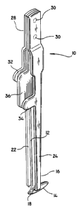
FIG. 1 is a view illustrating the dural closing
surgical forceps according to the present disclosure;
FIG. 2 is a partial cross-sectional view of the
surgical forceps of FIG. 1;
FIG. 3 is a partial view illustrating a distal end
of the surgical forceps of FIG. 1 in an open position;
FIG. 4 is a partial view illustrating a distal end
of the surgical forceps of FIG. 1 with a central member
inserted within a durotomy incision; and
FIG. 5 is a partial view illustrating a distal end
of the surgical forceps of FIG. 1 in a closed position.
DETAILED DESCRIPTION OF PREFERRED EMBODIMENTS
The preferred embodiments of the apparatus and
method disclosed herein are discussed in terms of dural
closing surgical forceps and procedures for using the
same. It is envisioned, however, that the disclosure is
applicable to a wide variety of procedures including,
but, not limited to both open and minimally invasive
-8-
CA 02289595 1999-11-05
WO 98/49949 PCTIUS98/09087
procedures inc:Luding endoscopic and arthroscopic
procedures wherein access to the surgical site is
achieved through a cannula or small incision.
In the discussion which follows, the term
"proximal", as is traditional, will refer to the portion
of the structure which is closer to the operator, while
the term "disterl" will refer to the portion which is
further from the operator.
The following discussion includes a description of
the dural closiLng surgical forceps utilized in closing a
dural incision followed by a description of the preferred
method for using the dural closing forceps in accordance
with the preserit disclosure.
Reference will now be made in detail to the
preferred embodiments of the disclosure, which are
illustrated in the accompanying figures. Turning now to
the figures, wherein like components are designated by
like reference numerals throughout the various figures,
attention is first d:irected to FIGS. 1-4.
The dural closing forceps 10 include a central flat
member 12 and a transversely positioned foot glide member
14 along a dist:al end 16 thereof. Each side of the
central member 12 provides a surface 18 against which
each of the incised or torn dural margins 20 can be held
against. Opposite central member 12 are two slender
extension members or blades 22 and 24 having distally
located project:ions or teeth 25 to engage the incised
dural margins or edges 20 and hold them tightly against
-9-
CA 02289595 1999-11-05
WO 98/49949 PCT/US98/09087
surface 18 of central member 12.
Extension members 22 and 24 each include a
projecting surface 32 and 34, respectively, which acts as
a finger pad against which force, i.e. finger force from
a surgeon, will be applied when operating the dural
forceps 10 of the present disclosure. Similarly, central
member 12 includes a projection surface 36 which is
disposed between finger pads 32 and 34. The finger pads
32, 34 and 36 of the present disclosure may be of any
suitable size and shape which will enable a surgeon to
easily manipulate the dural forceps 10 and may also
include a textured outside surface area to provide for
better frictional adherence.
As is shown in FIGS. 1 and 2, finger pads 32 and 34
are alternately spaced along opposing sides of finger pad
36. Finger pad 36 is preferably larger than finger pads
32 and 34 so as to provide an opposing surface for finger
pads 32 and 34 when extension members 22 and 24 are
manipulated to a closed position. In use the finger pads
32, 34 and 36 permit the surgeon's thumb to oppose either
or both of the extension members 22 and 24 against
central member 12. A surgeon's thumb and corresponding
index finger (preferably of the left hand) may be placed
on opposing surfaces of finger pads (32 and 36), (34 and
36) or (32 and 34) dependant on the particular
manipulation of the members 12, 22 and 24 required. With
the dural forceps being manipulated by one hand (left), a
clip applier, as previously discussed, may be used to_
-10-
CA 02289595 1999-11-05
WO 98/49949 PCT/US98/09087
apply clips 26 to the re-approximated dural margins 20
with the other hand (right).
Central member 12 and both extension members 22
and 24 are firnlly bonded together at the proximal end 28
by rivets 30 although other known bonding techniques such
as welding are also contemplated. This bonding of the
members 12, 22 and 24 maintain a cantilever relationship
between all thr.ee cornponents which allow the members 12,
22 and 24 to be rigid along the proximal end 28 but
flexible along the d:Lstal end 16 of dural forceps 10.
With particular reference to FIG. 3, the distal end
16 of dural forceps :L0 is shown in a relaxed or open
position. Extension members 22 and 24 are preferably
slightly shorter in :Length with respect to central member
12 although variations in the lengths of any member 12,
22 and 24 is obvious:Ly contemplated. Extension members
22 and 24 include projections or teeth 25 which are used
to grasp tissue, i.e., dural margins 20, together before
a clip 26 is applied. Central member 12 includes foot
glide 14 which includes a smooth intradural tissue
contacting surf:ace (not shown). Foot glide member 14
glides against the underside of the re-approximated
durotomy to act. as a barrier, keeping the nerve
filaments, membranous fragments and other intradural
material from f:lushing or protruding out through the
durotomy defect.. The foot glide 14 is preferably
transverse to t.he longitudinal axis of central member 12
and includes a rear portion 38 and a forward portion 40
-11-
CA 02289595 1999-11-05
WO 98/49949 PCT/US98/09087
generally separated by central member 12. The rear
portion 38 of foot glide 14 is generally shorter in
length than the forward portion 40. As is seen in FIGS.
4 and 5, a longer forward portion 40 ensures that the
nerve filaments and membranous fragments are adequately
covered by foot glide 14 prior to the application of
clips 26.
With particular reference to FIG 4, members 12, 22
and 24 are shown in an intermediate dural margins closing
position. The foot glide 14 of central member 12 is
shown holding the freely movable nerve filaments and
fragments of membranes away from the dural margins 20.
One edge 42 of the incised dural margins 20 is grasped by
the toothed portion 25 (not shown) of extension member 22
and pinched against surface 18 of central member 12.
Extension member 24 and corresponding toothed projection
is in a relaxed or open position.
As is shown in FIG. 5, the dural forceps 10
according to the present disclosure are in a fully closed
20 position with extension member 24 holding one incised
dural margin 20 against surface 18 of central member 12
and extension member 22 holding a second incised dural
margin 20 likewise against opposing surface 18 of central
member 12. In this closed position, the dural margins 20
25 are properly re-approximated without any of the nerve
filaments, membranous fragments or intradural tissue
protruding through the durotomy incision line. Once re-
approximated, the dural margins 20 are held together with
-12-
CA 02289595 1999-11-05
WO 98/49949 PCT/US98/09087
the applicatior.L of c7-ips 26 at small separated intervals,
preferably in the rarige of 2-3 mm. After application of
a clip 26, the three cantilevered components (central
member 12 and extension members 22 and 24) are relaxed or
opened to thereby release the grasp on the dural margins
20. The dural forceps 10 is subsequently moved further
along the durotomy incision line permitting progressive
clip 26 applications.
The dural closure forceps 10 according to the
present disclosure is capable of holding in position the
two incised dural margins or edges 20 while keeping nerve
filaments and fragmer.Lts of membranes from erupting out
from the dural margins 20 as dural closure clips 26 or
sutures are applied. Therefore, a surgeon with only one
hand is capable of holding the dural margins 20 and the
nerve filaments and fragments of membranes in the proper
relationship for closing the dural margins 20 of the
durotomy.
The operative steps involved with closure of an
incised dura utilizing the dural forceps 10 of the
present disclosure will now be discussed. The method
described below will discuss a method of closing an
incised dura post the performance of a standard durotomy
procedure. The method will also be discussed with
respect to a pa:rticular sequence, i.e., finger pads 32,
34 and 36 and corresponding hand and finger positions,
although alternate opposite sequences are obviously
contemplated.
-13-
CA 02289595 1999-11-05
WO 98/49949 PCT/US98/09087
Upon completion of a durotomy and upon commencing of
the procedure involved in closing the incised dura, the
dural forceps 10 according to the present disclosure are
grasped by a surgeon in one hand with finger pad 32 and
finger pad 36 being held against an index finger and
corresponding thumb, respectively. Using an additional
pair of simple forceps in an opposite hand, the surgeon
brings one cut dural margin 20 into the space above the
foot glide 14 and between the central member 12 and
extension member 22. The principal thumb then forces
finger pad 36 against finger pad 32 bringing the toothed
projections 25 of extension member 22 and surface 18 of
central member 12 together to thereby firmly grasp the
one cut dural margin 20 of the durotomy. Similarly, the
second cut dural margin 20 is then brought against
opposite surface 18 of central member 12. The principal
thumb is then slipped downward against finger pad 34 of
extension member 24 to thereby pinch the second cut dural
margin 20 against the surface 18 of central member 12.
At this point, the nerve filaments and free membrane
fragments are kept from flowing or protruding into the
re-approximated dural margins 20 by the foot glide 14, as
is shown in FIG. 5. Once the dural margins 20 are re-
approximated, clips 26 are applied in the same manner as
described earlier. After applying the clips 26, the clip
applier remains in a closed position thereby providing a
grasping force to the re-approximated dural margins 20
and serving as a temporary forceps while the dural _
-14-
CA 02289595 1999-11-05
WO 98/49949 PCT/US98/09087
forceps 10 are relaxed (opened) and moved a short
distance, i.e., about 2 or 3 mm, along the durotomy
margins 20 to commence the next re-approximation and
clipping maneuver.
It will be understood that various modifications may
be made to the embodiments disclosed herein. For
example, the dural forceps 10 may be fabricated from
either a surgical grade steel or other known surgical
alloys. Also, the dural forceps 10 may be fabricated
from a plastic resin making it both less expensive and
readily disposable. Therefore, the above description
should not be construed as limiting, but merely as
exemplifications of preferred embodiments. Those skilled
in the art will envision other modifications within the
scope and spirit of the claims appended hereto.
-15-