Note: Descriptions are shown in the official language in which they were submitted.
CA 02289665 1999-11-12
WO 98/56808 PCT/US98/12334
PROTEIN RECOVERY BY CHROMATOGRAPHY FOLLOWED BY FILTRATION UPON A CHARGED LAYER
BACKGROUND OF THE INVENTION
Field of the Invention
This invention relates generally to protein recovery. In particular, it
pertains to recovery of a
polypeptide, wherein the polypeptide is exposed to an immobilized reagent
which binds to, or
modfies, the polypeptide.
Description of Related Art
The large-scale, economic purification of proteins is increasingly an
important problem for the
biotechnology industry. Generally, proteins are produced by cell culture,
using either mammalian or
bacterial cel! lines engineered to produce the protein of interest by
insertion of a recombinant plasmid
containing the gene for that protein. Since the cell lines used are living
organisms, they must be fed
with a complex growth medium, containing sugars, amino acids, and growth
factors, usually supplied
from preparations of animal serum. Separation of the desired protein from the
mixture of compounds
fed to the cells and from the by-products of the cells themselves to a purity
sufficient for use as a
human therapeutic poses a formidable challenge.
Procedures for purification of proteins from cell debris initially depend on
the site of
expression of the protein. Some proteins can be caused to be secreted directly
from the cell into the
surrounding growth media; others are made intracellularly. For the latter
proteins, the first step of a
purification process involves lysis of the cell, which can be done by a
variety of methods, including
mechanical shear, osmotic shock, or enzymatic treatments. Such disruption
releases the entire
contents of the cell into the homogenate, and in addition produces subcellular
fragments that are
difficult to remove due to their small size. These are generally removed by
differential centrifugation or
by filtration. The same problem arises, although on a smaller scale, with
directly secreted proteins
due to the natural death of cells and release of intracellular host celt
proteins in the course of the
protein production run.
Once a clarified solution containing the protein of interest has been
obtained, its separation
from the other proteins produced by the cell is usually attempted using a
combination of different
chromatography techniques. These techniques separate mixtures of proteins on
the basis of their
charge, degree of hydrophobicity, or size. Several different chromatography
resins are available for
each of these techniques, allowing accurate tailoring of the purification
scheme to the particular
protein involved. The essence of each of these separation methods is that
proteins can be caused
either to move at different rates down a long column, achieving a physical
separation that increases
as they pass further down the column, or to adhere selectively to the
separation medium, being then
differentially eluted by different solvents. fn some cases, the desired
protein is separated from
impurities when the impurities specifically adhere to the column, and the
protein of interest does not,
that is, the protein of interest is present in the "flow-through."
As part of the overall recovery process for the protein, the protein may be
exposed to an
immobilized reagent which binds to or modifies the protein. For example, the
protein may be
subjected to affinity chromatography wherein an immobilized reagent which
binds specifically to the
protein, such as an antibody, captures the antibody and impurities pass
through the affinity
chromatography column. The protein can be subsequently eluted from the column
by changing the
CA 02289665 1999-11-12
WO 98/56808 PCT/US98/12334
conditions such that the protein no longer binds to the immobilized reagent.
The immobilized reagent
may also be an enzyme which modifies the protein. Sahni et al., Anal. Biochem.
193:178-185 (1991)
and Voyksner et aL, Anal. Biochern. 188:72-81 (1990) describe immobilized
proteases.
Another type of purification process is filtration. Filtration of fine
particle size contaminants
from fluids has been accomplished by the use of various porous filter media
through which a
contaminated composition is passed such that the filter retains the
contaminant. Retention of the
contaminant may occur by mechanical straining or electrokinetic particle
capture and adsorption. In
mechanical straining, a particle is retained by physical entrapment when it
attempts to pass through a
pore smaller than itself. In the case of electrokinetic capture mechanisms,
the particle collides with a
surface within the porous filter and is retained on the surface by short range
attractive forces. To
achieve electrokinetic capture, charge modifying systems can be used to alter
the surface charge
characteristics of a filter (see, e.g., W090/11814). For example, where the
contaminant to be
removed is anionic, a cationic charge modifier can be used to alter the charge
characteristics of the
filter such that the contaminant is retained by the filter.
There is a need in the art for improved methods for recovering polypeptides,
especially those
poiypeptides produced by recombinant techniques.
SUMMARY OF THE INVENTION
Accordingly, the invention provides a method for recovering a polypeptide
comprising: (a)
exposing a composition comprising a polypeptide to a reagent which binds to,
or modifies, the
poiypeptide, wherein the reagent is immobilized on a solid phase; and then (b)
passing the
composition through a filter bearing a charge which is opposite to the charge
of the reagent in the
composition, so as to remove leached reagent from the composition. Preferably
the charge
characteristics of the polypeptide in the composition in step (b) are such
that the polypeptide passes
through the filter and preferably the filter is placed in line with the
composition exposed to the reagent
as in step (a). In one embodiment of the invention, the polypeptide to be
treated in step (a) is a
precursor polypeptide and the immobilized reagent is a protease (e.g. pepsin)
which removes a
precursor domain (e.g. a ieucine zipper dimerization domain) from the
polypeptide.
The invention also provides a method for recovering a polypeptide comprising
removing a
leached reagent from a composition comprising the polypeptide and the leached
reagent by passing
the composition through a filter bearing a charge opposite to that of the
leached reagent, wherein the
leached reagent was previously immobilized on a solid phase.
In yet a further embodiment, the invention provides a method for modifying a
precursor
antibody comprising a leucine zipper dimerization domain, comprising exposing
the precursor
antibody to a protease immobilized on a solid phase such that the protease
removes the leucine
zipper from the precursor antibody. This method optionally further comprises
passing the antibody
free of the leucine zipper through a positively charged filter placed in line
with antibody which has
been exposed to the immobilized protease.
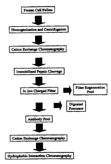
The anti-CD18 purification process is an example of a process in which an
immobilized
reagent is required to remove a leucine zipper dimerization domain from the
anti-CD18 antibody
precursor. The antibody precursor is initially purified using ABX cation
exchange chromatography
before the leucine zipper domain is removed by digestion with pepsin. The
amount of pepsin
_2_
CA 02289665 2005-O1-12
necessary to completely remove the leucine zipper from the antibody precursor
is considerable. A ratio
of 1 mg of pepsin per 20 mg of antibody is necessary to carry out the
digestion over a reasonable period
of time. Treatment like this will leave a large amount of pepsin to be removed
in the remaining steps of
the anti-CD 18 purification process (Figure 7). Quick removal of pepsin was
found to be beneficial,
since excessive exposure to pepsin resulted in overdigestion of the anti-CD 18
antibody, with
significant loses of intact product. In order to effectively control the
amount of pepsin added to the anti-
CD 18 precursor antibody, and effectively eliminate any traces of pepsin that
can persist through the
purification process, two methods were implemented into the anti-CD18 antibody
purification process.
First, to considerably reduce the amount of pepsin added to the ABX purified
antibody precursor pool,
pepsin was immobilized on a solid phase (i.e. coupled to control pore glass
beads (CPG) and packed
into a column). The digestion reaction was then carried out by flowing the
antibody precursor pool
through the pepsin-CPG column. This procedure limited the amount of pepsin
added into the antibody
precursor pool. Nevertheless, a further problem was identified in that pepsin
was found to leach from
the solid phase. A small amount of pepsin leaching from the solid phase was
found to be sufficient to
cause overdigestion of the anti-CD18 antibody, resulting in a reduction in
product yields. To overcome
this problem of pepsin leaching from the solid phase, a positively charged
filter was placed in line with
the effluent from the pepsin-CPG column. The filter was found to remove all
pepsin leaching from the
solid phase, thereby preventing overdigestion of the antibody precursor.
Pepsin is an acidic protein
with a low pl. Therefore at pH 4, the pH of the digestion step, pepsin
remained negatively charged and
bound strongly to the positively charged filter. The use of a charged filter
instead of a resin to remove
leachables was found to be advantageous, since filters are compact and capable
of very high flow rates
with minimal backpressure. A filter can be implemented in line without the
need to perform a separate
recovery step, therefore reducing process complexity and time.
In various embodiments, there is provided a method for recovering a
polypeptide comprising:
(a) exposing a composition comprising a polypeptide to a reagent which binds
to, or modifies, the
polypeptide, wherein the reagent is immobilized on a solid phase; and then (b)
passing an effluent
comprising the polypeptide eluted from or modified by the immobilized reagent,
and any reagent
leached from the solid phase, through a filter bearing a charge which is
opposite to the charge of the
reagent in the composition at the pH of the composition, so as to remove
leached reagent from the
effluent, the charge characteristics of the polypeptide in the composition
being such that the
polypeptide passes through the filter.
In various embodiments, there is provided a method for recovering a
polypeptide comprising
removing a leached reagent from a composition comprising the polypeptide and
the leached reagent by
passing the composition through a filter bearing a charge opposite to that of
the leached reagent at the
pH of the composition, wherein the leached reagent was previously immobilized
on a solid phase.
In various embodiments, there is provided a method for modifying a precursor
antibody
comprising a leucine zipper comprising exposing the precursor antibody to a
protease immobilized on a
solid phase such that the protease removes the leucine zipper from the
precursor antibody.
-3-
CA 02289665 2005-O1-12
It is envisaged that negatively and positively charged filters can be used to
solve problems
associated with leaching of formerly immobilized reagents in other recovery
processes.
BRIEF DESCRIPTION OF THE DRAWINGS
Figures lA and 1B depict the amino acid sequence of rhuMAb CD18 heavy chain
(Figure lA;
SEQ ID NO:1) and light chain (Figure 1B; SEQ ID N0:2). The sequence in italics
in Figure lA {SEQ
ID N0:3) is that of the leucine zipper.
Figures 2A and 2B depict intact antibody (Ab) and a variety of antibody
fragments (F(ab')Z,
Fab', light chain and Fd'). Heavy chains are depicted in white and light
chains are hatched. The two
disulfide bonds that form between two heavy chains are shown as -ss-. Figure
2B shows pepsin
cleavage of the rhuMAb CD 18 precursor to yield rhuMAb CD 18, free of the
leucine zipper.
Figure 3 depicts the structure of plasmid pS 1130 used to produce rhuMAb CD 18
of the
example below.
Figures 4A and 4B depict the full sequence of the pS 1130 expression cassette
(SEQ ID NO:S).
Figure 5 shows derivation of the 49A5 production cell line.
Figure 6 is a schematic of the fermentation process for rhuMAb CD 18.
Figure 7 is a flow diagram depicting the purification steps for rhuMAb CD 18.
-3a-
CA 02289665 1999-11-12
WO 98/56808 PCT/US98/I2334
DETAILED DESCRIPTION OF THE PREFERRED EMBODIMENTS
Definitions:
As used herein, "polypeptide" refers generally to peptides and proteins having
more than
about ten amino acids. Preferably, the polypeptide is a mammalian protein,
examples of which
include renin; a growth hormone, including human growth hormone and bovine
growth hormone;
growth hormone releasing factor; parathyroid hormone; thyroid stimulating
hormone; lipoproteins;
alpha-1-antitrypsin; insulin A-chain; insulin B-chain; proinsulin; follicle
stimulating hormone; calcitonin;
iuteiniztng hormone; glucagon; clotting factors such as factor VIIIC, factor
IX, tissue factor, and von
WiUebrands factor; anti-clotting factors such as Protein C; atrial natriuretic
factor; lung surfactant; a
plasminogen activator, such as urokinase or human urine or tissue-type
plasminogen activator (t-PA);
bombesin; thrombin; hemopoietic growth factor; tumor necrosis factor-alpha and
-beta;
enkephalinase; RANTES (regulated on activation normally T-cell expressed and
secreted); human
macrophage inflammatory protein (MIP-1-alpha); a serum albumin such as human
serum albumin;
Muellerian-inhibiting substance; relaxin A-chain; relaxin B-chain; proreiaxin;
mouse gonadotropin-
associated peptide; a microbial protein, such as beta-lactamase; DNase; IgE; a
cytotoxic T-
lymphocyte associated antigen (CTLA), such as CTLA-4; inhibin; activin;
vascular endothelial growth
factor (VEGF); receptors for hormones or growth factors; Protein A or D;
rheumatoid factors; a
neurotrophic factor such as bone-derived neurotrophic factor (BDNF),
neurotrophin-3, -4, -5, or -6
(NT-3, NT-4, NT-5, or NT-6), or a nerve growth factor such as NGF-p; platelet-
derived growth factor
(PDGF); fibroblast growth factor such as aFGF and bFGF; epidermal growth
factor (EGF);
transforming growth factor (TGF) such as TGF-alpha and TGF-beta, including TGF-
ail, TGF-~2, TGF-
p3, TGF-p4, or TGF-a5; insulin-like growth factor-I and -II (IGF-I and IGF-
II); des(1-3)-IGF-I (brain
IGF-I), insulin-tike growth factor binding proteins (IGFBPs); CD proteins such
as CD3, CD4, CDB,
CD19 and CD20; erythropoietin; osteoinductive factors; immunotoxins; a bone
morphogenetic protein
(BMP); an interferon such as interferon-alpha, -beta, and -gamma; colony
stimulating factors (CSFs),
e.g., M-CSF, GM-CSF, and G-CSF; interteukins (ILs), e.g., IL-1 to IL-10;
superoxide dismutase; T-cell
receptors; surface membrane proteins; decay accelerating factor; viral antigen
such as, for example, a
portion of the AIDS envelope; transport proteins; homing receptors;
addressins; regulatory proteins;
integrins such as CD11a, CDl1b, CD1lc, CD18, an lCAM, VLA-4 and VCAM; a tumor
associated
antigen such as HER2, HER3 or HER4 receptor; and fragments and/or variants of
any of the above-
listed polypeptides.
A "variant" or "amino acid sequence variant" of a starting polypeptide is a
polypeptide that
comprises an amino acid sequence different from that of the starting
polypeptide. Generally, a variant
will possess at least 80% sequence identity, preferably at least 90% sequence
identity, more
preferably at least 95% sequence identity, and most preferably at least 98%
sequence identity with
the native potypeptide. Percentage sequence identity is determined, for
example, by the Fitch et al.,
Proc. NatL Acad. Sci. USA 80:1382-1386 (1983), version of the algorithm
described by Needteman et
al., J. Mol. Biol. 48:443-453 (1970), after aligning the sequences to provide
for maximum homology.
Amino acid sequence variants of a potypeptide are prepared by introducing
appropriate nucleotide
changes into DNA encoding the potypeptide, or by peptide synthesis. Such
variants include, for
example, deletions from, and/or insertions into and/or substitutions of,
residues within the amino acid
CA 02289665 2003-07-31
sequence of the polypeptide of interest. Any combination of deletion,
insertion, and substitution is
made to arrive at the final construct, provided that the final construct
possesses the desired
characteristics. The amino acid changes also may alter post-translatlonal
pros~sses of the
polypeptide, such as changing the number or position of glycosylation sites.
Methods for generating
amino acid sequence variants of poiypepttde~s era describ~d in US Pat
5,534,815,
In preferred embodiments of the invention, the polypeptide iS a rOCOmbinAnt
polypeptide. A
"recombinant polypeptide" is ons which has been produced in a host cell which
has been transformed
or transfected with nucleic acid encoding the polypeptide, or produces the
polypeptlde as a result of
homologous recombination. "Transformation" and "transfection" are used
interchangeably to refer to the
process of introducing nudelc add irto a cell. FdIowIng transfom'~aiton or
trartstection, d'te nucleic acid may
integrate into the host cell genome, or may exist as an extrachromosomal
element. The "host cell'
includes a cell in in vitro cell culture as well a cell within a host animal.
Methods for recombinant
production of polypeptides are described In US Pat 6,534,615,
15. A 'precursor polypeptide" herein is a polypeptide to which fs fused on~ or
more precursor domains,
e.g. where the precursor domain" !s part of a polypeptide chain of the
polypeptldp or fs covalently
attached to the polypaptide by a chemical linker, for example. The'preeursor
domain' may be an amino acid
residue or polypeptIde. For example, the precursor domain may b~ a
dimerization
domain such as a leucine zipper, an amino acid sequence such as polyglutamle
acid which bears a
negative d~arge and another amino aad sequence such as polylysine which bears
a positive charge, or
a peptide helix bundle comprising a helix. a turn dnd another helix; an
epitope tag useful, e.g., in
purification of the polypeptide of interest; en amino acid residing pr peptide
at the amino or carboxy
terminus of the polypsptid~ which is dosirod to be removed to generate a
homogenous polypeptide
2s preparation; a N-terminal methionine, an artifact of production of the
polypeptlde in recombinaant cell
culture; a pre, pro or prepro domain of a mature polypeptide (e.g. the pro
domain of prothrombin,
wherein removal of the pro domain generates the biologically activA mature
thrombin molecule); a
polylysine polypeptide; an enzyme such as glutathione transferase; or the Fc
region of an Intact
antibody which is removed to generate an F(ab')z.
An "epitope tag" polypeptide has enough residues to provide an apitope against
which an antibody
thereagainst can be mad9, yet is short enough such that it dons not interfere
with activity of th~
polypeptide to which It Is fused. The Qpitopo tap prAferably is sufficiently
unique s0 that the
antibody thereagainst does not substantially cross-react with other epitopes.
Suitable epitope tag
polypeptldes generally have at IeaSt 6 amino acid residues and usually between
about 8-50 amino
ss acid residues (preferably between about 9-30 residues). Examples Include
the flu HA tag pofypeptide and
its urtlibody 12CA5 (Field et at. Mot CeIG Biol. 8:2159-2166 (1988}); the a-
myc tag and the 6F9, 3C7,
BE1 p, G4, B7 and 9Ei0 andbodles iherebo (Evan ef nL, n4ot. Cell. 8101.
5(12):3610-3618 (1985)); and the
Herpes Simplex virus glycoprotain D (gD) tag and its antibody (Paborsky et aL,
i'roloi»
Eaginsertru3 3(6):547-553 (1990)).
The term "antibody' is used in the broadest sense and specifically covers
monoclonal ar>titxxf3es
(rxduding full length monoclonal antibodies), pplycbnal ty'llibot~e~s,
muttispeafic antihoct~es
-5
CA 02289665 1999-11-12
WO 98/56808 PCT/US98/12334
(e.g., bispeciflc antibodies), and antibody fragments so long as they exhibit
the desired biological
activity.
The antibody herein is directed against an "antigen" of interest. Preferably,
the antigen is a
biologically important polypeptide and administration of the antibody to a
mammal suffering from a
disease or disorder can result in a therapeutic benefit in that mammal.
However, antibodies directed
against nonpolypeptide antigens (such as tumor-associated glycolipid antigens;
see US Patent
5,091,178) are also contemplated. Where the antigen is a polypeptide, it may
be a transmembrane
molecule (e.g. receptor) or ligand such as a growth factor. Exemplary antigens
include those
polypeptides discussed above. Preferred molecular targets for antibodies
encompassed by the
present invention include CD polypeptides such as CD3, CD4, CDB, CD19, CD20
and CD34;
members of the ErbB receptor family such as the EGF receptor, HER2, HER3 or
HERO receptor; cell
adhesion molecules such as LFA-1, Mac1, p150,95, VLA-4, ICAM-1, VCAM and
av/(i3 integrin
including either a or a subunits thereof (e.g. anti-CDl1a, anti-CD18 or anti-
CDllb antibodies); growth
factors such as VEGF; IgE; blood group antigens; flk2/flt3 receptor; obesity
(OB) receptor; mpl
receptor; CTLA-4; polypeptide C etc. Soluble antigens or fragments thereof,
optionally conjugated to
other molecules, can be used as immunogens for generating antibodies. For
transmembrane
molecules, such as receptors, fragments of these (e.g. the extracellular
domain of a receptor) can be
used as the immunogen. Alternatively, cells expressing the transmembrane
molecule can be used as
the immunogen. Such cells can be derived from a natural source (e.g. cancer
cell lines) or may be
cells which have been transformed by recombinant techniques to express the
transmembrane
motecule.
The term "monoclonal antibody" as used herein refers to an antibody obtained
from a
population of substantially homogeneous antibodies, ie., the individual
antibodies comprising the
population are identical except for possible naturally occurring mutations
that may be present in minor
amounts. Monoclonal antibodies are highly specific, being directed against a
single antigenic site.
Furthermore, in contrast to conventional (polyclonal) antibody preparations
which typically include
different antibodies directed against different determinants (epitopes), each
monoclonal antibody is
directed against a single determinant on the antigen. The modifier
"monoclonal" indicates the
character of the antibody as being obtained from a substantially homogeneous
population of
antibodies, and is not to be construed as requiring production of the antibody
by any particular
method. For example, the monoclonal antibodies to be used in accordance with
the present invention
may be made by the hybridoma method first described by Kohler et al., Nature
256:495 (1975), or
may be made by recombinant DNA methods (see, e.g., U.S. Patent No. 4,816,567).
In a further
embodiment, "monoclonal antibodies" can be isolated from antibody phage
libraries generated using
the techniques described in McCafferty et aL, Nature, 348:552-554 (1990).
Clackson et al., Nature,
352:624-628 (1991) and Marks et al., J. Mol. BioL, 222:581-597 (1991) describe
the isolation of
murine and human antibodies, respectively, using phage libraries. Subsequent
publications describe
the production of high affinity (nM range) human antibodies by chain shuffling
(Marks et aL,
BiolTechnology, 10:779-783 (1992)), as well as combinatorial infection and in
vivo recombination as a
strategy for constructing very large phage libraries (Waterhouse et aL, Nuc.
Acids. Res., 21:2265-
2266 (1993)). Thus, these techniques are viable alternatives to traditional
monoclonal antibody
-6-
CA 02289665 1999-11-12
WO 98/56808 PCT/US98/12334
hybridoma techniques for isolation of monoclonal antibodies. Alternatively, it
is now possible to
produce transgenic animals (e.g., mice) that are capable, upon immunization,
of producing a full
repertoire of human antibodies in the absence of endogenous immunoglobulin
production. For
example, it has been described that the homozygous deletion of the antibody
heavy-chain joining
region (J~,) gene in chimeric and germ-line mutant mice results in complete
inhibition of endogenous
antibody production. Transfer of the human germ-line imrnunoglobulin gene
array in such germ-line
mutant mice will result in the production of human antibodies upon antigen
challenge. See, e.g.,
Jakobovits et al., Proc. Natl. Acad. Sci USA, 90:2551 (1993); Jakobovits et
al., Nature, 362:255-258
(1993); Bruggermann et aL, Year in Immuno., 7:33 (1993); and Duchosal et aL
Nature 355:258
(1992).
The monoclonal antibodies herein specifically include "chimeric" antibodies
(immunoglobuiins) in which a portion of the heavy and/or light chain is
identical with or homologous to
corresponding sequences in antibodies derived from a particular species or
belonging to a particular
antibody class or subclass, while the remainder of the chains) is identical
with or homologous to
corresponding sequences in antibodies derived from another species or
belonging to another
antibody class or subclass, as well as fragments of such antibodies, so long
as they exhibit the
desired biological activity (U.S. Patent No. 4,816,567; and Morrison et al.,
Proc. Natl. Acad. Sci. USA
81:6851-6855 (1984)).
The term "hypervariable region" when used herein refers to the amino acid
residues of an
antibody which are responsible for antigen-binding. The hypenrariable region
comprises amino acid
residues from a "complementarity determining region" or "CDR" (i.e. residues
24-34 (L1), 50-56 (L2)
and 89-97 (L3) in the light chain variable domain and 31-35 (H1), 50-65 (H2)
and 95-102 (H3) in the
heavy chain variable domain; Kabat et al., Sequences of Polypeptides of
Immunological Interest, 5th
Ed. Public Health Service, National Institutes of Health, Bethesda, MD.
(1991)) andlor those residues
from a "hypervariable loop" (i.e. residues 26-32 (L1), 50-52 (L2) and 91-96
(L3) in the fight chain
variable domain and 26-32 (H1), 53-55 (H2) and 96-101 (H3) in the heavy chain
variable domain;
Chothia and Lesk J. MoL Biol. 196:901-917 (1987)). "Framework" or "FR"
residues are those variable
domain residues other than the hypervariable region residues as herein
defined. The CDR and FR
residues of the H52 antibody of the example below are identified in Eigenbrot
et al. Polypeptides:
Structure, Function and Genetics 18:49-62 (1994).
"Humanized" forms of non-human (e.g., murine) antibodies are chimeric
antibodies which
contain minimal sequence derived from non-human immunoglobulin. For the most
part, humanized
antibodies are human immunoglobufins (recipient antibody) in which residues
from a hypervariable
region of the recipient are replaced by residues from a hypervariable region
of a non-human species
(donor antibody) such as mouse, rat, rabbit or nonhuman primate having the
desired specificity,
affinity, and capacity. In some instances, Fv framework region (FR) residues
of the human
immunoglobulin are replaced by corresponding non-human residues. Furthermore,
humanized
antibodies may comprise residues which are not found in the recipient antibody
or in the donor
antibody. These modifications are made to further refine antibody performance.
In general, the
humanized antibody will comprise substantially all of at least one, and
typically two, variable domains,
in which all or substantially all of the hypervariable loops correspond to
those of a non-human
_7_
CA 02289665 1999-11-12
WO 98/56808 PCT/US98/12334
immunoglobulin and all or substantially all of the FR regions are those of a
human immunoglobulin
sequence. The humanized antibody optionally also will comprise at least a
portion of an
immunoglobulin constant region (Fc), typically that of a human immunoglobuiin.
The choice of human variable domains, both light and heavy, to be used in
making the
humanized antibodies is very important to reduce antigenicity. According to
the so-called "best-fit"
method, the sequence of the variable domain of a rodent antibody is screened
against the entire
library of known human variable-domain sequences. The human sequence which is
closest to that of
the rodent is then accepted as the human framework (FR) for the humanized
antibody (Sims et al., J.
lmmunol., 151:2296 (1993); Chothia et al., J. Mol. 8iol., 196:901 (1987)).
Another method uses a
particular framework derived from the consensus sequence of all human
antibodies of a particular
subgroup of light or heavy chains. The same framework may be used for several
different humanized
antibodies (Carter et al., Proc. Natl. Acad. Sci. USA, 89:4285 (1992); Presta
et al., J. Immnol.,
151:2623 (1993)).
It is further important that antibodies be humanized with retention of high
affinity for the
IS antigen and other favorable biological properties. To achieve this goal,
according to a preferred
method, humanized antibodies are prepared by a process of analysis of the
parental sequences and
various conceptual humanized products using three-dimensional models of the
parental and
humanized sequences. Three-dimensional immunoglobulin models are commonly
available and are
familiar to those skilled in the art. Computer programs are available which
illustrate and display
probable three-dimensional conformational structures of selected candidate
immunoglobulin
sequences. Inspection of these displays permits analysis of the likely role of
the residues in the
functioning of the candidate immunoglobulin sequence, i.e., the analysis of
residues that influence the
ability of the candidate immunoglobulin to bind its antigen. In this way, FR
residues can be selected
and combined from the recipient and import sequences so that the desired
antibody characteristic,
such as increased affinity for the target antigen(s), is achieved. In general,
the CDR residues are
directly and most substantially involved in influencing antigen binding.
In a preferred embodiment of the invention, the antibody is an antibody
fragment which is
preferably human or humanized (see above discussion concerning humanized
antibodies).
"Antibody fragments" comprise a portion of a full length antibody, generally
the antigen
binding or variable region thereof. Examples of antibody fragments include
Fab, Fab', F(ab')2, and Fv
fragments; diabodies; linear antibodies; single-chain antibody molecules; and
multispecific antibodies
formed from antibody fragments. Various techniques have been developed for the
production of
antibody fragments. Traditionally, these fragments were derived via
proteolytic digestion of intact
antibodies (see, e.g., Morimoto et al., Journal of Biochemical and Biophysical
Methods 24:107-117
(1992) and Brennan et al., Science, 229:81 (1985}). However, these fragments
can now be produced
directly by recombinant host cells. For example, the antibody fragments can be
isolated from the
antibody phage libraries discussed above. Alternatively, Fab'-SH fragments can
be directly recovered
from E. coli and chemically coupled to form F(ab')2 fragments (Carter et al.,
Bio/Technology 10:163-
167 (1992)). In another embodiment as described in the Example below, the
F(ab')2 is formed using
the leucine zipper GCN4 to promote assembly of the F(ab')2 molecule. According
to another
_g_
CA 02289665 1999-11-12
WO 98/56808 PCT/US98/12334
approach, F(ab')2 fragments can be isolated directly from recombinant host
cell culture. Other
techniques for the production of antibody fragments will be apparent to the
skilled practitioner. In
other embodiments, the antibody of choice is a single chain Fv fragment
(scFv). See WO 93/16185.
"Single-chain Fv" or "sFv" antibody fragments comprise the VH and VL domains
of antibody,
wherein these domains are present in a single polypeptide chain. Generally,
the Fv polypeptide
further comprises a polypeptide linker between the VH and VL domains which
enables the sFv to form
the desired structure for antigen binding. For a review of sFv see Pluckthun
in The Phamtacology of
Monoclonal Antibodies, vol. 113, Rosenburg and Moore eds. Springer-Verlag, New
York, pp. 269-315
( 1994).
The term "diabodies" refers to small antibody fragments with two antigen-
binding sites, which
fragments comprise a heavy chain variable domain (VH) connected to a light
chain variable domain
(VL) in the same polypeptide chain (V H - V L). By using a linker that is too
short to allow pairing
between the two domains on the same chain, the domains are forced to pair with
the complementary
domains of another chain and create two antigen-binding sites. Diabodies are
described more fully in,
for example, EP 404,097; WO 93/11161; and Hollinger et al, Proc. Natl. Acad.
Sci. USA 90:6444-
6448 (1993).
The expression "linear antibodies" when used throughout this application
refers to the
antibodies described in Zapata et al. Polypeptide Eng. 8(10):1057-1062 (1995).
Briefly, these
antibodies comprise a pair of tandem Fd segments (VH-CH1-VH-CHi) which form a
pair of antigen
binding regions. Linear antibodies can be bispecific or monospecific.
"Multispecific antibodies" have binding specificities for at least two
different epitopes, where
the epitopes are usually from different antigens. While such molecules
normally will only bind two
antigens (i.e. bispecific antibodies, BsAbs), antibodies with additional
specificities such as trispecific
antibodies are encompassed by this expression when used herein. Examples of
BsAbs include those
with one arm directed against a tumor cell antigen and the other arm directed
against a cytotoxic
trigger molecule such as anti-FcyRl/anti-CD15, anti-p185HER2/FcyRlll (CD16),
anti-CD3/anti-
malignant B-cell (1D10), anti-CD3lanti-p185HER2, anti-CD3/anti-p97, anti-
CD3lanti-renal cell
carcinoma, anti-CD3/anti-OVCAR-3, anti-CD3/t--D1 (anti-colon carcinoma), anti-
CD3/anti-melanocyte
stimulating hormone analog, anti-EGF receptoNanti-CD3, anti-CD3/anti-CAMA1,
anti-CD3/anti-CD19,
anti-CD3IMoV18, anti-neural cell ahesion molecule (NCAM)/anti-CD3, anti-folate
binding protein
(FBP)/anti-CD3, anti-pan carcinoma associated antigen (AMOC-31 )lanti-CD3;
BsAbs with one arm
which binds specifically to a tumor antigen and one arm which binds to a toxin
such as anti-
saporin/anti-Id-1, anti-CD22/anti-saporin, anti-CD7/anti-saporin, anti-
CD38/anti-saporin, anti-CEA/anti-
ricin A chain, anti-interferon-a(IFN-a.)lanti-hybridoma idiotype, anti-
CEA/anti-vinca alkaloid; BsAbs for
converting enzyme activated prodrugs such as anti-CD30/anti-alkaline
phosphatase (which catalyzes
conversion of mitomycin phosphate prodrug to mitomycin alcohol); BsAbs which
can be used as
fibrinolytic agents such as anti-fibrin/anti-tissue plasminogen activator
(tPA), anti-fibrin/anti-urokinase-
type plasminogen activator (uPA); BsAbs for targeting immune complexes to cell
surface receptors
such as anti-low density lipoprotein (LDL)lanti-Fc receptor (e.g. FcyRl,
FcyRll or FcyRlll); BsAbs for
use in therapy of infectious diseases such as anti-CD3lanti-herpes simplex
virus (HSV), anti-T-cell
-9-
CA 02289665 1999-11-12
WO 98/56808 PCT/US98/12334
receptor:CD3 complexlanti-influenza, anti-FcyR/anti-H1V; BsAbs for tumor
detection in vitro or in vivo
such as anti-CEA/anti-EOTUBE, anti-CEA/anti-DPTA, anti-p185HER2/anti-hapten;
BsAbs as vaccine
adjuvants; and BsAbs as diagnostic tools such as anti-rabbit IgG/anti-
ferritin, anti-horse radish
peroxidase (HRP)/anti-hormone, anti-somatostatin/anti-substance P, anti-
HRP/anti-FITC, anti-
s CEAlanti-~i-galactosidase. Examples of trispecific antibodies include anti-
CD3/anti-CD4/anti-CD37,
anti-CD3/anti-CD5/anti-CD37 and anti-CD3/anti-CD8/anti-CD37. Bispecific
antibodies can be
prepared as full length antibodies or antibody fragments (e.g. F(ab')2
bispecific antibodies).
Methods for making bispecific antibodies are known in the art. Traditional
production of full
length bispecific antibodies is based on the coexpression of two
immunoglobulin heavy chain-light
chain pairs, where the two chains have different specificities (Millstein et
al., Nature, 305:537-539
(1983)). Because of the random assortment of immunoglobulin heavy and light
chains, these
hybridomas (quadromas) produce a potential mixture of 10 different antibody
molecules, of which only
one has the correct bispecific structure. Purification of the correct
molecule, which is usually done by
affinity chromatography steps, is rather cumbersome, and the product yields
are low. Similar
procedures are disclosed in WO 93/08829, and in Traunecker et aL, EMBO J.,
10:3655-3659 (1991 ).
According to a different approach, antibody variable domains with the desired
binding
specificities (antibody-antigen combining sites) are fused to immunoglobulin
constant domain
sequences. The fusion preferably is with an immunoglobulin heavy chain
constant domain,
comprising at least part of the hinge, CH2, and CH3 regions. If is preferred
to have the first heavy-
chain constant region (CH1) containing the site necessary for light chain
binding, present in at least
one of the fusions. DNAs encoding the immunoglobulin heavy chain fusions and,
if desired, the
immunoglobulin light chain, are inserted into separate expression vectors, and
are co-transfected into
a suitable host organism. This provides for great flexibility in adjusting the
mutual proportions of the
three polypeptide fragments in embodiments when unequal ratios of the three
polypeptide chains
used in the construction provide the optimum yields. It is, however, possible
to insert the coding
sequences for two or ail three polypeptide chains in one expression vector
when the expression of at
least two poiypeptide chains in equal ratios results in high yields or when
the ratios are of no
particular significance.
In a preferred embodiment of this approach, the bispecific antibodies are
composed of a
hybrid immunogiobulin heavy chain with a first binding specificity in one arm,
and a hybrid
immunoglobulin heavy chain-light chain pair (providing a second binding
specificity) in the other arm.
It was found that this asymmetric structure facilitates the separation of the
desired bispecific
compound from unwanted immunoglobuiin chain combinations, as the presence of
an
immunoglobulin light chain in only one half of the bispecific molecule
provides for a facile way of
separation. This approach is disclosed in WO 94/04690. For further details of
generating bispecific
antibodies see, for example, Suresh et al., Methods in Enzymology, 121:210
(1986).
According to another approach described in W096/27011, the interface between a
pair of
antibody molecules can be engineered to maximize the percentage of
heterodimers which are
recovered from recombinant cell culture. The preferred interface comprises at
feast a part of the CH3
domain of an antibody constant domain. In this method, one or more small amino
acid side chains
-10-
CA 02289665 1999-11-12
WO 98/56808 PCT/US98/12334
from the interface of the first antibody molecule are replaced with larger
side chains (e.g. tyrosine or
tryptophan). Compensatory "cavities" of identical or similar size to the large
side chains) are created
on the interface of the second antibody molecule by replacing large amino acid
side chains with
smaller ones (e.g. alanine or threonine). This provides a mechanism for
increasing the yield of the
heterodimer over other unwanted end-products such as homodimers.
Bispecific antibodies include cross-linked or "heteroconjugate" antibodies.
For example, one
of the antibodies in the heteroconjugate can be coupled to avidin, the other
to biotin. Such antibodies
have, for example, been proposed to target immune system cells to unwanted
cells (US Patent No.
4,676,980), and for treatment of HIV infection (WO 91100360, WO 921200373, and
EP 03089).
Heteroconjugate antibodies may be made using any convenient cross-linking
methods. Suitable
cross-finking agents are well known in the art, and are disclosed in US Patent
No. 4,676,980, along
with a number of cross-linking techniques.
Techniques for generating bispecific antibodies from antibody fragments have
also been
described in the literature. For example, bispecific antibodies can be
prepared using chemical
linkage. Brennan et al., Science, 229: 81 (1985) describe a procedure wherein
intact antibodies are
proteolytically cleaved to generate F(ab')2 fragments. These fragments are
reduced in the presence
of the dithiol complexing agent sodium arsenite to stabilize vicinal dithiols
and prevent intermolecular
disulfide formation. The Fab' fragments generated are then converted to
thionitrobenzoate {TNB)
derivatives. One of the Fab'-TNB derivatives is then reconverted to the Fab'-
thiol by reduction with
mercaptoethylamine and is mixed with an equimolar amount of the other Fab'-TNB
derivative to form
the bispecific antibody. The bispecific antibodies produced can be used as
agents for the selective
immobilization of enzymes.
Recent progress has facilitated the direct recovery of Fab'-SH fragments from
E. toll, which
can be chemically coupled to form bispecific antibodies. Shalaby et al., J.
Exp. Med., 175: 217-225
{1992) describe the production of a fully humanized bispecific antibody
F{ab')2 molecule. Each Fab'
fragment was separately secreted from E. toll and subjected to directed
chemical coupling in vitro to
form the bispecific antibody.
Various techniques for making and isolating bispecific antibody fragments
directly from
recombinant cell culture have also been described. For example, bispecific
antibodies have been
produced using leucine zippers. Kostelny et al., J. immunol., 148(5):1547-1553
(1992). The leucine
zipper peptides from the Fos and Jun proteins were linked to the Fab' portions
of two different
antibodies by gene fusion. The antibody homodimers were reduced at the hinge
region to form
monomers and then re-oxidized to form the antibody heterodimers. This method
can also be utilized
for the production of antibody homodimers. The "diabody" technology described
by Hollinger et al.,
Proc. Natl. Acad. Sci. USA, 90:6444-6448 (1993) has provided an alternative
mechanism for making
bispecific antibody fragments. The fragments comprise a heavy-chain variable
domain (VH)
connected to a light-chain variable domain (V~) by a linker which is too short
to allow pairing between
the two domains on the same chain. Accordingly, the VH and V~ domains of one
fragment are forced
to pair with the complementary V~ and VH domains of another fragment, thereby
forming two antigen-
CA 02289665 1999-11-12
WO 98/56808 PCT/US98/12334
binding sites. Another strategy for making bispecific antibody fragments by
the use of single-chain Fv
(sFv) dimers has also been reported. See Gruber et aL, J. Immunol., 152:5368
(1994).
Antibodies with more than two valencies are contemplated. For example,
trispecific
antibodies can be prepared. Tutt et al. J. ImmunoL 147: 60 (1991 ).
By "recovering a polypeptide" is meant obtaining a polypeptide preparation
from a "pre-
recovery preparation" by purifying the pre-recovery preparation (see below) or
by modifying a
precursor polypeptide to generate a form of the polypeptide which is free of
the precursor domain.
By "purifying" a composition comprising an polypeptide and one or more
contaminants is
meant increasing the degree of purity of the polypeptide in the composition by
removing (completely
or partially) at least one contaminant from the composition. A "purification
step" may be part of an
overall purification process resulting in an "essentially pure" composition,
which is used herein to refer
to a composition comprising at least about 90% by weight of the polypeptide of
interest, based on
total weight of the composition, preferably at least about 95% by weight.
"Essentially homogeneous"
herein refers to a composition comprising at least about 99% by weight of
polypeptide of interest,
based on total weight of the composition.
The "reagent" of interest herein is a compound or composition (preferably a
polypeptide)
which is able to bind to and/or modify a polypeptide of interest. A "leached"
reagent is one which has
come free from the solid phase. The reagent may, for example, bind to the
polypeptide as is the case
for "capture reagents" used in affinity purification methods. Examples of such
"capture reagents"
include protein A or protein G for capturing polypeptides such as antibodies
and immunoadhesins;
antibodies which can be used for affinity purification of polypeptides; a
iigand binding domain of a
receptor for capturing a ligand thereto; a receptor binding domain for
capturing a receptor or a
fragment thereof binding protein (e.g. IGFBPs such as IGFBP-3 and growth
hormone binding proteins
(GHBPs)); and immunoadhesins. Alternatively, or in addition, the reagent may
modify the polypeptide
of interest. For example, the reagent may chemically or physically alter the
polypeptide. By
"chemical alteration" is meant modification of the polypeptide by, e.g., bond
formation or cleavage
resulting in a new chemical entity. By "physical alteration" is meant changes
in the higher order
structure of the polypeptide. Enzymes are examples of reagents which can
chemically and/or
physically modify the polypeptide. The preferred enzyme is a protease (e.g.
for removing one or more
precursor domains from a precursor polypeptide). A "protease" is an enzyme
which can hydrolyze a
polypeptide. Examples of proteases include pepsin, cathepsin, trypsin, papain,
eiastase,
carboxypeptidases, aminopeptidases, subtilisin, chymotrypsin, thermolysin, Vg
protease, prolinase
and other endo- or exopeptidases.
By "solid phase" is meant a non-aqueous matrix to which a reagent can adhere.
The solid
phase may be a purification column, a discontinuous phase of discrete
particles, a membrane or filter.
Examples of materials for forming the solid phase include polysaccharides
(such as agarose and
cellulose); and other mechanically stable matrices such as silica (e.g.
controlled pore glass),
poly(styrenedivinyl)benzene, polyacrylamide, ceramic particles and derivatives
of any of the above. In
preferred embodiments, the solid phase comprises controlled pore glass beads
retained in a column.
In certain embodiments, the solid phase is coated with a reagent (such as
glycerol) which is intended
to prevent nonspecific adherence of contaminants to the solid phase.
-12-
CA 02289665 1999-11-12
WO 98/56808 PCT/US98/12334
The reagent discussed above may be "immobilized" on or in the solid phase by
forming a
covalent bond between a functional group of the reagent and a reactive group
on the surface of the
solid phase. In other embodiments, the reagent is "immobilized" on the solid
phase by adsorption and
ionic binding or may be entrapped in the solid phase, e.g., within cells or
lattice type polymers or
microcapsules (See Holenberg and Roberts in Enzymes as Drugs John Wiley & Sons
NY (1981),
pages 396-411 ). The reagent should essentially retain its ability to bind to
andlor modify the
polypeptide of interest once immobilized to the solid phase. Reagent
immobilization may be achieved
by matrix activation. Briefly, this generally involves first activating the
solid phase by a specific
chemical reaction depending on the surface chemistry and then immobilizing the
reagent by
combining it with the activated solid phase. Activation of the solid phase can
involve activation of
hydroxyl groups (e.g. cyanogen bromide activation of the solid phase);
carboxyl groups (e.g. using N-
hydroxybenzotriazole in the presence of a water-soluble carbodiimide); acyl
hydrazide (using, e.g.,
glutaraldehyde to generate aldehyde groups); amines (using, e.g., nitrous
acid, phosgene and
thiosphosgene, or cyanogen bromide); or acrylonitrile. In another embodiment,
the reagent may be
immobilized using a cross-linking agent (i.e. the reagent is immobilized
indirectly to the solid phase)
such as zero-length cross-linkers (e.g. carbodiimide, Woodward's reagent K,
chioroformates and
carbonyldiimidazole); homobifunctional cross-linkers (e.g. glutaraldehyde,
chloroformates and
carbonyldiimidazole, heterocyclic halides, divinylsulfone, quinones and
transition metal ions);
heterobifunctional cross-linkers including, for example, monohalogenacetyl
halide, epichlorohydrin as
well as amino and thiol group-directed reagents. In yet a further embodiment,
the reagent is cross-
linked to the solid phase through a carbohydrate chain. To achieve this, the
sugar moieties may be
first oxidized to aldheydes which form Schiff bases with either
ethylenediamine or glycyltyrosine.
Sodium borohydride may be used to stabilize the bonds. The derivatized
glycoprotein is immobilized
to the solid phase. For a review of immobilization techniques, see Wong, S.
Chemistry of Protein
Conjugation and Cross-Linking CRC Press lnc., Boston (1991).
A "leucine zipper" is a peptide (often about 20-40 amino acid residues long)
having several
repeating amino acids, in which every seventh amino acid is a leucine residue.
Such leucine zipper
sequences form amphipathic a-helices, with the leucine residues lined up on
the hydrophobic side for
dimer formation. Leucine zippers may have the general structural formula known
as the heptad
repeat (Leucine-X1-X2-X3,-X4-X5-X6;SEG1 ID N0:4)n, where X may be any of the
conventional 20
amino acids, but is most likely to be amino acids with tight a-helix forming
potential, for example,
alanine, valine, aspartic acid, glutamic acid and lysine, and n may be three
or greater, although
typically n is 4 or 5. Examples of leucine zippers herein include the Fos-Jun
leucine zipper (O'Shea et
al. Science 245:646 (1989)) which may be used for forming heterodimers (e.g.
bispecific antibodies);
the GCN4 leucine zipper from yeast (Landschulz et aL Science 240:1759-1764
{1988)) which may be
used for forming homodimers (e.g. monospecific antibodies, as in the example
below); and leucine
zippers found in other DNA-binding proteins, such as ClEBP and c-myc, as well
as variants of any of
these.
The term "filter" when used herein refers to a porous filter media through
which an aqueous
phase can pass but which retains one or more contaminants. The filter can be
formed from a variety
of materials such as cellulose fibers, including, e.g. cellulose acetate
(SARTOBINDn" membrane
-13
CA 02289665 1999-11-12
WO 98/56808 PCT/US98/12334
adsorbers by Sartorius); silica based particulate; fibrous and particulate
filter elements; nylon
membranes or any combination of these. The filter of interest herein is a
"charged filter" (i.e. positively
or negatively charged) which means that it bears an overall net positive
charge or an overall net
negative charge. This may be achieved, for example, by attaching "charge
modifying groups" to the
filter. Anionic charge modifiers include water soluble polymers having anionic
functional groups such
as carboxyl, phosphorous, phosphoric, sulfonic groups (US Pat No. 4,604,208).
Cationic charge
modifiers include melamine formaldehyde cationic colloid (US Pat No.
4,077,113), inorganic cationic
colloidal silica (US Pat. No. 4,305,782), polyamido-polyamine epichlorohydrin
cationic resin,
polyamine epichlorohydrin. The filter is preferably one which allows high flow
rates, without sacrificing
binding capacity (as opposed to bead based columns, for example). Various
configurations of the
filter are contemplated, such as multilayer modules and spiral wound
arrangements.
A "buffer' is a solution that resists changes in pH by the action of its acid-
base conjugate
components. An "equilibration buffer" is that used to prepare a solid phase
for loading the polypeptide
of interest. The "loading buffer" is that which is used to load the
composition comprising the
polypeptide and contaminants onto the solid phase. Often, the equilibration
and loading buffers are
the same. The "elution buffer" is used to elute the polypeptide from the solid
phase.
As used herein, the term "immunoadhesin" designates antibody-like molecules
which
combine the "binding domain" of a heterologous "adhesin" polypeptide (e.g. a
receptor, iigand or
enzyme) with the effector functions of an immunogfobulin constant domain.
Structurally, the
immunoadhesins comprise a fusion of the adhesin amino acid sequence with the
desired binding
specificity which is other than the antigen recognition and binding site
(antigen combining site) of an
antibody (i.e. is "heterologous") and an immunoglobulin constant domain
sequence. The
immunoglobulin constant domain sequence in the immunoadhesin is preferably
derived from y1, y2, or
y4 heavy chains since immunoadhesins comprising these regions can be purified
by protein A
chromatography (Lindmark et al., J. lmmunol. Meth. 62:1-13 (1983)).
The term "ligand binding domain" as used herein refers to any native cell-
surface receptor or
any region or derivative thereof retaining at least a qualitative ligand
binding of a corresponding native
receptor. In a specific embodiment, the receptor is from a cell-surface
polypeptide having an
extracellular domain which is homologous to a member of the immunoglobulin
supergenefamily.
Other receptors, which are not members of the immunoglobulin supergenefamily
but are nonetheless
specifically covered by this definition, are receptors for cytokines, and in
particular receptors with
tyrosine kinase activity (receptor tyrosine kinases), members of the
hematopoietin and nerve growth
factor receptor superfamilies, and cell adhesion molecules, e.g. (E-, L- and P-
) selectins.
The term "receptor binding domain" is used to designate any native ligand for
a receptor,
including cell adhesion molecules, or any region or derivative of such native
iigand retaining at least a
qualitative receptor binding ability of a corresponding native ligand. This
definition, among others,
specifically includes binding sequences from ligands for the above-mentioned
receptors.
Modes for CartVinQ Out the Invention
The invention herein provides a method for modifying a polypeptide andlor
purifying a
polypeptide from a composition comprising the polypeptide and one or more
contaminants. The
composition is generally one resulting from the recombinant production of the
polypeptide, but may be
-14-
CA 02289665 2003-07-31
that resulting from production of the polypoptido by poptide synthesis (or
other synthetic means) or
the polypeptide may be purified from a native source of the polypsptido.
Preferably the polypeptide is an
antibody, e.g. One which binds the CD18 antigen.
For recombinant production of the polypeptide, the nucleic acid encoding it is
isolated and
insortod into a repltcable vector tar further cloning (amplification of the
DNA) or for expression. DNA
encoding the polypeptide is readily isolated and sequenced using conventional
procedures (e.g.,
where the polypeptidu is an antibody by using oligonucleotide probes that are
capable of binding
Specifically to genes enaoc~ng the heavy and light chains d the antibody).
Many vecEol's are available. The vector
corrrponertts generally include, but art not limited to, one or more of the
following: a signal
~ 0 soqu~ncp, an origin of replication, one or more marker genes, an enhancer
element, a promoter, and a
transcription termination sequence
Suitable boat c111s for ckxring or expressing the DNA in the vectors herein
ar~9 the prokaryote, yeast, or
higher eukaryote cells described above. Suitable prokaryotes for this purpose
include
f5 eubacteria, such as Oram-negative or Gram-positive organisms, for example,
Enterobacteriacoae
Such as Escherlchla, eg., E. call, Enterobacter, Erwlnia. Klebvslella,
Proteus, Salmonella. e.g.,
Salmonella typhlmulium, Serratla, e.g., Serratia marcescans, and Sh)pella, as
watt as Bacill! such as 8.
subtilis and B, iichsnjormls (cg., 9. ifcheru;~'nmvs 41 P disclosed in DD
268,710 published 12 April
1989), Pseudomorras such as P. aerugfnosa, and Strlpfomyces. t7ne preferred E.
call cloning host is
LO ~, call 284 (ATCC 31,446), although other stralna such as E. golf B, ir.
call X177B (ATCC 31,537), and
E, Cell W31 10 (A1"CC 27,32;5) are surtable. example are iAUStrati~a rather
than liritinp,
1n addition to prokaryotes, eukaryotic microbes such a: faamontous fungi or
yeast are suitable cloning
or expression hosts for polypeptide encoding vectors. Saxharornyces
cers3vlsiae, or common baker's
yeast, is the most commonly used among lower eukaryotic host microorganisms.
However, a
25 number of other genera, spsci~s, and Strains are commonly available and
useful herein, such as
Schizosaccharomyces pombe; Kluyveromyces hosts such as, o.g.. IG lactis, K.
fragills (ATCC 12,424),
K. bulyariws (ATCC 18,045). K. wickeramii (ATCC 24,178), K, waltfl (ATCC
56,500), K. drosophilarum
(ATCC 36,906), x. trurmototerans, and K. marxianus; yarrowia (EP 402,228);
Pkhia pastoris (EP
183,070); Candida; TiichodemNl reasfa (EP 244,234); Neuraspora crease;
30 Schwanniomyces such as SChwaJfn(omyceS occidente6's; and filamentous fungi
such as, e.g.,
NeuroSpora. Penicflllum, Tolypodadfum, and AsperglNus hosts such as A.
nfdulans and A. niger.
Suitable host cells for the expression of glycosylated polypeptide are derived
from
multicoilular organisms. Examples of Invert~brat~ cells include plant and
insect celig. Numerous
baculoviral strains and variants and corresponding permissive Insect host
Coils from hosts such as
35 Spodoptera fruylperda (caterplliar), Aedes aegypN (mosquito}, Aede9
albqpJCftJ,s (mosquito), Drosophfla
melano~aster (fruitfy}, and Bombyx marl have been identified. A variety of
viral strains for transfection
are publicly available, e.g., the L-1 variant of Autographs callfomke NPV and
the Bm-5 strain of
J3pmayx marl NPV, and such viruses may be used as the virus herein according
to the present
invention, partioutariy for transfoction of Spodoptera lrupiperda cells. Plant
cAli cultures of
qOcatton, cam, potato, soybean, petunia, ~toma6D, and Eobaxo can also be
utileed as hoses
-15-.
CA 02289665 1999-11-12
WO 98/56808 PCT/US98/I2334
However, interest has been greatest in vertebrate cells, and propagation of
vertebrate cells in
culture (tissue culture) has become a routine procedure. Examples of useful
mammalian host cell
lines are monkey kidney CV1 line transformed by SV40 (COS-7, ATCC CRL 1651);
human embryonic
kidney line (293 or 293 cells subcloned for growth in suspension culture,
Graham et al., J. Gen ViroL
36:59 (1977)); baby hamster kidney cells (BHK, ATCC CCL 10); Chinese hamster
ovary cells/-DHFR
(CHO, Urlaub et aL, Proc. Natl. Acad. Sci. USA 77:4216 (1980)); mouse sertoli
cells (TM4, Mather,
Biol. Reprod. 23:243-251 (1980)); monkey kidney cells (CV1 ATCC CCL 70);
African green monkey
kidney cells (VERO-76, ATCC CRL-1587); human cervical carcinoma cells (HELA,
ATCC CCL 2);
canine kidney cells (MDCK, ATCC CCL 34); buffalo rat liver cells (BRL 3A, ATCC
CRL 1442); human
lung cells (W138, ATCC CCL 75); human liver cells (Hep G2, HB 8065); mouse
mammary tumor
(MMT 060562, ATCC CCL51); TRI cells (Mather et al., Annals N. Y. Acad. Sci.
383:44-68 (1982));
MRC 5 cells; FS4 cells; and a human hepatoma line (Hep G2).
Host cells are transformed with the above-described expression or cloning
vectors for
polypeptide production and cultured in conventional nutrient media modified as
appropriate for
inducing promoters, selecting transformants, or amplifying the genes encoding
the desired
sequences.
The host cells used to produce the polypeptide of this invention may be
cultured in a variety of
media. Commercially available media such as Ham's F10 (Sigma), Minimal
Essential Medium
((MEM), (Sigma), RPM/-1640 (Sigma), and Dulbecco's Modified Eagle's Medium
((DMEM), Sigma)
are suitable for culturing the host cells. In addition, any of the media
described in Ham et ai., Meth.
Enz. 58:44 (1979), Barnes et aL, Anal. Biochem.102:255 (1980), U.S. Pat. Nos.
4,767,704; 4,657,866;
4,927,762; 4,560,655; or 5,122,469; WO 90/03430; WO 87/00195; or U.S. Patent
Re. 30,985 may be
used as culture media for the host cells. Any of these media may be
supplemented as necessary with
hormones andlor other growth factors (such as insulin, transferrin, or
epidermal growth factor), salts
(such as sodium chloride, calcium, magnesium, and phosphate), buffers (such as
HEPES),
nucleotides (such as adenosine and thymidine), antibiotics (such as
GENTAMYCINT~"drug), trace
elements (defined as inorganic compounds usually present at frnal
concentrations in the micromolar
range), and glucose or an equivalent energy source. Any other necessary
supplements may also be
included at appropriate concentrations that would be known to those skilled in
the art. The culture
conditions, such as temperature, pH, and the tike, are those previously used
with the host cell
selected for expression, and will be apparent to the ordinarily skilled
artisan.
When using recombinant techniques, the polypeptide can be produced
intracellulariy, in the
periplasmic space, or directly secreted into the medium. If the polypeptide is
produced intracelluiarly,
as a first step, the particulate debris, either host cells or lysed cells
(e.g. resulting from
homogenization), is removed, for example, by centrifugation or
ultrafiitration. Where the polypeptide
is secreted into the medium, supernatants from such expression systems are
generally first
concentrated using a commercially available protein concentration filter, for
example, an Amicon or
Millipore Pellicon ultrafiltration unit.
The polypeptide is then subjected to one or more purification steps. Examples
of purification
procedures include fractionation on an ion-exchange column, hydrophobic
interaction
chromatography (e.g. on phenyl sepharose), ethanol precipitation, Reverse
Phase HPLC,
-16-
CA 02289665 2005-04-19
r
chromatography on silica, chromatography on heparin SepharoseT"", anion
exchange chromatography,
cation exchange chromatography (e.g. on a Bakerbond ABX column or SPSepharose
HP column),
chromatofocusing, SDS-PAGE, ammonium sulfate precipitation, hydroxylapatite
chromatography, gel
electrophoresis, dialysis, and affinity chromatography (e.g. using
protein A, protein G, an antibody, a speaflc substrate, ligand or antigen as
the capture reag~t).
In one embodiment of the invention, the recovery step involves exposing a
composition
comprising the polypeptide (and optionally one or more contaminants) to a
solid phase to which is
immobilized a reagent which binds to, or modifies, the polypeptide. This step
may be at the start or
end or anywhere in a sequence of recovery steps for the polypeptide. In one
embodiment, the solid
phase is packed in a column and the immobilized reagent captures the
polypeptide. In another
embodiment, the reagent chemically and/or physically modifies the polypeptide
and is immobilized on
the solid phase which is, e.g., packed in a column, and the composition is
passed through the column. For
example, the polypeptide may comprise a precursor domain which the immobilized
regent
removes as part of the recovery process. In the example below, the precursor
polypeptide was an
antibody with a leucine zipper dimerization domain which was removed by
immobilized pepsin in the
recovery process. Following this step, the solid phase (e.g. chromatography
column) may be
regenerated using techniques applicable for regenerating such a solid phase.
tt has been discovered herein that leaching of the immobilized reagent from
the solid phase
can occur and this can result in decreased yields and/or contamination of the
polypeptide preparation
following this step. In particular, in the example below, it was found that
the pepsin could teach from a column to
which it was immobilized and result in digestion of the antibody following
removal of the leucine
zipper, thereby reducing yields of functional antibody.
In order to obviate this problem, the invention provides a step following
exposure of the
composition to the immobilized reagent as discussed above. This involves
passing the composition
comprising the polypeptide and leached reagent (and optionally one or more
further contaminants)
through a filter bearing a charge which is opposite to the charge of the
reagent at the pH of the
composition, so as to remove leached reagent from the composition. The filter
may be positively
charged to remove contaminants that are negatively charged at the pH of the
composition, such as
acidic proteases, protein A, protein G or other reagents that can leach from
affinity columns.
Alternatively, the filter may be negatively charged to remove contaminants
that are positively charged at
the pH of the composition, such as basic proteases. Preferably, the charge
characteristics of the
polypeptide of interest in the composition passed through the filter am such
that the polypeptide is not
significantly retained by the filter and passes therethrough. The ability of
the leached reagent to tHnd to
the filter and the polypeptide to pass through it vases deperxting on the pH
of the composition
passing though the filter. To determine which filter to use (i.e. positively
or negatively charged filter), one
may investigate the pl of tile leached reagent and, optionally, the pl of the
polypeptide exposed to the
immobilized reagent as discussed above. In one embodiment (e.g. as in the
example below), the pH of
the composition will be such that the leached reagent and polypeptide already
have opposite net
charges. In another embodiment, it may be beneficial to adjust the pH of the
composition to be
40passed through the charged filter such that the leached reagent and
polypeptide have opposite
charges. Such alteration of the pH of the composition may serve to increase
binding of oppositely
-17-
CA 02289665 2003-07-31
charged contaminants to the filter and/or decrease binding of the polypepdde
of interest to the fitter,
Other modifications of the composition to achieve the same effect are
envisaged herein. Following
any optional modibcations of th~ composition, a filter may be selected which
has a charge opposite to
that of the leached restgerrt to be removed from the compoNtion.
In a preferred embOdhllent of ttto Invention, the 111ter Is phrold 1n one" wHh
the eMu~d tread as in the previous
stop (l.e. the effluent flows directly though the filter). This can be
achieved by conneciing the filter
directly to the column effluent port, before the effluent is collected intp a
pool tank. The ftltor rnay be
regenerated using techniques applicable to the type of filter used.
Tha polypeptid~ preparation may be subjected to addItlonal purification, if
necessary.
Exemplary further- purtiicatbn steps have bean dlScussed above. The pdypeptide
thus rred may be
formulated in a pharmaceutically acceptable carrier and i8 used for vartous
diagnostic, therapeutic or
other uses known for such molecules.
The following examples are Offered by way of illustration and not by way of
limitation.
16 ,j)~
This example concerns an antibody (rhuMAb CD18) produced as a preCUrlor
polypeptide
with a leucine zipper domain which is removed during the purification process
of the instant invention.
Recombinant humanized anti-CD18 antibody {rhuMAb CD18) having the amino acid
sequence shown In
Figure 1A (heavy chain; SEI~ ID N0:1) and Figure 113 (fight chain; SEQ ID
N0:2) Was created by
20humanization of the murlne monoclonal antibody muMAb M52 (Hildreth et aG J.
Immurrolo8y
134:3272-3280 (1985)).
Recombinant production of rhuMAb CD18: Plasmid pS1130 was constructed to
direct
production of iho rhuMAb CD18 precursor molecule in E, colt. The procurlor is
cleaved during th6
purlncation process 17y the protease p~lpsin to yield rhuMAb CDlt3. rhuMAb
CD18 is an F(ab')2
25 mot~cule composed of 2 different pepijdes (light and heavy chains) linked
by disulfide bonds. The Fo
region of intact antibodies normally holds the 2 Fab arms together (Figure
2A), so when Fab' Is
produced in E. coif very little F{ab')2 is formed. Fusion of a yeast GCN4
leucine zipper dimerization
domain to the G-terminus of an Fab' substitutes for the Fc region and avows
for efficient t=(ab')2
production In E. coil. The GCN4 leucine zipper domains interact to form stable
dimeric structures
30(parallll Coiled cells) that hold the hinge region cysteins residues of two
heavy chains together so that
the two native int~rcriain disulfide bonds can form. This results in formation
of F(ab')2 complexes that
are covalently linked by disulfide bonds. ThA leucine zipper domairlS are
later removed from the
rhuMAb CD18 precursor during the purification process using the protease
poplin, which clAaves
uniformly betwl~n the 2 leucine residues of the hinge. This results in the
formation of the rhuMAb
~CD18 F(ab')2 molecule (Fi~ure 2B).
Plasmid pS1130 (Figure 8) is based on the well characterized plasmid pBR322
with a 2143
by axpresslon cassette (Figure 4) inserted into the EcoRl restriction site.
Plasmld pSi 130 is resistant
to both tetraayclina and (3-lactam antibloitcs. The expression cassette
contains air single copy of each
gene linked in tandem. Transadption of each gene into a singly dicistronic
mRNA fs directed by the E.
4pcoli phcA promoter (Charrg et af. Gene 44:121-125 (1986)) and ends at the
phage lamda tn terminator
-1&
CA 02289665 1999-11-12
WO 98/56808 PCT/US98/12334
(Scholtissek and Grosse Nucleic Acids Research 15:3185 (1987)). Translation
initiation signals for
each chain are provided by E. toll STII (heat stable enterotoxin) (Picken et
al. Infection and Immunity
42:269-275 (1983)) Shine-Dalgarno sequences. Translation of each chain begins
with a 23 residue
STII signal peptide that directs translocation of the peptides across the
cytopfasmic membrane into
the periplasmic space (SEG1 ID NOs: 6 and 7). The STII signal peptide is then
removed by the E. toll
leader peptidase. The light and heavy chains fold into their native
conformations after secretion into
the periplasm and associate into the rhuMAb CD18 precursor, a covalently
linked F(ab')2 (Figure 2B).
The leucine zipper domain is cleaved from the precursor during the
purification process {see below) to
yield rhuMAb CD18 (Figure 2B). The cell line used in the production of rhuMAb
CD18 is 49A5,
derived from E. toll cell line W3110 (ATCC 27,325} as shown in Figure 5. The
fermentation
procedure takes place as shown in Figure 6. Production of rhuMAb CD18
precursor occurs when the
medium becomes depleted in phosphate, typically 30-60 hours after inoculation.
Purification of rhuMAb CD18 precursor from the E. toll cell paste was as
follows.
Homogenization and Centrifugation: Frozen cell pellets containing anti-CD18
precursor
antibody, were dissolved in about 3 volumes of extraction buffer (120 mM MES,
5mM EDTA
buffer, pH 6) heated to 30 - 40'C. This resulted in a suspension with a pH
between about 5.4 and 6.5.
This suspension was passed twice through a Gaulin homogenizer at 5500 to 6500
psi and kept below
20'C with a heat exchanger. 5% polyethyleineimine (PEl) (w/v), pH 6 was added
to the homogenate
to a final concentration of 0.2 % PEI. The mixture was incubated for about one
hour ai 2-8~C. About
one volume of extraction buffer (120 mM MES, 5 mM EDTA, pH 6 ) was added
before the solids were
removed by centrifugation at 15,280g. The clear supernatant was conditioned to
a conductivity of less
than 3 mohms by the addition of cold water.
ion Exchange Chromatography: The conditioned supernatant was loaded onto a
cation
exchange column (ABX column; Mallinckrodt Baker, Inc., NJ, USA) equilibrated
in 50
mM MES, pH 6Ø The column was washed with the equilibration buffer and the
anti-CD18 precursor
was eluted with a linear gradient from 50 mM MES, pH 6.0 to 50 mM MES, 100 mM
sodium
citrate, pH 6Ø The column was monitored by absorbance at 280 nm, and the
eluate was collected in
fractions. The appropriate fractions were pooled based on analytical ration
exchange hydrophobic
liquid chromatography (HPLC). After use, the ration exchange column was
regenerated using 3.0 M
guanidine HCI, 20 mM HEPES buffer, pH 7.4, followed by 1% acetic acid, 120 mM
phosphoric acid.
The column was stored in 1 % acetic acid, 120 mM phosphoric acid.
Precursor digestion: Pepsin (Sigma, M0, USA) was chemically coupled to
controlled pore
glass (CPG) by Bioprocess Ltd., UK. The CPG was activated with NalO,, followed
by reduction of
schiff base formation between CPG and pepsin using NaBH3CN.
The ration exchange anti-CD18 precursor antibody pool of the previous step was
diluted with
50 mM MES, 36 mM sodium citrate, pH 4.0 to a concentration of approximately 2
g/L. The pool was
then adjusted to pH 4 by addition of 2 M citric acid and flowed through a
column containing
immobilized pepsin (pepsin-CPG) previously equilibrated with 50mM MES, 36mM
sodium citrate pH
4Ø This procedure removed the zippers from the hinge region while leaving
intact F(ab')2. After use,
the pepsin column was regenerated with 0.12% aqueous HCI, pH 1.5 and stored in
100 mM sodium
acetate, 150 mM sodium chloride, 0.01 % Thimerosal, 50 % glycerol, pH 4.5.
-19-
CA 02289665 2005-O1-12
WO 98156808 PCT/US98112334
Anion exchange filtration: The effluent from the pepsin-CPG column was passed
directly in
line through an anion exchange Sartobind D membrane (Sartorius, Goettingen,
West Germany). The
generated anti-CD18 F(ab')2 antibody flows through the membrane while pepsin
and other negatively
charge impurities bind strongly to the membrane. The membrane was regenerated
using 50 mM
MES, 36 mM sodium citrate, 1 M sodium chloride, pH 4.0 and was stored in 0.1 N
sodium hydroxide.
Analysis of the digestion reaction: Digestion of the anti-CD18 precursor
antibody was
analyzed by HPLC ration-exchange chromatography on a BAKERBONDT~" carboxy-
sulfon (CSX) 50 x
4.6 mm column (J. T. Baker Phillipsburg, NJ) maintained at 55 °C. The
polypeptides were eluted
using an increasing linear gradient from pH 6.0 to pH 8.0 at a flow rate of 4
mllmin using a detection
wavelength of 280 nm. Buffer A contained 16 mM of each HEPES1PIPES/MES, pH 6.0
and Buffer B
contained 16 mM of each HEPESIPIPES/MES, pH 8Ø For the separation of
digested and undigested
anti-CD18 precursor antibody, a linear gradient was run for 10 min from 40% B
to 100% B.
Pepsin analysis: The amount of pepsin leached from the pepsin-CPG column was
determined by reverse phase HPLC analysis and by pepsin ELISA analysis.
For HPLC analysis, a TosoHass TSK-Phenyl (7.5 x 75 mm) column was monitored
with 90 °!°
solvent A (0.1 % TFA in water) and 10 % solvent B (0.1 % TFA in acetonitrile).
Upon 75 ~g sample
injection, a 30 minute gradient from 10 % to 25 °h solvent B was
initiated; the flow rate was 1 mUmin,
and the temperature was maintained at 55 °C throughout.
For the EL1SA, a sandwich ELISA was pertormed. Polyclonal goat anti-pepsin
antibodies
were used to coat a 96-well microtiter plate. Pepsin containing samples and
standards were
incubated in the coated wells. The sandwich was completed with biotinylated-
goat-anti-pepsin. Prior
to biotinylation, the second antibodies were affinity purified using CPG-
pepsin. The immunological
complexes were detected in the plates using streptavidin-alkaline phosphatase
and p-nitrophenyi
phosphate substrate. Absorbance at 405 nm was measured in a microtiter plate
reader. Standards
cover the range of 33.3 uglml down to 0.5 pgJml in 2-fold dilutions. Dilutions
were made for the
samples (pure sample or diluted 1:2, 1:4, and 1:8). Samples were also spiked
at the level of 10 pg/ml
with pepsin and assayed as samples. The detection limit of the assay was 1
pgJml. A 4-parameter
logistic curve fit to the data produced an acceptable standard curve.
Cation exchange chromatography: The pool was diluted to give a conductivity of
approx. 7
mohms by the addition of water. The pool was applied to a ration exchange
column (SP Sepharose
High Performance; SPHP) equilibrated in 25mM MES, 60mM acetic acid, pH 4Ø
The SP Sepharose
column was washed with 25 mM MES, 75 mM sodium acetate pH 5.6 and eluted in a
linear gradient
of 75-110 mM sodium acetate in 25 mM MES pH 5.6. The column eluate was
monitored at 280 nm
and the eluate fractions were pooled based on analytical ion exchange HPLC.
The SP Sepharose
column was regenerated in 25 mM MES, 400mM sodium acetate pH 5.6 followed by a
wash with
0.5% sodium hydroxide. The column was stored in 0.1% NaOH.
Hydrophobic Interaction Chromatography (H1C): The pooled fraction from the SP
sepharose column was diluted with the addition of 3.OM ammonium sulphate, 25
mM MES pH 6.0 at a
ratio of 0.26 liters per liter of pool. This was then passed through a HIC
column (phenyl sepharose FF
- low substitution) previously equilibrated in 0.625 M ammonium sulphate, 25
mM MES pH 6Ø After
loading, the column was washed with the same buffer used in the equilibration
and the rhuMAb CD18
-20-
CA 02289665 1999-11-12
WO 98/56808 PCT/US98/12334
eluted in 0.375M ammonium sulphate, 25 mM MES pH 6Ø The eluate was monitored
at 280 nm and
the fractions are collected based on analytical reversed phase HPLC. The HIC
column was
regenerated in 25 mM MES, pH 6.0, followed by a wash in 0.5% NaOH. The column
was stored in
0.1 % NaOH.
RESULTS
Two separate large scale purification runs were performed (see Figure 7). The
purification
process started with E. coli cell paste containing anti-CD18 precursor
antibody, and completed with
the anti-CD18 F(ab')2 lacking the leucine zipper dimerization domain. During
both purification runs,
digestion of the antibody precursor molecule was performed by passing
partially purified anti-CD18
precursor antibody through a pepsin-CPG column. Digestion was monitored by SDS
PAGE and
analytical canon exchange HPLC. The total amount of pepsin leached from the
pepsin-CPG column
was determined by measuring pepsin in the digested precursor antibody pool
after the CPG-pepsin
digestion and filtration step and in the anion exchange membrane regeneration
pool. Regeneration of
the membrane was performed by eluting pepsin and contaminants attached to the
membrane using
50 mM MES, 36 mM sodium citrate, 1 M sodium chloride, buffer pH 4.0 (see
Figure 7). The effective
removal of pepsin throughout the purification steps was monitored by Western
blots using purified
goat anti-pepsin antibodies and quantitated using the ELISA method.
The results of the reverse phase HPLC analysis are shown in Table 1. In the
first run, pepsin
was detected in both the anion exchange membrane regeneration pool at a
concentration of 40 ~g/ml
and in the digested precursor antibody pool after the CPG-pepsin and
filtration step at a concentration
of 48.3 ~g/ml. By adding the total concentration of pepsin in both pools it
was determined that 13.4 g
of pepsin leached from the CPG-pepsin column during the digestion step in the
first run. The data
also revealed that the amount of filtration area used to remove leached pepsin
was not enough at the
flow rates and pH used in the first run. Nevertheless, the membrane was able
to remove 21 % of the
total amount of pepsin leached from the pepsin-CPG column. Since the digested
precursor antibody
pool contained 10.6 g of leached pepsin that was not removed by the membrane,
the purification
yields from the pepsin-CPG digestion step and the SPHP step were low; 77 and
53%, respectively.
Also, pepsin was detected in the SPHP pool by Western blot analysis.
-21-
CA 02289665 1999-11-12
WO 98/56808 PCT/US98/12334
TABLE 1
RUN#1 Pepsin concentration
Pepsin digested Ab pool ~ 48.3 ~g/ml
Pepsin digested Ab pool 220 L
volume
Total amount of pepsin 10.6 g
Ab pool 40.4 pglmi
Membrane regeneration
pool
Membrane regeneration 70 L
volume
Total amount of pepsin 2.g g
Membrane pool
RUN#Z Pepsin concentration
Pepsin digested Ab pool 0
Pepsin digested Ab pool 630 L
volume
Total amount of pepsin 0
Ab pool
Membrane regeneration 230 ~g/ml
pool
Membrane regeneration 10 L
volume
Total amount of pepsin 2.3 g
Membrane
pool
After the final purification step (Phenyl sepharose), pepsin was not detected
by ELISA (Table
2) or by Western blot analysis. In the second run, the filtration area of the
anion exchange membrane
was doubled from 11,000 cm2 to 22,000 cm2. Pepsin was detected only in the
anion exchange
regeneration pool at a concentration of 230 ~g/ml. Pepsin was not detected in
the digested precursor
antibody pool, after the CPG-pepsin digestion and filtration steps. The total
amount of pepsin leached
by the CPG-pepsin resin was 2.3 g. This value is 17 % of the total amount of
leached pepsin detected
during the first run. Pepsin was not detected by reverse phase, pepsin ELISA
or Western blots
through the remaining purification steps of the second run. As a result of
completely removing pepsin
from the digested precursor pool, the purification yields from the pepsin-CPG
digestion step and the
SPHP were improved to 97 and 90%, respectively.
TABLE 2
Sample Pepsin Values
(mean of 2 reps.) [ug/ml]
Abx pool < .5, <.5
Q pool run 1 7.4
Q pool run 2 < .5, <.5
SPHP Pool run < .5, <.5
1
SPHP Pool run < ,5, <.5
2
HIC pool run < .5, <.5
1
HIC pool run < .5, <.5
2
Form. product < .5, <.5
run 1
Form. product < ,5, <.5
run 2
Placebo formulation< .5, <.5
The results of these experiments demonstrate that the use of a positively
charged membrane
in tine immediately after the immobilized pepsin digestion step was
advantageous. When pepsin was
-22-
CA 02289665 1999-11-12
WO 98/56808 PCT/US98/12334
not completely removed by the membrane from the digested precursor antibody
pool, decreased
yields of functional antibody were obtained. Without being bound to any one
theory, this was probably
the result of overdigestion by the remaining pepsin in the pool. Furthermore
when pepsin is not
completely removed by the positively charged membrane it was detected in the
SPHP pool by
Western blots. In the second run, leached pepsin was completely removed by the
membrane. As a
result the recovery yields for the pepsin digestion step and the SPHP cation
exchange steps
improved. Introduction of the anion exchange membrane improved the anti-CD18
purification process
in two fundamental ways. First yields were improved by effectively removing
pepsin from the CPG
digestion pool, preventing further digestion. Second the overall efficiency
and reproducibility of the
process was improved by removing pepsin and other negatively charged
contaminants early in the
process.
-23-
CA 02289665 1999-11-12
WO 98/56808 PCT/US98/12334
SEQUENCE LISTING
(1) GENERAL INFORMATION:
(i) APPLICANT: Genentech, Inc.
(ii) TITLE OF INVENTION: Protein Recovery
(iii) NUMBER OF SEQUENCES: 7
(iv) CORRESPONDENCE ADDRESS:
(A) ADDRESSEE: Genentech, Inc.
(B} STREET: 1 DNA Way
(C) CITY: South San Francisco
(D) STATE: California
(E) COUNTRY: USA
(F) ZIP: 94080
(v) COMPUTER READABLE FORM:
(A) MEDIUM TYPE: 3.5 inch, 1.44 Mb floppy
disk
(B) COMPUTER: IBM PC compatible
(C) OPERATING SYSTEM: PC-DOS/MS-DOS
(D) SOFTWARE: WinPatin (Genentech)
(vi) CURRENT APPLICATION DATA:
(A) APPLICATION NUMBER:
(B) FILING DATE:
(C) CLASSIFICATION:
(viii) ATTORNEY/AGENT INFORMATION:
(A) NAME: Schwartz, Timothy R.
(B) REGISTRATION NUMBER: 32171
(C) REFERENCE/DOCKET NUMBER: P1105R1PCT
(ix) TELECOMMUNICATION INFORMATION:
(A) TELEPHONE: 650/225-7467
(B) TELEFAX: 650/952-9881
(2) INFORMATION FOR SEQ ID NO:1:
(i) SEQUENCE CHARACTERISTICS:
(A) LENGTH: 241 amino acids
(B) TYPE: Amino Acid
(D) TOPOLOGY: Linear
(xi) SEQUENCE DESCRIPTION: SEQ ID NO:1:
Glu Val Gln Leu Val Glu Ser Gly Gly Gly Gln Pro
Leu Val Gly
1 5 10 15
Gly Ser Leu Arg Leu Ser Cys Ala Thr Ser Thr Phe
Gly Tyr Thr
20 25 30
Glu Tyr Thr Met His Trp Met Arg Gln Ala Lys Gly
Pro Gly Leu
35 40 45
Glu Trp Val Ala Gly Ile Asn Pro Lys Asn Thr Ser
Gly Gly His
50 55 60
Asn Gln Arg Phe Met Asp Arg Phe Thr Ile Ser Val Asp Lys Ser
-24-
CA 02289665 1999-11-12
WO 98/56808 PCT/US98/12334
65 70 75
Thr Ser Thr Ala Tyr Met Gln Met Asn Ser Leu Arg Ala Glu Asp
80 85 90
Thr Ala Val Tyr Tyr Cys Ala Arg Trp Arg Gly Leu Asn Tyr Gly
95 100 105
Phe Asp Val Arg Tyr Phe Asp Val Trp Gly Gln Gly Thr Leu Val
110 115 12 0
Thr Val Ser Ser Ala Ser Thr Lys Gly Pro Ser Val Phe Pro Leu
125 130 135
Ala Pro Ser Ser Lys Ser Thr Ser Gly Gly Thr Ala Ala Leu Gly
140 145 150
Cys Leu Val Lys Asp Tyr Phe Pro Glu Pro Val Thr Val Ser Trp
155 160 165
Asn Ser Gly Ala Leu Thr Ser Gly Val His Thr Phe Pro Ala Val
170 175 180
Leu Gln Ser Ser Gly Leu Tyr Ser Leu Ser Ser Val Val Thr Val
185 190 195
Pro Ser Ser Ser Leu Gly Thr Gln Thr Tyr Ile Cys Asn Val Asn
200 205 210
His Lys Pro Ser Asn Thr Lys Val Asp Lys Lys Val Glu Pro Lys
215 220 225
Ser Cys Asp Lys Thr His Thr Cys Pro Pro Cys Pro Ala Pro Glu
230 235 240
Leu
241
(2) INFORMATION FOR SEQ ID N0:2:
(i) SEQUENCE CHARACTERISTICS:
(A) LENGTH: 214 amino acids
(B) TYPE: Amino Acid
(D) TOPOLOGY: Linear
(xi) SEQUENCE DESCRIPTION: SEQ ID N0:2:
Asp Ile Gln Met Thr Gln Ser Pro Ser Ser Leu Ser Ala Ser Val
1 5 10 15
Gly Asp Arg Val Thr Ile Thr Cys Arg Ala Ser Gln Asp Ile Asn
20 25 30
Asn Tyr Leu Asn Trp Tyr Gln Gln Lys Pro Gly Lys Ala Pro Lys
35 40 45
Leu Leu Ile Tyr Tyr Thr Ser Thr Leu His Ser Gly Val Pro Ser
50 55 60
-25-
CA 02289665 1999-11-12
WO 98/56808 PCT/US98/12334
Arg Phe Ser Gly Ser Gly Ser Gly Thr Asp Tyr Thr Leu Thr Ile
65 70 75
Ser Ser Leu Gln Pro Glu Asp Phe Ala Thr Tyr Tyr Cys Gln Gln
85 90
Gly Asn Thr Leu Pro Pro Thr Phe Gly Gln Gly Thr Lys Val Glu
95 100 I05
Ile Lys Arg Thr Val Ala Ala Pro Ser Val Phe Ile Phe Pro Pro
110 I15 120
Ser Asp Glu Gln Leu Lys Ser Gly Thr Ala Ser Val Val Cys Leu
125 130 135
Leu Asn Asn Phe Tyr Pro Arg Glu Ala Lys Val Gln Trp Lys Val
140 145 I50
Asp Asn Ala Leu Gln Ser Gly Asn Ser Gln Glu Ser Val Thr Glu
155 160 165
Gln Asp Ser Lys Asp Ser Thr Tyr Ser Leu Ser Ser Thr Leu Thr
170 175 180
Leu Ser Lys Ala Asp Tyr Glu Lys His Lys Val Tyr Ala Cys Glu
185 190 195
Val Thr His Gln Gly Leu Ser Ser Pro Val Thr Lys Ser Phe Asn
200 205 210
Arg Gly Glu Cys
214
(2) INFORMATION FOR SEQ ID N0:3:
(i) SEQUENCE CHARACTERISTICS:
(A) LENGTH: 36 amino acids
(B) TYPE: Amino Acid
(D) TOPOLOGY: Linear
(xi) SEQUENCE DESCRIPTION: SEQ ID N0:3:
Leu Gly Gly Arg Met Lys Gln Leu Glu Asp Lys Val Glu Glu Leu
1 5 10 15
Leu Ser Lys Asn Tyr His Leu Glu Asn Glu Val AIa Arg Leu Lys
20 25 30
Lys Leu Val Gly Glu Arg
35 36
(2) INFORMATION FOR SEQ ID N0:4:
(i) SEQUENCE CHARACTERISTICS:
(A) LENGTH: 7 amino acids
(B) TYPE: Amino Acid
(D) TOPOLOGY: Linear
(xi) SEQUENCE DESCRIPTION: SEQ ID N0:4:
-26-
CA 02289665 1999-11-12
WO 98/56808 PCT/US98/12334
Leu Xaa Xaa Xaa Xaa Xaa Xaa
1 5 7
(2) INFORMATION FOR SEQ ID N0:5:
(i) SEQUENCE CHARACTERISTICS:
{A) LENGTH: 2143 base pairs
(B) TYPE: Nucleic Acid
(C) STRANDEDNESS: Single
(D) TOPOLOGY: Linear
(xi) SEQUENCE DESCRIPTION: SEQ ID N0:5:
GAATTCAACT TCTCCATACT TTGGATAAGG AAATACAGAC 50
ATGAAAAATC
TCATTGCTGA GTTGTTATTT AAGCTTTGGA GATTATCGTC 100
ACTGCAATGC
TTCGCAATAT GGCGCAAAAT GACCAACAGC GGTTGATTGA 150
TCAGGTAGAG
GGGGCGCTGT ACGAGGTAAA GCCCGATGCC AGCATTCCTG 200
ACGACGATAC
GGAGCTGCTG CGCGATTACG TAAAGAAGTT ATTGAAGCAT 250
CCTCGTCAGT
AAAAAGTTAA TCTTTTCAAC AGCTGTCATA AAGTTGTCAC 300
GGCCGAGACT
TATAGTCGCT TTGTTTTTAT TTTTTAATGT ATTTGTAACT 350
AGAATTCGAG
CTCGCCGGGG ATCCTCTAGA GGTTGAGGTG ATTTTATGAA 400
AAAGAATATC
GCATTTCTTC TTGCATCTAT GTTCGTTTTT TCTATTGCTA 450
CAAACGCGTA
CGCTGATATC CAGATGACCC AGTCCCCGAG CTCCCTGTCC 500
GCCTCTGTGG
GCGATAGGGT CACCATCACC TGTCGTGCCA GTCAGGACAT 550
CAACAATTAT
CTGAACTGGT ATCAACAGAA ACCAGGAAAA GCTCCGAAAC 600
TACTGATTTA
CTATACCTCC ACCCTCCACT CTGGAGTCCC TTCTCGCTTC 650
TCTGGTTCTG
GTTCTGGGAC GGATTACACT CTGACCATCA GCAGTCTGCA 700
ACCGGAGGAC
TTCGCAACTT ATTACTGTCA GCAAGGTAAT ACTCTGCCGC 750
CGACGTTCGG
ACAGGGCACG AAGGTGGAGA TCAAACGAAC TGTGGCTGCA 800
CCATCTGTCT
TCATCTTCCC GCCATCTGAT GAGCAGTTGA AATCTGGAAC 850
TGCCTCTGTT
GTGTGCCTGC TGAATAACTT CTATCCCAGA GAGGCCAAAG 900
TACAGTGGAA
GGTGGATAAC GCCCTCCAAT CGGGTAACTC CCAGGAGAGT 950
GTCACAGAGC
AGGACAGCAA GGACAGCACC TACAGCCTCA GCAGCACCCT 1000
GACGCTGAGC
AAAGCAGACT ACGAGAAACA CAAAGTCTAC GCCTGCGAAG 1050
TCACCCATCA
GGGCCTGAGC TCGCCCGTCA CAAAGAGCTT CAACAGGGGA 1100
GAGTGTTAAG
-27-
CA 02289665 1999-11-12
WO 98/56808 PCT/US98/12334
CTGATCCTCT ACGCCGGACGCATCGTGGCGCTAGTACGCA 1150
AGTTCACGTA
AAAACGGTAT CTAGAGGTTGAGGTGATTTTATGAAAAAGAATATCGCATT1200
TCTTCTTGCA TCTATGTTCGTTTTTTCTATTGCTACAAACGCGTACGCTG1250
AGGTTCAGCT GGTGGAGTCTGGCGGTGGCCTGGTGCAGCCAGGGGGCTCA1300
CTCCGTTTGT CCTGTGCAACTTCTGGCTACACCTTTACCGAATACACTAT1350
GCACTGGATG CGTCAGGCCCCGGGTAAGGGCCTGGAATGGGTTGCAGGGA1400
TTAATCCTAA AAACGGTGGTACCAGCCACAACCAGAGGTTCATGGACCGT1450
TTCACTATAA GCGTAGATAAATCCACCAGTACAGCCTACATGCAAATGAA1500
CAGCCTGCGT GCTGAGGACACTGCCGTCTATTATTGTGCTAGATGGCGAG1550
GCCT~AACTA CGGCTTTGACGTCCGTTATTTTGACGTCTGGGGTCAAGGA1600
ACCCTGGTCA CCGTCTCCTCGGCCTCCACCAAGGGCCCATCGGTCTTCCC1650
CCTGGCACCC TCCTCCAAGAGCACCTCTGGGGGCACAGCGGCCCTGGGCT1700
GCCTGGTCAA GGACTACTTCCCCGAACCGGTGACGGTGTCGTGGAACTCA1750
GGCGCCCTGA CCAGCGGCGTGCACACCTTCCCGGCTGTCCTACAGTCCTC1800
AGGACTCTAC TCCCTCAGCAGCGTGGTGACCGTGCCCTCCAGCAGCTTGG1850
GCACCCAGAC CTACATCTGCAACGTGAATCACAAGCCCAGCAACACCAAG1900
GTCGACAAGA AAGTTGAGCCCAAATCTTGTGACAAAACTCACACATGCCC1950
GCCGTGCCCA GCACCAGAACTGCTGGGCGGCCGCATGAAACAGCTAGAGG2000
ACAAGGTCGA AGAGCTACTCTCCAAGAACTACCACCTAGAGAATGAAGTG2050
GCAAGACTCA AAAAGCTTGTCGGGGAGCGCTAAGCATGCGACGGCCCTAG2100
AGTCCCTAAC GCTCGGTTGCCGCCGGGCGTTTTTTATTGTTAA 2143
(2) INFORMATION ID N0:6:
FOR SEQ
(i) SEQUENCE CHARACTERISTICS:
(A) LENGTH: 237 amino
acids
(B) TYPE: Amino Acid
(D) TOPOLOGY: Li near
(xi) SEQUENCE DESCRIPTION:
SEQ ID N0:6:
Met Lys Lys Asn a Phe Met Phe Phe
Ile Al Leu Leu Val
Ala Ser
-23 -20 -15 -10
Ser Ile Ala Thr a Tyr Met Thr Ser
Asn Al Ala Asp Gln
Ile Gln
-5 1 5
Pro Ser Ser Leu Val Thr Thr
Ser Ala Ser Val Ile
Gly Asp Arg
10 15 20
-28-
CA 02289665 1999-11-12
WO 98/56808 PCT/US98/12334
Cys Arg Ala Ser Gln Asp Ile Asn Asn Tyr Leu Asn Trp Tyr Gln
25 30 35
S Gln Lys Pro Gly Lys Ala Pro Lys Leu Leu Ile Tyr Tyr Thr Ser
40 45 50
Thr Leu His Ser Gly Val Pro Ser Arg Phe Ser Gly Ser Gly Ser
55 60 65
Gly Thr Asp Tyr Thr Leu Thr Ile Ser Ser Leu Gln Pro Glu Asp
70 75 SO
Phe Ala Thr Tyr Tyr Cys Gln Gln Gly Asn Thr Leu Pro Pro Thr
85 90 95
Phe Gly Gln Gly Thr Lys Val Glu Ile Lys Arg Thr Val Ala Ala
100 105 110
Pro Ser Val Phe Ile Phe Pro Pro Ser Asp Glu Gln Leu Lys Ser
115 120 125
Gly Thr Ala Ser Val Val Cys Leu Leu Asn Asn Phe Tyr Pro Arg
130 135 140
2S
Glu Ala Lys Val Gln Trp Lys Val Asp Asn Ala Leu Gln Ser Gly
145 150 155
Asn Ser Gln Glu Ser Val Thr Glu Gln Asp Ser Lys Asp Ser Thr
160 165 170
Tyr Ser Leu Ser Ser Thr Leu Thr Leu Ser Lys Ala Asp Tyr Glu
175 180 185
3S Lys His Lys Val Tyr Ala Cys Glu Val Thr His Gln Gly Leu Ser
190 195 200
Ser Pro Val Thr Lys Ser Phe Asn Arg Gly Glu Cys
205 210 214
(2) INFORMATION FOR SEQ ID N0:7:
(i) SEQUENCE CHARACTERISTICS:
(A) LENGTH: 300 amino acids
(E) TYPE: Amino Acid
(D) TOPOLOGY: Linear
(xi) SEQUENCE DESCRIPTION: SEQ ID N0:7:
Met Lys Lys Asn Ile Ala Phe Leu Leu Ala Ser Met Phe Val Phe
-23 -20 -15 -10
Ser Ile Ala Thr Asn Ala Tyr Ala Glu Val Gln Leu Val Glu Ser
-5 1 5
SS
Gly Gly Gly Leu Val Gln Pro Gly Gly Ser Leu Arg Leu Ser Cys
10 15 20
Ala Thr Ser Gly Tyr Thr Phe Thr Glu Tyr Thr Met His Trp Met
-29-
CA 02289665 1999-11-12
WO 98/56808 PCTNS98/12334
25 30 35
Arg Gln Ala Pro Gly Lys Gly Leu Glu Trp Val Ala Gly Ile Asn
40 45 50
Pro Lys Asn Gly Gly Thr Ser His Asn Gln Arg Phe Met Asp Arg
55 60 65
Phe Thr Ile Ser Val Asp Lys Ser Thr Ser Thr Ala Tyr Met Gln
70 75 80
Met Asn Ser Leu Arg Ala Glu Asp Thr Ala Val Tyr Tyr Cys Ala
85 90 95
IS Arg Trp ArgGlyLeu AsnTyrGlyPhe AspValArgTyr PheAsp
100 105 110
Val Trp GlyGlnGly ThrLeuValThr ValSerSerAla SerThr
115 120 125
Lys Gly ProSerVal PheProLeuAla ProSerSerLys SerThr
130 135 140
Ser Gly GlyThrAla AlaLeuGlyCys LeuValLysAsp TyrPhe
145 150 155
Pro Glu ProValThr ValSerTrpAsn SerGlyAlaLeu ThrSer
160 165 170
Gly Val HisThrPhe ProAlaValLeu GlnSerSerGly LeuTyr
175 180 185
Ser Leu SerSerVal ValThrValPro SerSerSerLeu GlyThr
190 195 200
Gln Thr TyrIleCys AsnValAsnHis LysProSerAsn ThrLys
205 210 215
Val Asp LysLysVal GIuProLysSer CysAspLysThr HisThr
220 225 230
Cys Pro ProCysPro AlaProGluLeu LeuGlyGlyArg MetLys
235 240 245
Gln Leu GluAspLys ValGluGluLeu LeuSerLysAsn TyrHis
250 255 260
Leu Glu AsnGluVal AlaArgLeuLys LysLeuValGly GluArg
265 270 275 277
-30-