Note: Descriptions are shown in the official language in which they were submitted.
CA 02289864 1999-11-17
CAPILLARY ELECTROPHORETIC APPARATUS
BACKGROUND OF THE INVENTION
Feld of the Invention
The present invention relates to a capillary electrophoretic apparatus
for separating and analyzing a biopolymer such as protein or nucleic acid.
Such a capillary electrophoretic apparatus is applied to sequence
determination for DNA. The capillary electrophoretic apparatus for DNA
sequence determination electrophoreses a DNA fragment sample prepared by
labeling a primer or a terminator with a fluorochrome and detects fluorescence
from the DNA fragment sample during electrophoresis for determining the
base sequence.
Description of the Prior Art
A DNA sequencer having high sensitivity, a high speed and high
throughput is necessary for sequence determination for DNA such as a human
genome having long base sequence. As an example, capillary electrophoresis
employing a capillary column charged with a gel in place of slab gel
electrophoresis employing a flat plate type slab gel is proposed. With such a
capillary column, a sample can not only be readily handled or injected but
also
2 0 electrophoresed at a high speed and detected in high sensitivity as
compared
with the slab gel. If a high voltage is applied to the slab gel, a band is
spread
or a temperature gradient is caused due to influence by Joulean heat
However, the capillary column hardly causes such a problem and can perform
detection in high sensitivity with small band spreading even if perForming
2 5 high-speed electrophoresis with application of a high voltage.
A mufti-capillary electrophoretic apparatus prepared by arranging a
plurality of capillary columns is also proposed.
An automatic DNA sequencer utilizes a fluorochrome for identifying
the four types of bases forming DNA. A Rhodamine derivative such as R6G,
3 0 R-110 or ROX or a fluorescein derivative such as FAM is utilized as the
fluorochrome. An argon ion laser unit having dominant wavelengths of 488.0
nm and 514.5 nm is utilized as a laser beam source.
1
CA 02289864 1999-11-17
However, both of the wavelengths of 488.0 nm and 514.5 nm are
separate from the absorption maximum wavelengths of the fluorescein
derivatives and the Rhodamine derivatives. While fluorescein derivatives
having an absorption maximum wavelength of 493.5 nm are not much reduced
in efficiency, the Rhodamine derivatives, which are excited at 514.5 nm
although its absorption maximum wavelength is 550 nm, has inferior efficiency.
In order to solve this problem, there has been made an attempt
(referred to as an energy transfer method) of introducing both of the
fluorescein derivative and the Rhodamine derivative into the same molecule
when using the Rhodamine derivative as a label thereby improving the
efficiency of the Rhodamine derivative through the principle of energy
transfer.
The energy transfer method is superior to general methods, but yet
has the following problems:
It is technically difficult to introduce a plurality of fluorochromes
into the same molecule, and the cost is increased following this difficulty.
2. Extreme influence is exerted by Raman scattering since the
excitation wavelength is in the visible region. When excited at 488 nm, a
Raman scattering line of water around 516 nm forms background noise of a
channel detecting the fluorescein derivative having a fluorescence maximum
2 0 at 510 nm to reduce an S-N (signal~to-noise) ratio.
3. Influence is exerted by Rayleigh scattering to readily reduce the
S-N ratio.
Considering a mufti-capillary electrophoretic apparatus in which a
plurality of capillary columns are so arranged that a plurality of samples are
2 5 injected into the capillary columns and simultaneously electrophoresed in
all
capillary columns, one ends of the plurality of capillary columns defining a
sample injection side are two-dimensionally arranged and fixed by a sample
injection side holder while the other ends defining a detection side are
aligned
with each other on a plane and fixed by a detection side holder for forming a
3 0 capillary array. The detection side holder is provided with a slot along
the
arrangement of the capillary columns, and parts of the capillary columns
exposed through the slot define a part to be detected. When separating and
2
CA 02289864 1999-11-17
detecting a sample containing a DNA fragment labeled in four types with a
fluorescent material, excitation light is applied to the part to be detected
for
detecting fluorescence generated from sample components electrophoresed
to the part to be detected thereby identifying the sample components.
The prior art employs an epi-optical system comprising a condenser
lens condensing and projecting excitation light onto each capillary column on
the part to be detected and receiving the fluorescence generated from the
sample electrophoresed in the capillary column as an objective lens for
projecting the excitation light and receiving the fluorescence through the
same
lens as an excitation~light receiving optical system. The objective lens is
scanned along a straight line parallel to the plane of arrangement of the
capillary columns on the part to be detected and perpendicular to the
electrophoresis direction, thereby detecting the fluorescence as to all
capillary
columns.
In such an optical system, the objective lens is preferably arranged in
proximity to the part to be detected for collecting the maximum amount of
fluorescence in consideration of detection sensitwity. Therefore, the
condenser lens having a short focal length is employed as the objective lens.
When employing the condenser lens having a short focal length as the
2 0 objective lens, the amount of collected fluorescence is reduced to reduce
the
detection sensitivity if the position of the part to be detected of the
capillary
array slightly deviates in the direction of application of the excitation
light
Therefore, high working accuracy is required when preparing the detection
side holder and fixing the capillary columns to the detection side holder.
2 5 In the mufti-capillary electrophoretic apparatus, the capillary columns
are fixed to cassette holders on a sample introduction side and the detection
side. The cassette holders two-dimensionally arrange the capillary columns
on the sample introduction side and planarly align the same with each other on
the detection side.
3 0 When charging each capillary column with a polymer, one end of the
capillary column is stuck into and fixed to an elastic member such as a rubber
stopper or fixed to a dedicated holder for polymer charging with an adhesive
3
CA 02289864 1999-11-17
for filling up a clearance. The polymer is charged into the capillary column
by
fixing the elastic member or the dedicated holder to a vessel storing the
polymer so that the end of the capillary column is dipped in the polymer,
sealing the vessel and pressurizing the vessel with a pump for press~Flling
the
polymer into the capillary column, or by connecting the elastic member or the
dedicated holder to a pump, dipping another end of the capillary column into
the polymer and decompressing the capillary column with the pump for inhaling
the polymer into the capillary column.
When charging the polymer into the capillary column by press-filling or
inhaling in the method of sticking and fixing the capillary column into and to
the
elastic member, pressure resistance of the elastic member may be so
insufficient that the polymer cannot be smoothly charged into the capillary
column. On the other hand, in the method of fixing the capillary column to
the dedicated holder for polymer charging with an adhesive, it may be
impossible to smoothly charge the polymer into the capillary column due to
insufficient supply of the adhesive, to result in an inferior manufacturing
yield.
SUMMARY OF THE INVENTION
A first objective of the present invention is to perform efficient
detection in a capillary electrophoretic apparatus.
A second objective of the present invention is to provide a capillary
cassette capable of reliably charging all capillary columns with polymers with
a
high yield in a mufti-capillary electrophoretic apparatus.
A first aspect of the present invention for performing efficient
detection comprises detection means exciting a fluorochrome bonded to a
sample component as a label for making the same fluoresce and detecting the
generated fluorescence without influence by Raman scattering or Rayieigh
scattering. In a capillary electrophoretic apparatus according to the present
invention, the detection means applies excitation light having a wavelength
3 0 longer than the fluorescent wavelength of the fluorochrome, excites the
fluorochrome by multiphoton absorption and detects fluorescence generated
from the fluorochrome. In other words, this aspect utilizes a multiphoton
4
CA 02289864 1999-11-17
absorption method of applying light (the excitation light having a longer
wavelength than the fluorescent wavelength of the fluorochrome) having
energy of one photon smaller than excitation energy for the fluorochrome to
the sample bonded with the fluorochrome and making the fluorochrome absorb
multiphotons thereby exciting the fluorochrome and making the same
fluoresce.
In the muttiphoton absorption method, both a fluorescein derivative
and a Rhodamine derivative can be excited with a common laser wavelength.
Therefore, it is not necessary to introduce a plurality of fluorochromes into
the
same molecule.
The excitation wavelength used in the multiphoton absorption method
may be set in a range from 400 nm to 2 ,u m, and preferably is set in the near
infrared region of at least 600 nm. With the excitation wavelength of at least
600 nm, most of the Raman scattering line outgoing from the wavelength is
Stokes Raman scattered light having a wavelength of at least 600 nm.
Therefore, the Raman scattering light does not form background noise in
detection of fluorescence from fluorescein or Rhodamine.
Furthermore, the intensity of Rayleigh scattering is in inverse
proportion to the sixth power of the wavelength, and hence the excitation
light,
2 0 of a longer wavelength region exceeding 600 nm utilized in the multiphoton
absorption method is superior to an argon laser beam for suppressing Rayleigh
scattering.
Thus, the fluorescence from the fluorochrome can be detected in a
high S-N ratio while suppressing influence by Raman scattering and Rayleigh
2 5 scattering by comprising the detection means employing the muttiphoton
absorption method, applying the light of a wavelength longer than the
fluorescent wavelength of the fluorochrome to the fluorochrome for exciting
the same and detecting the fluorescence thereof.
A second aspect of the present invention for perForming efficient
3 0 detection is to relieve requirement for working accuracy at the time of
preparing a detection side holder and fixation of capillary columns to the
detection holder, fix the position of a part to be detected of a capillary
array to
5
CA 02289864 1999-11-17
the apparatus with excellent reproducibility, and suppress reduction of
detection sensitivity. in a mufti-capillary electrophoretic apparatus The
mufti-capillary electrophoretic apparatus to which this aspect is applied
comprises a capillary array in which a plurality of capillary columns are so
arranged that one ends defining a sample injection side are fixed by a sample
injection side holder, the other ends defining a detection side are aligned
with
each other on a plane and fixed by a detection side holder and a part to be
detected is provided on the position of the detection side holder, a multi-
capillary array electrophoresis part to which the sample injection side holder
and the detection side holder are fixed so that samples are injected into the
capillary columns, the ends on the sample injection side are dipped into a
buffer solution, the ends on the detection side are dipped into another buffer
solution and an electrophoresis voltage is applied through both buffer
solutions
for performing electrophoresis in all capillary columns, and a detection part
applying light to the part to be detected of the capillary array and detecting
light affected by interaction with the samples. According to this aspect, the
mufti-capillary array electrophoresis part includes a detection side holder
fixing
part fixing the detection side holder and a parallelism adjusting mechanism
adjusting the parallelism between the detection part and the part to be
2 0 detected.
Detection can be performed in constant sensitivity regardless of the
position of the part to be detected by adjusting the parallelism between the
detection part and the part to be detected by the parallelism adjusting
mechanism.
2 5 The detection system of the detection part may be either a scanning
system of sequentially detecting the capillary columns one by one on the part
to be detected or an image system of collectively capturing the capillary
columns on the part to be detected as an image.
The detection part in the scanning system comprises an epi-optical
3 0 system condensing and projecting light onto one of the capillary columns
on
the part to be detected while receiving light affected by interaction with the
samples and a scanning mechanism reciprocally moving the epi-optical system
6
CA 02289864 1999-11-17
along a straight line parallel to the plane of arrangement on the part to be
detected of the capillary array and perpendicular to the electrophoresis
direction, and the parallelism adjusting mechanism adjusts the parallelism
between a scanning axis of the epi-optical system and the part to be detected
in this case.
In the parallelism adjustment, the scanning system fixes the detection
side holder to the detection side holder fixing part, thereafter drives the
scanning mechanism to reciprocate the epi-optical system in the direction
perpendicular to the electrophoresis direction, and adjusts the parallelism
between the scanning axis of the epi-optical system and the part to be
detected by the parallelism adjusting mechanism on the basis of a current
detection signal of the detection part
The image system can be provided with an imaging optical system and
a line sensor described in, for example, U.S. Patent No. 5534703. In this
case,
the parallelism adjusting mechanism may adjust an optical axis of the imaging
optical system.
A mode of the parallelism adjusting mechanism is preferably a gate
adjusting mechanism adjusting a mounting angle of the detection side holder
fixing part by rotation of a screw. Consequently, the parallelism between the
2 0 scanning axis of the epi-optical system and the part to be detected can be
adjusted in a simple structure through a simple operation.
Another mode of the parallelism adjusting mechanism preferably
comprises an actuator automatically adjusting a gate angle of the detection
side holder fixing part in correspondence to the detection signal at the time
of
2 5 scanning the epi-optical system. Consequently, a burden on an operator can
be reduced.
Furthermore, the detection part preferably comprises an epi-optical
system condensing and projecting light onto each capillary column on the part
to be detected and receiving light afFected by interaction with the samples,
3 0 and a scanning mechanism reciprocally moving the epi-optical system along
a
straight line perpendicular to the electrophoresis direction while
automatically
adjusting the distance between the part to be detected and the epi-optical
7
CA 02289864 1999-11-17
system in correspondence to a detection signal at the time scanning the same
along the straight line. Consequently, the distance between the part to be
detected and the epi-optical system can be rendered suitable without
providing a parallelism adjusting mechanism.
The detection side holder fixing part preferably comprises a detection
position member arranged between the part to be detected and the epi-
optical system, having an opening on a position corresponding to the part to
be
detected and having a plane in contact with one surFace of the part to be
detected and a detected part pressing member having a plane pressing the
part to be detected against the detection position member from a side
opposite from the detection position member. , Consequently, the plurality of
capillary columns of the part to be detected can be fixed onto the plane of
the
detection position member with exceptional reproducibility.
The inventive mufti-capillary electrophoretic apparatus according to
this aspect fixes the part to be detected of the capillary array onto a plane
of a
movable plate by the detected part pressing member and thereafter adjusts a
gate angle of the movable plate by a gate adjusting mechanism so that the
parallelism between the part to be detected and the scanning axis of the epi-
optical system can be adjusted, whereby requirement for working accuracy in
2 0 preparation of the detection side holder and fixation of the capillary
columns to
the detection side holder can be relaxed, the position of the part to be
detected can be fixed to the apparatus with excellent reproducibility, and
reduction of detection sensitivity can be suppressed.
A capillary cassette according to the present invention capable of
2 5 reliably charging all capillary columns with polymers in an excellent
yield is a
capillary cassette in which a plurality of capillary columns used in a multi-
capillary electrophoretic apparatus are bundled so that first ends thereof are
cylindrically bundled by a sleeve and clearances between the sleeve and the
capillary columns and between the capillary columns are sealed with a filler.
3 0 The one ends of the capillary columns cylindrically bundled by the
sleeve have a cylindrical outer shape and hence can be readily mounted on a
polymer charger in an airtight manner, whereby the capillary columns can be
8
CA 02289864 1999-11-17
readily charged with the polymers through a high pressure.
The sleeve is preferably prepared by shrinking a shrinkable member,
and is preferably a heat shrinkable tube.
The capillary column ends can be most densely and cylindrically
bundled by passing the one ends of the plurality of capillary columns through
the sleeve formed by a shrinkable member and thereafter shrinking the sleeve.
When previously applying the filler to the capillary column surFaces on
positions con-esponding to the shrinkable member, the clearances between
the capillary columns can be filled up with the filler without failure in the
process of bundling the capillary columns. When employing a heat-shrinkable
tube as the sleeve, the capillary column ends.can be bundled by simply heating
the same with a dryer or the like.
A mounting member for mounting the sleeve on the polymer charger
in an airtight manner is preferably mounted on the sleeve.
The capillary column ends cylindrically bundled by the sleeve can be
handled similarly to, for example, a pipe of a liquid chromatograph. For
example, if a mounting member such as a ferrule, is mounted on the sleeve
when charging the capillary columns with the polymers, the capillary column
ends can be fixed to the polymer charger in an airtight manner.
2 0 It is preferable that a part to be detected in which the capillary
columns are aligned with each other is provided on the side of the
cylindrically
bundled ends while the capillary columns are two-dimensionally arranged to
define a sample injection part on the side of the other ends.
It is possible to dip the capillary column ends in various sample
2 5 solutions respectively in sample injection for simultaneously injecting
samples
into the respective capillary columns by two-dimensionally arranging the
capillary column ends opposite from the cylindrically bundled ends. Thus, the
capillary columns can be reliably charged with the polymers in an exceptional
yield. Furthermore, it is possible to apply a electrophoresis voltage across
all
3 0 capillary columns after sample injection for simultaneously separating and
detecting the samples in the respective capillary columns.
The foregoing and other objects, features, aspects and advantages of
9
CA 02289864 1999-11-17
the present invention will become more apparent from the following detailed
description of the present invention when taken in conjunction with the
accompanying drawings.
BRIEF DESCRIPTION OF THE DRAWINGS
Fig. 1 is a schematic perspective view showing one embodiment
applying the present invention to a mufti-capillary electrophoretic apparatus;
Fig. 2 is a conceptual diagram showing one embodiment of detection
means of the embodiment;
Fig. 3 is a side sectional view schematically showing another
embodiment;
Fig. 4 is a front sectional view showing an exemplary capillary array
mounted on the embodiment;
Fig. 5 is a left side elevational view of the capillary array;
Fig. 6 is a top plan view of the capillary array;
Fig. 7A is a schematic side sectional view showing an optical system in
the embodiment, Fig. 7B is a schematic perspective view showing a lens panel
employed in Fig. 7A, and Fig. 7C is a schematic perspective view showing a
filter panel employed in Fig. 7A;
2 0 Fig. 8A is a schematic sectional view of one embodiment of a
detection side holder fixing part and its periphery in the embodiment as
viewed
from above, Fig. 8B is a schematic sectional view taken along the line B-B' in
Fig. 8A and Fig. 8C is a schematic sectional view taken along the line C-C' in
Fig. 8A, while Fig 8A is taken along the lines A-A' in Figs. 8B and 8C;
2 5 Figs. 9A to 9C are a schematic top plan view, a schematic front
elevational view and a schematic right side sectional view showing a movable
plate in the detection side holder fixing part respectively,
Fig. 10 is a perspective view of a capillary cassette of one embodiment
in which a plurality of capillary columns are arranged;
30 Fig. 11 is a model diagram showing a procedure of bundling capillary
column ends with a heat shrinkable tube;
Figs. 12A and 12B are sectional views taken along the lines A-A' and
CA 02289864 1999-11-17
B-B' showing sections of detection side capillary column ends of the
embodiment;
Fig. 13 is a schematic sectional view showing a state of fixing a
mounting member for a polymer charger to the capillary column ends; and
Fib 14 is a schematic perspective view showing one embodiment of a
mufti-capillary electrophoretic apparatus to which the embodiment is applied.
DESCRIPTION OF THE PREFERRED EMBODIMENTS
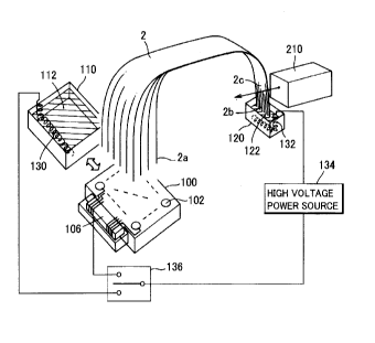
Fig. 1 is a schematic perspective view showing one embodiment
applying the first aspect of the present invention to a mufti-capillary
electrophoretic apparatus, which comprises detection means exciting a
fluorochrome bonded to a sample component as a label for making the same
fluoresce and detecting the fluorescence without influence by Raman
scattering or Rayleigh scattering. Fig. 2 is a conceptual diagram showing one
embodiment of the detection means of this embodiment This figure shows
application to a four~color label DNA sequencer.
A pair of reservoirs 110 and 120 store electrophoresis buffer solutions
112 and 122 respectively, while electrodes 130 and 132 are provided in the
buffer solutions 112 and 122 respectively.
2 0 Respective wells 102 of a sample plate 100 store samples bonded with
fluorochromes having different fluorescent wavelengths such as a fluorescein
derivative and a Rhodamine derivatwe in correspondence to the four types of
end bases of DNA fragments. The sample plate 100 is formed with a wiring
pattern, in which electrodes are arranged in the wells 102 respectively and
2 5 connected to a high-voltage wiring cable through a connector part 106
respectively.
A high-voltage switching part 136 switchably connects the reservoir
110 and the sample plate 100 so that wiring can be switched, and an
electrophoretic high-voltage power source 134 is connected between the
3 0 high-voltage switching part 136 and the electrode 132 provided in the
other
reservoir 120 for switching and applying voltages for sample injection and
electrophoresis.
11
CA 02289864 1999-11-17
In sample injection, one ends 2a of capillary columns forming a
capillary array 2 are inserted one by one into the wells 102 of the sample
plate
100, and after the sample injection, the ends 2a are switched to the reservoir
110 to be dipped in the buffer solution 112. The other ends 2b of the
capillary
columns are dipped in the buffer solution 122 of the other reservoir 120. The
second ends 2b are provided with a part to be detected 2c irradiated with
excitation light from an optical measuring part 210 detecting the samples by
fluorescence so that the fluorescence is measured.
The capillary column ends 2a of the capillary array 2 have two-
dimensional arrangement corresponding to the arrangement of the wells 102 of
the sample plate 100, while the capillary columns are aligned with each other
on the part to be detected 2c and irradiated with the excitation light from a
direction perpendicular to the plane of the arrangement of the capillary
columns.
A moving mechanism (not shown in Fig. 1 ) switches and arranges the
sample plate 100 and the reservoir 110 as indicated by the broad arrow so that
either one selectively comes into contact with the first ends 2a of the
capillary
columns.
The optical measuring part 210 comprises, for example, a laser beam
2 0 source 212 such as a mode-locked titanium sapphire laser unit having a
repetition rate of 78 MHz, a pulse width of 120 to 150 fs, an oscillation
wavelength of 700 to 900 nm and an average output of about 1 W. The
energy of one photon of its laser beam is smaller than excitation energy for
the
fluorochromes.
2 5 A dichroic mirror 214 reflecting laser beam is provided on the optical
path of the laser beam from the laser beam source 212, so that the laser beam
is reflected by the mirror 214 and applied to the part to be detected 2c of
the
capillary array 2 through a lens 216.
Light from the part to be detected 2c is transmitted to the dichroic
3 0 mirror 214 through the lens 216. Fluorescence included in the light is
transmitted through the dichroic mirror 214 and thereafter separated into
prescribed wavelength regions by dichroic mirrors 218, 222 and 226. The
12
CA 02289864 1999-11-17
dichroic minor 214 transmits light having a shorter wavelength than the laser
wavelength of the excitation light in the light from the lens 216. The light
transmitted through the dichroic minor 214 is transmitted to the dichroic
minor 218 so that light having a wavelength of not more than 510 nm is
transmitted through the dichroic minor 18 and incident upon and detected by
a photomuftiplier tube (PMT) 220, while light having a wavelength longer than
510 nm is reflected by the dichroic minor 218 and transmitted to the dichroic
minor 222. In the light from the dichroic minor 218, light having a wavelength
of not more than 560 nm is reflected by the dichroic minor 222 and incident
upon and detected by a photomultiplier tube 224, while light having a
wavelength longer than 560 nm is transmitted through the dichroic minor 222
and transmitted to the dichroic mirror 226. In the light transmitted through
the dichroic minor 222, light having a wavelength of not more than 580 nm is
reflected by the dichroic minor 226 and incident upon and detected by a
photomuitiplier tube 228, while light having a wavelength longer than 580 nm
is
transmitted through the dichroic minor 226 and incident upon and detected by
a photomultiplier tube 230.
The optical measuring part 210 is scanned so that the excitation light
reciprocates horizontally across the plane of arrangement of the capillary
2 0 columns on the part to be detected 2c for successively detecting all
capillary
columns. However, illustration of a scanning mechanism is omitted.
Operations of this embodiment shall now be described with reference
to Figs. 1 and 2.
In sample injection, the one ends 2a of the capillary columns are
2 5 dipped one by one in the wells 102, while the other ends 2b of the
capillary
columns are collectjvely dipped in the buffer solution 122 of the reservoir
120.
The high-voltage switching part 136 is connected to the sample plate 100, and
the electrophoretic high-voltage power source 134 applies a high voltage
between the wells 102 and the reservoir 120. The samples in the wells 102
3 0 are injected into the capillary columns.
After the sample injection, application of the high voltage is
temporarily stopped and the moving mechanism moves the sample plate 100
13
CA 02289864 1999-11-17
and the reservoir 110, thereby dipping the one ends 2a of the capillary
columns
on a sample side into the buffer solution 112 of the reservoir 110. Thereafter
a high voltage is applied between the reservoirs 110 and 120 for performing
electrophoretic separation. The voltage for sample injection into the
capillary
columns and a electrophoresis power supply voltage are, for example, 30 kV
and a current capacity is 10 to 30 mA.
Separated sample components successively pass through the part to
be detected 2c, and are detected by the optical measuring part 210 at this
time.
The laser beam from the laser beam source 212 is applied to the part
to be detected 2c through the dichroic mirror 214 and the lens 216 so that the
fluorochromes bonded to the samples absorb multiphotons and are excited to
fluoresce. The optical measuring part 210 captures the fluorescence so that
the photomuttiplier tubes 220, 224 and 228 detect fluorescence having a
wavelength of not more than 510 nm, fluorescence having a wavelength longer
than 510 nm and not more than 560 nm, fluorescence having a wavelength
longer than 560 nm and not more than 580 nm and fluorescence having a
wavelength longer than 580 respectively.
Base sequence can be determined by bonding the fluorescence
2 0 having a wavelength of not more than 510 nm, the fluorescence having a
wavelength longer than 510 nm and not more than 560 nm, fluorescence
having a wavelength longer than 560 nm and not more than 580 nm and the
fluorescence having a wavelength longer than 580 to DNA fragment samples
for the respective bases respectively.
2 5 Since a Raman scattering line generated in the part to be detected 2c
due to irradiation with the laser beam has a wavelength at least 700 nm, it
does
not form background noise in fluorescence detection. Furthermore, the
intensity of Rayleigh scattering is advantageously smaller than that of a
conventional argon laser beam. In addition, four types of fluorochromes can
3 0 be efficiently excited with a single laser wavelength due to the
muttiphoton
absorption method, whereby it is not necessary to introduce a plurality of
fluorochromes into the same molecule.
14
CA 02289864 1999-11-17
The optical measuring part 210 is not restricted to this embodiment
but may have any system so far as the same can excite fluorochromes
bonded to samples by muttiphoton absorption for making the same fluoresce
and detect the fluorescence.
While the present invention is applied to a mufti-capillary
electrophoretic apparatus in this embodiment, the present invention is also
applicable to electrophoresis employing a single capillary column.
Fig. 3 is a side sectional view schematically showing one embodiment
applying the second aspect of the present invention to a multi-capillary
electrophoretic apparatus, and its mufti-capillary array electrophoresis part
comprises a detection side holder fixing part fixing a detection side holder
and
a parallelism adjusting mechanism adjusting the parallelism between a
detection part and a part to be detected.
A capillary array 2 is formed by arranging a plurality of capillary
columns charged with gels of separation media. One ends (lower ends) 2a of
the capillary columns defining a sample injection side are two-dimensionally
arranged and fixed by a sample injection side holder 4 to come into contact
with a sample in a sample injection reservoir or a bufFer solution in a lower
reservoir for electrophoresis. The other ends 2b of the capillary columns
2 0 forming the capillary array 2 define a detection side on which the
capillary
columns are aligned with each other by a detection side holder 6, and comes
into contact with an upper reservoir buffer solution. A part to be detected 2c
is provided on the detection side (2b side) of the capillary array on a
position
where the capillary columns are aligned with each other and supported by the
2 5 detection side holder 6.
Figs. 4, 5 and 6 are a schematic front elevational view, a schematic left
side elevational view, and a schematic top plan view showing an exemplary
capillary array mounted on this embodiment
In the sample injection side holder 4, a rubber plate 4d of silicone
30 rubber holding and fixing glass capillary columns into holes is held
between
resin holder plates 4a and 4b for two-dimensionally arranging the capillary
columns and integrated by fixed screws 4c. The holder plates 4a and 4b are
CA 02289864 1999-11-17
provided with holes for receiving the capillary columns on 16 by 24 portions
in
con-espondence to the positions of holes of a 384-hole microplate used for
sample introduction. The diameters of the holes of the holder plates 4a and
4b are set larger than the outer diameters of the capillary columns. The
capillary columns passing through the holder plates 4a and 4b and the rubber
plate 4d held therebetween are held in the holes of the rubber plate 4d by
elasticity of rubber, to be airtightly frxed to the holder 4.
The detection side holder 6 holds the capillary columns closely
arranged on a plane by a holder plate 6a from below and by a rubber plate 6d
of
silicone rubber from above. In order to press and fix the capillary columns
against and to the holder plate 6a with the rubber plate 6d, holder plate 6b
is
provided for fixing the rubber plate 6d to the holder plate 6a on both side
portions of the arrangement of the capillary columns. Fixed screws 6c fix the
holder plates 6a and 6b to each other.
The total length of each capillary column is about 500 nm, and the part
to be detected 2c is provided on a position of about 400 nm from the end of
the sample injection side. In order to form a detection window on the part to
be detected 2c, the holder plates 6a and 6b and the rubber plate 6d are
provided with elliptic openings 8 extending in the direction of the
arrangement
2 0 of the capillary columns so that the openings 8 overlap with each other on
the
part to be detected 2c. Signal detection in electrophoresis is performed
through the openings 8.
The mufti-capillary electrophoretic apparatus according to the present
invention is provided on the detection part with location pins 44a guiding the
2 5 holder 6 to a fixed position as described later, and the holder 6 is
provided with
location holes 44b receiving the location pins 44a.
Each capillary column is made of quartz glass or borosilicate glass, and
has an outer diameter of 200 to 300 ~,m and an inner diameter of 75 to 100
~,m. The outer periphery of the capillary column is preferably covered with a
3 0 film of a non-rfluorescent material such as Si02 not fluorescing or
fluorescing
to an extent not hindering fluorescence measurement with excitation light of
ultraviolet to near infrared regions. In this case, the film may not be
removed
16
CA 02289864 1999-11-17
on the part to be detected 2c. If the capillary column has a fluorescing resin
film, the film is removed on the part to be detected 2c.
The capillary columns are charged with a polyacrylamide gel, a linear
acrylamide gel, a polyethylene oxide (PEO) gel and the like as gels of
separation media. Samples containing four types of DNA fragments labeled
with four types of fluorescent materials selected from FAM, JOE, TAMRA,
ROX, R6G, R-110 and the like varied with the end bases are injected into the
capillary columns respectively and simultaneously electrophoresed.
Referring again to Fig. 3, an argon gas laser unit 10 is provided as an
excitation light source for exciting the labeling fluorescent materials. The
argon gas laser unit 10 is a muki-line type unit having an output of 40 to 100
mW and simultaneously oscillates laser beams having wavelengths of 488 nm,
514.5 nm and the like.
When applying the mufti-capillary electrophoretic apparatus shown in
Fig. 3 to an apparatus utilizing multiphoton absorption of the first aspect, a
mode-locked titanium sapphire laser unit generating a laser beam having a
longer wavelength than fluorescence generated by a labeled fluorochrome is
used as the excitation light source in place of the argon gas laser unit 10.
The energy of one photon of the laser beam is smaller excitation energy for
2 0 the fluorochrome.
An optical system 12 applying the laser beam from the laser unit 10 to
the part to be detected 2c of the capillary array 2 as excitation light and
detecting fluorescence from the part to be detected 2c is an epi-optical
system shown in Fig. 7A in detail. Numeral 16 denotes a mirror
2 5 perpendicularly applying a laser beam 14 from the laser unit 10 to a
surface of
the part to be detected 2c of the capillary array 2, numeral 18 denotes a
tunnel mirror having a hole on its center for transmitting the excitation
light
beam through the hole and reflecting the fluorescence on a minor surface, and
numeral 20 denotes an objective lens consisting of a condenser lens
3 0 condensing and projecting the excitation light onto a single capillary
column
and receiving fluorescence generated from a sample migrating in the capillary
column. The objective lens 20 projects the excitation light and receives the
17
CA 02289864 1999-11-17
fluorescence by the same lens, and forms the epi-optical system. The minor
surface of the tunnel minor 18 reflects the fluorescence collected by the
objective lens 20.
Numeral 22 denotes an optical filter blocking an excitation light
component from the reflected light and transmitting the fluorescence, numeral
24 denotes a pinhole slit for limiting a detection field, and numeral 26
denotes a
diaphragm lens imaging the fluorescence transmitted through the optical filter
22 on the position of the pinhole slit 24. A fluorescing point in the
capillary
column is imaged on the position of the pinhole slit 24, thereby forming a
confocal optical system. An edge filter or colored glass can be employed as
the optical fitter 22 for removing the excitation light The pinhole slit 24
reduces the detection field for preventing invasion of stray light from
adjacent
capillary columns.
n order to divide the fluorescent image on the pinhole slit 24 into four
luminous fluxes, a lens panel 28 shown in Fig. 7B is arranged. The lens panel
28 can be manufactured as that prepared by cutting single lenses and sticking
the same to each other or a glass molding. A filter panel 30 formed by
difFerent spectroscopic filters for respective labeling fluorescent materials
shown in Fig. 7C is arranged on optical paths of the four luminous fluxes. The
2 0 filter panel 30 is a bandpass fitter, which is formed by arranging four
types of
filters having difFerent wavelength characteristics corresponding to the
labeling
fluorescent materials on the respective optical paths in parallel with each
other. The transmission wavelengths of the respective fitters correspond to
light emission wavelengths of the fluorescent materials labeling fragment
2 5 samples whose end bases are A (adenine), G (guanine), C (cytosine) and T
(thymine). Four photomultiplier tubes 32 are arranged on the respective
optical paths for detecting fluorescence transmitted through the fitters.
he epi-optical system 12 including the minor 16, the tunnel minor 18,
the objective lens 20, the optical filter 22, the pinhole slit 24, the
diaphragm
3 0 lens 26, the lens panel 28, the filter panel 30 and the photomultiplier
tubes 32 is
mounted on a stage of a scanning mechanism 34, and reciprocally moved along
a straight line (perpendicular to the plane in Fig. 3 and vertical in Fig. 7A)
18
CA 02289864 1999-11-17
parallel to the plane of the part to be detected 2c of the capillary array 2
and
perpendicular to the electrophoresis direction, in order to detect
fluorescence
from all capillary columns on the part to be detected 2c. The laser beam 14
is incident upon the mirror 16 in parallel with a scanning direction of the
epi-
optical system 12, so that the optical axis of the laser beam 14 is not
fluctuated by scanning of the epi-optical system 12.
Fig. 8A is a sectional view of one embodiment of the detection side
holder fixing part and its periphery as viewed from above, Fig. 8B is a front
sectional view taken along the line B-B' in Fg. 8A and Fig. 8C is a side
sectional view taken along the line C-C' in Fib 8A, while Fg. 8A is taken
along
the lines A-A' in Figs. 8B and 8C. Figs. 8A and 8B omit illustration of an
upper electrode 58 and a detection side capillary end pressing member 60, and
Fig. 8A also omits illustration of a detected part pressing member 62.
A movable plate 36 is provided on a position for fixing the detection
side holder 6. Figs. 9A, 9B and 9C are a top plan view, a front sectional view
and a right side elevational view showing the movable plate 36 respectively.
The movable plate 36 is formed by a substrate 36a and a detection
position plate 36b. The substrate 36a is provided with a slot 38 in a
direction
where the epi-optical system 12 is scanned. The detection position plate
2 0 36b slightly smaller in dimension than the opening 8 of the detection side
holder 6 is arranged on the slot 38. An epi-optical system scanning groove
40 is formed in the detection position plate 36b on the substrate 36a side,
and
a slot defining a light application window 42 is formed on the bottom surFace
of
the scanning groove 40. The objective lens 20 side of the epi-optical system
2 5 12 is arranged in the slot 38 and the scanning groove 40 and scanned along
the slot 38.
Two location pins 44a are arranged on positions of the substrate 36a
corresponding to the location holes 44b of the holder 6. The holder 6 is
correctly arranged on the movable plate 36 by registering the positions of the
3 0 location pins 44a and the location holes 44b and those of the detection
position plate 36b and the opening 8 of the holder 6.
Furthermore, set screws 46 are arranged on the substrate 36a on
19
CA 02289864 1999-11-17
positions corresponding to four comers of the holder 6. The height for
arranging the holder 6 can be adjusted by rotating the set screws 46 and
adjusting the length of the set screws 46 projecting from the substrate 36a.
Furthermore, the substrate 36a is provided with a gate angle adjusting
mechanism formed by two gate adjusting screws 48a passing through the
substrate 36a and a gate adjusting supporting point pin 48b provided on a side
opposed to the side provided with the gate adjusting screws 48a and opposite
to the detection position plate 36b. The parallelism between the movable
plate 36 and a scanning axis of the epi-optical system 12 can be adjusted by
rotating the gate adjusting screws 48a.
The holder 6 is fixed to the movable plate 36 by fastening two clamps
50 provided in the vicinity of both ends of the movable plate 36.
An upper reservoir 52 storing a bufFer solution for dipping the other
ends 2b of the capillary columns forming the capillary array 2 and a cover 54
covering upper portions of the movable plate 36 and the upper reservoir 52 are
provided in the vicinity of the movable plate 36. The cover 54 can be
opened/closed along a cover switching shaft 56.
The upper electrode 58 covered with a cylindrical insulating member is
mounted on the cover 54 and comes into contact with the buffer solution of
2 0 the upper reservoir 52 in the state covered with the cover 54. A capillary
array end pressing member 60 is arranged on the cover 54, for bending the
other ends 2b of the capillary columns forming the capillary array 2 toward
the
upper reservoir 52 and dipping the same in the bufFer solution.
The cover 54 is provided with the detected part pressing member 62
2 5 on a position corresponding to the part to be detected 2c. The pressing
member 62 is formed with a plane smaller in dimension than the opening 8 of
the holder 6, and a rubber plate 64 of silicone rubber is stuck to this plane.
Four rod members 66 provided on the cover 54 mount the pressing member
62 to be slidable in a direction perpendicular to the plane of the part to be
3 0 detected 2c. Springs 68 are arranged on the rod members 66 between the
cover 54 and the pressing member 62 respectively, so that the pressing
member 62 presses the part to be detected 2c against the detection position
CA 02289864 1999-11-17
plate 36b with appropriate pressure through the silicone rubber plate 64 when
the cover 54 is closed after arranging the holder 6 on the movable plate 36.
The detection side holder fixing part according to the present
invention is formed by the movable plate 36, the gate adjusting screws 48a,
the
gate adjusting supporting point pin 48b, the clamps 50 and the detected part
pressing member 62.
As shown in Fig. 3, the detection side holder 6 is fixed in an
electrophoresis chamber 66. A lower electrode 68 is mounted on a lower
portion of the chamber 66, to come into contact with a buffer solution in a
lower reservoir and communicate with the lower ends 2a of the capillary
columns forming the capillary array 2 when the buffer solution in a sample
injection reservoir or the lower reservoir for electrophoresis is pushed up to
a
position coming into contact with the lower ends 2a of the capillary columns.
A sample injection voltage or an electrophoresis voltage is applied between
the
buffer solutions in both reservoirs from a high-voltage power source through
the electrodes 58 and 68. For example, the power supply voltage is 30 kV
and a current capacity is 10 to 30 mA.
The reservoir for electrophoresis and a sample injection reservoir 70
are arranged in a horizontal plane and supported on an X-Z sample stage 72
2 0 under the ends 2a of the capillary columns on the sample injection side of
the
capillary array 2. The X-Z sample stage 72 perForms movement in a
horizontal direction (X direction: perpendicular to the plane of Fig. 3) for
locating either reservoir under the ends 2a and movement in a vertical
direction (Z direction: vertical in Fig. 3) for bringing the buffer solution
in the
2 5 reservoir into contact with the ends 2a or separating the former from the
latter by a sample stage moving mechanism 74.
A sample titer plate 76 formed with wells corresponding to the
arrangement of the ends 2a of the capillary columns is placed on the reservoir
70. Bottoms of the wells pass through the sample titer plate 76, membranes
3 0 are formed on the bottoms and samples are adsorbed on the membranes of
the wells. The bufFer solution in the reservoir 70 comes into contact with the
membranes, and the sample injection voltage is applied to the ends 2a of the
21
CA 02289864 1999-11-17
capillary columns from the lower electrode 68 through the buffer solution.
Operations of fixing the detection side holder 6 to the movable plate
36 and adjusting the parallelism between the plane of the part to be detected
2c and the scanning axis of the epi-optical system 12 shall now be described.
The capillary array 2 having the part to be detected 2c charged with a
fluorochrome is prepared so that the holder 6 for the capillary array 2 is
arranged on the movable plate 36 by opening the cover 54 and registering the
positions of the location pins 44a and the location holes 44b and clamped and
fixed by the clamps 50. Thus, the part to be detected 2c of the capillary
array 2 can be fixed to the apparatus with excellent reproducibility.
The cover 54 is closed so that the pressing member 62 presses the
part to be detected 2c against the detection position plate 36b and fixes the
same onto the light application window 42 along the plane of the detection
position plate 36b while the pressing member 60 dips the ends 2b of the
capillary columns of the capillary array 2 in the buffer solution of the upper
reservoir 52.
The epi-optical system 12 is scanned and an image formed on the
pinhole slit 24 is observed to determine whether or not the distance between
the part to be detected 2c and the objective lens 20 is proper. If the
distance
2 0 between the part to be detected 2c and the objective lens 20 is improper,
the
gate adjusting screws 48a are rotated for adjusting the distance between the
part to be detected 2c and the objective lens 20. Thus, requirement for
working accuracy in preparation of the detection side holder 6 and formation
of the capillary array 2 can be relieved and reduction of detection
sensitivity
2 5 can be suppressed. Furthermore, since the distance between the part to be
detected 2c and the epi-optical system 12 can be adjusted, this
electrophoretic apparatus is adaptive to various outer diameters of the
capillary columns arranged on the capillary array 2. In addition, outer
diameter tolerance by manufacturing lot difference of the capillary columns
3 0 can be allowed.
While this embodiment employs two gate adjusting screws and the
gate adjusting supporting point pin as the gate angle adjusting mechanism, the
22
CA 02289864 1999-11-17
present invention is not restricted to this but a gate angle may be
automatically adjusted in correspondence to a detection signal at the time of
scanning the epi-optical system by employing an actuator such as a
piezoelectric element, for example.
Alternatively, an actuator moving the epi-optical system along the
optical axis of applied light may be provided in place of the gate angle
adjusting
mechanism for automatically adjusting the distance between the part to be
detected and the epi-optical system.
Fig. 10 is a perspective view of a capillary cassette of one embodiment
am~nging a plurality of capillary columns.
A plurality of capillary columns 102 of a, capillary array 2 are arranged,
sample injection sides are fixed by a cassette holder 4 and detection sides
are
fixed by a cassette holder 6 and a heat shrinkable tube 80 on an end to form a
capillary cassette 9. One ends 2a of the capillary columns 102 define a
sample injection part and are two-dimensionally arranged and fixed by the
cassette holder 4. The other ends 2b of the capillary columns 102 forming
the capillary cassette 9 are planarly aligned with each other, fixed by the
cassette holder 6, and cylindrically bundled by a shrank heat shrinkable tube
80. A detection window is formed on the cassette holder 6, and portions of
2 0 the capillary columns 2 located on the detection window define a part to
be
detected 2c.
For example, the outer diameter of each capillary column 102 is 300 to
400 ~,m,.
Figs.11 A to 11 C are model diagrams showing a procedure of bundling the ends
2b of the capillary columns 102 by the hea~shrinkable tube 80. Figs. 12A
and 12B are sectional views taken along the lines A-A' and B-B' in Fig. 11 C.
These figures show the capillary columns 102 in a reduced number.
Manufacturing is performed along the following sequence:
(A) After aligning the plurality of capillary columns 102 with each other,
a filler 12 such as an epoxy resin adhesive or a silicon compound is applied
to
surface portions of the capillary columns 102 on prescribed positions from the
ends 2b.
23
CA 02289864 1999-11-17
(B) The ends 2b of the capillary columns 102 are bundled and inserted
into the heat shrinkable tube 80, which in tum covers the positions to which
the filler 12 is applied.
(C) The heatshrinkable tube 80 is heated and shrunk. The inner
diameter of the heat shrinkable tube 80 is reduced, the capillary columns 102
adhere to each other, and clearances therebetween are filled up with the
filler
12 so that the capillary columns 102 are cylindrically bundled in an airtight
manner. Clearances between the heat-shrinkable tube 80 and the capillary
columns 102 and between the capillary columns 102 are sealed with the filler
12, and both ends of the heat-shrinkable tube 80 do not communicate with
each other except in the capillary columns 102.
Fig. 13 is a model diagram showing a method of fixing a mounting
member to a polymer charger to the ends 2b of the capillary columns 102.
The capillary columns 102 bundled by the heat-shrinkable tube 80 are
inserted into a ferrule 86 serving as the mounting member for the polymer
charger along with the heatshrinkable tube 80 and fixed by a screw 88 so that
each capillary column 102 attains sufficient pressure resistance in polymer
injection.
It is preferable to set the outer diameter of the shrank heat-shrinkable
2 0 tube 80 receiving the capillary columns 102 to a pipe outer diameter of a
liquid
chromatograph such as X1.6 (outer diameter of 1.6 mm), ~2 or ~3 by adjusting
the outer diameters of the capillary columns 102, the number of the capillary
columns 102 and the thickness of the shrank heat shrinkable tube 80.
Consequently, an existing ferrule employed for a liquid chromatograph or the
2 5 like can be employed. When preparing a dedicated female, the capillary
columns 102 may be bundled in response to the inner diameter of the
dedicated ferrule.
Irregularities of the bundled capillary columns 102 are removed due to
the thickness of the heat-shrinkable tube 8, and hence the thickness of the
3 0 shrunk heat shrinkable tube 80 is preferably increased.
In polymer charging, the ferrule 86 is connected to and mounted on a
connection part of the polymer charger, for charging the capillary columns 2
24
CA 02289864 1999-11-17
with polymers by press~filling or suction.
Fig. 14 is a schematic perspective view showing one embodiment of a
mufti-capillary electrophoretic apparatus to which the capillary cassette 9
shown in Fig. 10 is applied. Fig 14 omits illustration of the cassette holders
4
and 6. This mufti-capillary electrophoretic apparatus is identical to that
shown in Fig. l except the structure on the side of the other ends 2b, and
hence redundant description is omitted.
The ends 2b of the capillary columns are bundled by the heat
shrinkable tube 80, and dipped in a bufFer solution 122 in a reservoir 120
along
with the heat shrinkable tube 80.
Operations in electrophoretic separation are also identical to those in
Fig. 1.
Such a mufti-capillary electrophoretic apparatus preferably comprises
an automatic polymer charging mechanism for reusing the capillary columns
102 by discharging used polymers from the capillary columns 102 and charging
new polymers.
Although the present invention has been described and illustrated in
detail, it is clearly understood that the same is by way of illustration and
example only and is not to be taken by way of limitation as the spirit and
scope
2 0 of the present invention are limited only by the terms of the appended
claims.