Note: Descriptions are shown in the official language in which they were submitted.
CA 02289961 1999-11-12
1
DESCRIPTION
A Gene Causative of Rheumatoid Arthritis,
Method for Diagnosing Rheumatoid Arthritis,
and Method for Identifying Causative Factors of Rheumatoid Arthritis
TECHNICAL FIELD
The present invention relates to the genes causative of rheumatoid arthritis,
a
method for diagnosing rheumatoid arthritis using the mutations of these genes
as the
indicators, and a method for identifying the causative factors of rfieumatoid
arthritis.
BACKGROUND OF THE INVENTION
The aspects of arthritis and joint damage causing rheumatoid arthritis,
particularly the pathological courses thereof, have been elucidated gradually
through
various research works. Because many of the autoimmune diseases including
rheumatoid arthritis are induced by the concomitant participation of numerous
causative
factors and are then exacerbated progressively to the stage of apparent
diseases,
however, the interactive mechanism per se of such riumerous factors should be
elucidated for accurate characterization and appropriate therapeutic
management of
the disease. The prevalent rheumatoid arthritis is about 1%(N. Engl. J. Med.,
322: 1277-
1289, 1990), but the frequency of the disease is about 8 times increased in
the siblings
of the patients with the disease (Cell, 85: 311-318, 1996). Hence, it is
predicted that a
certain genetic factor may serve as one of the causative factors_
Nevertheless,
molecular genetic technology arid genetic engineering technology for general
use for
CA 02289961 1999-11-12
2
identifying of genetic factors of diseases never function effectively in case
of
autoimmune diseases, because the onset of autoimmune diseases is never induced
via
such a biologically simple mechanism as abnormal amplification of one mutated
gene
as in the case of cancer. Conventional strategies of traditional genetics for
the
elucidation of the fundamental genetic pathogenesis of diseases have
demonstrated
distinctively that autoimmune diseases are caused by genetic multi-factor
inheritance,
but the strategies were apparently insufficient. As has been described above,
none of
the genes involved in rheumatoid arthritis or none of the loci of the genes on
chromosome have absolutely been evidenced so far.
Altematively, the linkage analysis method and the positional cloning method by
means of polymorphic markers have opened an inriovative progress recently in
the field
of research works on genetic diseases. By using these methods, not only the
chromosomal locations of disease genes previously never characterized have
been
identified but also the causative genes of numerous diseases have been
isolated and
assayed (Experimental Medicine, Vol. 12, No. 6: 80-85, 1994). More recently,
the
causative gene of type I diabetes mellitus has been isolated (Nature, 171: 130-
136,
1994), owing to a sib-pair analysis method comprising usage of the linkage
analysis
using a microsatellite marker as the polymorphic marker (Nature, 359: 794-801,
1992;
Nature Genet., 7: 246-336) and the analysis of patient pedigrees as one of the
procedures of traditional Qwetics. Thus, it becomes possible that genes
causing
diseases hardly curable because of the current absence of any effective
therapeutic
means, including autoimmune diseases, will be identified.
In such circumstances and the latest progress of the research works, the
present application has been submitted. It is an object of the invention to
provide the
genes causative of rheumatoid arthritis, the chromosomal locations of these
genes
CA 02289961 1999-11-12
3
firstly being specified, a method for diagnosing rheumatoid arthritis using
the mutations
of these genes as the indicators, and a method for identifying the causative
factors of
rheumatoid arthritis.
DISCLOSURE OF THE INVENTION
In accordance with the application of the invention to overcome the
aforementioned problems, the following individual genes are provided.
1. A gene causative of rheumatoid arthritis, which gene is located within 1
centimorgan from a DNA sequence on human chromosome 1 to which the
microsatellite markers D1S214 and/or D1S253 are hybridized.
2. A gene causative of rheumatoid arthritis, which gene is located wlthin 1
centimorgan from a DNA sequence on human chromosome 8 to which the
microsatellite marker D8S556 is hybridized.
3. A gene causative of rheumatoid arthritis, which gene is located within 1
centimorgan from a DNA sequence on human chromosome X to which the
microsatellite markers DXS1001, DXS1047, DXS1205, DXS1227 and/or DXS1232 are
hybridized.
In accordance with the application, a method for diagnosing rheumatoid
arthritis,
comprising amplifying the genomic DNA of a subject by PCR method using as
primer at
least one of microsatelliteooarkers D1S214, D1S253, D8S556, DXSIOOI, DXS1047,
DXS1205, DXS1227 and DXS1232, and then comparing the resulting PCR products
with the PCR products prepared in the same manner using the genomic DNA of a
normal control.
In accordance with the application, furthermore, a method for identifying the
causative factors of rheumatoid arthritis, comprising amplifying the genomic
DNA of a
CA 02289961 1999-11-12
4
subject by PCR using as primer at least one of microsateqite markers D1 S214,
D1 S253,
DSS556, DXS1001, DXS1047, DXS1205, DXS1227 and DXS1232, and then
comparing the resulting PCR products with the PCR products prepared in the
same
manner using the genomic DNA of a normal control.
BRIEF DESCRIPTION OF THE DRAWINGS
Fig. I is a map of microsatellite markers for use in identifying the loci of
the
inventive genes on the chromosomes 1 to 5;
Fig. 2 is a map of microsatellite markers for use in identifying the loci of
the
inventive genes on the chromosomes 6 to 15;
Fig. 3 is a map of microsatellite markers for use in ideritifying the loci of
the
inventive genes on the chromosomes 16 to 22, and chromosome X;
Fig. 4 shows one example of the gel electrophoresed PCR products;
Fig. 5 shows one example of the Genotyper analysis of the PCR products;
Fig. 6 depicts the results of the MLS values of the analyzed microsatellite
markers, as plotted vs the chromosomes I to 6;
Fig. 7 depicts the results of the MLS values of the analyzed microsatellite
markers, as plotted vs the chromosomes 7 to 12;
Fig. 8 depicts the results of the MLS values of the analyzed microsatellite
markers, as plotted vs the-chromosomes 13 to 18;
Fig. 9 depicts the results of the MLS values of the analyzed microsatellite
markers, as plotted vs the chromosomes 19 to X; and
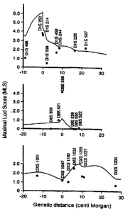
Fig. 10 graphically shows the relationship between the MLS values of the
individual microsatellite markers at multiple marker sites including the
marker sites
indicating the inventive disease genes and the distances (in unit centimorgan)
thereof
CA 02289961 2007-11-29
from the target disease genes on the chromosome 1(upper panel), the chromosome
8
(middle panel) and the X chromosome (lower panel).
BEST MODE FOR CARRYING OUT THE INVENTION
5 The method for identifying the genes of the present invention is now
described in detail hereinafter.
The genes of the invention comprise plural genes having been identified of the
chromosomal loci by the linkage analysis of patients with rheumatoid arthritis
and their
families with blood relationship. More specifically, the inventors have
determined the
toci of all the genes with relation to the disease sensitivity to rheumatoid
arthritis, by
using the DNA polymorphism (polymorphism of the length of CA repeat sequence)
of
microsatellite marker genes over the entire lengths of human chromosomes_ The
method is specifically described below.
1 . Extraction of genomic DNA
A set of Patient A and another Patient B, both with rheumatoid arthritis at
the
stage II or higher of joint damage satisfying the dinical standard of American
Rheumatism Association, and a normal sibling member C was first prepared; and
then,
such 35 families were analyzed as subjects. From the Individuals blood was
drawn
peripheral blood (10 mi), using EDTA; the blood was then graduaily mixed with
20 ml
buffer I[0.32 M sucrose, 5 % v/v Tritori X-100, 5 mM MgC12, 12 mM Tris HCI (pH
7,6)],
to solubilize the cell membrane. After centrifugation, precipitated nuclei
reacted with
buffer II [4 M guanidine thiocyanate, 12 mM EDTA, 375 mM NaCI, 0.5 % sodium N-
lauroyl sarcosinate, 0.1 M 13-mercaptoethanol, 12 mM Tris HCI (pH 7.6)) to
solubilize the
nuclear membrane to extract DNA by ethanol precipitation.
2. PCR amplification and sizing of micxosatellite DNA
* Trademark
CA 02289961 1999-11-12
6
Using the extracted genomic DNA as template, microsatellite marker genes
corresponding to the chromosomal loci shown in Figs. 1 to 3 were amplified by
PCR
using fluorescently labeled primers (manufactured by Perkin-Elmer Co.).
Herein, the
marker D1S502 was not used because of the technical toughness for DNA
amplification.
For detailed analysis of the region HLA-D, individual genes of D6S299, D6S265
and
D6S273 were amplified in place of D8D276. In addition to the amplification of
the
microsatellite DNAs, genes in the vicinity of the region HLA-DRBI were also
amplified
by PCR using restriction fragment length polymorphism (RFLP) markers; in
total, 359
marker sites were examined.
The composition of the PCR solution (15 i) for microsatellite DNA
amplification
was as follows; DNA (30 ng), primers in mixture (0.2 M), dNTP (each 0.2 mM),
DNA
polymerase (1 unit), MgC12 (2.5 mM) and 1 x PCR buffer II. Amplification was
conducted under the following conditions; denaturation at 94 C for 10
minutes,
denaturation (94 C for 30 seconds), annealing at 55 C for 1 minute and
extension at
72 C for 2 minutes; the program was repeated for 27 cycles, but the last
extension was
conducted at 72 C for 5 minutes.
Individual PCR products labeled with 6-FAM. TET or HEX were electrophoresed,
together with TAMURA-labeled size standards, on one gel panel (4 %
acrylamide/6 M
urea) in a DNA sequencer (ABl 337; manufactured by Applied BioSystems, Co.
Ltd.).
Fig. 4 shows one exaroWe of the electrophoresis. According to the method,
electrophoretic error could be reduced greatly, because the peaks, sizes and
regions of
DNA fragments were analyzed with reference to the size standards pattemed.
Subsequently, the individual marker genes were sized on the basis of the
positions of
the fluorescent images incorporated on a computer system, to determine how the
genes
derived from parents could be inherited per one pedigree. Furthermore, the
_ .~.~..~.,
CA 02289961 2007-11-29
7
electrophoretic results were subjected to gene scanning analysis and
subsequent sizing
with an analysis software Genotyper Fig. 5 shows one example of the analysis
results
with Genotyper*
3. Linkage analysis
The sib-pair analysis method using linkage analysis by means of microsatellite
marker has already been known in the identification of the causative gene of
type I
diabetes mellitus (Proc. Natl. Acad. Sci., 92: 8560-8565, 1995). However, the
method
cannot be applied as it is with no modification to subjects with rheumatoid
arthritis.
The reason is that general sib-pair analysis method determines the genetic
mode of IBD
(identical by descent) between a patient and both the parents thereof and that
both the
parents of individual patients with rheumatoid arthritis as one of senile
diseases are
dead in most of the cases at the onset of the disease so no definite IBD value
can be
determined in such cases.
In accordance with the invention, 35 families composed of Patient A, Patient B
and a normal sibling member C were analyzed for IBD determination. More
specifically, the IBD value Is designated as 1 provided that both of the
affected sibling
members are endowed with the essential gene "a" of a parent; IBD = 2 provided
that
the afflicted sibling members share individual alleles in common and that the
individual
alleles are imparted from either one of the parents. When both the parents are
already
dead with no possibility of typing of the genes of the parents. IBD cannot
definitely be
defined. Provided that the distribution of a certain arbitrary gene marker in
a race
population as a analysis subject is defined, the IBD value nevertheless can be
detemiined by using the frequency of the allotype of the gene. Provided that
the
apparent IBD agreement between patient A, patient B and a normal sibling
member C is
defined as (IBD between A and B, IBD between A and C and IBD between B and C)
and
* Trademark
CA 02289961 1999-11-12
8
that a certain arbitrary gene as a target is defined as "a" while others are
defined as "a",
all the possibilities are shown as 27 cases as in Table 1.
[Table 1)
Case 1: (ua, aa, aa), (az, aa, ai), (aa, a8, aa)
Case 2: (aa, aa. a2), (aa, aZ, aa)
Case 3: (aa, aa, aa), (f+b, aa, as), (aa, aP, aa), (a&, aa, aa)
Case 4: (aa, aa, ae), (aa, aA, aa), (fla, aa, az), (aa, aa, ara)
Case G: (aa, aa, aa), (Ard, aa, a8), (aa, EtZ, ae d), (aA, aa, [12), (ae1, aa,
a.d),
(I a, al1, as), (na, a2, a2), (aA, aa. d
Case G: (aA, aa, aS), (&a, an, aa)
Case 7: (Ae, ad, 2a), (aa, aa, 2a), (ai, aa, aa), (rdA, aA, aa)
According to the formula 1 of Holmans & Clayton (Am. J. Hum. Genet. 57:
1221-1232, 1995) provided that the allotype frequency of the gene "a" is
defined as
"Pa", the Lod value (L value) can be determined as follows.
Formula 1
Dai-erital )[z';r<i ( P)~ .ppr ~ aiJected pairl P
1,EP
For example, the L values of three sets of Case I are calculated as L,,. L,:
and Lõ by
the following formulas 2, 3 and 4, respectively.
Formula 2
Lis = P. zo 4- 1/2 I'.'(1+P.)zi + 1/4I'.2(1+P.)2 z:
Formula 3
L,a = Pj=' zo + 1/2 PbB (1+P,)zi + 114Pi= (1+I'j)2zs
CA 02289961 1999-11-12
9
Formula 4
Li3 = 3P,'P,2zo + 1/2P,Pt(1+2P.P2) zi + P.P, (1+1/2P,P,)z:
In the same manner, the L values from Lr1 to L72 can be caicuiated. By the
same calculation on all subjects, the L value of the gene "a" in a population
can be
calculated according to the formula S.
Formula 5
rr! nll u. 72
L
By subsequently permitting the variabies ZO, Z, and Z2 to vary throughout the
ranges under provisions of Zo -5; 1/2, Zfl <_ 1/2 Z, and 4 + Z, + Z2 = 1, the
maximum (L.)
of the L values can be determined. Under the provision of no emergence of any
relationship between these markers and the actual gene, alterriativeiy, the L
value (L,wõ)
can be calculated according to the formula 6, provided that Zo = 0.25, Z, =
0.50 and Zz =
0.25_
Formuia 6
Finally, the maximum Lod value (Maximal Load (Lod?) Score: MLS) can be
calculated according to the formula 7.
Formula 7
A1LS = los'~"~ ~n~rr
Figs. 6 to 9 depict the results of the MLS values of all the microsateliite
markers
of 359 in total as plotted vs each chromosome. First, herein, marker sites
with MLS
CA 02289961 1999-11-12
values around 3.0 are supposed to significantly correspond to the causative
genes as
well. In other words, the MLS value indicates the probability of the emergence
of the
relationship between one of the markers and one of the causative genes
compared with
the accidental emergence thereof and is represented in a logarithmic figure
based on
5 log,a; at MLS = 3.0, the relationship is supposed to be present at a
probability 1000-fold
the probability of the accidental emergence thereof. More specifically, it is
indicated
that the microsatellite markers D1 S214, D1S253, D8S556 and DXS1232 at
extremely
large MLS values around 3.0 are present very closely to the causative genes.
Fig. 10 graphically shows the relationship between the MLS values of the
10 individual microsatellite markers at multiple marker sites including the
four marker sites
and the distances (in centimorgan) thereof from the disease genes on the
chromosome
1(upper panel), the chromosome 8 (middle panel) and the X chromosome (lower
panel).
As shown in Fig. 10, a target disease gene of rheumatoid arthritis is present
at a
position very close to the sites of the microsatellite markers 01S214 and/or
D1S253 on
human chromosome 1. Similarly, the disease genes are present at a position
very
close to the site marked with the microsatellite marker D8S556 on human
chromosome
8 and at a position very close to the sites marked with microsatellite markers
DXS1001,
DXS1047, DXS1205, DXS1227 and/or DXS1232 on human X chromosome.
The disease genes of rheumatoid arthritis in accordance with the invention are
present at such specific chromosomal loci as described above (genes present at
least
at one or more of positions within 1 centimorgan from the 8 sites marked
with the
microsatellite markers): once the coding regions and DNA sequences thereof are
identified and determined by known methods such as positional cloning, the
disease
genes can make profound contributions to the establishment of an effective
therapeutic
method thereof. The PCR amplification and analysis of the microsatellite genes
used
CA 02289961 1999-11-12
11
in accordance with the invention are applicable to the diagnosis of rheumatoid
arthritis
and the identification of the causative factors thereof. More specifically, by
amplifying
the genome DNA of a patient with the potential onset of the disease by PCR
using tho
markers corresponding to the chromosomal sites as primers and comparing thQ
resulting amplified products with the genome DNA of a normal control by the
analysii,
with Genotyper as shown in Fig. 5, the potential subsequent onset of the
disease can
be estimated at a high precision.
INDUSTRIAL APPLICABILITY
The invention provides the disease genes of human rheumatoid arthritis, a
method for diagnosing rheumatoid arthritis using the mutations of these
disease genes,
as the indicators, and a method for determining the causative factors thereof.
These.
aspects of the invention are applicable to the development of pharmaceutical
medicines
and therapeutic strategies.
~.