Note: Descriptions are shown in the official language in which they were submitted.
CA 02290316 1999-11-24
NOVEL SOURCE OF
ELEUTHEROBIN AND RELATED ANTIMITOTIC DITERPENES
BACKGROUND OF THE INVENTION
Antimitotic compounds interfere with the dynamic assembly and disassembly of w-
and ~-tubulin into microtubules causing cells to arrest in mitosis. Prolonged
arrest in mitosis
eventually leads to cell death, often by apoptosis. Two chemical classes of
antimitotic agents,
the vinca alkaloids (vinblastine, vincristine, and vinorelbine) and the
taxanes (paclitaxel and
docetaxel), are clinically useful anticancer drugs. Most known antimitotic
agents induce
mitotic arrest by inhibiting the polymerization of tubulin into microtubules.
This is the
mechanism of the vinca alkaloids and rhizoxin.
Paclitaxel was the first chemical entity shown to cause mitotic arrest by
stabilizing
microtubules against depolymerization. Four additional chemotypes that have
paclitaxel-like
effects were later identified. These include the myxobacterium metabolites
epothilones A and
B, the marine sponge metabolites discodermolide, laulimalide, and
isolaulimalide, and the soft
coral metabolite eleutherobin shown below as Compound 1. Ojima et al. (1999)
Proc. Natl.
2 o Acad. Sci. USA 96:4256-4261, propose a common pharmacophore for the
microtubule
stabilizing compounds that effectively accommodates nonataxel, paclitaxel,
discodermolide,
eleutherobin, and the epothilones. This model predicts that three regions of
eleutherobin
(boxes A, B, and C below) are important for binding to tubulin (Me = methyl;
Ac = acetyl).
N OH
~i o
N
s' OH
2' ~ O ~ OAc
,~ OMe O
3 0 O~,11~ ~s
CA 02290316 1999-11-24
-2-
The majority of known antimitotic natural products were initially isolated
because they
exhibited potent in vitro cytotoxicity. Only subsequent detailed mechanism of
action studies
revealed that they arrested cells in mitosis and interfered with tubulin
assembly and
disassembly dynamics. For example, rhizoxin is a 16-membered ring macrolide
first isolated
in 1984 and determined to be very cytotoxic. Only later was rhizoxin shown to
cause the
accumulation of cells in mitosis. Sarcodictyins A-D were the first members of
the
eleutherobin class of compounds to be identified (see: D'Ambrosio, M., et al.
(1987) Helv.
Chim. Acta. 70:2019-2027; and, (1988) Helv. Chim. Acta. 71:964-976), their
paclitaxel-like
properties being recognized only later. Eleutherobin was originally isolated
from the soft
coral Eleutherobia sp. (possibly E. albiflora) collected in Western Australia
(see: Lindel, T.
et al. (1997) J. Am. Chem. Soc. 119:8744-8745; and, international patent
application
published May 23, 1996 under WO 96/14745).
Using a new cell-based antimitotic assay, the inventors herein have
demonstrated
potent antimitotic activity in extracts of various marine organisms providing
an abundant new
source of naturally occurring antimitotic diterpenes. Microscopic examination
of cells
arrested in mitosis by the extracts show tubulin bundling, similar to the
effects of paclitaxel.
Bioassay guided fractionation of extracts of marine organisms as described
herein, led
to the isolation of eleutherobin 1 (as shown above) and the new antimitotic
diterpenes shown
below, including desmethylelcutherobin 2, desacetyleleutherobin 3,
isoeleutherobin A 4,
2 0 Z-eleutherobin 5, caribaeoside 6, and caribaeolin 7.
Me ME
N ORS N
~N ~ O ~°
N
z, OR, ORi I O _
~s
O ~1~0~ ~OMi R
~ 5 O._
» H-~~~~
9
OH
2 R~ = Ac; R2 =Rg =R4 =_ H: e2'.3' (E)
O
3 R~ =R2 =R3 =_ H; R4 =Me: e2~.3~ (E)
4 R~ =R3 =H; R2 =Ac; R4 =Me; 02~~3~ (E) s ~ H
OAc
5 R~ =Ac. Rp =R3 =H; R4 =Me, e2'.3' (~
7 R=Ac
CA 02290316 1999-11-24
-3-
SUMMARY OF THE INVENTION
This invention provides the use of organisms previously not known to certain
antimitotic diterpenes to prepare purified or partially purified antimitotic
diterpenes.
This invention provides a method to obtain antimitotic diterpenes wherein an
extract
of organisms of the order Gorgonacea or the order Alcyoniidae in a polar
solvent is subjected
to fractionation to separate such diterpenes from compounds lacking
antimitotic activity.
Fractionation may include any suitable process for separation of diterpene
compounds. The
antimitotic diterpenes may comprise one or more of the compounds identified as
Compounds
1-7 above.
A polar solvent as used in extracts of organisms according to this invention
may be
any organic polar solvent such as an alcohol, acetone or an acetate compound
(eg. ethyl
acetate; EtOAc). Mixtures of such solvents with water may be used, the ratios
to be
determined by procedures known in the art. The most preferred organic polar
solvent is
methanol (MeOH).
Preferred fractionation procedures are chromatographic. Preferably, several
chromatography procedures will be performed, with each procedure intended to
separate
compounds according to differing parameters such as: solubility (eg. gradient
elution), and
molecular size (eg. by use of a molecular sieve such as a SephadexTM gel). A
suitable
2 o gradient elution chromatography procedure involves elution of compounds
from a substrate
(eg. a silica bed in a column) by application of mixed solvents having varying
ratios of
solvent components (eg. reversed or normal phase; vacuum or flash liquid
chromatography).
For example, applied solvents may have varying ratios of a polar solvent (eg.
MeOH) to
either: a different polar solvent (eg. EtOAc or H20), or a non-polar solvent
(eg. hexane).
2 5 Selection of appropriate bed substrates and elution profiles as well as
chromatography bed
design may be done using standard laboratory procedures and protocols, or the
specific
procedures described herein may be employed. Purification may also be
accomplished by
using high pressure liquid chromatography (HPLC) which may be used to
particular
advantage as a final step in purification. In some cases, purification by
crystallization of
3 0 compounds from solution may be accomplished.
CA 02290316 1999-11-24
-4-
Fractionation of compounds in this invention may be guided by monitoring for
particular chemical or physical characteristics of desired or undesired
compounds.
Monitoring for the specific characteristics of such compounds as described
herein may be
carried out using standard procedures, such as determination of
melting/decomposition
temperature or by spectroscopic methods (including mass spectrometry, UV
spectrometry and
nuclear magnetic resonance (NMR)). For example, the unique UV chromophore of
eleutherobin may be used to monitor the presence of that compound in fractions
obtained as
the method of this invention is carried out .
The method of this invention may also be guided by the use of any suitable
assay for
antimitotic activity. Presence or absence of antimitotic compounds in crude
extracts of
selected organisms of the above-mentioned orders may be determined prior to
the
performance of the method of this invention. Further, such an assay may be
used to monitor
the presence of desired compounds in fractions obtained during performance of
the method of
this invention. Preferred assays for antimitotic activity are the cell based
assays described
herein.
This invention also provides an assay for antimitotic activity comprising:
(a) applying a sample to be tested for antimitotic activity to cells which are
capable of mitosis in culture;
(b) culturing said cells for a time sufficient for such cells to undergo
mitosis;
2 0 (c) fixing said cells on a substrate and treating said cells to increase
said cells'
permeability to an antibody; and
(d) applying a mitotic cell-specific antibody to the cells of (c) and
detecting
binding of said antibody within said cells.
The cells are fined using any suitable method for the type of cell and the
substrate.
2 5 Formaldehyde is a common fixative. Permeability may be increased by
treatment with an
alcohol and/or a detergent. A preferred method of detecting binding of the
antibody is to also
apply to the cells of (c), a second antibody capable of binding to the mitotic
cell-specific
antibody, wherein the second antibody is linked to a detectable indicator.
After removal of
unbound antibodies from the cells, the presence of bound mitotic cell-specific
antibody is
3 o detected by determining the presence of the detectable indicator. When the
detectable
CA 02290316 1999-11-24
-$-
indicator is an enzyme, its presence is determined by determining the presence
of a product of
the reaction that is catalyzed by the enzyme.
This invention also provides novel antimitotic diterpenes and pharmaceutical
preparations thereof, wherein the diterpenes have the formula:
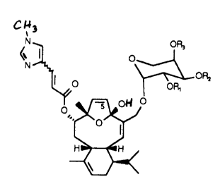
~ ~3
OR3
I o
I
O OR,
o ~oH i
2 o and wherein R1, R2 and R3 may independently be H or an acyl group having
from 1-6 carbon
atoms. Preferably, such acyl groups are acetyl. The diterpenes of this
invention include salts
(preferably pharmaceutically acceptable salts) and also include isomers of
both the Z and E
configurations. Compounds of this invention include desmethyleleutherobin 2 as
described
herein. Compounds of this invention may be isolated from natural sources as
described
2 5 herein or may be synthesized from an intermediate prepared by total
synthesis using
conventional starting materials or obtained by reduction and glycosylation of
sarcodictyin A
(see: WO 96/14745). Alternatively, the intermediate used in the preparation of
compounds
of this invention may be eleutherobin with appropriate substitutions at R~-3
being done using
conventional procedures such as the acetylation procedure described in the
Examples below.
CA 02290316 1999-11-24
-6-
BRIEF DESCRIPTION OF DRAWINGS
Figure 1 (A and B) are graphs showing mitotic arrest of MCF-7 cells by
different
concentrations of paclitaxel, as determined by mitotic spreads and microscopy
(Fig. lA) and
the ELISA (~) and ELICA(1) assays described herein (Fig. 1B).
Figure 2 (A and B) are graphs showing incidence of mitotic arrest of MCF-7
cells
using the indicated compounds as determined by the ELICA essay described
herein.
Figure 3 (A and B) are panels in a schematic diagram showing a fractionation
procedure according to an exemplified embodiment of this invention. E.
caribaeorum is
1 o homogenized to produce a crude extract. The crude extract is subjected to
fractionation
procedures including reversed and normal phase chromatography followed by
HPLC, to
produce antimitotic diterpenes and other compounds.
DETAILED DESCRIPTION OF EMBODIMENTS OF THE INVENTION
Previously, the only known natural source of eleutherobin was a species of
soft coral
from Western Australia (see: Lindel, T. et al. [supra]. This invention
provides an abundant
new source of eleutherobin and other antimitotic diterpenes from taxomical
orders of
coral-like organisms much different from the order comprising the soft coral
described by
2 o Lindel, T. et al. Using assays specifically adapted to detect antimitotic
compounds, it has
now been determined that organisms of the order Gorganacea and the order
Alcyoniidae
produce such antimitotic compounds. Such organisms include different species
of the genus
Rumphella (family Gorgoniidae); Mopsea whiteleggei and Muricellisis Sp. a
(family Isididae);
Subergorgia Sp. 1 cf Mollis and Subergorgia Mollis (geog. variant) (family
Subergorgiidae),
2 5 Junceella sp. d. Verrucella Sp. b and Ctenosella regia (family
Ellisellidae); Sinularia Sp. c,
Sinularia Sp. h (Order Alcyoniidae).
A preferred source of antimitotic compounds according to this invention is
Erythropodium caribaeorum, a gorgonian coral which is found in abundance and
has been
grown in aquarium. Gorgonian corals are found in all tropical and sub-tropical
regions,
30 particularly the Caribbean. They may be readily identified (for example,
see: Bayer, F.M.;
CA 02290316 1999-11-24
_7_
"The Shallow-Water Octocorallia of the West Indian Region" (1961) Martinus
Nighoff; The
Hague, at page 65 and 75-77 for Erythropodium). E. caribaeorum may be
collected in
abundance in Caribbean waters, including United States waters. An analysis of
toxic or
defensive compounds of the latter species has been reported. The latter
investigation included
fractionation and HPLC analysis of terpene compounds but did not reveal the
presence of the
diterpenes disclosed herein (Fenical, W. and Pawlik, J.R. (1991) Mar. Ecol.
Prog. Ser.
75:1-8).
Assays suitable for detection of antimitotic compounds are preferably based on
the use
of antibodies specific for mitotic cells, such as those described in the
international patent
1o application published April 1, 1999 under WO 99/15157. The assay will
typically employ
cells which regularly divide in culture (eg. cancer cells). A known
antimitotic compound
such as nocodazole may be used as a control.
In the assay, determination of the cells which proceed to mitosis is carried
out using
any of the known immunological methods by employing antibodies which have
specificity for
mitotic cells. Monoclonal antibodies demonstrating such specificity are known
and include
MPM-2 which was raised against mitotitc HeLa cells and recognizes phospho-
epitopes that
are highly conserved in mitotic proteins of all eukaryotic species. Other
examples are the
monoclonal antibodies recognizing phospho~pitopes in the paired helical
filament proteins
(PHF) found in brain tissue of patients suffering from Alzheimer's disease as
described
2 0 in: PCT International Application published July 4, 1996 under No. WO
96/20218; and,
Vincent et al. (1996) "The Journal of Cell Biology", 132:413-425. The examples
in this
specification make use of the antibody TG-3 described in the latter two
references, which may
be obtained from Albert Einstein College of Medicine of Yeshiva University,
Bronx, New
York.
2 5 The TG-3 monoclonal antibody, originally described as a marker of
Alzheimer's
disease, is highly specific for mitotic cells. Flow cytometry shows that TG-3
immunofluorescence is > 50-fold more intense in mitotic cells than in
interphase cells. In
Western blots, the antibody reacts with a 105-kDa protein identified as a
mitotically
phosphorylated form of nucleolin, that is present in abundance in extracts of
cells treated
3 o for 20 hours with the antimitotic agent nocodazole but present at only low
levels in extracts
CA 02290316 1999-11-24
_g_
from cycling MCF-7 cells. Densitometric scanning of the bands on Western blots
in these
examples show a 27-fold difference in intensity between nocodazole-treated and
untreated
cells, corresponding well to the difference in the number of mitotic cells in
the two
samples: 80% for the nocodazole-treated sample and 3% for the untreated
sample, as
measured by microscopy.
TG-3 also recognizes mitotic cells in ELISA. In the ELISA assay, the cells may
be
grown in mufti-well plates, lysed and transferred to protein-binding ELISA
plates for
adsorption to the plastic surface. The antigen may be detected by incubating
with TG-3
antibody, an HRP-conjugated secondary antibody and performing a colorimetric
determination of HRP activity.
Immunological methods useful for determination of mitotic cells in this assay
include
any method for determining antibody-antigen binding, including:
immunocytochemistry
(eg. immunofluorescence), flow cytometry, immunoblotting, and ELISA. Several
immunological methods are described in detail in examples herein as well as in
Vincent, I.
et al. [supra] . Other immunological procedures not described herein are well-
known in the
art and may be readily adapted for use in this assay. However, high throughput
testing of
samples may best be achieved by use of ELISA or the ELICA assay described
herein.
Pharmaceutical preparations containing compounds of this invention may be
prepared
as for similar preparations containing eleutherobin, paclitaxel, etc. In the
case of compounds
2 0 of this invention capable of salt formulation, pharmaceutically acceptable
salts (eg. HCI salt)
may be used to advantage to permit administration of the compound in an
aqueous solvent. A
preferred mode of administration would be intravenous to achieve a circulating
concentration
of the drug as predicted from its activity using standard methodology.
2 5 EXAMPLES
Sample Collection and Extract Preparation. Specimens of marine invertebrates
were collected by hand, using scuba, from cold temperate waters of the Pacific
Ocean along
the coast of British Columbia (Canada), from tropical Pacific Ocean reefs off
Motupore and
3 0 Madang in Papua New Guinea, and from tropical waters off the Island of
Dominica in the
CA 02290316 1999-11-24
-9-
Caribbean. Samples were deep frozen on site and transported over dry ice.
Voucher samples
of each invertebrate are stored in methanol at -20~C at The University of
British Columbia,
Vancouver, B.C. Canada, for taxonomic identification. Marine microorganisms
were
isolated from the invertebrates on site using marine culture media, and pure
cultures were
grown as a lawn on solid agar marine media in 10 cm petri plates for several
days and then
freeze-dried.
Extracts of invertebrates were prepared by homogenizing in methanol
approximately
200 g of each sample. The homogenates were filtered and concentrated to
dryness in vacuo
to give a gummy residue. Extracts of microorganisms were prepared by
extracting the
freeze-dried culture (cells and agar) multiple times with dry
methanol/acetone, followed by
lyophilization. A small amount of each extract was dissolved in DMSO for the
antimitotic
screening assay.
Cell Culture and Treatment. Human breast carcinoma MCF-7 cells were cultured
as monolayers. The cells were seeded at 10,000 per well of 96-well polystyrene
tissue
culture plates (Falcon) in 200 pl medium and were allowed to grow overnight.
Crude
extracts of marine organisms were then added at about 10 pg/ml or 1 pg/ml,
from 1000-fold
stocks in dimethylsulfoxide (DMSO). Untreated samples received an equivalent
amount of
DMSO and several as negative controls. Cells treated with 100 ng/ml nocodazole
(Sigma),
2 o from a 1000-fold stock in DMSO, served as positive controls. Cells were
incubated for
16-20 hours. The relative number of cells in mitosis was then determined by
microscopy by
enzyme-linked immunosorbent assay (ELISA) or by enzyme-linked cytochemical
assay
(ELICA), as described below.
2 5 ELISA of Mitotic Cells. After incubation with marine organism extracts,
the cell
culture medium was withdrawn carefully using a pipetor. Rounded-up mitotic
cells remained
attached to the plates. The cells were lysed by adding 100 p,l of ice-cold
lysis buffer (1 mM
EGTA pH 7.4, 0.5 mM phenylmethylsulfonyl fluoride) and by pipeting up-and-down
ten
times. The cell lysates were transferred to 96-well PolySorpT"" plates (Nunc)
and dried
3 o completely in a stream of air at about 37~C from a hair dryer. Vacant
protein binding sites
CA 02290316 1999-11-24
- 10-
were blocked by adding a 200 pl of antibody buffer (10 mM Tri-HCl pH 7.4, 150
mM NaCI,
0.1 mM phenylmethylsulfonyl fluoride, 3 % (w/v) dried nonfat milk (Carnation))
per well for
1 hour at room temperature. This was removed and replaced with 100 pl antibody
buffer
containing 0.1-0.5 ~g/ml TG-3 monoclonal antibody. After 16-20 hour incubation
at 4~C,
the antibody solution was removed and the wells were rinsed twice with 200 p,l
10 mM
Tris-HCl pH 7.4, 0.02% Tween 2OT"". Horseradish peroxidase (HRP) conjugated
goat
anti-mouse IgM secondary antibody (Southern Biotechnology Associates) was
added at a
500-fold dilution. After overnight incubation at 4~C, the antibody solution
was removed and
the wells were rinsed three times with 200 pl 10 mM Tris-HCl pH 7.4, 0.02%a
Tween 2OT"".
l0 Finally, 100 pl of 120 mM Na2HP04, 100 mM citric acid (pH 4.0) containing
0.5 pg/ml
2,2'-azino-bis(3-ethylbenzthiazoline-6-sulfonic acid) and 0.01 % hydrogen
peroxide was added
for 1 hour at room temperature and absorbance at 405 nm was determined using a
Dynex
MRXTM plate reader.
ELICA of Mitotic Cells. After incubation with marine extracts, the medium was
withdrawn carefully using a pipetor and 100 pl of 10 mM Tris-HCl (pH 7.4) 150
mM NaCI,
containing 3 .7 % formaldehyde was added to fix the cells for 30 minutes at
4~C. The fixative
was removed and replaced with 100 pl of cold (-20~C) methanol for 5 minutes to
permeabilize the fixed cells. The methanol was removed and the wells were
rinsed briefly
2 o with 200 ~l antibody buffer. Then, 100 ul antibody buffer containing 0.1-
0.15 pg/ml TG-3
monoclonal antibody and HRP-conjugated goat anti-mouse IgM secondary antibody
at a
500-fold dilution, was added to 16-20 hours at 4~C. The plates were washed
twice with
200 pl 10 mM Tris-HCl pH 7.4, 0.02% Tween 2OT"". Then, 100 ~l of 120 mM
Na2HP04,
100 mM citric acid (pH 4.0) containing 0.5 pg/ml
2 5 2,2'-azino-bis(-3ethylbenzthiazoline-6-sulfonic acid) and 0.01 % hydrogen
peroxide was added
for 1 hour at room temperature and the absorbance at 405 nm was measured.
Screens for Antimitotic Agents. MCF-7 cells were incubated for 20 hours with
different concentrations of the antimitotic drug paclitaxel, and the
proportion of cells
CA 02290316 1999-11-24
-11-
arrested in mitosis was measured by counting mitotic cells in the microscope,
and by
ELISA. Paclitaxel induced mitotic arrest in a concentration-dependent manner
with
half-maximal activity at 10 nM measured by microscopy (Fig. lA) and at 4 nM
measured
by ELISA (Fig. 1B, ice)
While the ELISA is accurate and reliable, it requires transferring cell
lysates to
ELISA plates and many solution changes. The ELICA assay is faster and easier
to use for
drug screening. This assay, combining some features of ELISA and the
"cytoblot"
technique (Stockwell, B.R. et al. (1999) Chemistry and Biology 6:71-93),
reduces the time
of the procedure and the number of steps by half and does not require transfer
of samples
1o to ELISA plates. In this procedure, the cells are fixed with formaldehyde
in their
microtiter culture plate and permeabilized with methanol and detergents. The
TG-3
primary antibody and HRP-conjugated secondary antibody may be added
sequentially but
are preferably added simultaneously. Colorimetric detection of HRP activity
remains
unchanged. The new assay is termed Enzyme-Linked Immuno-Cytochemical Assay
(ELICA).
Dose-dependent arrest of cells in mitosis by paclitaxel was detected by ELICA
with
half maximal activity at 1.5 nM (Fig. 1B, (1). ELICA provided a higher signal
at low
paclitaxel concentrations and a lower signal at high concentrations as
compared to ELISA.
The differences may result from higher non-specific staining of interphase
cells because of
2 o reduced washing and from lower specific staining of mitotic cells because
of fixation and
reduced antibody incubation times. ELICA consistently showed a difference in
absorbance
of 1 unit between cells treated or not with antimitotic agents at
concentrations causing
maximal mitotic arrest, allowing unambiguous detection of mitotic cells.
Measurements
obtained by ELICA consistently showed smaller standard deviations than
obtained by
2 5 ELISA, because the reduced number of manipulations reduced experimental
variation.
Thus, ELICA is particularly suited for rapid screening of large numbers of
extracts and the
ELISA assay may be preferred for precise quantitation of antimitotic activity.
ELISA was first used to screen a small selection of crude extracts from marine
microorganisms. Of 264 extracts tested, 261 showed no activity, giving
absorbance
3 o readings not statistically different from those of untreated cells (0.270
~ 0.051). Three
CA 02290316 1999-11-24
-12-
extracts showed strong activity, with absorbance readings of 1.135, 1.437 and
1.245, close
to the values obtained with nocodazole as a positive control.
Over 2000 crude extracts of marine sponges, tunicates, gorgonians, starfish,
and
nudibranchs were then 'screened, initially by ELISA and later by ELICA. This
screen
identified 16 additional extracts with antimitotic activity. The positive
extracts were
retested by counting mitotic figures in the microscope and all were found to
arrest cells in
mitosis.
Identification of Rhizoxin Analogs. Marine bacterial isolate MK7020 collected
off
1 o the coast of British Columbia, was identified as a Pseudomonas sp. by gas
chromatographic
analysis of cellular fatty acids. Two active compounds (A and B shown below)
were
purified by chromatographic procedures using the ELISA to guide fractionation.
The two
other microbial extracts were found to be independent isolates of the same
Pseudomonas
species and contained the same active compounds as MK7020.
0
Compound A is identical to WF-1360, a previously reported analog of the
antimitotic agent rhizoxin (Kiyoto, S. et al. (1986) J. Antibiot. (Tokyo)
39:762-772; and,
Iwaski, S. (1986) Chem. Pharm. Bull. 34:1387-1390). Compound A showed half
maximal
antimitotic activity (ICSO) at 52 nM as determined by ELISA. Compound B is a
~,-lactone
seco hydroxy acid analog of rhizoxin, not previously known to be naturally
occurring and
which had an ICso of 8 nM.
_..
CA 02290316 1999-11-24
-13-
Identification of New Eleutherobin Analogs. An extract of octocoral
Erythropodium caribaeorum collected from shallow reefs near Dominica also
showed
antimitotic activity. Eight active compounds were isolated and their chemical
structures
elucidated, as described below.
Freshly collected specimens of E, caribaeorum were frozen on site and
transported to
Vancouver over dry ice. Thawed samples (5.3 kg wet wt.) were extracted
multiple times
with MeOH and the combined MeOH extracts were concentrated to a gum in vacuo.
Fractionation of the crude gum (280 g) by sequential application of vacuum
reversed phase
flash (gradient elution: 80:20 H20 / MeOH to MeOH in 10 % increments), normal
phase
flash (gradient elution: EtOAc to 80:20 EtOAc / MeOH in 2 % increments), and
normal
phase high performance liquid chromatographies (HPLC) (eluent: 93:7 CH2CIz /
MeOH)
gave pure samples of 1 (50 mg), 2 (7 mg), 3 (6 mg), 4 (3 mg), and 5 (2 mg).
Compounds 6
(1 mg) and 7 (1 mg) partially decomposed on silica gel and were isolated using
only vacuum
reversed phase flash chromatography and cyano bonded phase HPLC (eluent:
56:42:2
EtOAc / hexane / (iPr)2NH). Figures 3A and 3B show the sequence of procedures
used to
isolate eleutherobin, its analogs and other compounds from E. caribaeorum.
One major compound was identified as eleutherobin 1. Novel compounds 2-7
described above were also identified. Desmethyleleutherobin 2 differs from
eleutherobin
by the presence of a hydroxyl instead of a methoxyl at C-4.
Desacetyleleutherobin 3
2 0 retains the arabinose, but not the 2" acetyl substituent of eleutherobin.
Isoeleutherobin A 4
has an acetyl group at the 3" position instead of the 2" position. Z-
eleutherobin 5 is the
geometric isomer of eleutherobin at the C-2' to C-3' double bond of the C-8 N-
(6)'-
methylurocanic acid ester side chain. Caribaeoside 6 differs from eleutherobin
by the
addition of a hydroxy at C-11 of the tricyclic core, and a double bond at C-12
to C-13
2 5 instead of C-11 to C-12, thereby altering the cyclohexene ring.
Caribaeolin 7 differs from
caribaeoside by the presence of a -CH20C0-CH3 substituent in the C-3 side
chain. One
further compound was also recovered and identified as the known compound,
sarcodictyin
A (shown below) which differs from eleutherobin by replacement of the C-15 ~-
linked
2"-O-acetyl-D arabinopyranose side chain of eleutherobin with a methyl ester
and
3 0 replacement of the C-4 methoxyl with a hydroxyl group.
CA 02290316 1999-11-24
-14-
~a
L
O:M G
15
The antimitotic activity profile of the above-described compounds as
determined by
ELICA is shown in Figure 2. Eleutherobin has an ICso of 100 nM. The ICSO of
Z-eleutherobin is 250 nM. Desmethyleleutherobin and isoeleutherobin A were
more potent
than eleutherobin, with an ICso of 20 nM and 50 nM, respectively.
Desacetyleleutherobin
2 o was less potent, with an ICso of 400 nM. Sarcodictyin A showed lower
activity, with an
ICSO of 2 pM. Caribaeoside and caribaeolin were considerably less potent, with
an ICSO of
~M for both compounds.
Characterization of Antimitotic Compounds
All NMR data for the E. caribaeorum diterpenes was recorded in DMSO-d6 at
500 MHz. Eleutherobin 1 was identified by comparison of its spectroscopic data
with the
values reported by Lindel, T. et al. [supra]. The UV chromophore for
eleutherobin
is: UV (MeOH) ~,max (log e) - 29 nm (3.8). Eleutherobin crystals were obtained
which
CA 02290316 1999-11-24
-15-
decomposed at 258 -- 260°C.
Desmethyleleutherobin 2 was isolated as a clear oil that gave a [M + H]+ ion
in the
HRFABMS at m/z 643.32230 appropriate for a molecular formula of C34H~NZOIo
(OM - 1.21 ppm), that differed from the molecular formula of eleutherobin by
the loss of
CH2. The 'H NMR spectrum of 2 differed from the 1H NMR spectrum of
eleutherobin 1
only by the absence of a methyl resonance at ~ g 3.10 that could be assigned
to the C-4
methoxy substituent. 2D NMR data obtained for 2 was in agreement with an
assignment of
a hydroxyl group at C-4.
Desacetyleleutherobin 3 was isolated as a clear oil that gave a [M + H]+ ion
at m/z
615.32813 in the HRFABMS corresponding to a molecular formula of C33H~N209
(OM - 0.05 ppm), that differed from the formula of eleutherobin by the loss of
C2H20.
The 'H NMR spectrum of 3 showed a strong resemblance to the 'H NMR spectrum of
eleutherobin except for the absence of a methyl singlet at ~ 2 ppm that could
be assigned to
an acetyl residue and the chemical shifts of the resonances assigned to the
arabinose
protons. Acetylation of the abrabinose fragment of 3 with acetic anhydride in
pyridine
converted it to triacetyleleutherobin, which was identical to
triacetyleleutherobin prepared
by acetylation of eleutherobin using the same reaction conditions. Preparation
of
triacetyleleutherobin by acetylation of eleutherobin was described in WO
96/14745.
Isoeleutherobin A 4, isolated as a clear oil, gave a [M + H]+ ion at m/z
657.33834
2 o in the HRFABMS corresponding to a molecular formula of C35H48NZOlo (~M -
0.58 ppm),
which was identical to the molecular formula of eleutherobin. Comparison of
the 'H 1D
and 2D NMR data for isoeleutherobin A 4 with the data for eleutherobin showed
that the
molecules differed only in the position of acetylation on the arabinose
fragment. COSY
correlations observed between resonances at g 3.38 and 3.62 (both broad
2 5 doublets: J = 11.5 Hz), assigned to the C-5" methylene protons, and a
methine at g 3.83
(H-4" : m) showed that the acetate was not a C-4" . The H-4" resonance in turn
showed a
COSY correlation to a resonance at g 4.80 (dd, J = 10.1, 2.5 Hz), assigned to
H3", which
was significantly deshielded relative to the corresponding H3" resonance (g
3.73) in
eleutherobin 1. Therefore, isoeleutherobin A was assigned structure 4.
Acetylation with
CA 02290316 1999-11-24
-16-
acetic anhydride in pyridine converted isoeleutherobin A 4 to
diacetyleleutherobin 8,
confirming the assigned structure of 4.
Z-Eleutherobin 5 gave a [M + H]+ ion at m/z 657.33830 in the HRFABMS
appropriate for a molecular formula of C35H~Nz0lo (OM - 0.65 ppm), again
identical to
the molecular formula of eleutherobin. Comparison of the NMR data obtained for
5 with
the data for eleutherobin showed that the molecules differed only in the
configuration of the
023' olefin. In the 'H NMR spectrum of Z-eleutherobin 5, the uroconic acid
olefinic
proton resonances appeared at g 5.95 (H-2') and 6.94 (H-3') with a coupling
constant of
12.6 Hz, whereas in the spectrum of eleutherobin, they were found at g 6.35 (H-
2') and
l0 7.35 (H-3') with a coupling constant of 15.6 Hz. The NMR sample of Z-
eleutherobin 5
partially isomerized over time to eleutherobin, confirming the assigned
structure.
Caribaeoside 6, obtained as a colorless glass, gave a [M + H]+ ion in the
HRFABMS at m/z 673.33474 appropriate for a molecular formula of C35H4gN20u
(OM - 1.64 ppm), that only differed from the molecular formula of eleutherobin
1 by the
presence of one additional oxygen atom. Analysis of NMR data obtained for
caribaeoside 6
revealed that it was a diterpene glycoside with the same N-(6')-methyluroconic
acid and
2"-O-acetylarabinose substituents that are attached to the central core of
eleutherobin. A
number of features of NMR data revealed that caribaeoside and eleutherobin
differed in the
C-11 to C-13 regions of their diterpene cores. The C-17 olefinic methyl
resonance at
2 0 g 1.47 and the H-12 olefinic methine resonance at g 5.27 in the ~H NMR
spectrum of
eleutherobin (DMSO-d6) were both missing in the 1H NMR spectrum of
caribaeoside 6. In
their place, the 'H NMR spectrum of 6 had a singlet methyl resonance at g 0.82
and a pair
of -coincidentally chemical shift equivalent olefinic methine resonances at g
5.52 (H-12 and
H-13). The two proton olefinic resonance at g 5.52 showed correlations in the
HMQC
2 5 spectrum to carbon resonance at g 125.6 (C-13) and 137.5 (C-12). HMBC
correlations
observed between the Me-17 singlet at g 0.82 and the C-12 olefmic resonance at
g 137.5, a
quaternary carbon resonance at g 68.5, and a methine resonance at g 45.8 (HMQC
to
g 2.06) confirmed the proximity of Me-17 and C-12 and indicated that there was
a hydroxyl
substituent at C-11 and a methine carbon at C-10. A pair of overlapping
doublet (6H) at
CA 02290316 1999-11-24
-17-
g 0.93 - 0.95, that showed COSY correlations to a methine resonance at g 1.68,
were
assigned to the Me-19 and Me-20 isopropyl protons, and a multiplet at g 4.00,
that showed
COSY correlations to an olefinic doublet at g 5.38 (H-2) and a methine
resonance at g 2.06
(H-10), was assigned to H-1. The H-1 resonance in the spectrum of 6 had a
chemical shift
and multiplicity nearly identical to the H-1 resonance in eleutherobin (g
3,88), consistent
with the proposal that the C-1, C-2, C-10, and C-14 centers in 6 were
identical to the
corresponding sites in 1. ROESY and scalar coupling constant data established
the relative
stereochemistry about the cyclohexene ring in caribaeoside 6. The resonances
assigned to
H-1 (g 4.00) and H-2 (g 5.38) in 6 had chemical shifts and a vicinal coupling
constant
(J + 9.7 Hz) nearly identical with their counterparts in eleutherobin (g H-1,
3.88; H-2,
5.39: J = 9.4 Hz), indicating that the dihedral angle between them in 6 was
essentially
identical to that in 1. ROESY correlations observed between the isopropyl
methyl proton
resonances at g 0.93 - 0.95 and the H-1 (g 4.00) and H-10 (g 2.06) resonances
in 6,
demonstrated that the isopropyl group, H-1, and H-10 are on the same face of
the
molecule, as in eleutherobin. The Me-17 resonance at g 8.02 in 6 showed a
strong ROESY
correlation to the H-2 (g 5.38) resonance demonstrating that Me-17 and C-2 are
cis.
Models indicate that the Me-17 protons can sit in the shielding region of the
~'-~3 olefin,
consistent with their unusually shielded chemical shift of g 0.82.
Caribaeolin 7 was isolated as a clear oil that gave a [M + H]+ ion in the
2 o HRFABMS at m/z 541.29111 corresponding to a molecular formula of C~H~N20~
(~M - 0.49 ppm). Analysis of the 1D and 2D 1H detected NMR data obtained for 7
showed that it contained the diterpene core and N-(6')-methyluroconic acid
fragments that
constitute the aglycon of caribaeoside 6, but was missing the arabinose sugar
residue.
COSY and ROESY correlations were observed between an olefinic methine
resonance at
2 5 g 5.37, assigned to H-2, and a broad two proton singlet at g 4.46,
assigned to the H-15
methylene protons. HMBC correlations were observed between a carbonyl
resonance at
g 169.8 and both the H-15 methylene proton resonance at g 4.46 and a singlet
methyl
resonance at g 1.97. These HMBC correlations demonstrated that in caribaeolin,
a C-15
acetyl substituent was present in place of the C-15 arabinose sugar residue
found in
3 p caribaeoside. Strong ROESY correlations were observed between the Me-17
resonance at
CA 02290316 1999-11-24
-18-
g 0.77 and the H-2 olefinic proton resonance at g 5.37 indicating that Me-17
and C-2 were
cis to each other as in caribaeoside 6, again accounting for the unusually
shielded nature of
the Me-17 proton resonance. Additional ROSEY correlations observed between the
C-19 / C-20 isopropyl methyl proton resonance at g 0.94 - 0.95 and the H-1 (g
4.01) and
H-10 (g 2.08) resonances confirmed that the isopropyl group, H-1 and H-10 were
all on the
same face of the molecule.
The significant decrease in antimitotic potency of caribaeoside 6 relative to
eleutherobin 1, resulting from introduction of a hydroxyl group at C-11 and
migration of
the olefin to the Q12.13 position, alters both the shape and polarity of
region B of the
1o proposed pharmacophore. The Ojima pharmacophore proposal suggests that
changes in the
C-11 to C-13 region of eleutherobin would have an impact on the ability of
analogs to
stabilize tubulinpolymers.
Replacement of the arabinose fragment in caribaeoside 6 with a simple acetate
reside
(Compound 7) results in no additional loss of potency. Altering the ~2'~3'
configuration (a
change in the A region of the pharmacophore) has little effect (Compound 5),
while
alterations in the arabinose fragment (representing changes in the C region of
the
pharmacophore) can either enhance (Compound 4) or decrease potency (Compound
3).
Changing the C-4 substituent from the methoxyl of eleutherobin to hydroxyl, an
alteration
that is outside of the Ojima pharmacophore biding regions is now shown by this
invention
2 o to result in an increase in potency.
Using the fractionation and assay procedures described above, similar
antimitotic
extracts were obtained from various other species from the order Gorgonacea as
well as
species from the order Alcyoniidae.
All publications, patents and patent applications referred to herein are
hereby
2 5 incorporated by reference. While this invention has been described
according to particular
embodiments and by reference to certain examples, it will be apparent to those
of skill in
the art that variations and modifications of the invention as described herein
fall within the
spirit and scope of the attached claims.