Note: Descriptions are shown in the official language in which they were submitted.
CA 02290439 1999-11-15
WO 98/52499 PCT/US98/10354
-1-
TRIAL FEMORAL PROSTHESIS FOR USE IN
KNEE JOINT REPLACEMENT SURGERY
The present invention relates to orthopedic surgical
instrumentation, and more particularly to an improved
femoral trial prosthesis apparatus having particular
utility in knee joint replacement surgery (particularly
revision surgical cases) wherein a previous femoral
prosthesis has been removed by a surgeon. Even more
particularly, the present invention relates to an improved
posterior stabilized-type femoral trial apparatus for
preparing a patient's femur to receive a posterior
stabilized femoral prosthesis wherein a trial body carries
a module selected from a kit of modules, each module
including cutting and rasping surfaces that extend
longitudinally and a stem portion for accepting a stem
member from a kit of various stem members of differing
sizes and diameters.
When a surgeon removes a previous femoral implant, it
is known in the art as a "revision" case. A surgeon must
remove that previous femoral implant and replace it with a
new implant. However, often the patient has weakened or
reduced bone tissue for attachment.
In the case of revision femoral implant surgery,
surgeons often use a posterior stabilized-type femoral
implant. Such a posterior stabilized femoral implant is
sold by Smith & Nephew of Memphis, Tennessee as part of the
Genesis Total Knee System or the Profix Total Knee System.
The surgeon may attempt to use a trial prosthesis to
first determine the appropriate size and shape of the final
prosthesis to be implanted.
CA 02290439 1999-11-15
WO 98/52499 PCT/US98/10354
-2-
The present invention provides a posterior stabilized
femoral trial that resects the bone in the distal end of
the femur to prepare the posterior stabilized box housing
as the trial is driven into place. The present invention
provides a posterior stabilized femoral trial apparatus for
preparing a patient's femur to receive a posterior
stabilized femoral prosthesis. The apparatus of the
present invention includes a trial body that has proximal
and distal portions, the distal portion of the trial body
having an articulating surface that articulates with a
patient's tibial component or with a patient's tibia.
The femoral articulating surface includes anterior,
distal, and posterior condylar portions.
The trial body also has a proximal surface that
includes a plurality of flat intersecting surfaces,
preferably five (5) of such flat intersecting surfaces.
The present invention provides a module that fits the
trial body at the proximal surface, the module including a
rasping surface that extends longitudinally along a
proximal to distal plane that is generally parallel to an
anterior to posterior plane.
The module is removably attachable to the trial body
at the proximal surface.
The apparatus includes portions that extend laterally
on a medial to lateral line, attaching to the medial and
lateral sides of the trial body. A slot on the distal
surface of the trial body receives the module.
Cutting surfaces are provided on the trial body at the
condylar surfaces.
A plurality of cutting guide slots extend from the
proximal to the distal surface of the body and along medial
CA 02290439 1999-11-15
WO 98/52499 PCT/US98/10354
-3-
and lateral lines. There are preferably three sets of
cutting guide slots including distal cutting guide slots
and anterior chamfer cutting guide slots.
An opening of the trial body is placed in between the
condylar portions and extends anteriorly a partial distance
toward the anterior portion of the trial body, the opening
being bordered on the rear by a transverse bar that spans
in between the condylar portions.
Fasteners enable the module to be attached to and
removed from the trial body.
The module includes a pair of flange portions that
engage the body, the flange portions having openings
through which threaded fasteners can be placed for
attaching the module to the trial body.
The trial body includes a projecting portion that
extends away from the flange portion along a generally
proximal to distal line.
The module includes a frustoconically-shaped
projecting portion that can receive a selected stem
extension, a kit being provided with several stem
extensions of differing lengths and diameters. The module
includes wall portions, at least one of which has a rasping
surface thereon. In the preferred embodiment, the lateral
wall portion carries the rasping surface.
In the preferred embodiment, the stem connector forms
an angle of less than ninety degrees (90°) with the plane of
the flange portions thus providing a valgus adjustment for
the stem connector and stem extensions when the trial
prosthesis is then placed on the patient's distal femur.
For a further understanding of the nature, objects,
and advantages of the present invention, reference should
CA 02290439 1999-11-15
WO 98/52499 PCT/US98/10354
-4-
be had to the following detailed description, read in
conjunction with the following drawings, wherein like
reference numerals denote like elements and wherein:
Figure 1 is a perspective view of the preferred
embodiment of the apparatus of the present invention
illustrating the trial prosthesis body portion thereof;
Figure 2 is a proximal view of the preferred
embodiment of the apparatus of the present invention
illustrating the trial prosthesis body;
Figure 3 is a distal view of the preferred embodiment
of the apparatus of the present invention illustrating the
trial prosthesis body;
Figure 4 is a side view of the preferred embodiment of
the apparatus of the present invention illustrating the
trial prosthesis body;
Figure 5 is a posterior view of the preferred
embodiment of the apparatus of the present invention
illustrating the trial prosthesis body;
Figure 6 is a perspective view of the preferred
embodiment of the apparatus of the present invention
illustrating the trial prosthesis body and a selected
posterior stabilized module;
Figure 7 is a side view of the preferred embodiment of
the apparatus of the present invention shown after
placement on a patient's distal femur that is shown in
phantom lines;
Figure 8 is a partial perspective view of the
preferred embodiment of the apparatus of the present
invention illustrating the module portion thereof;
Figure 9 is a side view of the module of Figure 8;
Figure 10 is another side view of the module of Figure
CA 02290439 1999-11-15
WO 98/52499 PCT/US98/10354
_5-
8 showing the lateral side thereof;
Figure 11 is an anterior view of the module of Figures
8-10;
Figure 12 is a distal view of the module of Figures 8-
11;
Figure 13 is a proximal view of the module of Figures
8-12; and
Figures 14-16 are perspective views of the preferred
embodiment of the apparatus of the present invention shown
with a trial coupler, trial stems of differing lengths, and
trial wedge.
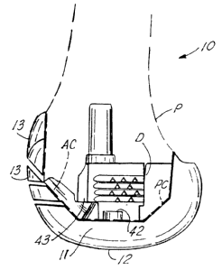
Figures 1-7 show the preferred embodiment of the
apparatus of the present invention designated by the
numeral 10 in Figures 6-7.
Femoral trial apparatus 10 includes a trial body 11
(Figures 1-7~ having a distal surface 12 that includes
various articulating portions including an anterior
articulating surface 13, condylar surfaces 14, 15, and
cutting edges 16, 17 at the condylar surfaces 14, 15.
Trial body 11 can provide a plurality of five flat
surfaces 18, 19, 20, 21, 22 that are used to form a load
transfer interface with a patient's surgically prepared
femur F. The femur F is surgically prepared as shown in
Figure 7 to provide five cut surfaces. These surgically
cut surfaces on femur F correspond in shape and placement
to the surfaces 18, 19, 20, 21, and 22 of trial body 11.
' When the femur F is surgically cut (see Figure 7), the
surgeon forms a plurality of five cut surfaces including:
a distal D cut surface, anterior A and posterior P cut
surfaces, and anterior and posterior chamfer cut surfaces
AC, PC. When a surgeon performs a revision surgical case,
CA 02290439 1999-11-15
WO 98/52499 PCT/US98/10354
-6-
these cut surfaces are already present on the femur from a
previous surgery. When the surgeon removes the previous
implant in a revision case, the femur F resembles the shape
shown in phantom lines in Figure 7. The surgeon then uses
the trial apparatus 10 to reshape the distal femur F and to
determine the correct size for a final prosthesis to be
used in the revision surgery, replacing the old removed
implant. The five surgically prepared surfaces are
reshaped somewhat using trial body 11. The cutting
surfaces 16, 17 shave bone from the posterior condylar cut
surface P of the femur F. Cutting edges 68A & 69A and
rasping surface 71 on the module 50 also cut and shave
bone.
In Figure 1, a slot or recess 23 is provided for
receiving a module 50. The module 50 is shown in Figures
8-13. The module 50 is assembled to implant body 11 in
Figures 6-7. Implant body 10 provides a pair of spaced
apart openings 24, 25. An opening or open space 26 as
shown in Figure 2 is provided in between the recess
portions 23A, 23B.
In Figure 3, anterior groove 27 extends along the
anterior portion of trial body 11, copying the shape of the
femoral prosthesis to be used after the trial 10 has been
employed by the surgeon to arrive at a correct size. Each
recessed portion 23A, 23B provides a flat surface 28, 29
respectively. Each flat surface 28, 29 is surrounded
respectively by a sidewall portion 30, 31.
Holes 32, 33 are provided at flat surface 20 for
providing alignment with module 50 at correspondingly
shaped openings 72, 73 of module 50. Post 34 spans between
condylar portions 14, 15. Holes 32, 33 receive a
CA 02290439 1999-11-15
WO 98/52499 PCT/US98/10354
_7_
correspondingly shaped peg 82 on a trial wedge 77.
Threaded fastener 81 of wedge 77 fits threaded opening 24
' or 25 (see Figure 14?.
A plurality of cutting guide slots are provided for
cutting bone tissue at the patient's distal femur. Often
bone is missing from the femur in the distal area. Bone
has often been worn or eroded away in revision cases. The
surgeon cleans up by cutting some bone from either the
medial or lateral distal surface using a selected cutting
guide slot 34A, 34B, 35A, 35B, 36A, 36B. Cutting guide
slots 34A and 34B can be used to track and guide a cutting
blade, saw, or the like during a cutting of tissue from the
patient's distal femoral surface. Similarly, guide slots
35A, 35B guide a cutting blade during a cutting of the
distal femur. The cutting guide slots 36A, 36B are chamfer
cutting guide slots for making anterior chamfer cuts on the
patient's distal femur.
Raised portions 37, 38 form a thickened reinforcement
of trial body 11 at the cutting guide slots 34A, 35A, 36A
and 34B, 35B, 36B. The raised portion 38 has a flat
surface 43 for receiving the surface 58 of module 50.
Raised portion 38 is defined by flat wall sections 39, 40,
41 (Figure 1). Module 50 aligns with and abuts flat
surfaces 39, 40, 41 of trial body 11. A generally
rectangular slot 26 accepts module 50. Surface 58 of
module 50 rests upon surface 43 of body 11.
As seen in Figures 6-7 and 14-16, module 50 attaches
to trial body 11 using threaded fasteners 42, 44. The
fasteners 42, 44 pass through respective openings 53, 54 of
flanges 51, 52 and then threadably engage internally
threaded openings 24, 25 respectively of trial body 11. If
CA 02290439 1999-11-15
WO 98/52499 PCT/US98/10354
_g_
a trial wedge 77 is to be attached, a threaded fastener
passes through an opening in the trial wedge and through
the opening 53 or 54 of module 50 before forming a threaded
attachment to an opening 24 or 25. If a trial wedge 77 is
attached, the peg 82 of trial wedge 77 registers in slot 72
or 73 of module 50 and then into opening 32 or 33 of trial
body 11.
The construction of module 50 is shown more
particularly in Figures 8-13. Module 50 has a lower end
portion 55 in the form of flanges 51, 52. Flange 51 has a
flat surface 57 that fits surface 29 of the recess portion
23A of trial body 11. Flange 52 has a flat surface 56 that
fits surface 28 of the recess portion 29 of trial body 11.
Module 50 has stem connector 60 with a flat surface 61
at the free end of stem 60. Stem 60 has a cylindrical base
64, tapered transition 63 and frustoconical surface 62 that
enables a trial coupler 78 (see Figures 14-16) to
preferably form a taper lock connection with stem 60 at
surface 62.
Trial coupler 78 can have upper projecting portion 79
that connects (e.g., a threaded connection) to a
correspondingly threaded socket at the lower end of each
stem 74, 75, 76. A selected stem extension 74, 75 or 76
could then be selectively connected to trial coupler 78 by
a surgeon. In this manner, the trial prosthesis 10 of the
present invention enables a surgeon to vary the length and
diameter of the stem portion of a trial body when fitting
the trial body 11 and an attached trial coupler and/or a
selected stem extension 74, 75 or 76 to a patient's
surgically prepared femur. Coupler 78 has a lowermost
socket 80 that corresponds in size and shape to stem
CA 02290439 1999-11-15
WO 98/52499 PCT/US98/10354
_g_
connector 60 so that a taper lock connection can be formed
between stem connector 60 and socket 80. In Figure 11,
stem connector 60 is inclined with respect to a plane 65
defined by surfaces 56, 57. The inclination is indicated
by arrow 66 in Figure 11. Angle 66 can be between 80° - 90°
for example, enabling the central axis 67 of stem connector
60 to compensate for the valgus angle of the patient's
intramedullary canal at the distal femoral region.
Likewise, a generally cylindrically shaped stem extension
74, 75 or 76 affixed to connector 60 will form an angle 66
with the plane 65 of surfaces 56, 57. The surfaces 56, 57
register against flat surfaces 28, 29 of trial body 11.
The surfaces 28, 29 are parallel to the surgically cut
distal surface D of the patient's distal femur F (see
Figure 7). Also, the surfaces 28, 29 (and 56, 57) are
parallel to surface 20 of implant body 11 that fits against
the surgically cut distal surface D of femur 11.
Stem connector 60 attaches to flanges 51, 52
respectively with side walls 68, 69. The side walls 68, 69
have sharp cutting edges 68A, 69A proximally. The side
wall 69 has a plurality of teeth 70 forming a rasping
surface 71. This rasping surface 71 is helpful in revision
cases as it cuts away excess bone as the surgeon taps or
hammers the trial prosthesis 10 onto the distal femur F
until the trial 10 reaches the position in Figure 7.
Surface 69 is provided with rasping surface 71 because it
' is the surface that will likely engage excess bone due to
the angle 66 of inclination of connector 60 (and its
attached stem extension 74, 75 or 76) with respect to plane
65.
The following is a list of suitable parts and
CA 02290439 1999-11-15
WO 98/52499 PCT/US98/10354
-10-
materials for the
various elements
of the preferred
embodiment of the present invention.
PARTS LIST
Part Number Description
10 femoral trial apparatus
11 trial body
12 distal surface
13 anterior surface
14 condylar surface
1015 condylar surface
16 cutting edge
17 cutting edge
18 flat surface
19 flat surface
1520 flat surface
21 flat surface
22 flat surface
23 recess
23A recess portion
2023B recess portion
24 opening
25 opening
26 open space
27 groove
2528 flat surface
29 flat surface
30 side wall
31 side wall
32 hole
3033 hole
34 post
CA 02290439 1999-11-15
WO 98/52499 PCT/US98/10354
-11-
34A cutting guide
34B cutting guide
35A cutting guide
35B cutting guide
36A cutting guide
36B cutting guide
37 raised portion
38 raised portion
39 flat wall section
40 flat wall section
41 flat wall section
42 threaded fastener
43 flat surface
44 threaded fastener
50 module
51 flange
52 f lange
53 opening
54 opening
55 lower end portion
56 flat surface
57 flat surface
58 anterior flat surface
60 stem
61 flat surface
62 frustroconical surface
63 transition section
64 cylindrical section
65 plane
66 angle
67 central axis of stem
CA 02290439 1999-11-15
WO 98/52499 PCT/US98/10354
-12-
68 side wall
68A cutting edge
69 side wall
69A cutting edge
70 rasp teeth
71 rasping surface
72 U-shaped slot
73 U-shaped slot
74 stem extension
75 stem extension
76 stem extension
77 trial wedge
78 trial coupler
79 projecting threaded portion
80 socket
81 threaded fastener
82 peg
A anterior cut surface
AC anterior chamfer cut surface
D distal cut surface
PC posterior chamfer cut surface
P posterior cut surface
F femur
U.S. patent application Serial No. 08/861,094, filed
May 21, 1997, is incorporated herein by reference.
The foregoing embodiments
are presented by
way of
example only; the
scope of the present
invention is to be
limited only by the following claims.