Note: Descriptions are shown in the official language in which they were submitted.
CA 02290743 1999-11-22
WO 98/52543 PCT/US98/10626
SEMI-INTERPENETRATING OR INTERPENETRATING POLYMER
NETWORKS FOR DRUG DELIVERY AND TISSUE ENGINEERING
Background of the Invention
The United States government has certain rights in this invention by
virtue of grant AR08387 NIH Fellowship 23464 to Robert S. Langer.
This application claims priority to U.S. Serial No. filed April 11,
1997, Express Mail Label No. EM290166797US.
The present invention is generally in the area of using polymeric
semi-interpenetrating and interpenetrating polymer network compositions and
photocrosslinkable polymeric hydrogels in medical treatments, especially joint
resurfacing and plastic surgery and delivery of drugs.
Congenital Defects
Many congenital defects, especially in the urogenital areas, require
surgical correction. Examples include treatment of reflux and urinary
incontinence. WO 94/25080 by Massachusetts Institute of Technology
describes the use of injectable polysaccharide-cell compositions for
delivering
isolated cells by injection, which then form new tissue that is effective as a
bulking agent. The polymers that are described crosslink ionically, as a
function of ionic strength, temperature, pH, or combinations thereof. WO
96/40304 by Reprogenesis describes similar applications of polymeric
hydrogels formed by covalent crosslinking, for example, by
photopolymerization of the injected polymer-cell suspension using a catheter
or during surgery.
Craniofacial contour deformities
Craniofacial contour deformities, whether traumatic, congenital, or
aesthetic, currently require invasive surgical techniques for correction.
Furthermore, deformities requiring augmentation often necessitate the use of
alloplastic prostheses which suffer from problems of infection and extrusion.
Correction of these defects and irregularities remain a difficult and
controversial problem. Sims, et at., reported in Plastic Reconstructive
CA 02290743 1999-11-22
WO 98/52543 PCT/US98/10626
-2-
Sur e 98:845 (1996), the formation of new cartilage from injected
polyethylene oxide-cell suspensions, and suggested that this technology would
be useful in plastic surgery.
Replacement or Repair of Cartilaginous Surfaces
The aging population, especially of those active in sports and in jobs
creating stress on joints, have little recourse at this time for repair or
replacement of cartilage. Arthroscopic surgery can be used to remove torn
cartilage but highly invasive and painful surgery is required for repair or
replacement of a joint having little cartilage left. In most cases a
prosthetic
device must be used to replace the entire joint, following destruction of the
smooth cartilaginous surface which normally allows for free movement of the
abutting joint surfaces. As described in U.S. Patent No. 5,514,378 to
Vacanti, et al., it has been proposed to create new joint surfaces using a
synthetic polymeric mesh seeded with chondrocytes, which forms new
cartilage as the polymer degrades. Although this is promising, the seeded
mesh must still be implanted surgically.
There is a need for improved injectable polymer-cell compositions
which are biocompatible and biodegradable for delivering isolated cells by
injection. There is a further need for less invasive means of covalently
crosslinking polymer-cell suspensions following injection.
Accordingly, it is an object of the present invention to provide
methods and compositions for injection of cells to form cellular tissues and
cartilaginous structures, based on interpenetrating networks of synthetic
polymers.
It is a further object of the invention to provide improved
compositions to form cellular tissues and cartilaginous structures including
non-cellular material which will degrade and be removed to leave tissue or
cartilage that is histologically and chemically the same as naturally produced
tissue or cartilage.
CA 02290743 1999-11-22
WO 98/52543 PCT/US98/10626
-3-
It is another object of the present invention to provide compositions
for and a method for covalent crosslinking a polymer-cell suspension for
formation of new tissue following injection.
Sununary of the Invention
Compositions for tissue engineering and drug delivery have been
developed based on solutions of two or more polymers which form semi-
interpenetrating or interpenetrating polymer networks upon exposure to active
species following injection at a site in a patient in need thereof. The
polymers crosslink to themselves but not to each other; semi-interpenetrating
networks are formed when only one of the polymers crosslink. The resulting
viscous solutions retain the biologically active molecules or cells at the
site of
injection until release or tissue formation, respectfully, occurs.
As a result of studies conducted with polymer-cell suspensions
forming interpenetrating polymer networks, it has been determined that
polymer solutions can be formulated wherein the active species is provided
by exposure of the polymer solution to an exogenous source of active
species, typically electromagnetic radiation, preferably light. Studies
demonstrate that light will penetrate through skin, body fluids (such as
synovial fluid) and membranes and polymerize the polymer solutions. The
polymer solutions can be crosslinked ionically or covalently, to form a
hydrogel, semi-interpenetrating polymer network or an interpenetrating
polymer network.
Brief Description of the Drawings
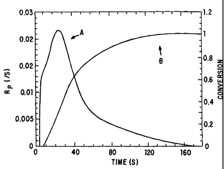
Figure 1 graphically depicts the rate of polymerization, A, and
percent conversion of a polymerizable solution to a polymer matrix, B, over
time with a photoactive initiator and ultraviolet light.
CA 02290743 1999-11-22
WO 98/52543 PCT/US98/10626
-4-
Figure 2 graphically sets for the calculated rate (time in minutes) of
photopolymerization of a polymerizable solution under the subcutaneous, A,
subcutaneous and fat, B, and subcutaneous and fat and muscle, C, layers of
rat skin based upon the penetration of light through these layers.
Figure 3 graphically depicts the effect of succinic acid (SAD)
concentration on the equilibrium swelling volume of a polymer, for polymers
of molecular weight 1000 and 3400.
Figure 4 graphically depicts the effect of succinic acid concentration
(17%, 43%, 51%, and 0% SAD) on release of serum albumin (percent) from
a polymer over time (days).
Figure 5 graphically depicts the effect of succinic acid concentration
(19% and 21 % SAD) on the release of rhodamine from a polymer (percent
over time in days).
Detailed Description of the Invention
Techniques of tissue engineering employing biocompatible polymer
scaffolds hold promise as a means of creating alternatives to prosthetic
materials currently used in plastic surgery and joint repair or replacement,
as
well as formation of organ equivalents to replaced diseased, defective, or
injured tissues.
Interpenetrating networks ("IPN") are defined as networks where two
components are crosslinked, but not to each other. As described herein, in
one embodiment, semi-interpenetrating networks of synthetic and/or natural
polymers are used as the polymeric support for cells to be. injected. Semi-
interpenetrating networks are defined as solutions that include two
independent components, where one component is a crosslinked polymer and
the other component is a non-crosslinked polymer. The crosslinked polymer
preferably constitutes between about 10 and 90 % by weight of the semi-
interpenetrating network composition.
CA 02290743 1999-11-22
WO 98/52543 PCT/US98/10626
-5-
The semi-interpenetrating polymer networks are preferably prepared
from hydrophilic polymers. In one embodiment, the polymer networks
include a hydrophilic polymer with groups crosslinkable with active species
and/or ionically crosslinkable groups, and a hydrophilic polymer with no
active species or ionically crosslinkable groups.
In a second embodiment, cells are suspended in a polymer solution
which can be crosslinked by active species generation, preferably by
photoactivation. The suspension is injected at the site where new tissue is to
be formed. Light is then applied externally to the skin to crosslink the
injected polymer. This method is based on the discovery that combinations
of polymers and photoinitiators (in a concentration not toxic to the cells,
less
than 0.1 % by weight, more preferably between 0.05 and 0.01 % by weight
percent initiator) will crosslink upon exposure to light equivalent to between
one and 3 mWatts/cm2 applied to the skin of nude mice. Although discussed
herein principally with regard to administration of a light source external to
the skin, this should be interpreted as equally applicable to light applied
through tissues, for example, from a catheter in a blood vessel adjacent to a
tissue where the polymer-cell suspension has been injected, or in the synovial
space adjacent to a cartilaginous surface to be repaired or replaced with
injected polymer-cell suspension.
Polymer Compositions
The polymer compositions can consist solely of covalently
crosslinkable polymers, as described in W096/40304, in combination with an
effective but non-toxic about of photoinitiator to allow crosslinking using
radiation provided by an external source, or blends of covalently and
ionically crosslinkable or hydrophilic polymers which when exposed to
radiation form semi-interpenetrating networks having cells suspended therein.
Ionically crosslinkable and Hydrophilic Polymers
As used herein, "hydrophilic polymers" are defmed as polymers with
a solubility of at least ten grams/liter of an aqueous solution at a
temperature
CA 02290743 2006-06-14
Jun-14-06 04:22pm From-Osler
+6132352867 T-711 P.007/012 F-888
-6-
of between aboixt 0'and 50' C. Aqueous solutions can include small amouncs
of water-soluble organic solvents, such as dimethylsulfoxide,
d"unethylformamide, alcohols, acetone, and/or glymes.
Suitable hydrophilic polymers include synthetic polymers such as
poly(ethylene glycol), poly(ethylene oxide), partially or fuiiy hydrolyzed
poly(vinyl alcohol), poly(vinylpyrrolidone), poly(ethyioxazoline),
poly(ethylerae oxide)-co-poly(propylene oxide) block copolymers (poloxamers
and meroxapols), poloxamines, carboxymethyl cellulose, and
hydroxyalkylated celluloses such as hydroxyethyl cellulose an.d
methylhydroxypropyl cellulose, and natural polymers such as poiypesptide,ti,
polysaccharides or carbohydrhtes such as Ficolllb polysucrose, . hyaluxonic
acid, dextran, heparan sulfate, chondroitin sulfate, heparin, or alginate, and
proteins such as gelatin, collagen, albumin, or ovalburain or copolyrners or
blends thereof. As used herein, "celluloses" Includes cellulose arxi
derivatives of the types described above; "dextran" includes dexcran and
similar derivatives thereof.
Examples of materials which can be used to form a hydrogel include
modified aIginates. Alginate is a carbohydrate polymer isolated from
seaweed, which can be crosslinked to form a hyclrogel by exposure to a
divalent cation such as calc.ium, as described, for example in WO 94/250$0,
Alginate is
ionically crosslinlced in the presence of divalent cations, in water, at roum
temperature, to form a b.ydrogel matxix. ' Modified alginate deriv<<tives may
be synthesized which have an impmved ability to form hydrogels. The use
of alginate as the srarting material is advantageous because it is =available
from more than one source, and is available in good purity and
characterization. As used herein, the term "modified alginates" refers to
cheanically modified alginates with modified hydrogel properties. Naturally
occurring alginate may be chemically modified to produce alginate polymer
derivatives that degrade more qulckly. For example, alginate may be
CA 02290743 1999-11-22
WO 98/52543 PCT/US98/10626
-7-
chemically cleaved to produce smaller blocks of gellable oligosaccharide
blocks and a linear copolymer may be formed with another preselected
moiety, e.g. lactic acid or E-caprolactone. The resulting polymer includes
alginate blocks which permit ionically catalyzed gelling, and oligoester
blocks
which produce more rapid degradation depending on the synthetic design.
Alternatively, alginate polymers may be used wherein the ratio of
mannuronic acid to guluronic acid does not produce a firm gel, which are
derivatized with hydrophobic, water-labile chains, e.g., oligomers of E-
caprolactone. The hydrophobic interactions induce gelation, until they
degrade in the body.
Additionally, polysaccharides which gel by exposure to monovalent
cations, including bacterial polysaccharides, such as gellan gum, and plant
polysaccharides, such as carrageenans, may be crosslinked to form a
hydrogel using methods analogous to those available for the crosslinking of
alginates described above. Polysaccharides which gel in the presence of
monovalent cations form hydrogels upon exposure, for example, to a solution
comprising physiological levels of sodium. Hydrogel precursor solutions also
may be osmotically adjusted with a nonion, such as mannitol, and then
injected to form a gel.
Polysaccharides that are very viscous liquids or are thixotropic, and
form a gel over time by the slow evolution of structure, are also useful. For
example, hyaluronic acid, which forms an injectable gel with a consistency
like a hair gel, may be utilized. Modified hyaluronic acid derivatives are
particularly useful. As used herein, the term "hyaluronic acids" refers to
natural and chemically modified hyaluronic acids. Modified hyaluronic acids
may be designed and synthesized with preselected chemical modifications to
adjust the rate and degree of crosslinking and biodegradation. For example,
modified hyaluronic acids may be designed and synthesized which are
esterified with a relatively hydrophobic group such as propionic acid or
benzylic acid to render the polymer more hydrophobic and gel-forming, or
CA 02290743 1999-11-22
WO 98/52543 PCTIUS98/10626
-8-
which are grafted with amines to promote electrostatic self-assembly.
Modified hyaluronic acids thus may be synthesized which are injectable, in
that they flow under stress, but maintain a gel-like structure when not under
stress. Hyaluronic acid and hyaluronic derivatives are available from
Genzyme, Cambridge, MA and Fidia, Italy.
Other polymeric hydrogel precursors include polyethylene oxide-
polypropylene glycol block copolymers such as PluronicsTM or TetronicsTM,
which are crosslinked by hydrogen bonding and/or by a temperature change,
as described in Steinleitner et al., Obstetrics & Gynecology, 77:48-52 (1991);
and Steinleitner et al., Fertility and Sterility, 57:305-308 (1992). Other
materials which may be utilized include proteins such as fibrin, collagen and
gelatin. Polymer mixtures also may be utilized. For example, a mixture of
polyethylene oxide and polyacrylic acid which gels by hydrogen bonding
upon mixing may be utilized. In one embodiment, a mixture of a 5% w/w
solution of polyacrylic acid with a 5% w/w polyethylene oxide (polyethylene
glycol, polyoxyethylene) 100,000 can be combined to form a gel over the
course of time, e.g., as quickly as within a few seconds.
Water soluble polymers with charged side groups may be crosslinked
by reacting the polymer with an aqueous solution containing ions of the
opposite charge, either cations if the polymer has acidic side groups or
anions if the polymer has basic side groups. Examples of cations for cross-
linking of the polymers with acidic side groups to form a hydrogel are
monovalent cations such as sodium, divalent cations such as calcium, and
multivalent cations such as copper, calcium, aluminum, magnesium,
strontium, barium, and tin, and di-, tri- or tetra-functional organic cations
such as alkylammonium salts. Aqueous solutions of the salts of these cations
are added to the polymers to form soft, highly swollen hydrogels and
membranes. The higher the concentration of cation, or the higher the
valence, the greater the degree of cross-linking of the polymer. Additionally,
the polymers may be crosslinked enzymatically, e.g., fibrin with thrombin.
CA 02290743 1999-11-22
WO 98/52543 PCT/US98/10626
-9-
Suitable ionically crosslinkable groups include phenols, amines,
imines, amides, carboxylic acids, sulfonic acids and phosphate groups.
Aliphatic hydroxy groups are not considered to be reactive groups for the
chemistry disclosed herein. Negatively charged groups, such as carboxylate,
sulfonate and phosphate ions, can be crosslinked with cations such as calcium
ions. The crosslinking of alginate with calcium ions is an example of this
type of ionic crosslinking. Positively charged groups, such as ammonium
ions, can be crosslinked with negatively charged ions such as carboxylate,
sulfonate and phosphate ions. Preferably, the negatively charged ions contain
more than one carboxylate, sulfonate or phosphate group.
In the embodiment wherein modified alginates and other anionic
polymers that can form hydrogels which are malleable are used to
encapsulate cells, the hydrogel is produced by cross-linking the polymer with
the appropriate cation, and the strength of the hydrogel bonding increases
with either increasing concentrations of cations or of polymer.
Concentrations from as low as 0.001 M have been shown to cross-link
alginate. Higher concentrations are limited by the toxicity of the salt.
The preferred anions for cross-linking of the polymers to form a
hydrogel are monovalent, divalent or trivalent anions such as low molecular
weight dicarboxylic acids, for example, terepthalic acid, sulfate ions and
carbonate ions. Aqueous solutions of the salts of these anions are added to
the polymers to form soft, highly swollen hydrogels and membranes, as
described with respect to cations.
A variety of polycations can be used to complex and thereby stabilize
the polymer hydrogel into a semi-permeable surface membrane. Examples of
materials that can be used include polymers having basic reactive groups such
as amine or imine groups, having a preferred molecular weight between
3,000 and 100,000, such as polyethylenimine and polylysine. These are
commercially available. One polycation is poly(L-lysine); examples of
synthetic polyamines are: polyethyleneimine, poly(vinylamine), and poly(allyl
CA 02290743 1999-11-22
WO 98/52543 PCT/US98/10626
-10-
amine). There are also natural polycations such as the polysaccharide,
chitosan.
Polyanions that can be used to form a semi-permeable membrane by
reaction with basic surface groups on the polymer hydrogel include polymers
and copolymers of acrylic acid, methacrylic acid, and other derivatives of
acrylic acid, polymers with pendant SO3H groups such as sulfonated
polystyrene, and polystyrene with carboxylic acid groups.
These polymers can be modified to contain active species polymerizable
groups and/or ionically crosslinkable groups. Methods for modifying
hydrophilic polymers to include these groups are well known to those of skill
in the art.
The polymers may be intrinsically biodegradable, but are preferably
of low biodegradability (for predictability of dissolution) but of
sufficiently
low molecular weight to allow excretion. The maximum molecular weight to
allow excretion in human beings (or other species in which use is intended)
will vary with polymer type, but will often be about 20,000 daltons or below.
Usable, but less preferable for general use because of intrinsic
biodegradability, are water-soluble natural polymers and synthetic equivalents
or derivatives, including polypeptides, polynucleotides, and degradable
polysaccharides.
The polymers can be a single block with a molecular weight of at
least 600, preferably 2000 or more, and more preferably at least 3000.
Alternatively, the polymers can include can be two or more water-soluble
blocks which are joined by other groups. Such joining groups can include
biodegradable linkages, polymerizable linkages, or both. For example, an
unsaturated dicarboxylic acid, such as maleic, fumaric, or aconitic acid, can
be esterified with hydrophilic polymers containing hydroxy groups, such as
polyethylene glycols, or amidated with hydrophilic polymers containing
amine groups, such as poloxamines.
CA 02290743 2006-06-14
Jun-14-06 04:22pm From-Qslar
+ 6132352861 T-T12 P.008/012 F-888
-11-
Cyyalentlv Crosslink,able Polvroer Solutions
covaiently crossliftble hydrogei precursors also are useful. For
exam.ple, a water soluble polyamine, such as ch3tosan, can be cross-linked
with a water soluble dlisothiocyanate, such as poYyethylene glycol
diisothiocyanate. The isothiocyanates will react with the amines to form a
chemically crosslinked gel. Aldehyde reactions with amtaes, e.g., with
polyethylene glycol dialdehyde also may be utiiized. A hydroxylated water
soluble polymer also may be utilized.
Alternatively, polymers may be utilized which include substituents
which are crosslinked by a radical reaction upon contact with a radical
initiator. For example, polymers including etb.ylenically unsaturated group.y
which can be photochemically crosslinked may be utilized, as ttisclosext in
WO 93/17669.
In this embodiment, water soluble macromers that include at least one watWr
soluble region, a biodegradable region, and at least two free radical-
polymerizable regions, are provided. The macromers ar$ polymerized by
exposure of the polymerizable regions to free radicals generated, for
example, by photosensitive chemicals and or light. Examples of these
macromers are PEG-oligolactyl-acrylates, wherein the acrylate groups are
polymerized using radical initiating systems, such as an eosin dye, or by
brief exposure to ultraviolet or v3sible light. Additionally, water soluble
polymers which include cinnamoyl groups which rnay be photoctteraieally
crosslinked may be utilized, as disalosed in Matsuda et at., ASElID Tran.s.,
38:154-157 (1992).
The term "active species polymerizable gmp" is defuied as areactive
fuactional group that has the capacity to form additional covalent bonds
resulting in polymer interitnking upon exposure to active species. Active
species include free radicals, cations, and anions. Suitable free radical
polymerizable groups include ethylenically unsaturated groups (i.e., vinyl
groups) such as vinyl ethers, allyl groups, unsaturated monocarboxylic acids,
CA 02290743 1999-11-22
WO 98/52543 PCT/US98/10626
-12-
unsaturated dicarboxylic acids, and unsaturated tricarboxylic acids.
Unsaturated monocarboxylic acids include acrylic acid, methacrylic acid and
crotonic acid. Unsaturated dicarboxylic acids include maleic, fumaric,
itaconic, mesaconic or citraconic acid. In one embodiment, the active
species polymerizable groups are preferably located at one or more ends of
the hydrophilic polymer. In another embodiment, the active species
polymerizable groups are located within a block copolymer with one or more
hydrophilic polymers forming the individual blocks. The preferred
polymerizable groups are acrylates, diacrylates, oligoacrylates,
dimethacrylates, oligomethacrylates, and other biologically acceptable
photopolymerizable groups. Acrylates are the most preferred active species
polymerizable group.
In general, the polymers are at least partially soluble in aqueous
solutions, such as water, buffered salt solutions, or aqueous alcohol
solutions. Methods for the synthesis of the other polymers described above
are known to those skilled in the art. See, for example Concise
Encyclopedia of Polymer Science and Polymeric Amines and Ammonium
Salts, E. Goethals, editor (Pergamen Press, Elmsford, NY 1980). Many
polymers, such as poly(acrylic acid), are commercially available. Naturally
occurring and synthetic polymers may be modified using chemical reactions
available in the art and described, for example, in March, "Advanced
Organic Chemistry," 4th Edition, 1992, Wiley-Interscience Publication, New
York.
Preferably, the hydrophilic polymers that include active species or
crosslinkable groups include at least 1.02 polymerizable or crosslinkable
groups on average, and, more preferably, each includes two or more
polymerizable or crosslinkable groups on average. Because each
polymerizable group will polymerize into a chain, crosslinked hydrogels can
be produced using only slightly more than one reactive group per polymer
(i.e., about 1.02 polymerizable groups on average). However, higher
CA 02290743 1999-11-22
WO 98/52543 PCT/US98/10626
-13-
percentages are preferable, and excellent gels can be obtained in polymer
mixtures in which most or all of the molecules have two or more reactive
double bonds. Poloxamines, an example of a hydrophilic polymer, have four
arms and thus may readily be modified to include four polymerizable groups.
Photoinitiators
Polymerization is preferably initiated using photoinitiators.
Photoinitiators that generate a active species on exposure to UV light are
well
known to those of skill in the art. Active species can also be formed in a
relatively mild manner from photon absorption of certain dyes and chemical
compounds.
These groups can be polymerized using photoinitiators that generate
active species upon exposure to UV light, or, preferably, using long-
wavelength ultraviolet light (LWUV) or visible light. LWUV and visible
light are preferred because they cause less damage to tissue and other
biological materials than UV light. Useful photoinitiators are those which
can be used to initiate polymerization of the macromers without cytotoxicity
and within a short time frame, minutes at most and most preferably seconds.
Exposure of dyes and cocatalysts such as amines to visible or LWUV
light can generate active species. Light absorption by the dye causes the dye
to assume a triplet state, and the triplet state subsequently reacts with the
amine to form a active species which initiates polymerization.
Polymerization can be initiated by irradiation with light at a wavelength of
between about 200-700 nm, most preferably in the long wavelength
ultraviolet range or visible range, 320 nm or higher, and most preferably
between about 365 and 514 nm.
Numerous dyes can be used for photopolymerization. Suitable dyes
are well known to those of skill in the art. Preferred dyes include
erythrosin,
phloxime, rose bengal, thonine, camphorquinone, ethyl eosin, eosin,
methylene blue, riboflavin, 2,2-dimethyl-2-phenylacetophenone, 2-methoxy-2-
phenylacetophenone, 2,2-dimethoxy-2-phenyl acetophenone, other
CA 02290743 1999-11-22
WO 98/52543 PCT/US98/10626
-14-
acetophenone derivatives, and camphorquinone. Suitable cocatalysts include
amines such as N-methyl diethanolamine, N,N-dimethyl benzylamine,
triethanol amine, triethylamine, dibenzyl amine, N-benzylethanolamine, N-
isopropyl benzylamine. Triethanolamine is a preferred cocatalyst.
Photopolymerization of these polymer solutions is based on the
discovery that combinations of polymers and photoinitiators (in a
concentration not toxic to the cells, less than 0.1 % by weight, more
preferably between 0.05 and 0.01 % by weight percent initiator) will crosslink
upon exposure to light equivalent to between one and 3 mWatts/cm2 applied
to the skin of nude mice.
Figure 1 demonstrates the extent of conversion of a polymer solution
over time as compared to the rate of polymerization.
Source of Cells
Cells can be obtained directed from a donor, from cell culture of cells
from a donor, or from established cell culture lines. In the preferred
embodiment, cells of the same species and preferably immunological profile
are obtained by biopsy, either from the patient or a close relative, which are
then grown to confluence in culture using standard conditions and used as
needed. If cells that are likely to elicit an immune reaction are used, such
as
human muscle cells from immunologically distinct individual, then the
recipient can be immunosuppressed as needed, for example, using a schedule
of steroids and other immunosuppressant drugs such as cyclosporine.
However, in the most preferred embodiment, the cells are autologous.
In the preferred embodiments, cells are obtained directly from a
donor, washed and implanted directly in combination with the polymeric
material. The cells are cultured using techniques known to those skilled in
the art of tissue culture. Cells obtained by biopsy are harvested and
cultured,
passaging as necessary to remove contaminating cells. Isolation of
chondrocytes and muscle cells is demonstrated in WO 94/25080, the
disclosure of which is incorporated herein.
CA 02290743 1999-11-22
WO 98/52543 PCT/US98/10626
-15-
Cell attachment and viability can be assessed using scanning electron
microscopy, histology, and quantitative assessment with radioisotopes. The
function of the implanted cells can be determined using a combination of the
above-techniques and functional assays. For example, in the case of
hepatocytes, in vivo liver function studies can be performed by placing a
cannula into the recipient's common bile duct. Bile can then be collected in
increments. Bile pigments can be analyzed by high pressure liquid
chromatography looking for underivatized tetrapyrroles or by thin layer
chromatography after being converted to azodipyrroles by reaction with
diazotized azodipyrroles ethylanthranilate either with or without treatment
with P-glucuronidase. Diconjugated and monoconjugated bilirubin can also
be determined by thin layer chromatography after alkalinemethanolysis of
conjugated bile pigments. In general, as the number of functioning
transplanted hepatocytes increases, the levels of conjugated bilirubin will
increase. Simple liver function tests can also be done on blood samples,
such as albumin production.
Analogous organ function studies can be conducted using techniques
known to those skilled in the art, as required to determine the extent of cell
function after implantation. For example, islet cells of the pancreas may be
delivered in a similar fashion to that specifically used to implant
hepatocytes,
to achieve glucose regulation by appropriate secretion of insulin to cure
diabetes. Other endocrine tissues can also be implanted. Studies using
labelled glucose as well as studies using protein assays can be performed to
quantitate cell mass on the polymer scaffolds. These studies of cell mass can
then be correlated with cell functional studies to determine what the
appropriate cell mass is. In the case of chondrocytes, function is defmed as
providing appropriate structural support for the surrounding attached tissues.
This technique can be used to provide multiple cell types, including
genetically altered cells, within a three-dimensional scaffolding for the
efficient transfer of large number of cells and the promotion of transplant
CA 02290743 1999-11-22
WO 98/52543 PCT/US98/10626
-16-
engraftment for the purpose of creating a new tissue or tissue equivalent. It
can also be used for immunoprotection of cell transplants while a new tissue
or tissue equivalent is growing by excluding the host immune system.
Examples of cells which can be implanted as described herein include
chondrocytes and other cells that form cartilage, osteoblasts and other cells
that form bone, muscle cells, fibroblasts, and organ cells. As used herein,
"organ cells" includes hepatocytes, islet cells, cells of intestinal origin,
cells
derived from the kidney, and other cells acting primarily to synthesize and
secret, or to metabolize materials.
Biologically Active Materials added to the Polymer Suspensions.
The polymer solutions can be used for drug delivery. Examples of
materials to be incorporated into the polymer solutions are proteins,
polysaccharides, nucleic acid molecules, and synthetic organic or inorganic
molecules. These may be useful for therapeutic, prophylactic or diagnostic
purposes. Drugs may include antibiotics, antivirals, chemotherapeutic
agents, anti-angiogenic agents, hormones, drugs having an effect on vascular
flow, anti-inflammatories, and many others routinely used.
The polymeric matrix can be combined with humoral factors to
promote cell transplantation and engraftment. For example, the polymeric
matrix can be combined with angiogenic factors, antibiotics,
antiinflammatories, growth factors, compounds which induce differentiation,
and other factors which are known to those skilled in the art of cell culture.
For example, humoral factors could be mixed in a slow-release form
with the cell-polymer suspension prior to formation of implant or
transplantation. Alternatively, the hydrogel could be modified to bind
humoral factors or signal recognition sequences prior to combination with
isolated cell suspension.
Blends of Ionically and Covalently Crosslinkable Polymers
In a preferred embodiment, the polymer solution is formed of two or
more polymers, which crosslink to form a semi-interpenetrating network.
CA 02290743 1999-11-22
WO 98/52543 PCT/US98/10626
-17-
For example, the blend could include PEO, which is ionically crosslinkable,
and diamethacrylated PEO, in a range of between 10 and 40% by weight
covalently crosslinkable polymer in the preferred embodiment. Alternatively,
blends of two covalently crosslinkable polymers can be used, selected on the
basis that they form a network of crosslinked homopolymers, not to each
other. Advantages of the semi-interpenetrating networks include that the
diffusion of non-crosslinked polymer can provide advantages degradation
properties, and enhance mechanical properties, especially for use in plastic
surgery.
Cell Suspensions
Preferably the polymer is dissolved in an aqueous solution, preferably
a 0.1 M potassium phosphate solution, at physiological pH, to a
concentration forming a polymeric hydrogel. The isolated cells are
suspended in the polymer solution to a concentration of between 1 and 50
million cells/ml, most preferably between 10 and 20 million cells/ml.
Methods of Implantation
In the preferred embodiment, the molecules to be delivered or cells
are mixed with the polymer solution and injected directly into a site where it
is desired to implant the molecules or cells, prior to crosslinking of the
polymer to form the hydrogel matrix.
The site, or sites, where molecules or cells are to be injected is
determined based on individual need, as is the requisite amount of molecules
or number of cells. For cells having organ function, for example,
hepatocytes or islet cells, the mixture can be injected into the mesentery,
subcutaneous tissue, retroperitoneum, properitoneal space, and intramuscular
space. For formation of cartilage, the cells are injected into the site where
cartilage formation is desired. One could also apply an external mold to
shape the injected solution. Additionally, by controlling the rate of
polymerization, it is possible to mold the cell-hydrogel injected implant like
CA 02290743 1999-11-22
WO 98/52543 PCT/US98/10626
-18-
one would mold clay. Alternatively, the mixture can be injected into a mold,
the hydrogel allowed to harden, then the material implanted.
The suspension can be injected via a syringe and needle directly into a
specific area wherever a bulking agent is desired, i.e., a soft tissue
deformity
such as that seen with areas of muscle atrophy due to congenital or acquired
diseases or secondary to trauma, bums, and the like. An example of this
would be the injection of the suspension in the upper torso of a patient with
muscular atrophy secondary to nerve damage.
The suspension can also be injected as a bulking agent for hard tissue
defects, such as bone or cartilage defects, either congenital or acquired
disease states, or secondary to trauma or burns. An example of this would
be an injection into the area surrounding the skull where a bony deformity
exists secondary to trauma. The injunction in these instances can be made
directly into the needed area with the use of a needle and syringe under local
or general anesthesia.
The suspension could also be injected percutaneously by direct
palpation, such as by placing a needle inside the vas deferens and occluding
the same with the injected bulking substance, thus rendering the patient
infertile. The suspension could also be injected through a catheter or needle
with fluoroscopic, sonographic, computed tomography, magnetic resonance
imaging or other type of radiologic guidance. This would allow for
placement or injection of this substance either by vascular access or
percutaneous access to specific organs or other tissue regions in the body,
wherever a bulking agent would be required.
Further, this substance could be injected through a laparoscope or
thoracoscope to any intraperitoneal or extraperitoneal or thoracic organ. For
example, the suspension could be injected in the region of the gastro-
esophageal junction for the correcting of gastroesophageal reflux. This could
be performed either with a thoracoscope injecting the substance in the
esophageal portion of the gastroesophageal region, or via a laparoscope by
CA 02290743 1999-11-22
WO 98/52543 PCT/US98/10626
-19-
injecting the substance in the gastric portion of the gastroesophageal region,
or by a combined approach.
The material can also be used to treat vesicoureteral reflux. In
addition to its use for the endoscopic treatment of reflux, the system of
injectable autologous muscle cell may also be applicable for the treatment of
other medical conditions, such as urinary and rectal incontinence, dysphonia,
plastic reconstruction, and wherever an injectable permanent biocompatible
material is needed. Methods for using an injectable polymer for delivering
isolated cells via injection are described for example in WO 94/25080.
In addition to the use of the cell-polymer suspension for the treatment
of reflux and incontinence, the suspension can also be applied to
reconstructive surgery, as well as its application anywhere in the human body
where a biocompatible permanent injectable material is necessary. The
suspension can be injected endoscopically, for example through a
laryngoscope for injection into the vocal chords for the treatment of
dysphonia, or through a hysteroscope for injection into the fallopian tubes as
a method of rendering the patient infertile, or through a proctoscope, for
injection of the substance in the perirectal sphincter area, thereby
increasing
the resistance in the sphincter area and rendering the patient continent of
stool.
This technology can be used for other purposes. For example,
custom-molded cell implants can be used to reconstruct three dimensional
tissue defects, e.g., molds of human ears could be created and a chondrocyte-
hydrogel replica could be fashioned and implanted to reconstruct a missing
ear. Cells can also be transplanted in the form of a thee-dimensional
structure which could be delivered via injection.
Application of Active Species Generators
In the preferred embodiment using photopolymerizable polymers, the
light is applied externally to the tissue where the polymer suspension has
been injected. Biologically active molecules or cells are suspended in a
CA 02290743 1999-11-22
WO 98/52543 PCT/US98/10626
-20-
polymer solution which can be crosslinked by active species generation,
preferably by photoactivation. The suspension is injected at the site where
new tissue is to be formed or drug to be released. Light is then applied
externally to the skin to crosslink the injected polymer. This method is based
on the discovery that combinations of polymers and photoinitiators (in a
concentration not toxic to the cells, less than 0.1 % by weight, more
preferably between 0.05 and 0.01 % by weight percent initiator) will crosslink
upon exposure to light equivalent to between one and 3 mWatts/cm2 applied
to the skin of nude mice. Although discussed herein principally with regard
to administration of a light source external to the skin, this should be
interpreted as equally applicable to light applied through tissues, for
example,
from a catheter in a blood vessel adjacent to a tissue where the polymer-cell
suspension has been injected, or in the synovial space adjacent to a
cartilaginous surface to be repaired or replaced with injected polymer-cell
suspension.
The depth of penetration can be controlled by the wavelength of the
light utilized to cause the photopolymerization. For example, visible light
penetrates deeper through tissue than UV light. Penetration through tissue
can range from microns to one cm, 1 cm occurring with visible light. In a
preferred embodiment, radiation of 200 to 700. nm wavelength is used to
create active species and polymerize the network.
A minimum of 0.01 mW/cm2 intensity is needed to induce
polymerization. Maximum light intensity can range from one to 1000
mW/cm2 depending upon the wavelength of radiation. Higher light
intensities can be exposed to tissue for example, with longer wavelength,
visible light which causes less tissue/cell damage than shortwave UV light.
In dental applications, blue light (470-490 nm) is used at intensities of 100
to
400 mW/cmZ clinically.
The intensity of the radiation is controlled to minimize cell exposure
in the case of injection of polymer-cell suspensions. In the nude mouse, the
CA 02290743 2006-06-14
Jun-14-06 04:22pm From-Osler +6132352867 T-712 P.009/012 F-888
-21-
cells were exposed to 1 to 3 mW/cm2 UVA light. By knowing the thicioness
of the tissue and the deorease in radiation intensity'as it passes through
tissuo; one can predict and control the light intensity to which the cells are
= exposed. It is desiratile to have the cell-polymer suspension exposed to
light
of the minimum intensity needed to cause the formation of active species and
polymerization.
The teachings of the cited publications are indicative of the level of
skill and the general knowledge of those sldAed in the art.
Where appropriate, the following definitioits are to be used.
"Electromagnetic Radiation" as used herein refers to energy waves of
the eleetromagnetic spectrum inaluding, but not limited to, x-ray,
ulCraviolct,
visible, infrared, far xnftared, microwave and radio-frequency.
"Visible light" as used herein refers to energy waves having a
wavelength of at least approximately 4.0 x 10'5 cm.
"Ultraviolet light" as used herein refers to energy waves having=a -
wavelength of at least approximately 1_0 x 104 cm but less than 7.0 x 14'~
cm.
"Ultraviolet light" as used herein refers to energy waves having a
wavelength of at least approximately 1.0 x 10'5 cm but less than 4.0 x 10¹¹
ein.
"Blue light" as used herein refers to energy waves having a
wavelength of at least appzoximately 4.5 x 10'5 em but less thau 4.9 x 10⁶
cm.
"Radiation source" as used herein refers to a source of radiation (as
defined above). Examples include, but are not limited to, lamps, the surt,
= blue lamps, and ultraviolet lamps.
The pre~sent invention will be further understood by referen+:e to the
following non-limiting examples.
)Jxample 1: Ex Vivo Lxperiments
CA 02290743 1999-11-22
WO 98/52543 PCT/US98/10626
-22-
Ex vivo experiments were performed to obtain quantitative data on
polymerization rates and depth of polymerization under skin, fat and muscle.
First, light intensity was measured in the skin. Skin was harvested
from a rat and photopolymerization was tested under the epidermis and
dermis with no subcutaneous fat, with subcutaneous fat and intramuscular
under the skin and fat.
TABLE 1: Experimentally measured light intensity at different levels in
the rat skin and the effect of wavelength on the light
penetration.
Injection Site UVA Blue Light
(skin thickness) (% light transmitted) (% light transmitted)
Subcutaneous 4. 0 % 11.6%
(= 1mm)
Subcutaneous Fat 1.6% 4.8%
(= 1.6mm)
Subcutaneous Fat 0.7% 1.9%
and Muscle
(5.0 mm)
After light intensity was determined, the ability to induce
photopolymerization under various thickness skin layers was assessed.
Poly(ethylene glycol) (MW 3400, Polysciences, Warrington, PA), end
capped with a methacrylate group at both ends (Shearwater Polymers,
Huntsville, AL) was polymerized with ultraviolet, blue and visible light as
described in U.S. Patent No. 5,567,435 to Hubbel et al., herein incorporated
by reference. A differential scanning calorimeter equipped with a
CA 02290743 1999-11-22
WO 98/52543 PCT/US98/10626
-23-
photocalorimeter accessor (Perkin Elmer, Norwalk, CT) was used to
photopolymerize under the skin layers and obtain polymerization rates.
Figure 2 predicts the rate of photopolymerization of a polymerizable
solution under the subcutaneous, A, subcutaneous and fat, B, and
subcutaneous and fat and muscle, C, layers of rat skin. Hence, the
polymerization time can be varied from seconds to minutes depending on the
wavelength of light, depth of injection, intensity of the incident light, and
initiator type and concentration. Even with minimal transmittance of light
(e.g., 100 mW/cm2 at the skin surface, but approximately 0.6 mW/cm2 at the
intramuscular layer), polymerization occurs. In essence, polymerization is
feasible if the intensity of light reaching the injected polymer solution is
at
least 0.01 mW/cm2. The main influence of light attenuation is the increase in
polymerization time and decrease in polymerization rate.
Example 2: In Vivo Experiments
Nude mice were injected subcutaneously with a polymerizable solution
containing DMA as described in Example 1 and exposed to UVA light from
a tanning bed at an intensity of 3-5 mW/cm2 for four minutes. The resulting
hydrogel was palpated and determined to have polymerized from a liquid to a
solid. Controls not exposed to light did not polymerize. In order to further
confirm polymerization, the mice were sacrificed and the hydrogel and
surrounding skin and tissue were excised. Polymerization was confirmed by
swelling the hydrogel in water.
Example 3: Drug Delivery Vehicle
The cogelation of the methacrylated mixed anhydride of succinic acid
and poly(ethylene oxide) and dimethacrylate (PEOD) is useful for extending
the release of hydrogels. This increases the crosslinking density of PEO
networks. A labile anhydride bond in addition to the ester bonds attaching
the methacrylate groups of PEO is present in the resulting hydrogels
CA 02290743 1999-11-22
WO 98/52543 PCT/US98/10626
-24-
increasing the mechanisms and rate by which hydrolytic degradation may
occur.
This example describes the creation of a photopolymerized succinic
acid anhydride/PEO polymer and release of compounds from this polymer.
The example is divided as follows: A) mixing succinic acid with
polymerizable methacrylate groups, B) mixing release compound with a
polymerizable solution of succinic dimethacrylate and PEOD, C) testing
swelling, and D) measuring release over time. PEOD was also cogelled with
1,2-(dihydroxyethylene)bisacrylamide and .Diallyl-tartardiamide.
A. Making Succinic Dimethacrylate (SAD)
Succinic acid was dissolved in anhydrous dimethyl sulfoxide (DMSO,
Aldrich, Steizenhofen, Germany) and an excess of methacrylic anhydride
(Aldrich, Steizenhofen, Germany) was added. The reaction mixture was
purged with argon and heated to 40 C for 24 h. The reaction mixture was
cooled to room temperature and precipitated by adding to a lOx excess of
ether. The precipitate was vacuum filtered and dried under vacuum.
Infrared spectroscopy was used to monitor substitution of the succinic
acid carboxylic acid groups. Comparison of the infrared spectra of succinic
acid before and after reaction with methacrylic anhydride shows the
disappearance of the wide acid peak centered at 3100crt7' (i. e. , showing the
disappearance of the succinic acid carboxylic acid groups).
B. Mixing Release Compound In a Polymerizable Solution
PEOD of MW's 1000 (Polysciences, Warrington, PA) and 3400
(Shearwater Polymers, Huntsville, AL) were utilized as a polymerizeable
solution. Varying percentages of PEOD and succinic dimethacrylate (SAD)
were dissolved in water to form a 50/50% w/v polymerizable solution using
approximately 100 mg polymerizable solution. Polymerizable solutions
CA 02290743 1999-11-22
WO 98/52543 PCT/US98/10626
-25-
containing more than 40% succinic dimethacrylate were heated on a hot plate
for 2-4 seconds before photopolymerization in order to dissolve the SAD.
Bovine serum-albumin (BSA, Sigma, Steizenhofen, Germany) was
added to the 50/50 % w/v polymerizable solution solutions and vortexed. The
polymer solution was subsequently exposed to UV radiation (EFOS
Ultracure) in 3 mL PBS and in the presence of HPK (a radical photoactive
initiator) for approximately 10 seconds.
The gels were incubated at 37 C. At various time points the PBS
was removed and frozen while 3 mL fresh PBS was added. Rhodamine was
encapsulated in a similar fashion.
C. Measuring Swell Volume
The equilibrium swelling volume, Q, correlates to the crosslinking
density of a hydrogel. The higher the crosslinking density of a hydrogel, the
less volume of water (or other solvent) the network is able to absorb. Gels
were swollen in 3 mL phosphate buffered saline (PBS). Swollen weights
increased and stabilized after 2 days. The equilibrium swelling volume, Q
was calculated using the 2 day swelling weight.
Q was calculated for hydrogels ranging from 0 to 70% SAD using
PEOD of MW 3400 and MW 1000. As predicted, as SAD concentration
increases, Q decreases. Hydrogels synthesized from the lower MW PEOD
(1000) had lower Q values (Figure 3).
D. Measuring Release of Compound
The effect of varying SAD hydrogel concentrations on controlled
release of albumin was studied. Levels of BSA was quantified using a
micro-BSA assay (Pierce) and release was observed by fluorimetry. Figure 4
shows release profiles for hydrogels with 0, 15, 17, 43 and 51 % SAD for up
to 40 days. All gels exhibited an initial fast release for the first 10 days.
The percent of albumin released at the 10 day time point varied with SAD.
CA 02290743 1999-11-22
WO 98/52543 PCT/US98/10626
-26-
The 43 and 51 % SAD gels released only 25 % of the encapsulated albumin
while the 0% SAD gel released 50%. After 10 days the 0% SAD gel
released very low levels of albumin while the gels with SAD released on
average 1% albumin per day for up to 40 days.
Rhodamine (MW 479) was used to study the release of a small
molecule for the initial fast release 10 day phase. Hydrogels with 20% SAD
released 70-90% of the encapsulated rhodamine during this time period
(Figure 5).
Thus, cogelation of dimethacrylate succinic acid and poly(ethylene
oxide) yields a hydrogel which can slowly release molecules such as
rhodamine and albumin. Varying the concentration of succinic acid in the
hydrogels is a means to further control the hydrogel swelling and release
characteristics.
Example 4: Ex vivo and In vivo Tissue Engineering
In this example, the use of photopolymerizable solutions for tissue
engineering was studied ex vivo and in vivo. For both the ex vivo and in vivo
studies, articular cartilage from the knee, hip and shoulder joints of a pig
were dissected for underlying bone, cut into small chips and isolated by
incubation with collagenase. Chondrocytes were isolated by differential
centrifugation. The isolated chondrocytes were washed and cell number was
determined using a hemacytometer.
A. Ex vivo
While the polymers PEO and PEOD can be used in vivo and shows
good biocompatibility, the biocompatibility of the initiator was examined in
vitro using bovine chondroycytes. Although the initiator PHK is approved
for dental applications, the initiator was examined without the presence of
polymer to ascertain the toxicity of the radical produced to initiate a
photopolymerization. The PEOD is very reactive towards the radical so the
CA 02290743 1999-11-22
WO 98/52543 PCT/US98/10626
-27-
lack of polymer would make all radicals available to damage cells (a worst
case scenario). Three initiator concentrations (0.1, 0.05 and 0.01 %(w/w))
were examined.
Chondrocyte isolated from calf shoulders were maintained ex vivo in
cell culture medium (DMEM) at 37 C and 5% C02. Cells were passaged
approximately once a week. Approximately 10,000 cells per well in an 8-
well tissue culture plate were seeded. 1-hydroxycylcohexyl-phenyl-ketone
(HPK) was added to DMEM to reach 0.1, 0.05 and 0.01 % (w/w)
concentrations. The cells were incubated for 24 hours in the present of the
initiator to examine possible acute cell toxicity. The cells were then exposed
to UVA radiation (2 mW/cm2) for 3 minutes to activate the photoactive
initiator.
To determine the effect of photoactive initiator concentration on cells
before exposure to light, cells were exposed to a concentration of 0%(w/w),
0.1 %(w/w), 0.05 %(w/w), and 0.01 %(w/w). Little difference is observed
between all cases showing that the initiator does not appear to be toxic
before
exposure to light.
The effect of photoactive initiator concentration on cells after
exposure to light for four days was determined by exposing cells to a
concentration of 0% (w/w), 0.1% (w/w), 0.05% (w/w), and 0.01% (w/w).
The cells were compared morphologically and in relation to cell proliferation
for 4 days. After light exposure comparable to that which is needed in vivo
to cause polymerization (3 minutes at 1-3 mW/cm2), extensive cell death is
observed at an initiator concentration of 0.1 %. Variable damage is observed
at 0.05% and no damage was observed morphologically between the control
and the 0.01 % cells. Four days after light exposure the 0.01 % and control
cells had shown similar proliferation.
Further analysis was made between controls which were a) not
exposed to initiator or UVA, b) cells exposed to UVA only and c) cells
exposed to UVA in the presence of 0.01 % PHK. The effect of photoactive
CA 02290743 1999-11-22
WO 98/52543 PCT/US98/10626
-28-
initiator on cells when exposed to no light or photoactive initiator, light
only,
0.01 % light and initiator, and 24 hours after light exposure was determined.
This dose of initiator does not appear to alter proliferation and morphology.
B. In vivo
The time of degradation of polymer scaffold for tissue engineering is
important. While it is not necessary to understand the precise mechanism, it
is believed that the proliferating cells need room to multiply and form tissue
yet young tissue needs protection from mechanical forces in order to maintain
shape. Chondrocytes form neocartilage in only one week when injected
alone or with PEG. Therefore it is desirable to have a fast degrading or
eroding tissue engineering scaffold.
While space is needed for the growing tissue, structural support is
necessary to maintain construct shape in the presence of the various
mechanical forces of surrounding tissue including the skin. The desire for a
scaffold with a fast degrading component and one to maintain structural
integrity which is injectable led to the use of semi-interpenetrating networks
(semi-IPN).
In this example, the polymer used consisted of 35 % PEOD which
covalently reacts together to form a porous network in the presence of light.
The remaining 65 % consisted of PEO MW 100,000 which does not
chemically react to form a network, but is trapped within the network formed
by PEOD forming a semi-IPN.
This system provides a twofold degradation. The PEO can diffuse
form the network but the covalently connected PEOD must have ester bonds
broken before the PEOD chains can be released and excreted. This chemical
degradation is slow but may be accelerated by the production of enzymes
such as esterases by neocartilage.
For the in vivo studies, the chondrocyte were centrifuged and brought
up to a volume to make a concentration of 50 x 106 cells in 900 microliters.
CA 02290743 1999-11-22
WO 98/52543 PCT/US98/10626
-29-
A 35 % ratio of methacrylated to nonmethacrylated polymer (Shearwater
Sciences, Huntsville, AL) was used. 70 milligrams of PEOD (molecular
weight 3400, Shearwater Polymers, Huntsville, AL) and 130 milligrams of
PEO (molecular weight 100,000 Sigma Chemical, Steizenhofen, Germany)
were dissolved in 900 microliters of cells (50 x 106) and media and 100
microliters of 1 mg/ml PHK to form a 20% polymer solution.
Three athymic female mice (Massachusetts General Hospital, Boston,
MA) were anesthetized with methoxyflurane and 0. i milliliter or
polymer/chondrocyte solution was injected in four regions subcutaneously
using a 22 gauge needle. It was then necessary to show that the cells
survived injection and transdermal polymerization. A nude mouse was
injected twice with injections similar to those in the cell/polymer implants
described below. The mouse was then placed under a lamp emitting UVA
radiation. Mice received a light intensity of 1-3 mW/cm2 as measured by a
radiometer for 3 minutes. The polymer/chondrocyte construct could be
palpated to observe polymerization progression.
A mouse was sacrificed at each of one, two and three weeks and the
four constructs were removed and fixed in 10% phosphate buffered formalin
for 24 hours. Specimens were embedded in paraffined sections. The
sections were subsequently stained with hematoxylin and eosin (H&E) and
Safrinin 0 according to standard histological technique.
Four constructs per mouse were harvested at 1, 2 and 3 weeks. H&E
staining and Safranin 0 staining of the implants after one week shows that at
one week, islands of proliferating chondrocytes are observed. Safranin 0
stain shows the production of GAG, a product of differentiated chondrocytes.
At two weeks, the cells are surrounded by basophilic tissue similar to that of
neocartilage. Safranin 0 staining shows further production in GAG
compared to 1 week.
CA 02290743 1999-11-22
WO 98/52543 PCT/US98/10626
-30-
Modifications and variations will be obvious to those skilled in the art
from the foregoing detailed description. Such modifications and variations
are intended to come within the scope of the appended claims.