Note: Descriptions are shown in the official language in which they were submitted.
CA 02291092 1999-11-19
WO 98/55179 PCT/US98/10235
PERFUSION BALLOON AND RADIOACTIVE WIRE DELIVERY SYSTEM
Cross-Reference to Related Applications
This application is a continuation-in-part of copending U.S. Patent
Application Serial
No. 08/812,248, filed March 6, 1997, entitled PERFUSION BALLOON AND
RADIOACTIVE WIRE DELIVERY SYSTEM, which is a continuation-in-part of co-
pending
U.S. Patent Application Serial No. 08/782,471, filed January 10, 1997,
entitled
INTRAVASCULAR RADIATION DELIVERY SYSTEM, which is a continuation-in-part
of co-pending U.S. Patent Application Serial No. 08/608,655, filed February
29, 1996, the
1 o entire disclosures of which are herein incorporated by reference. This
application is related
to U.S. Patent No. 5,558,642, entitled DRUG DELIVERY CATHETER, also
incorporated
by reference.
Field of the Invention
The present invention relates generally to intralumenal or intravascular
catheters used
to delivery radiation inside a living body. More specifically, the present
invention relates to
radioactive perfusion balloon catheters for therapeutic purposes.
Background of the Invention
Intravascular diseases are commonly treated by relatively non-invasive
techniques
such as percutaneous transluminal angioplasty (PTA) and percutaneous
transluminal coronary
2 0 angioplasty (PTCA). These therapeutic techniques are well known in the art
and typically
involve use of a guide wire and a balloon catheter, possibly in combination
with other
intravascular devices. A typical balloon catheter has an elongate shaft with a
balloon attached
to its distal end and a manifold attached to the proximal end. In use, the
balloon catheter is
-1-
CA 02291092 1999-11-19
WO 98/55179 PCT/US98/10235
advanced over the guide wire such that the balloon is positioned adjacent a
restriction in a
diseased vessel. The balloon is then inflated and the restriction in the
vessel is opened.
Vascular restrictions that have been dilated do not always remain open. In
approximately 30% of the cases, a restriction reappears over a period of
months. The
mechanism of this restenosis is not understood. The mechanism is believed to
be different
from the mechanism that caused the original stenosis. It is believed that
rapid proliferation
of vascular smooth muscle cells surrounding the dilated region may be
involved. Restenosis
may be in part a healing response to the dilation, including the formation of
scar tissue.
Drug infusion near the stenosis has been proposed as a means to inhibit
restenosis.
l0 U.S. Patent No. 5,558,642 to Schweich, 3r. et al. describes drug delivery
devices and methods
for delivering pharmacological agents to vessel walls in conjunction with
angioplasty
Intravascular radiation, including thermal, light and radioactive radiation,
has been
proposed as a means to prevent or reduce the effects of restenosis. For
example, U.S. Patent
No. 4,799,479 to Spears suggests that heating a dilated restriction may
prevent gradual
restenosis at the dilation site. In addition, U.S. Patent No. 5,417,653 to
Sahota et al. suggests
that delivering relatively low energy light, following dilatation of a
stenosis, may inhibit
restenosis. Furthermore, U.S. Patent No. 5,199,939 to Dake et al. suggests
that intravascular
delivery of radioactive radiation may be used to prevent restenosis. While
most clinical
studies suggest that thermal radiation and light radiation are not
significantly effective in
2 0 reducing restenosis, some clinical studies have indicated that
intravascular delivery of
radioactive radiation is a promising solution to the restenosis enigma.
Since radiation prevents restenosis but will not dilate a stenosis, radiation
is preferably
administered during or after dilatation. European Patent No. 0 688 580 to
Verin discloses a
-2-
CA 02291092 1999-11-19
WO 98/55179 PCT/US98/10235
device and method for simultaneously dilating a stenosis and delivering
radioactive radiation.
In particular, Verin discloses a balloon dilatation catheter having an open-
ended lumen
extending therethrough for the delivery of a radioactive guide wire.
One problem associated with the open-ended lumen design is that bodily fluids
(e.g.,
blood) may come into contact with the radioactive guide wire. This may result
in
contamination of the guide wire bodily fluid and require the re-sterilization
or disposal of the
radioactive guide wire. To address these issues, U.S. Patent No. 5,503,613 to
Weinberger et
al. proposes the use of a separate closed-ended lumen in a balloon catheter.
The closed-ended
lumen may be used to deliver a radioactive guide wire without the risk of
contaminating the
blood and without the need to resterilize or dispose of the radiation source.
The closed-ended lumen design also has draw backs. For example, the addition
of a
separate delivery lumen tends to increase the overall profile of the catheter.
An increase in
profile is not desirable because it may reduce flow rate of fluid injections
into the guide
catheter and it may interfere with navigation in small vessels.
Another problem with both the open-ended and closed-ended devices is that
radiation
must travel through the fluid filled balloon in order to reach the treatment
site. While this is
not a problem for gamma radiation, it poses a significant problem for beta
radiation which
does not penetrate as well as gamma radiation. Beta radiation is considered a
good candidate
for radiation treatment because it is easy to shield and control exposure. In
larger vessels
2 0 (e.g., 0.5 cm or larger}, a fluid filled balloon absorbs a significant
amount of beta radiation
and severely limits exposure to the treatment site.
Other intravascular treatments, including delivery of radioactive radiation
have been
proposed as a means to prevent or reduce the effects of restenosis. Dake et
al. suggest
-3-
CA 02291092 1999-11-19
WO 98/55179 PCT/US98/10235
delivering radiation within the distal portion of a tubular catheter.
Fischell, in the publication
EPO 0 593 136 A1, suggests placing a thin wire having a radioactive tip near
the site of
vessel wall trauma for a limited time to prevent restenosis. Problems exist in
attempting to
provide uniform radiation exposure using a point or line source. Specifically,
as the radiation
varies inversely with the square of distance for a point source and inversely
with distance for
a line source laying off center near one vessel wall may significantly
overexpose the nearby
wall while underexposing the further away wall. This is especially critical
for beta radiation
which is absorbed by tissue and blood at a relatively short distance from the
source.
Bradshaw, in PCT publication WO 94/251 O6, proposes using an inflatable
balloon to
l0 center the radiation source wire tip. In PCT publication WO 96/14898,
Bradshaw et al.
propose use of centering balloons which allow blood perfusion around the
balloon during
treatment. U.S. Patent No. 5,540,659 to Tierstein suggests use of a helical
centering balloon,
attached to a catheter at points about the radiation source to allow perfusion
through the
balloon, between the balloon and radiation ribbon source.
Use of continuous centering balloons, having a beta radiation source within,
significantly attenuate the beta radiation when filled with inflation fluid
and they may also
allow the radiation source to "warp" when placed across curved vessel regions,
allowing the
balloon to bend but having the central radiation source lying in a straight
line between the two
ends. Segmented centering balloons may improve the warping problem but may
have
2 0 significant beta attenuation due to blood standing or flowing between the
beta source and
vessel walls. What remains to be provided is an improved apparatus and method
for
delivering uniform radiation to vessel interiors to inhibit restenosis. What
remains to be
-4-
CA 02291092 1999-11-19
WO 98/55179 PCT/US98/10235
provided is an improved perfusion catheter having radiation delivery and drug
infusion
capabilities.
Summaryof the Invention
The present invention includes devices and methods for providing radiation to
the
interior of human body vessels. Preferred devices include both devices having
spaced apart,
sparse helical windings and devices having tightly wound, closely spaced
helical or spiral
windings. Preferred sparsely wound devices include a helical perfusion
balloon, having at
least one helical strand configured into multiple windings having the windings
spaced apart
longitudinally. The preferred device includes a balloon assembly disposed at
the distal region
1 o of a catheter shaft, where the catheter shaft includes an inflation lumen,
a radiation wire
lumen, and a drug infusion lumen. In the distal region, the radiation wire
lumen can be
disposed above the shaft, making room for a distal, single-operator-exchange
guide wire
lumen. The spiral, inflatable windings are laced inside shaft through- holes
transverse to the
shaft longitudinal axis and preferably off center. Lacing the helical strand
through the shaft
secures the helical balloon to the shaft. Lacing the strands also provides
positions along the
shaft in between windings for the placement of drug infusion apertures.
Preferred devices
include a tubular sheath over the helical balloon and shaft distal region,
defining a perfusion
lumen outer wall. The sheath preferably is snugly attached to both the
exterior contours of
the individual helical balloon strand windings and the catheter shaft.
2 0 One sparsely wound device includes a closed end radiation tube extending
through a
substantial portion of the balloon. This device allows for use and re-use of
non-sterilized
radiation sources with the sterile catheter. Another device includes an open
ended radiation
tube terminating distally near the proximal end of the balloon and not
extending substantially
-S-
CA 02291092 1999-11-19
WO 98/55179 PCT/US98/10235
through the balloon. This device allows extension of a radiation wire or
source through the
balloon, without having a radiation wire tube within the perfusion lumen
within the balloon.
The open ended radiation wire tube embodiment provides greater perfusion cross-
sectional
area due to the lack of the additional tube within the perfusion flow area.
The open ended
embodiment can also provide a smaller, uninflated profile.
In devices supporting drug infusion, drug infusion apertures extend through
the
catheter shaft distal region between balloon strand windings. The infused drug
exits the
apertures into the inter-strand spaces outside the tubular sheath and contacts
the inside of the
enclosing blood vessel wall. The drug can spread around the outside of the
perfusion sheath
through the spiral shaped spaces created by the helical strand windings
underneath the tubular
sheath material. The confined space allows concentrated drug delivery against
the vessel wall.
It is believed the combined radiation and drug delivery can significantly
inhibit restenosis.
Preferred tightly wound or closely spaced helix devices include a helical,
perfusion
balloon, having at least one helical strand configured into multiple windings.
The helical
balloon adjacent windings are closely spaced or in contact when inflated so as
to have
insubstantial space separating them. The tight spiral windings or closely
spaced windings
improve centering of the catheter in the curved or tortuous vascular system
due to many more
balloon segments than lobed designs. The balloon is capable of being inflated
with a gas.
Using gas to inflate the balloon results in decreased absorption of radiation
by the inflated
2 0 balloon interior. The passage of beta radiation is especially improved by
use of a gas rather
than a liquid for inflation. Gas allows beta radiation to pass relatively
unhindered from beta
source to the balloon wall.
-6-
CA 02291092 1999-11-19
WO 98/55179 PCT/US98/10235
In a first closely spaced helix embodiment, the catheter device is a "single
operator
exchange" catheter suitable for use with a removable, preferably sheathed,
radiation source.
A second closely spaced helix embodiment includes an "over the wire" catheter
suitable for
use with a removable, preferably sheathed, elongate radiation source. Yet
another closely
spaced helix embodiment is a single operator exchange device having a
combination use
lumen partitioned into sterile and non-sterile portions by a permanent sheath
extending within
the catheter lumen. A guide wire can be inserted through the sterile portion,
and a radiation
source can be inserted through the non-sterile portion. Maintaining a non-
sterile portion
separate from contact with the patient allows for use of non-sterilized or non-
sterilizable
1 o radiation sources, while abating the risk of infection for the patient.
Radiation sources in the
sterilized portion can be re-used without sterilization, saving considerable
time and expense.
Single operator exchange devices according to the present invention can have a
proximal, extended entry lumen. This allows for retracting a guide wire distal
portion out of
the lumen area used in common by both the guide wire and the radiation source.
The
extended entry lumen is sufficiently long to allow the guide wire to maintain
position within
the catheter, when lying within, yet does not interfere with insertion of the
radiation source
through the length of the catheter.
In use, the above mentioned devices can be used for irradiation only, drug
infusion,
or for concurrent irradiation, drug infusion, and angioplasty. The devices can
be advanced
2 0 over a guide wire, the guide wire retracted, the balloon inflated and the
radiation source
inserted. After angioplasty and/or irradiation and/or drug infusion are
complete, the radiation
source can be retracted, the guide wire advanced, and the catheter retracted
over the guide
wire while maintaining the wire across the treated area.
CA 02291092 1999-11-19
WO 98/55179 PCT/US98/10235
The present invention also provides a radiation delivery system that permits
the use
of an open-ended delivery lumen without the risk of blood contamination and
without the
need to dispose of or resterilize the radiation source. In addition, the
present invention
provides a radiation delivery system that permits beta radiation to be
delivered through a
balloon without a significant decrease in radiation exposure to the treatment
site, even in large
vessels.
One embodiment of the present invention may be described as a catheter having
an
open-ended lumen, a radiation source disposed in the open-ended lumen of the
catheter and
a closed-end sheath surrounding the radiation source. The closed-end sheath
prevents blood
or other fluids from coming into contact with the radiation source so that
blood does not
contaminate the radiation source and it may be reused. The catheter may be a
balloon catheter
and may include a guide wire disposed in the open-ended lumen of the catheter.
The open-
ended lumen may be a full-length lumen or a partial-length lumen (e.g., a
rapid exchange
lumen). Preferably, the lumen is centered in the balloon for uniform radiation
delivery. The
catheter may also include a blood perfusion lumen under the balloon or around
the balloon.
The open-ended lumen in the catheter may have a reduced diameter adjacent the
distal end
of the catheter to prevent the radiation source from exiting the lumen.
Alternatively, the
closed-end sheath may have a ridge which abuts a corresponding restriction in
the open-end
lumen of the catheter to prevent the radiation source from exiting the lumen.
2 0 Another embodiment of the present invention may be described as a method
of
delivering radiation to a treatment site inside the vasculature of a patient
using the radiation
delivery system described above wherein the method includes the steps of (1)
inserting the
catheter into the vasculature of a patient; (2) inserting the radiation source
into the closed-end
_g_
CA 02291092 1999-11-19
WO 98/55179 PCT/US98/10235
sheath; (3) inserting the radiation source and the closed-end sheath into the
lumen of the
catheter such that the radioactive portion is positioned adjacent a treatment
site; and (4)
exposing the vascular wall to radiation from the radiation source.
Alternatively, the sheath
may be inserted into the catheter before the radiation source is loaded into
the sheath. The
method may also include the steps of {5) removing the radiation source from
the catheter; and
(6) removing the catheter from the patient. The catheter rnay be inserted into
the vasculature
over a guide wire and the guide wire may be removed from the catheter prior to
exposing the
vascular wall to radiation.
Yet another embodiment of the present invention may be described as a method
of
delivering radiation to a treatment site inside the vasculature of a patient
using a gas-filled
balloon catheter and a radiation source wherein the method includes the steps
of: (1) inserting
the catheter into the vasculature such that the balloon is adjacent to a
treatment site; (2)
inflating the balloon with a liquid or gas; (3) inserting the radiation source
into the catheter
such that the radioactive portion is adjacent to the balloon; and (4) exposing
the treatment site
to radiation from the radiation source through the gas in the balloon. The
balloon may be
inflated prior to or subsequent to inserting the radiation source. Preferably
beta radiation is
used, but other radioisotopes may be employed.
Brief Description of the Drawings
Fig. 1 is a partially sectioned side view of an embodiment of the present
invention;
2 0 Fig. 2 is a cross-sectional view taken at A-A in Fig. l;
Fig. 3 is a side view of an alternative embodiment of the present invention
including
a helical-shaped balloon;
-9-
CA 02291092 1999-11-19
WO 98/55179 PCT/US98/10235
Fig. 4 is a side view of an alternative embodiment of the present invention
including
a toroidal-serpentine-shaped balloon;
Figs. Sa, ~b and Sc are partially sectioned side views of an alternative
embodiment of
the present invention including a rapid-exchange guide wire lumen;
Fig. 6 is a partially sectioned side view of an alternative embodiment of the
present
invention including a perfusion lumen passing through the balloon;
Fig. 7 is a cross-sectional view taken at B-B in Fig. 6;
Fig. 8 is a cross-sectioned side view of an alternative sheath of the present
invention;
Fig. 9 is a lengthwise, longitudinal cross-sectional view of an single
operator exchange
catheter according to the present invention;
Fig. 10 is an enlarged, lengthwise longitudinal cross-sectional view of a
distal portion
of the catheter of Fig. 9;
Fig. 11 is a lengthwise, longitudinal cross-sectional view of an over-the-wire
catheter
according to the present invention;
Fig. 12 is a lengthwise, longitudinal cross-sectional view of a single
operator exchange
catheter having a sheath according to the present invention;
Fig. 13 is a lengthwise, longitudinal cross-sectional view of the catheter of
Fig. 12
having a guide wire inserted past the sheath;
Fig. 14 is a cross-sectional view of the catheter of Fig. 13 taken through 14-
14;
2 0 Fig. 15 is a fragmentary, side view of a sparsely wound balloon on a
radiation delivery
catheter;
Fig. 16 is a fragmentary, side view of the distal region of the catheter of
Fig. 15;
-10-
CA 02291092 1999-11-19
WO 98/55179 PCT/US98/10235
Fig. 17 is a cross-sectional view taken through line 17-17 in Fig. 15,
illustrating a
proximal catheter shaft cross-section;
Fig. 18 is a cross-sectional view taken through line 18-18 in Fig. 16,
illustrating a
distal catheter shaft cross-section;
Fig. 19 is a cross-sectional view taken through line 19-19 in Fig. 16,
projected through
one complete inflation coil revolution;
Fig. 20 is a cross-sectional view taken through line 20-20 in Fig. 16, shown
without
the inflation coil, illustrating infusion openings;
Fig. 21 is an enlarged fragmentary bottom view taken through line 21-21 in
Fig. 16,
illustrating an inflation coil laced through holes in the catheter shaft;
Fig. 22 is a fragmentary side view of a radiation wire member including a tube
with
radioactive coil; and
Fig. 23 is a fragmentary, side view of a catheter distal region having a
radiation wire
tube terminating proximate the proximal end of the inflation coil.
1 S Detailed Description of the Preferred Embodiments
Refer now to Figs. 1 and 2 which illustrate one embodiment of a radiation
delivery
system 10 of the present invention. Radiation delivery system 10 includes a
catheter 11
having an open-ended lumen 12 extending therethrough. A closed-ended sheath 13
surrounds
a radiation source 14 (such as a guide wire) disposed in the open-ended lumen
12. An after-
2 0 loader 22 may be connected to the proximal end of the radiation source 14
to advance and
retract the radiation source 14 and safely contain it when not in use.
The catheter 11 includes an inflatable balloon 15 having an interior 16 which
is in
fluid communication with an inflation lumen 17. The catheter 11 illustrated in
Figs. 1 and 2
-11-
CA 02291092 1999-11-19
WO 98/55179 PCT/US98/10235
has a coaxial shaft construction including an inner tube 23 and an outer tube
24. Other shaft
constructions may be employed such as a dual lumen shaft design illustrated in
Fig. 6. A
manifold 18 is connected to the proximal end of the catheter 11 and includes a
guide wire port
19 and a flush port 20 both of which are in fluid communication with the open-
ended lumen
S 12. The guide wire port may include a toughy-borst (not shown) to seal about
the proximal
end of the closed-end sheath 13. The manifold 18 also includes an inflation
port 21 which is
in fluid communication with the inflation lumen 17 and the interior 16 of the
balloon 15.
The closed-end sheath 13 preferably extends to the proximal end of the
catheter 11
and may include means for connection to the after-loader 22. The closed-end
sheath 13 may
1 o be formed of polyethylene, PTFE coated polyimide or other suitable
flexible material. The
closed-end sheath 13 may have a length of about 100 to 300 cm depending on the
length of
the catheter 11. A wall thickness between 0.0002 and 0.005 inches is preferred
to minimize
profile and radiation absorption.
As included with catheter 11 illustrated in Figs. 1 and 2, the open-ended
lumen 12,
1 S closed-ended sheath 13, radiation source 14, after loader 22 and toughy-
borst are also included
with catheters 31, 41, 51 and 61 as illustrated in Figs. 3, 4, 5 and 6,
respectively. In addition,
those skilled in the art will appreciate that the various features of each
catheter 11, 31, 41, 51
and 61 may be mixed and matched depending on the desired result. For example,
the rapid
exchange features of catheter S 1 may be incorporated into perfusion catheter
61, resulting in
2 o a perfusion rapid exchange catheter for the delivery of radiation. As
another example, the
centering balloon 35 or 45 may be contained inside balloon IS of catheters 11
and 61 to
provide a centering function, even in curved vasculature.
-12-
CA 02291092 1999-11-19
WO 98/55179 PCT/US98/10235
Refer now to Figs. 3 and 4 which illustrate alternative radiation delivery
catheters 31
and 41. Alternative catheters 31 and 41 may be used in place of catheter 11
for the radiation
delivery system 10 illustrated in Fig. 1. Except as described herein, the
design and use of
alternative catheters 31 and 41 is the same as catheter 11. Alternative
catheter 41 may be
made as described in co-pending U.S. Patent Application serial number
08/608,655 which is
incorporated herein by reference. Similarly, alternative catheter 31 may be
made as described
in the above-referenced case except that the balloon 35 is wound in a helical
shape rather than
a serpentine shape.
With reference to Fig. 3, alternative catheter 31 includes a helicaliy-shaped
balloon
35 which is wound around the distal end of the catheter 31. When the helically-
shaped
balloon 35 is inflated, a helically-shaped perfusion path 36 is defined
between the balloon 35,
the shaft 37 and the inside surface of the blood vessel. The blood perfusion
path 36 allows
blood to flow across the treatment site while the balloon 35 is inflated. In
addition, the
concentric and flexible helical shape of the inflated balloon 35 maintains the
distal portion of
1 S the catheter 31 centered in the vessel, even around turns in the
vasculature. Having the
catheter 31 centered in a vessel permits the uniform distribution of radiation
to the treatment
site.
The distal end of the shaft 37 may include a reduced diameter tip 38 with a
corresponding reduced inside diameter open-ended lumen (not visible). The
reduced inside
2 0 diameter permits a conventional guide wire to exit out the distal end of
the catheter 31 but
prohibits the sheath 13 and radioactive source wire 14 from exiting. This
assumes, of course,
that the sheath 13 or radioactive source wire 14 is larger than the guide
wire. A reduced
diameter tip may be included on any of the catheters described herein.
-13-
CA 02291092 1999-11-19
WO 98!55179 PCT/US98/10235
With reference to Fig. 4, alternative catheter 41 includes a toroidal-
serpentine-shaped
balloon 45. When the serpentine-shaped balloon 45 is inflated, a linear
perfusion path 44 is
defined between the balloon 45, the shaft 47 and the inside surface of the
blood vessel. The
blood perfusion path 44 allows blood to flow across the treatment site while
the balloon 45
is inflated. As with the helical balloon described above, the concentric and
flexible serpentine
shape of the inflated balloon 45 maintains the distal portion of the catheter
41 centered in the
vessel, even around turns in the vasculature. Having the catheter 41 centered
in a vessel
permits the uniform distribution of radiation to the treatment site. A further
advantage of the
serpentine-shaped balloon 45 is the relative linearity of the perfusion path
44 which tends to
minimize resistance to blood flow.
Catheter 41 may also include two radiopaque markers 46 to facilitate
radiographic
placement in the vasculature. The distal end of the shaft 47 may include a
reduced diameter
tip 48 with a corresponding reduced inside diameter open-ended lumen (not
visible). The
reduced inside diameter permits a conventional guide wire to exit out the
distal end of the
catheter 41 but prohibits the sheath 13 and radioactive source wire 14 from
exiting.
It is also contemplated that both the helical balloon 35 and the serpentine
balloon 45
may be covered with an elastomeric sleeve to aid in collapsing the balloon
35/45 upon
deflation. This sleeve would be connected to the shaft adjacent the proximal
and distal ends
of the balloon 35/45. It is further contemplated that this sleeve may include
perfusion holes
2 0 both proximally and distally to permit blood perfusion along the perfusion
path 36/44 defined
by the balloon 35/45. If a gas is used to inflate the balloon 35/45 in large
diameter vessels
(e.g., peripheral vasculature), it is preferred to not permit perfusion of
blood which would
-14-
CA 02291092 1999-11-19
WO 98/55179 PCT/US98/10235
otherwise absorb beta radiation. In such a situation, the sleeve would not
include perfusion
holes.
Refer now to Figs. 5a, 5b and 5c which illustrate a rapid-exchange embodiment
of the
present invention. Alternative catheter 51 may be used in place of catheter 11
for the radiation
delivery system 10 illustrated in Fig. 1. Except as described herein, the
design and use of
alternative catheter 51 is the same as catheter 11.
Rapid-exchange catheter 51 includes an elongate shaft 57 with a manifold 52
connected to the proximal end and a balloon 45 connected to the distal end.
Although catheter
51 is shown with a serpentine balloon 45 and a corresponding linear perfusion
path 44, any
of the balloon types described herein may be used.
The manifold 52 includes a balloon inflation port 53 which is in fluid
communication
with the balloon 45 via a conventional inflation lumen. A radiation source
entry port 54 is
also included in the manifold 52. The entry port 54 communicates with the open-
ended lumen
and permits the insertion of the sheath 13 and radiation source 14. The open-
ended lumen
terminates in a reduced diameter tip 58 which permits a conventional guide
wire 56 to exit out
the distal end of the catheter 51 but prohibits the sheath 13 and radioactive
source wire 14
from exiting.
The guide wire 56 enters the shaft 57 at the proximal guide wire tube 55. The
guide
wire tube 55 is located near the distal end of the catheter to permit catheter
exchange without
2 0 the need for an extension wire or wire trapping device. As best seen in
Fig. 5c, the guide wire
tube 55 has sufficient length such that the guide wire 56 may be pulled back
and out of the
open-ended lumen. In particular, the distance from the proximal end of the
guide wire tube
55 to the distal end of the catheter 51 is less than the length of the guide
wire extending
-15-
CA 02291092 1999-11-19
WO 98/55179 PCT/US98/10235
outside of the patient's body. With the guide wire pulled back, the
radioactive source wire 14
and the sheath 13 may be inserted into the entry port 54 to the distal end of
the catheter S I .
Refer now to Figs. 6 and 7 which illustrate an alternative perfusion catheter
6I.
Alternative catheter 61 may be used in place of catheter 11 for the radiation
delivery system
I 0 illustrated in Fig. I. Except as described herein, the design and use of
alternative catheter
61 is the same as catheter 1 I.
Perfusion catheter 61 includes an elongate shaft 67 with a manifold 18
connected to
the proximal end and a balloon 16 connected to the distal end. The shaft 67 is
a multi-lumen
type extrusion including an open-ended lumen 62 and an inflation lumen 63.
Inflation lumen
63 provides fluid communication between the inflation port 21 and the interior
of the balloon
16. Open ended lumen 62 is in communication with entry port 19 for the
insertion of a guide
wire (not shown) or the radioactive source 14 and sheath 13. A guide wire
extension tube 64
is connected to the distal end of the mufti-lumen shaft 67 and rigidly
connects to the distal end
of the balloon 15.
Catheter 61 includes a series of perfusion ports 65 which are in fluid
communication
with the distal portion of the open-ended lumen 62. The perfusion ports 65
permit blood to
flow across the treatment site via the open-ended lumen while the balloon 15
is inflated.
With reference now to Fig. 8, an alternative sheath 81 is illustrated.
Alternative sheath
81 may be used in place of sheath 13 for the radiation delivery system 10
illustrated in Fig.
2 0 1. Except as described herein, the design and use of alternative sheath 81
is the same as
sheath 13.
Sheath 81 includes a proximal portion 82 and a distal portion 83, wherein the
proximal
portion 82 includes a relatively thicker wall and larger outside diameter. The
thicker wall
-16-
CA 02291092 1999-11-19
WO 98/55179 PCT/US98/10235
tends to absorb radiation to reduce the amount of unwanted exposure,
particularly exposure
of the medical personnel. The larger outside diameter of the proximal portion
84 may be used
in conjunction with a corresponding restriction in the open-ended lumen 12 of
any of the
catheters described herein. Specifically, the leading edge or ridge 86 of the
proximal portion
82 may abut a mating restriction in the open-ended lumen 12 such that the
sheath 81 cannot
be advanced beyond that point. The leading edge 86 and the mating restriction
in the open-
ended lumen serve the same function as the reduced diameter tip described
previously and
may be used in lieu thereof. In other words, the leading edge 86 and the
mating restriction
in the open-ended lumen would permit a conventional guide wire 56 to exit out
the distal end
of the catheter but would prohibit the sheath 81 and radioactive source wire
14 from exiting
the distal end of the catheter.
The closed-end sheath 81 may include means for connection to the after-loader
22.
The closed-end sheath 81 may be formed of polyethylene, PTFE coated polyimide
or other
suitable flexible material. The closed-end sheath 81 may have a length of
about 100 to 300
cm depending on the length of the catheter 11. On the distal portion 83, a
wall thickness
between 0.0002 and 0.005 inches is preferred to minimize profile and radiation
absorption.
On the proximal portion 82, a wall thickness between 0.040 and 1.0 inches is
preferred to
maximize radiation absorption without significantly compromising profile. The
outside
diameter of the proximal portion 82 may be greater than the vascular access
size on the
2 o portion of the sheath 81 that remains outside the body. Once the radiation
source is inside the
body, the risk of exposure of beta radiation to medical personnel is
diminished.
-17-
CA 02291092 1999-11-19
WO 98/55179 PCT/US98/10235
Sheath 81 may also include a radiopaque marker 84 to facilitate radiographic
placement of the sheath 81 and radioactive wire 14. Such a radiopaque marker
84 may also
be included on sheath 13.
Sheath 81 may also include a series of annular magnets 85. Magnets 85 may be
used
to interact with a series of magnets connected to the catheter 11, 31, 41, 51
or 61 or a series
of magnets connected to a guide catheter (not shown). This general arrangement
is described
in more detail in PCT publication WO 95/21566 which is fully incorporated
herein by
reference. The interacting magnets provide a means to longitudinally control
and stabilize the
position of the radiation source relative to the patient and treatment site.
1 o In practice, catheters 11, 31, 41, 51 and 61 may be used to delivery
radiation to the
vascular wall in the following manner. After vascular access is established
and a guide
catheter is in position (if desired), the catheter 11/31/41/51/61 is inserted
into the patient with
the distal portion adjacent the treatment site. If a guide wire is used, the
guide wire may be
inserted prior to or simultaneously with the catheter. The balloon is then
inflated to a low
pressure sufficient to center the balloon in the vasculature and prevent
movement of the
catheter relative to the treatment site. Optionally, the balloon may first be
inflated to a higher
pressure in order to dilate the treatment site. If desired, the balloon may be
inflated with a gas
such as nitrogen, carbon dioxide or other non-toxic gas to minimize the
absorption of
radiation by the inflation media. After dilatation, the balloon is maintained
in an inflated
2 0 state, preferably at a low pressure, to center the catheter in the
vascular lumen. The sheath 13
is placed over the radiation wire 14, preferably ahead of time, and the two
are advanced into
the open-ended lumen using an after-loader system. Optionally, the sheath 13
is first loaded
into the open-ended lumen of the catheter and the proximal end of the sheath
is connected to
-18-
CA 02291092 1999-11-19
WO 98/55179 PCT/US98/10235
the after-loader, followed by insertion of the radioactive source wire 14. The
toughy-borst is
maintained sufficiently loose to allow advancement and may be locked to fully
seal about the
sheath 13 once the radiation wire 14 and sheath 13 are in the desired
position. If a guide wire
is used in the open-ended lumen, the guide wire is preferably retracted to
permit passage of
the radioactive wire 14 and sheath 13. If a rapid exchange catheter 51 is
used, the guide wire
is pulled back into the proximal guide wire tube 55. The vascular wall is then
exposed to
radiation (preferably beta radiation) for the desired period of time. The
radioactive wire 14
and sheath 13 are removed from the catheter 11/31/41/51/61 and the catheter is
removed from
the patient.
Fig. 9 illustrates a catheter 120 suitable for single operator exchange
according to the
present invention. Catheter 120 is illustrated attached to a manifold 122,
extending from a
proximal portion 126, to a distal portion I28, to a distal end 130. An
elongate catheter shaft
123 includes a proximal outer tube 158, an inner tube 154, an intermediate
outer tube 156, and
a necked inner tube 162. A perfusion head 136 is located near catheter distal
portion 128.
Perfusion head 136 includes a balloon 140 disposed about a perfusion tube 166
which defines
a perfusion lumen 164. Perfusion lumen 164 can transport blood from proximal
perfizsion
ports 138 through to distal perfusion ports 132. A proximal guide wire port
146 and extended
entry guide wire lumen 148 allow insertion of a guide wire (not shown) through
the catheter
and out distal port 134.
2 0 Referring now to Fig. i 0, an enlarged view of a proximal portion of
catheter 120 is
illustrated. Balloon 140 as illustrated, includes a single strand 142 formed
into a series of
helical windings 144 about perfusion lumen 164. Windings 144 are closely
adjacent
(preferably in contact when inflated) to each other, having little or no inter-
strand spacing, as
-19-
CA 02291092 1999-11-19
WO 98/55179 PCT/US98/10235
indicated at 145. An inflation lumen 1 S0, extending proximally from balloon
140, is in fluid
communication with the interior of balloon 140, indicated at 141. Helical
balloon 140 serves
to center perfusion lumen 164, and anything contained within, useful when the
balloon is
inflated in vessel curves or bends.
In use, a guide wire can be inserted within the vasculature of a patient and
advanced
to a stenosed site to be treated. Catheter 120 can then have the guide wire
proximal end
inserted through distal port 134, through the balloon portion, through
extended entry lumen
148, and proximally out proximal guide wire port 146. With the guide wire thus
threaded,
catheter perfusion head 136 can be advanced to the site to be treated. Once in
position, a gas
under pressure can be used to inflate balloon 140. Either before, during, or
after balloon
inflation, the guide wire can be partially retracted such that the guide wire
distal end is
generally near the distal end of extended entry lumen 148, indicated at 149.
The length of
extended entry lumen 148 is such that the guide wire is able to maintain its
position within
the extended entry lumen without falling out. The guide wire should not extend
distally so
far that it interferes with advancement of a radioactive source, discussed
below.
With the guide wire thus in position, a radioactive source can be advanced
from
catheter proximal portion 126 through shaft 123 past the distal end of inner
tube 154,
indicated at 149. A preferred radiation source is a beta emitter, but other
radiation sources are
contemplated and are within the scope of the invention. One preferred source
is Nickel-66.
2 0 The radioactive source can be advanced further, within perfusion lumen 164
within balloon
140. The radioactive source outside diameter is small enough, and perfusion
lumen inside
diameter large enough, that sufficient blood is able to perfuse around the
radioactive source
and through perfusion lumen 164.
-20-
CA 02291092 1999-11-19
WO 98/55179 PCT/US98/10235
With the radiation source thus disposed, the radiation is able to pass
relatively
unhindered through the gas filled interior 141 of balloon 140 to the
surrounding vessel walls.
In one method, the pressure is such that concurrent angioplasty and
irradiation are carried out.
In another method, only irradiation is performed, requiring lower gas
pressure. In either of
the aforementioned two methods, pressure is supplied sufficient to bring
balloon 140 into
close contact with the surrounding vessel walls. This excludes substantially
all of the blood
and external perfusing blood flow from between the balloon exterior and the
vessel walls.
This removal of interposing blood removes a source of beta radiation
attenuation.
Once the radiation exposure period is complete, the radiation source can be
1 o withdrawn, and the guide wire can be advanced distally once more. In a
preferred method,
the radiation source is enclosed in a sheath. This allows for use of a non-
sterile radiation
source. This allows for use and re-use of a radiation source without requiring
either
sterilization or disposal of the radiation source. Sterilization or disposal
is normally required
after use, as the elongate radiation source has been in contact with the
patients blood. This
contact contaminates the exposed radiation source, requiring either disposal
or subsequent
sterilization. The sheath can be deployed within the catheter prior to
radiation source
advancement or slid over the radiation source outside of the catheter, and the
sheathed source
inserted into the catheter as a unit.
Referring now to Fig. 11, an "over-the-wire" embodiment of the present
invention is
2 o illustrated. Catheter 121 is similar in many respects to catheter 120 of
Fig. 9, but having an
outer tube 157 having no proximal guide wire port suitable for "single
operator exchange".
Rather, catheter 121 is suitable for use over a guide wire, where the guide
wire extends from
proximal portion 126 through distal portion 128 and out distal port i 34.
-21-
CA 02291092 1999-11-19
WO 98/55179 PCT/US98/10235
In use, a guide wire is positioned near a site to be treated. Catheter 121 can
then be
advanced over the guide wire, positioning perfusion head 136 near the
treatment site. Inflation
gas can them be supplied via inflation lumen 150, inflating balloon 140
against the vessel
walls. The guide wire can be withdrawn proximally out of the catheter, either
before or after
balloon inflation. A radioactive source, preferably in a sheath, can then be
advanced distally
through the catheter, advancement stopping when the radioactive source distal
region is
disposed within balloon 140.
With the radioactive source disposed within the balloon, radiation treatment
can
continue for the appropriate time. The advantages of using a sheath, a gas
filled balloon, and
1 o a tight, helical balloon are described above with respect to the
embodiment of Fig. 9. Once
treatment is complete, the radiation source can be withdrawn.
Referring now to Fig. 12, a "single operator exchange" catheter 220 having a
fixed
sheath is illustrated. Catheter 220 is similar in many respects to catheter
120 of Fig. 9, with
some similar reference numerals omitted for clarity. Catheter 220 includes a
sheath 250
within shaft 123, sheath 250 having a proximal portion 252 and a distal
portion 254, and is
preferably fixed within shaft 123, using a method such as adhesive bonding. A
guide wire
222 is illustrated inserted into guide wire proximal entry port 146, lying
within extended entry
lumen 148. Guide wire 222 has a distal end 226, indicating inserted as far as
224 in Fig. 12.
2 o Fig. 13 illustrates catheter 220 of Fig. 12 having guide wire 222 inserted
distally past
distal port 134, to necked inner 162. In this configuration, catheter 220 can
be advanced or
retracted over guide wire 222. Sheath 250 is partially displaced radially by
the insertion of
the guide wire and does not interfere with guide wire insertion. Fig. 14
illustrates a cross
-22-
CA 02291092 1999-11-19
WO 98/55179 PCT/US98/10235
section of catheter 220 taken through I4-14 in Fig. 13, showing that flexible
sheath 250 is
partially displaced by guide wire 222 being inserted through catheter 220.
Both sheath 250
and guide wire 222 are shown within necked inner tube 162. The displacement of
sheath 250
is indicated also at 255 in Fig. 13. With guide wire 222 this far inserted, in
preferred
embodiments, there is insufficient room for insertion of an elongate
radioactive source
through to perfusion head 136.
Catheter 220 is used in a similar manner to catheter 120 of Fig. 9. Sheath 250
however is displaced by guide wire 222 during catheter advancement and
retraction, when the
radiation source is withdrawn sufficiently proximally so as to not interfere
with guide wire
1 o movement within the catheter. Sheath 250 is at least partially filled by
an elongate radiation
source during radiation exposure of the vessel site. When sheath 250 is
containing a radiation
source, guide wire 222 is withdrawn sufficiently proximally so as to not
interfere with
radiation source placement yet lying sufficiently within the extended entry
lumen 146 so as
maintain guide wire position within the catheter.
Sheath 252 is an illustration of one aspect of the invention, the partitioning
of a lumen
into sterile and non-sterile portions. In Fig. 12, sheath lumen 252 does not
have to be sterile,
since it is not in contact with blood. Shaft lumen 125 external to sheath 252
is sterile to
prevent patient exposure to infection. This partitioning, accomplished with a
flexible
partitioning means, allows dual, though not necessarily simultaneous, uses of
a lumen. The
2 0 distal portion of the lumen can be occupied by a disposable guide wire in
the sterile portion
during catheter advancement or retraction. The distal portion of the lumen can
be occupied
by a reusable, not necessarily sterile or sterilizable, radiation source once
the catheter is in
-23-
CA 02291092 1999-11-19
WO 98/55179 PCT/LJS98/10235
place. The catheter perfusion head 36 profile can thus be kept small by
allowing sufficient
lumen space for only the guide wire or the radiation source at one time, not
both.
Totally enclosing the radiation source in a sheath illustrates one embodiment
of the
invention. In another embodiment, the lumen is partitioned into sterile and
non-sterile
portions by dividing the lumen along a longitudinal axis with a flexible wall
or membrane,
the wall extending across an intermediate portion of the lumen. In this later
embodiment, the
sterile portion of the lumen is formed in part by a flexible wall and in part
by the usually more
rigid lumen walls. Furthermore, in one embodiment, this flexible wall need
extend
longitudinally only from near the guide wire proximal entry port to near the
lumen distal end.
The remaining proximal portion of the lumen need not be divided by the wall in
a single
operator exchange embodiment, where there is no need to insert a guide wire.
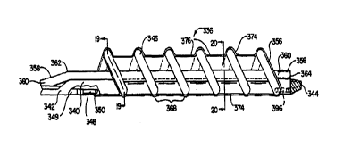
Fig. 15 illustrates a sparsely wound radiation delivery catheter 320 including
a tubular
shaft 322 having a proximal region 324 and a distal region 326, a manifold 328
disposed near
shaft proximal region 324, a balloon assembly 336 disposed on shaft distal
region 326, and
a distal tip 338. Shaft 322 includes a proximal shaft portion 352 and a distal
shaft portion 354
and is preferably formed of polyethylene. Manifold 328 includes a radiation
wire port 330,
an inflation port 332, and an infusion port 334. Radiation port 330 is used to
insert an
elongate radiation emitting member. Inflation port 332 is used to admit an
inflation fluid to
balloon assembly 336. Infusion port 334 can be used to infuse drugs through to
balloon
2 o assembly 336. The present invention can be made in accordance with the
drug delivery
catheters described in U.S. Patent No. 5,558,642, herein incorporated by
reference.
In one embodiment, a catheter according to the present invention includes
inflation
and radiation wire lumens, but no infusion lumen. Fig. 1 S illustrates a
preferred embodiment
-24-
CA 02291092 1999-11-19
WO 98/55179 PCT/US98/10235
catheter 320 having an infusion lumen as well. The inflation, radiation, and
infusion lumens
in preferred embodiments extend through shaft 322 to balloon assembly 336. A
preferred
embodiment includes a distal, single-operator-exchange guide wire lumen having
a proximal
port 342 and a distal port 344.
Referring now to Figs. 16, 19 and 20, Fig. 16 illustrates detail area 16 of
Fig. 15,
showing balloon assembly 336 in more detail in an inflated state. A radiation
wire tube 358
defines a radiation wire lumen 360, rising near radiation tube region 362 near
proximal guide
wire port 342 to accommodate entering guide wire tube 341 below, extending
through a
substantial portion of balloon assembly 336, and ending in a radiation wire
tube distal closed
end 364. Closed end 364 prevents fluid communication between bodily fluids and
radiation
wire lumen 360, allowing use and re-use of radiation sources within the closed
lumen without
sterilization. The closed lumen allows use of non-sterile sources within a
sterile catheter, as
the radiation source does not contact the blood stream and become
contaminated. In a
preferred embodiment, the radiation wire tube lies external to the catheter
shaft within the
balloon assembly, as illustrated by radiation wire tube distal portion 358
lying atop shaft distal
portion 354 in Figs. 16, I9 and 20. Radiation wire tube 358 can be formed of
polyimide or
PTFE. In a preferred embodiment, radiation wire tube 358 includes a distal
segment formed
of a collapsible polyolefin copolymer (POC) material within balloon assembly
336, enabling
increased perfusion when not occupied by a radiation wire.
2 0 Guide wire tube 341 extends from proximal entry port 342 through distal
guide wire
port 344. Guide wire tube 341 is preferably formed of polyethylene. In a
preferred
embodiment, guide wire lumen 340 lies within shaft distal portion 354. In
catheter 320, an
-25-
CA 02291092 1999-11-19
WO 98/55179 PCTNS98/10235
infusion lumen 366 is defined between the outside walls of guide wire tube 341
and the inside
walls of shafts 354 and 352, as illustrated by Figs. 17, 18, 19 and 20.
In the embodiment shown, a helical balloon is formed of at least one
inflatable helical
strand or coil 346 having multiple windings extends longitudinally over a
substantial portion
of balloon assembly 336. Balloon strand 346 is preferably formed of
polyolefin. Balloon
strand 346 is in fluid communication with an inflation lumen 349 within an
inflation tube 348
and preferably has a blind, distal termination 396. Inflation lumen 349
preferably lies within
shafts 352 and 354, as illustrated by inflation tube 348 lying within shafts
352 and 354.
Inflation tube 348 is preferably formed of polyimide. Balloon strand 346 can
be attached to
1 o inflation tube 348 as illustrated at 350. Balloon inflatable strand 346,
in an inflated state,
defines a perfusion lumen 356 therethrough, as indicated in Figs. 16, 19 and
20. Perfusion
lumen 356 does not lie uniformly around shaft 354 in a preferred embodiment,
but has shaft
354 lying to one side of the lumen and forming a boundary of the lumen, as
shown in Fig. 19.
Fig. 19, illustrating a section taken through a complete inflation coil
strand, shows the
perfusion lumen created by the inflation of coil 346. Perfusion lumen 356
allows perfusing
blood flow during radiation treatment. As illustrated by Figs. 19, 20 and 21,
distal shaft 354
has helix strand 346 secured by the lacing of strand 346 through through-holes
370. Fig. 21
illustrates in detail the securing of balloon strand 346 to shaft 354 using
holes 370. In the
embodiment shown, holes 70 form a pair aligned substantially transversely to
the longitudinal
2 o axis of the shaft. In another embodiment, the through-holes can be
oriented obliquely to the
shaft longitudinal axis, substantially aligned with the helix strands as they
approach the shaft.
This later embodiment may not be self securing and may require adhesive
bonding to the
shaft.
-26-
CA 02291092 1999-11-19
WO 98/55179 PCT/US98/10235
Lacing strand 346 repeatedly through shaft 354 removes shaft 354 to one side
of
perfusion lumen 356, creating a greater unobstructed area for perfusing blood
flow, compared
to placing shaft 354 within the center. Placing shaft 354 to one side by
threading strand 346
through pairs of holes in the shaft brings an exterior portion of the shaft
into fluid
communication with the space between strands 346. As illustrated in Fig. 20,
infusion holes
372, preferably located between strands 346, provide access from within
infusion lumen 366
to the vessel wall the catheter is disposed within.
Infusion holes 372 and infusion lumen 366 can be used to infuse local agents
in
conjunction with radiation treatment. Infused substances can include agents to
promote
healing and agents to enhance the effect of radiation treatment. In
particular, agents may be
infused to prevent hypoxia (oxygen deprivation) while the balloon is inflated
against vessel
walls. Oxygenating agents include the patient's own arterial blood, which may
be heparinized,
and water or saline, which may be heparinized. Oxygenated blood, saline, water
or other
fluids can be used. Peroxides such as hydrogen peroxide can also be used to
provide oxygen
to vessel walls. Applicants believe the agents enhance the effectiveness of
the radiation
treatment.
Catheter 320 can also have a tubular sheath 374 disposed over strand 346 as
illustrated
in Figs. 16 and 19. Sheath 374 is preferably formed of polyurethane elastomer.
Sheath 374
is preferably configured to hug the contours of strand 346 such that inter-
strand pockets 368
2 0 lie between the strands and also spiral around balloon assembly 336 as
does strand 346. If
sheath 374 lay straight between the outermost extent of strands 346, a
substantially straight-
walled cylindrical sheath would result, leaving less space between sheath and
vessel wall for
infusing drugs. As sheath 374 has inter-strand pockets 376, there is space for
drugs to
-27-
CA 02291092 1999-11-19
WO 98/55179 PCT/US98/10235
circulate and diffuse to contact the vessel walls. While a helical coil
without a sheath
provides some reduced flow, dead space for drug infusion near vessel walls, a
sheath
substantially insulates the vessel walls from perfusion flow and is the
preferred embodiment.
Referring now to Fig. 22, a radiation wire device 378 having a distal region
380 is
illustrated. A radioactive coil 382 is preferably wound about a radiation wire
support tube 384
having a lumen 386. Support tube 384 is preferably formed of polyimide, having
radioactive
wire 382 wound around distal region 380 and covered with a shrink wrap layer
388 preferably
formed of polyolefm copolymer.
In one embodiment, radiation wire support tube 384 is extremely flexible or
floppy
and incapable of being pushed alone through radiation wire lumen 360 from the
catheter
proximal end. In this embodiment, a radiation wire guide wire lumen 386 is
included within
tube 384, as illustrated in Fig. 22. A separate guide wire may be required for
this
embodiment, to guide the radiation emitting device through to the balloon
assembly. A guide
wire may be required to provide a pilot wire through the rise or bend 362 in
the radiation wire
tube, where the guide wire lumen enters the balloon assembly, where it may be
difficult to
push a flexible tube.
One embodiment includes perfusion holes proximal of coil 382, providing
perfusion
through lumen 386 when the guide wire is retracted. In this embodiment, the
guide wire can
be used to position the radiation member then retracted proximal of radiation
wire tube rise
2 0 362, lessening the obstruction to perfusion blood flow during irradiation.
The radiation
member having perfusion holes is optimally used in conjunction with an open
ended radiation
tube, described below. Radiation wire coil 382 preferably includes Yttrium-90
or Nickel-66,
-28-
CA 02291092 1999-11-19
WO 98/55179 PCT/US98/10235
high energy beta emitters. In another preferred embodiment, radiation wire 382
includes
Gadolinium-153, a gamma emitter.
Referring now to Fig. 23, another embodiment catheter 390 is illustrated.
Catheter
390 is similar to catheter 320, but has a radiation wire tube 392 with an open
distal end 394.
The resulting perfusion lumen 356 is still open to passage by the radiation
wire, which can
extend substantially through the balloon assembly, but without a supporting
tube in this distal
region. As can be visualized with Fig. 19, the removal of radiation wire tube
358 would
provide greater cross sectional area for perfusing blood flow within perfusion
lumen 356. The
greater cross sectional area would be especially significant during periods
when the radiation
l0 wire device itself is not within the perfusion lumen, as when the radiation
wire device lies
proximal of radiation wire tube bend 362. A device having no radiation wire
tube within the
inflatable balloon also provides a smaller profile for the balloon assembly in
the deflated state,
as can be illustrated by visualizing Fig. 19 without radiation wire tube 358.
The open ended
radiation wire lumen does allow contact between the radiation source and the
bodily fluids.
This may require sterilization or disposal of the radiation source after a
single use.
As previously stated, a preferred source of radiation for all embodiments of
the present
invention is the radioactive compound Nickel-66. Nickel-66 decays with a half
life of 2.28
days with only low energy beta emissions and no gamma emission into its
daughter element
Copper-66. Copper-66 then emits high energy beta radiation with a half life of
5.10 minutes
2 0 and decays into the stabile element Zinc-66. This two-step decay has a
particular advantage
in use in the catheters of the present invention.
The Nickel-66 acts as a Garner for the high energy copper decay allowing for
time to
transport the source to the end user, and also allows for disposal of the
device through
-29-
CA 02291092 1999-11-19
WO 98/55179 PCT/US98/10235
ordinary means in about 23 days. A Copper-66 source alone would decay quickly
and not be
useful without the parent Nickel. Nickel is low cost and has desirable
mechanical properties
in its pure form and in alloys, such as a Nickel Titanium alloy.
The Nickel-66 can be utilized in any of the embodiments disclosed herein.
Also, this
source or another source could be incorporated into an atherectomy device. An
exemplary
embodiment of an atherectomy device is disclosed by Auth et al., in U.S.
Patent No.
5,314,407, the disclosure of which is incorporated herein by reference. A
rotating ablative
burr assembly is utilized to remove a stenosis. This burr assembly can have
incorporated
therein a radiation emitting source. Thus, radiation treatment can occur
simultaneously with
1 o the atherectomy procedure.
Another preferred radiation source is Gadolinium-153. Gadolinium-I53 is a
composite gamma source which can provide low energy gammas to vessel intima
layer while
providing higher energy gammas to penetrate calcified plaques and reach the
adventitia.
Moderate shielding can be used with Gadolinium-153, allowing the treating
physician to
remain in the room with the patient during therapy. Another preferred source
of radiation can
include Yttrium-90, a high energy beta emitter.
Numerous advantages of the invention covered by this document have been set
forth
in the foregoing description. It will be understood, however, that this
disclosure is, in many
respects, only illustrative. Changes may be made in details, particularly in
matters of shape,
2 0 size, and arrangement of parts without exceeding the scope of the
invention. The inventions's
scope is, of course, defined in the language in which the appended claims are
expressed.
-30-